spectrophotometric analysis of crown discoloration … · j appl oral sci. 138 abstract ...
TRANSCRIPT
J Appl Oral Sci. 138
ABSTRACT
www.scielo.br/jaoshttp://dx.doi.org/10.1590/1678-7757201302254
Spectrophotometric analysis of crown discoloration induced by MTA- and ZnOE-based sealers
Konstantinos IOANNIDIS1, Ilias MISTAKIDIS2, Panagiotis BELTES3������������� �� �4
1- DDS, MSc, Specialist in Endodontics, London, United Kingdom.2- DDS, MSc, Department of Basic Dental Sciences, School of Dentistry, Aristotle University of Thessaloniki, Greece.3- Professor, Department of Endodontology, School of Dentistry, Aristotle University of Thessaloniki, Greece.4- PhD, Mathematician, Department of Mathematics, Aristotle University of Thessaloniki, Greece.
Corresponding address: Ioannidis Konstantinos - 300 Vauxhall Bridge Road, SW1V1AA - London - UK - Phone: 00447414979270 - e-mail: [email protected]
�������������������������������������!��"�!#��$��������������%�����"�!#��$���������
Crown discoloration can be induced by root canal sealer remnants following root canal treatment. Objective: The aim of this study was to evaluate chromatic alterations in
human tooth crowns induced by a Mineral Trioxide Aggregate-based sealer (MTA Fillapex®) and a commonly used ZnOE-based sealer (Roth-811). The tested null hypothesis was that the application of the materials did not induce clinically perceptible crown discoloration ������������� ����������������������������� ������ ���������!"���#���� �"���$� %������%�mandibular third molars were sectioned 1 mm below the cemento-enamel junction. The pulp chambers were chemomechanically debrided via the cervical access. The specimens were randomly assigned into three groups Group 1: MTA Fillapex, Group 2: Roth 811, Group 3: &�'���"����������#�!��� ���� ��**���� ������ �"� #�����*��+� �"�������������'� ������� �/�����#$����������"�0����456����7����$��������9������������/�������� � �;��#����<��'�a UV-VIS spectrophotometer equipped with integration sphere in the visual spectrum at baseline, 1 week, 1 and 3 months after material placement. Data were transformed into "��#�������������=>�>;>�����������*��� ����������$�� ��'����"��#���/������#���� ��?�������������������/���$�����*� �#���'��/�@/���*�0� �B&HJB�*� ���%����$KL�LN���"��������'��!�����P��#�����B����������������'��!����������������>��� �;>����*����$���*��������������7B������$�0�Q��#$�/���*���#�� ����/�"��%����"��#��� � ������0�� ������#*�������$���$��;�������������� ����������������� #���'������0$���*������$���� ������KW�XX������P���@X55�Q��#$%������������������'��!���� ����������=>��� ������������������'��!����increase in a* and b* chromatic parameters was measured, during all observation $���� ���P��#��������� "��#��� �0�� � � ���� �#*������� $���$��;������ �������� � ������ 5�/��+�����5KN�[N�������#�������B$$������������7B������$�0������������/������#��� ����minimal color alterations, while Roth 811 induced severe discoloration, in vitro. It could be suggested that, in terms of aesthetics, the use of MTA Fillapex appears to be favorable.
Key words: CIE color system. Discoloration. MTA Fillapex. Root canal sealer. Roth 811. Spectrophometry.
INTRODUCTION
Poor aesthetic appearance of endodontically treated teeth remains a challenging issue in clinical dentistry even nowadays. Interestingly, it has been reported that poor aesthetic appearance of �� ������ � ������ ��'��!������ ������� ���� $�������]�
quality of life10.A major etiological factor for the occurrence of
local intrinsic staining, especially in the cervical and middle third of the crown, is the presence of root �����!����'�*��������� ���������/���������������dentin of the pulp chamber16,17. In the long-term, core materials and sealers interact with dentin. Any
2013;21(2):138-44
J Appl Oral Sci. 139
change to the optical and chromatic properties of the dentinal structure is likely to cause an alteration in the outward appearance of the crown caused by ������'��������*�����'��� ���9����'�$��$������16,17.
Apart from thorough pulp chamber debridement, a reduction of the coronal aspect of the root canal !����'�;���/��������������"�0������z#��� ���������prevention of sealer-induced crown discoloration in the anterior aesthetic zone. However, sealer remnants cannot be always thoroughly removed from the pulp chamber and sometimes are present due to iatrogenic inadequate manipulations.
Despite the improvement of physicochemical, biomechanical and biological properties of endodontic sealers, the appearance of coronal discoloration is still evident in daily practice. Several laboratory studies have shown that some categories of sealers including ZnOE and epoxy-based sealers are capable of inducing moderate to severe crown discoloration17,24,25,29. In endodontics, ZnOE sealers are used in clinical practice for many decades and are regarded as a gold standard in several laboratory and clinical studies. These materials are regarded to be clinically satisfactory, providing reasonable seal in the root canal system13. However, after setting reaction, the formation of a weak porous mass leads to dissolution in contact /��������#��9#� �8. Due to gradual hydrolysis, the release of eugenol leads to long-lasting cytotoxicity and additional potential for sensitization6.
Recently, Mineral Trioxide Aggregate (MTA) Fillapex® was introduced as a new generation MTA-based sealer. The main concept for the development of MTA-based sealers is the exploitation of the physical and biological properties of MTA such as bioactivity12, biocompatibility21 and hard tissue conductivity22.
A developing amount of research data is becoming available upon MTA Fillapex, with regard to its physical and biological properties2,13,14,23,26. Considering the increasing demands for aesthetics, biomaterials should be chromatically stable, present optical properties similar to dental structures and not exert staining effects to hard dental tissues17. Recent reports showed that both White and Gray MTA formulations are capable of inducing tooth discoloration4,5. From that perceptive, it is mandatory for every new MTA-based material to be tested in terms of aesthetic and color objectives. Currently, there are no available studies investigating the potential of MTA Fillapex to induce color alterations to dental tissues.
The aim of this study was to evaluate the chromatic alterations in human tooth crowns induced by MTA Fillapex and Roth 811 root canal sealer. The null-hypothesis (Ho) to be tested was that the application of the materials did not induce clinically perceptible crown discoloration (Ho: CIE
����� ����������������19.
MATERIAL AND METHODS
Preparation of teeth������ !"�� �������� �0����� %� �#���� �"���$� %�
impacted and semi-impacted mandibular third molars free of cracks, fractures, caries, abrasions and discoloration due to systemic intrinsic causes were collected, according to the guidelines of good clinical practice (Department of Oral and Maxillofacial Surgery, School of Dentistry, Ethical Committee, Aristotle University of Thessaloniki, Greece).
Soft tissue was removed and the teeth were sectioned in the coronal third of the root complex, 1 mm below the buccal cemento-enamel junction. Sectioning was performed with the aid of a low-speed diamond-edge rotary saw microtome (Leica RM2255, Leica, Wetzlar, Germany). Access cavity preparation was not performed. Pulps were extirpated with a dental spoon and the internal axial walls of the pulp chambers were chemo-*���������� �;�� � � /���� �� ����*� !���� �&���#60-80) and 10 ml of sodium hypochlorite (2.5% w/w), through the apical access. Gentle reaming of the internal axial walls was performed through ���� ���������� ���/���� �"���� !��� ��z#���%� ����pulp chamber was irrigated with 2 ml of sodium ��$����������W�N��/�/�����!������'���0�W�*�K[�*����B�!����������/������*������� �#*���$���������(2.5% w/w) was performed at the end of the debridement. The pulp chambers of the specimens /���� !������ /���� � /���� N� *�� �������� ������� ���remove sodium hypochlorite remnants.
At the beginning of the experimental period, all crowns were transferred and stored in individually marked polyethlylene tubes containing distilled water up to the cervix of the crown in an incubator ��� ��456��� 7��� ������ �&K�N�� /���� ��� �*�������'�� ���������0$���*��������K5N�%�����$�����"����K5N�� �� � ���� ��'���"�� ������� '��#$� ��K5N���The materials to be evaluated were MTA Fillapex (Angelus, Londrina, Brazil) (Group 1) and a ZnOE based sealer, Roth 811 (Roth’s International, Chicago, IL) (Group 2) (Figure 1). The sealers were mixed and prepared according to the manufacturers’ instructions and were placed into the pulp chambers "���������"����������B�!�'���$�#''���/���#�� �to coat the internal axial walls with the sealers. The apical access was sealed with a thin layer of glass-ionomer cement (Ketac Cem Aplicap, 3M, Espe, Germany) in order to address microleakage and sealers solubility. Negative controls (Group 3) were ����������#*���� ��� ���*���� �#�!��� �
�%�����%����&�������!��$�����'����*!�������������!��!�#����+$��<���!��=!>?��+������������
2013;21(2):138-44

J Appl Oral Sci. 140
Measurement of crown chromatic alterationsA double-beam UV-Vis spectrophotometer
equipped with integrating sphere was used (UV-2401PC, Shimadzu Corporation, Kyoto, Japan) and a standardized mounting system was developed, utilizing a previously validated experimental model17. Standard D65 illumination was chosen (Commission Internationale de L’Eclairage 1978), as it corresponds approximately to the spectrum of midday daylight in Western/Northern Europe. The spectrophotometer was linked to a computer, /�������� � ������$��������9������#�"������the crowns, in visual spectrum (380-780 nm).
The obtained spectral curves in visual spectrum (380-780 nm) were transformed into L*, a* and b* values of the perceptually uniform CIE L*, a*, b* color space using a specialized computer software (Color Analysis UV-2401PC). L* values describe lightness, which range from black (0) to white (100), while a* values represent red (+80a*) to green (-80a*), and b* values represent yellow (+80b*) to blue (-80b*) color variations. Total ����� ���������� ����� /���� ��#���� � ��� ��'�to the equation:
��K���=i-L0*)2+(ai–a0*)2+(bi–b0*)2]½
The proposed acceptance for color matching � �$�� � ��� ����� ��# �� /��� ��� ���� ��� #�����(perceptibility threshold), beyond which the differences are clinically perceptible19. In dental �����%�/�������"��#�����������������5�#���%������color match occurs and any color differences cannot ;��� ����!� �;���� �$�� �����;���"���� in vitro27. However, color determination in clinical dentistry may become complicated by adjacent anatomic structures and lighting conditions. As a result, the proposed acceptance for color matching in dentistry has been set to 3.7 units (perceptibility threshold), beyond which differences are clinically visible19.
A standardized mounting system was developed for the customization and the reproducibility of the crown’s position. The cylindrical inner ���*�� � ��*����KW� *%� ������ ���'��KL�W� *��
of the black bakelite sample assembly that the �$����$����*�����/����z#�$$� �/���%�/���!��� �with black, non-polychromatic, thermo-plasticized ���������7������'#����#��������������/��/���!0� �within the silicone mass during its setting phase in order to construct individualized specimen carriers. The same individualized, silicone carrier for each specimen was used for measurements in all time intervals. The specimens were positioned in the in the circular opening of the aperture mask of the integrating sphere with the aid of an aligning system in order to ensure the reproducibility of the measured surface.
The dimensions of the polychromatic beam that illuminated the sample were 7x7 mm; thus the majority of the cervix and the crown surface was exposed and measured. The color appearance of the buccal surfaces of the crowns was measured in order to simulate their clinical appearance. Measurements were carried out by the same operator (I.M.) and at ambient temperature of 23±1°C.
The spectrophotometer was calibrated at each time interval using BaSO4 reference. Measurements were performed prior to the placement of the materials (baseline: t0) and consecutively 1 week (t1), 1 month (t2) and 3 months (t3) after sealer placement. All measurements were repeated twice �� � �"���'� �� ��� ���� ������ ����� ��������� ����between 2 measurements taken in a row exceeded the threshold of 1 DE unit, new measurements were obtained.
At the end of the 3rd month, three crowns of the experimental groups were randomly selected and longitudinally sectioned vertically to the middle of their mesio-distal dimension, on a bucco-lingual aspect, with the aid of a low-speed diamond-edge rotary saw microtome (Leica RM2255, Leica, Wetzlar, Germany). Digital images of the sectioned crown specimens were taken, in day-light conditions (Macro-Lense 100 mm, Canon EOS 1000D, Tokyo, Japan).
Group Samplesize (n)
Materials under��#�$
Manufacturer Composition
Group 1 15 MTA Fillapex Angelus, Londrina, Brazil
resins (salicylate, diluting, natural),bismuth trioxide, nanoparticulated silica,
mineral trioxideaggregate, pigments
Group 2(positivecontrol)
15 Roth 811 Roth’s International, Chicago, IL
Powder: Zinc oxide, bismouth bicarbonate, barium sulfate, dehydrated tetraboric sodium
Liquid: Chemically pure eugenol
Group 3(negative control)
15 �������
Figure 1- Study groups 1-3
>�� @ ����� �< @ �� ��H?Q<?��R���� �� ���
2013;21(2):138-44

J Appl Oral Sci. 141
Statistical analysisSample size was calculated after power analysis
based on the results of a pilot study, according to the equation nKW�<1-�/2 + z�)
2 �2 {1+(m���5��}/��2 with �KL�N%�*K�����$���� �*���#��*�����%��2K�%��KW�W%���$��KL�LN%�$�/��KL�X������������value of � was greater than the observed one in order to be close to the 3.7 threshold value and analogously done for the �2 )17. Accordingly the sample size to each group was calculated near to �K5W� �$��*����per group. In case of drop out during the experimental period, 3 specimens were � � ��������'��#$���K5N��
Two-way ANOVA with repeated measurements was used for data analysis of the values of CIE L*, a*, b* chromatic parameters and the total ����� ����������������7�����'��!������������� �interactions of the experimental factors were investigated with pairwise between-group and within-group comparisons, which were conducted with Bonferroni’s method. The overall analysis was performed with SPSS software (version 16.0, SPSS Inc., Chicago, Ill, USA). The level of statistical ��'��!����/����������$�0.05.
RESULTS
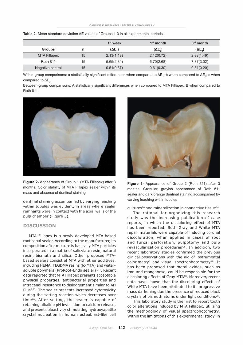
Tables 1 and 2 present the mean L*, a*, b* �� ����"��#����������'��#$�%� ������� ��*�� �����"����respectively. With regard to L*, a* and b* parameters, MTA Fillapex caused a statistically ��'��!����������������>����>�o-t3K5�L��%�$�L�LL5���� � ;>� ��;>� �o-t3KW�W�W%� $�L�LL5�� "��#���� =>�"��#��� ��*���� � ���;���� ��/�"��%� ���#������ ���values indicate that MTA Fillapex induced overall color changes below the human perceptibility threshold, ranging between 2.12-2.88 units. Roth 811 induced severe color changes exceeding the perceptibility threshold from the 1st week of �"��#������ ���KN�[N��� B� ������������� ��'��!������������ ��� �>� ���>�o-t3K@�%��N%� $�L�LL5�� �� �;>� ��;>� �o-t3K@�%L5�%� $�L�LL5�� "��#��� /���observed, while L* values statistically decreased ��=>�o-t3K�%�W[%�$�L�LL5������Q��#$��%�=>%��>%�;>�values remained stable in all time intervals.
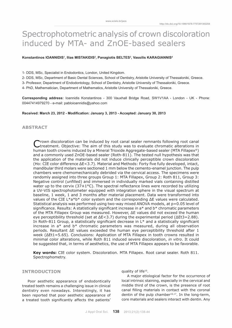
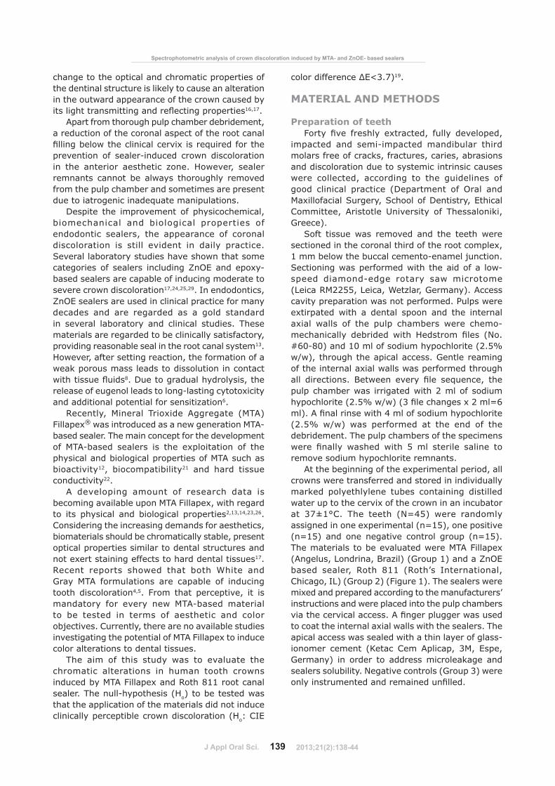
The macroscopic examination of the sectioned specimens of Group 1 (MTA Fillapex) showed stability of sealer color within its mass and absence of dentinal staining (Figure 2). On the contrary, in Group 2 (Roth 811) the set sealer displayed a granular, grayish appearance. Dark orange
baseline 1st week 1st month 3rd monthUV ?W�QX�%���&����
Groups n L0 L1 L2 L3
MTA Fillapex 15 84.95(3.14) 85.36(2.54) 84.35(3.10) 84.91(2.61)
Roth 811 15 84.21(1.98) 81.89(1.18) 79.38(1.82) 79.28(1.71)
���� ����������� 15 84.55(2.22) 84.77(2.26) 84.48(2.34) 84.55(2.17)
baseline 1st week 1st month 3rd monthUV ?W��X�%���&����
Groups n a0 a1 a2 a3
MTA Fillapex 15 0.15(0.68) 0.57(0.67) 0.77(0.68) 1.19(0.65)
Roth 811 15 0.01(0.79) 2.03(0.82) 2.71(1.00) 3.34(1.05)
���� ��������� 15 0.42(0.74) 0.39(0.66) 0.38(0.73) 0.41(0.73)
baseline 1st week 1st month 3rd monthUV ?W�+X�%���&����
Groups n b0 b1 b2 b3
MTA Fillapex 15 21.60(2.82) 23.36(2.21) 22.64(2.74) 23.89(2.35)
Roth 811 15 21.26(2.33) 25.67(2.36) 24.76(2.72) 25.28(2.89)
���� ��������� 15 20.62(3.11) 20.33(2.89) 20.49(2.94) 20.57(3.07)
Table 1- CIE L*, a*, b* mean standard deviation values of Groups 1-3, in all experimental periods
���������������������������������������������������������������������������0, b when compared to t1, c when compared to t2, d when compared to t3
����������������������������������������������������������������������������������!�������������������"����#$$
�%�����%����&�������!��$�����'����*!�������������!��!�#����+$��<���!��=!>?��+������������
2013;21(2):138-44
J Appl Oral Sci. 142
dentinal staining accompanied by varying leaching within tubules was evident, in areas where sealer remnants were in contact with the axial walls of the pulp chamber (Figure 3).
DISCUSSION
MTA Fillapex is a newly developed MTA-based root canal sealer. According to the manufacturer, its composition after mixture is basically MTA particles incorporated in a matrix of salicylate resin, natural resin, bismuth and silica. Other proposed MTA-based sealers consist of MTA with other additives, including HEMA, TEGDMA resins (lc-MTA) and water-soluble polymers (ProRoot-Endo sealer)7,11. Recent data reported that MTA Fillapex presents acceptable physical properties, antibacterial properties and intracanal resistance to dislodgement similar to AH Plus2,23. The sealer presents increased cytotoxicity during the setting reaction which decreases over time26. After setting, the sealer is capable of retaining alkaline pH levels due to calcium release, and presents bioactivity stimulating hydroxyapatite crystal nucleation in human osteoblast-like cell
cultures26 and mineralization in connective tissue14.The rational for organizing this research
study was the increasing publication of case reports, in which the discoloring effect of MTA has been reported. Both Gray and White MTA repair materials were capable of inducing coronal discoloration, when applied in cases of root and furcal perforation, pulpotomy and pulp revascularization procedures4,5. In addition, two ������ ��;����������# ������!�*� �����$��"��#��clinical observations with the aid of instrumental colorimetry1 and visual spectrophotometry18. It has been proposed that metal oxides, such as iron and manganese, could be responsible for the discoloring effects of Gray MTA18. Moreover, recent data have shown that the discoloring effects of White MTA have been attributed to its progressive mass darkening due the presence of reduced black crystals of bismuth atoms under light conditions28.
7������;����������# ���������!���������$����������color alterations induced by MTA Fillapex, utilizing the methodology of visual spectrophotometry. Within the limitations of this experimental study, in
Figure 3- Appearance of Group 2 (Roth 811) after 3 months. Granular, grayish appearance of Roth 811 sealer and dark orange dentinal staining accompanied by varying leaching within tubules
1st week 1st month 3rd monthGroups n UYZ1) UYZ2) UYZ3)
MTA Fillapex 15 2.13(1.18) 2.12(0.72) 2.88(1.49)
Roth 811 15 5.65(2.34) 6.70(2.68) 7.37(3.02)
Negative control 15 0.51(0.37) 0.61(0.30) 0.51(0.20)
Table 2-�%������������ ������&'� ��������+������$�/��������������������������
��������������������������������������������������������������������������&'1!�;�����������������&'2, c when �����������&'3
�������������������������<��������������������������������������������������%=<��������!�������������������Roth 811
Figure 2- Appearance of Group 1 (MTA Fillapex) after 3 months. Color stability of MTA Fillapex sealer within its mass and absence of dentinal staining
>�� @ ����� �< @ �� ��H?Q<?��R���� �� ���
2013;21(2):138-44
J Appl Oral Sci. 143
acceptance of the null hypothesis, MTA Fillapex did not induce clinically perceptible crown discoloration. In rejection of the null hypothesis, Roth 811 induced fast and severe discoloration and exceeded the perceptibility threshold 1 week after sealer placement.
The results of this study indicated that the tested sealer had minimum potential to induce color alterations in human teeth in vitro, since the measured color alterations did not exceed ���� �������� ����$���$��;������ ���� ����������7���descriptive analysis of the CIE L*, a*, b* chromatic $���*���������/� ������������������'��!���������change towards red and yellow after three months, while lightness was not affected. The macroscopic !� ��'����!�*� ����������������� � �����$�������color changes within its mass, maintaining its yellowish color throughout the experimental period. Dentinal staining was not evident as well. The increase of CIE a* and b* values of the crowns from ����!����/��+������"����'������*���;�������;#�� ����alterations in specimens’ optical properties due to the physical presence of the sealer in the internal dentinal surface.
Roth 811 sealer induced fast and severe discoloration, which was clinically perceptible 1 week following sealer placement. The chromogenic potential of ZnOE sealers has been attributed to the unstable chemical bond between ZnO and eugenol. Even after the end of the setting reaction, eugenol release leads to self-oxidation and becomes darker with time. The results of this study are in complete accordance with results of previous studies17,25,29.
Several methods have been proposed for the evaluation or measurement of sealer-induced discoloration, including visual technique and computer analysis of digital photos24,25,29. Inherent �;����"������ ����� �� �<������ ��!#������*���;��improved by the use of a spectrophotometer15. This methodology has been reported as accurate and reliable in dentistry for quantitative tooth color measurements17,20. A major advantage of visual spectrophotometry is that tooth color measurement ���;��� ��������*���#��*��������$��������9�����%�/���� ����;���������������9�������������*$������visual spectrum17.
Random errors in this study were minimized by strict control of the environmental factors along with multiple measurements and averaging. One of the main limitations of instrumental color measurement, however, is posed by systematic errors that can be attributed to variations in instrument geometrical design, metamerism and calibration techniques27.
The comparison of absolute tristimulus values found in that study with other studies is discouraged. Evaluation and comparison of total ����� ���������� ��0$����� �;�����%� ��/�"��%� ���well documented and considered as safe since
differential measurement is highly reproducible between instruments9. The results of this study are comparable to those of a previous study which was conducted utilizing the same methodology and under the same experimental conditions. After 3 months, non-perceptible total color differences ���������7B������$�0�/������*���������������"��#������ Q#���9�/� �P��+�%� �������%� ����� ���� =� �%�Germany) and Epiphany SE (Pentron Clinical Technologies, Wallingford, CT), indicating that they likewise exert minimal chromogenic effects17.
The results of this study do not directly represent the in vivo tooth discoloration potential of root canal sealers in good clinical practice. In this laboratory study, the investigation of the discoloration potential of root canal sealers was based on the generation ������/������������������;�����"��'�����'��!����amount of sealer in direct contact with the axial dentinal walls and several anatomical features of the pulp chamber17. However, the knowledge of the magnitude of the sealers’ chromogenic potential indicates that thorough cleaning measures are essential to prevent discoloration postoperatively.
Regardless of the minimal staining effects of new generation root canal sealers including MTA Fillapex, the clinician should always ensure thorough removal of sealer remnants. Apart from basic properties, such as biocompatibility and good sealing ability, it could be suggested that the chromogenic potential of sealers may also play an important role in �������'�$��$�������������!����'�*���������
CONCLUSION
Within the limitations of this experimental study, MTA Fillapex did not induce clinically perceptible crown discoloration. Roth 811 induced fast and severe discoloration and exceeded the perceptibility threshold 1 week after sealer placement. Although the incorporated MTA has proved chromogenic potential, MTA Fillapex posed minimal risk for potential staining effects. It could be suggested that, in terms of aesthetics, the use of MTA Fillapex appears to be favorable.
REFERENCES
1- Akbari M, Rouhani A, Samiee S, Jafarzadeh H. Effect of dentin bonding agent on the prevention of tooth discoloration produced by mineral trioxide aggregate. Int J Dent. 2012 [cited 2013 Feb 15];2012:563203. Available from: http://dx.doi.org/10.1155/2012/563203.2- Assmann E, Scarparo RK, Bottcher DE, Grecca FS. Dentin bond strength of two mineral trioxide aggregate-based and one epoxy resin-based sealers. J Endod. 2012;38:219-21.3- Barnett F, Trope M, Rooney J, Tronstad L. In vivo sealing ability of calcium hydroxide-containing root canal sealers. Endod Dent Traumatol. 1989;5:23-6.4- Belobrov I, Parashos P. Treatment of tooth discoloration after the use of white mineral trioxide aggregate. J Endod. 2011;37:1017-20.
�%�����%����&�������!��$�����'����*!�������������!��!�#����+$��<���!��=!>?��+������������
2013;21(2):138-44
J Appl Oral Sci. 144
5- Bortoluzzi EA, Araújo GS, Guerreiro Tanomaru JM, Tanomaru-Filho M. Marginal gingival discoloration by Gray MTA: a case report. J Endod. 2007;33:325-7.6- Brodin P. Neurotoxic and analgesic effects of root canal cements and pulp-protecting dental materials. Endod Dent Traumatol. 1988;4:1-11.7- Camilleri J, Mallia B. Evaluation of the dimensional changes of mineral trioxide aggregate sealer. Int Endod J. 2011;44:416-24. 8- Carvalho-Júnior JR, Guimarães LF, Correr-Sobrinho L, Pécora JD, Sousa-Neto MD. Evaluation of solubility, disintegration, and dimensional alterations of a glass ionomer root canal sealer. Braz Dent J. 2003;14:114-8.9- Douglas RD. Precision of in vivo colorimetric assessments of teeth. J Prosthet Dent. 1997;77:464-70.10- Dugas NN, Lawrence HP, Teplitsky P, Friedman S. Quality of life and satisfaction outcomes of endodontic treatment. J Endod. 2002;28:819-27.55@�Q�� ��!��Q%�7� ����%�?�;�����%��� ����%����$�����Q%����������Development of the foremost light-curable calcium-silicate MTA cement as root-end in oral surgery. Chemical-physical properties, bioactivity and biological behavior. Dent Mater. 2011;27:e134-57.5W@�Q�� ��!��Q%�7� ����%�7�����B%����������B$�����@���*��'��;������(bioactivity) of ProRoot MTA. Int Endod J. 2010;43:917-29.13- Gomes-Filho JE, Moreira JV, Watanabe S, Lodi CS, Cintra LT, Dezan E Jr, et al. Sealability of MTA and calcium hydroxide containing sealers. J Appl Oral Sci. 2012;20:347-51.14- Gomes-Filho JE, Watanabe S, Lodi CS, Cintra LT, Nery MJ, Otoboni JA Filho, et al. Rat tissue reaction to MTA FILLAPEX®. Dent Traumatol. 2012;28:452-6.15- Guan YH, Lath DL, Lilley TH, Willmot DR, Marlow I, Brook AH. The measurement of tooth whiteness by image analysis and spectrophotometry: a comparison. J Oral Rehabil. 2005;32:7-15.16- Ioannidis K, Beltes P, Lambrianidis T, Kapagiannidis D, Karagiannis V. Crown discoloration induced by endodontic sealers. Spectrophotometric measurement of Commission International de l’Eclairage’s L*, a*, b* chromatic parameters. Oper Dent. 2013. In press.17- Ioannidis K, Beltes P, Lambrianidis T, Kapagiannidis D, Karagiannis V. Validation and spectrophotometric analysis of crown discoloration induced by root canal sealers. Clin Oral Inv. 2013 [cited Feb 27]. Available from: http://dx.doi.org/10.1007/s00784-012-0850-x.
18- Ioannidis K, Mistakidis I, Beltes P, Karagiannis V. Spectrophotometric analysis of coronal discolouration induced by grey and white MTA. Int Endod J. 2013;46:137-44.19- Johnston WM, Kao EC. Assessment of appearance match by visual observation and clinical colorimetry. J Dent Res. 1989;68:819-22.20- Karamouzos A, Papadopoulos MA, Kolokithas G, Athanasiou AE. Precision of in vivo spectrophotometric colour evaluation of natural teeth. J Oral Rehabil. 2007;34:613-21.21- Mitchell PJ, Pitt Ford TR, Torabinejad M, McDonald F. Osteoblast biocompatibility of mineral trioxide aggregate. Biomater. 1999;20:167-73.22- Moretton TR, Brown CE Jr, Legan JJ, Kafrawy AH. Tissue reactions after subcutaneous and intraosseous implantation of mineral trioxide aggregate and ethoxybenzoic acid cement. J Biomed Mater Res. 2000;52:528-33.23- Morgental RD, Vier-Pelisser FV, Oliveira SD, Antunes FC, Cogo DM, Kopper PM. Antibacterial activity of two MTA-based root canal sealers. Int Endod J. 2011;44:1128-33.24- Parsons JR, Walton RE, Ricks-Williamson L. In vitro longitudinal assessment of coronal discoloration from endodontic sealers. J Endod. 2001;27:699-702.25- Partovi M, Al-Havvaz AH, Soleimani B. In vitro computer analysis of crown discolouration from commonly used endodontic sealers. Aust Endod J. 2006;32:116-9.26- Salles LP, Gomes-Cornélio AL, Guimarães FC, Herrera BS, Bao SN, Rossa-Junior C, et al. Mineral trioxide aggregate-based endodontic sealer stimulates hydroxyapatite nucleation in human osteoblast-like cell culture. J Endod. 2012;38:971-6.27- Seghi RR, Hewlett ER, Kim J. Visual and instrumental colorimetric assessments of small color differences on translucent dental porcelain. J Dent Res. 1989;68:1760-4.28- Vallés M, Mercadé M, Duran-Sindreu F, Bourdelande JL, Roig M. Color stability of white mineral trioxide aggregate. Clin Oral Inv. 2013 [cited Feb 27]. Available from: http://dx.doi.org/10.1007/s00784-012-0794-1.29- Van der Burgdt TP, Mullaney TP, Plasschaert AJM. Tooth discoloration induced by endodontic sealers. Oral Surg Oral Med Oral Pathol. 1986;61:84-9.
>�� @ ����� �< @ �� ��H?Q<?��R���� �� ���
2013;21(2):138-44