specificity determinants of the p1 and p7 plasmid centromere analogs
TRANSCRIPT

Proc. Natl. Acad. Sci. USAVol. 90, pp. 9228-9232, October 1993Genetics
Specificity determinants of the P1 and P7 plasmidcentromere analogs
(partition/segregation/protein-DNA interactions/parS/integration host factor)
FINBARR HAYES AND STUART J. AuSTIN*Laboratory of Chromosome Biology, Advanced BioScience Laboratories-Basic Research Program, National Cancer Institute-Frederick Cancer Research andDevelopment Center, Frederick, MD 21702
Communicated by Jon Beckwith, June 28, 1993
ABSTRACT The cis-acting parS sites of P1 and P7 aresimilar in sequence and promote active partition of theirrespective plasmid prophages to daughter cells when the cog-nate Par proteins are supplied. Forty of the 94 relevant basesdiffer between the P1 and P7 parS sites, and the protein-siteinteractions show complete species specificity. A method wasdeveloped to predict which subset of the differingparS bases isresponsible. When the four Pl bases thus identified weresubstituted into the P7 parS site, a complete switch to P1specificity was observed. The Pl-specific bases constitute twoCG dinucleotide elements situated 66 bp apart. They lie withinrepeats of the TCGCCA sequence implicated in secondarycontacts with the P1 ParB protein. The equivalent TC dinu-cleotides in the P7 site were found to be involved in P7specificity. However, three other P7 bases can also contribute,including two in the heptamer repeats primarily responsible forParB binding, and the P7-specific information shows someredundancy. The motifs containing the specificity dinucleotidesand the primary ParB binding (heptamer) sites bear no obviousrelationship of spacing or orientation to each other. For theParB protein to contact both types of motif at the same time,the topology of the interaction must be complex.
Bacterial plasmids maintained at low copy numbers haveacquired mechanisms to guarantee their faithful inheritancein growing cells (1). Critical among these are active partitionsystems expressly implicated in promoting segregationalstability of a number of low-copy-number plasmids (1-3).Bacteriophage P1 lysogenizes Escherichia coli as a highly
stable plasmid with a copy number that can be as low as twoper dividing cell (4, 5). This stability is dependent on thepresence of an active partition cassette consisting of twoessential genes, parA and parB, and an adjacent cis-actingsite, parS (6-8). The parS site is sufficient to promotepartition of a test plasmid if the protein products ofparA andparB (ParA and ParB, respectively) are both supplied in trans(9). ParB binds specifically to a series of heptamer motifs inparS (10), where it interacts cooperatively with the host-encoded integration host factor (IHF) (10-12). IHF bends theparS site (13), probably wrapping it around a ParB-IHFprotein core (11). ParA is an ATPase that is stimulated byParB and double-stranded DNA but has not been shown tobind directly to parS (14).The active partition system of the P7 prophage plasmid is
organized similarly and is homologous to that of P1, indicat-ing that these cassettes apparently are derived from a sharedprogenitor (15). However, the systems have diverged suffi-ciently that they demonstrate species specificity with respectto complementation, partition-mediated incompatibility, andpartition-site activities (15, 16). Although the relative posi-tions of the protein binding motifs in the P1 and P7 parS sites
The publication costs of this article were defrayed in part by page chargepayment. This article must therefore be hereby marked "advertisement"in accordance with 18 U.S.C. §1734 solely to indicate this fact.
are conserved perfectly (refs. 15 and 16; see Fig. 1) and thesites are 57% identical over the 94-bp sequences illustrated inFig. 1, the P1 parS site fails to work with the P7 proteins, andvice versa (ref. 16; see Fig. 1). Presumably, this specificity isdue to key macromolecular interactions in which the indi-vidual components of the P1 and P7 systems are not inter-changeable. The specific P1 nucleotides that determine sitespecificity must either be required for the cognate proteins tointeract correctly with the site, or alternatively, they specif-ically prevent the proteins of noncognate type from doing so.In either case, by pinpointing the elements that determine thisspecificity, it should be possible to define crucial steps in theactive partition pathway.
MATERIALS AND METHODSMedium, Enzymes, and Materials. The following antibiot-
ics were added to L medium (Sigma): chloramphenicol, 10,ug/ml; kanamycin, 50 ug/ml; spectinomycin, 25 p,g/ml.Sources of materials and enzymes have been described (16).
Bacterial Strains and Bacteriophage. E. coli HB101 (17) wasused as a host for plasmid propagation and in cloning pro-cedures. Partition-site assays used strains CC1531 (16),CC1532 (16), and CC1654. These strains are derivatives ofCC1572 [recA13 galK pro Strr (Axis6 indj)] (16) that harborpGB2, pALA480, and pALA283, respectively. Bacterio-phage kcI857-P1:5RA1005 (18) was maintained on strainYMC (19).
Plasmid DNA Procedures. Protocols for plasmid DNAmanipulation and sequencing have been described (16, 20).
Plasmids. Plasmids pALA283 (8) and pALA480 (16) con-tain the P1 and P7 parA and parB genes in the pSC101-basedvectors pLG338 (21) and pGB2 (22), respectively. PlasmidpALA1626 (16) is a P1 miniplasmid vector lacking the pargenes and the incA copy-number-control locus. It replicatesat a moderate copy number via the P1 origin (16). A set ofpALA1626-based framework plasmids was constructed con-taining portions of the P1 or P7 parS loci flanked by restric-tion enzyme sites. These allowed insertion of syntheticdouble-stranded oligonucleotides to regenerate altered parSsites while maintaining the continuity of the junction se-quences. For example, framework plasmid pALA1709 con-tains P1 parS regions II, III, and IV (see Fig. 1) into whichaltered region I sequences were cloned as synthetic oligonu-cleotides with Hpa I and BamHI compatible ends to giveplasmids pALA1735 and pALA1775. All plasmids containingaltered or hybrid parS regions were made by similar strate-gies, and their resulting parS sequences are shown in therelevant figures.
Partition-Site Assays. Details of the pickup partition assayhave been described (16, 23). Briefly, the parS site to betested was inserted into the elevated-copy-number P1 mini-
Abbreviation: IHF, integration host factor.*To whom reprint requests should be addressed.
9228
Proc. Natl. Acad. Sci. USA 90 (1993) 9229
plasmid pALA1626, and the resulting plasmid was incorpo-rated into bacteriophage AcI857-P1:5RA1005 by homologousrecombination. The phage provides the copy-number-controllocus, incA, which is absent from pALA1626, and the result-ant composite replicates at a low copy number (S.J.A.,unpublished data). The composite containing the clonedparSsite was introduced into a strain supplying the relevant Parproteins in trans. Its stability was estimated by measuringloss of the composite plasmid after e25 generations ofunselected growth.
RESULTSA Systematic Approach for Determining the Basis of Spec-
ificity in the Related P1 and P7 parS Sites. A protein-bindingsite can be regarded as a linear array ofmodular elements (thebases), which are recognized by specific features on thesurface of the cognate proteins. The P1 and P7 parS sites,although similar, must differ in one or more of these criticalcontacts. The P1 nucleotides that differ from those in the P7parS site must include all of the information responsible forP1 specificity. However, some of the bases unique to the P1parS site may contain no relevant information and couldtherefore be altered without effect. Other bases may becritical for function, but not for specificity, such that eitherthe P1 or P7 base is acceptable at that position. The keyinformation for P1 parS site specificity must reside in theremaining nucleotides, which cannot be changed, even to theP7 type, without loss of function. Thus, it should be possibleto identify the bases that determine P1 specificity by sys-tematic substitution of P7 bases into the P1 parS site andassaying for loss of P1 site function. The identity of thesebases can be confirmed by substituting them simultaneouslyinto an otherwise intact P7 parS site, which should nowswitch to P1 specificity. The key bases responsible for P7parS site specificity, which need not necessarily correspondto their P1 counterparts, could be determined by a reciprocalapproach.Approximate Localization of the Determinants for P1 parS
Specificity. Partition-site activity was measured by using thepickup partition assay (16). This assay detects both P1 and P7partition-site activities efficiently and defines homologousminimal P1 and P7 parS sites of similar lengths as illustratedin Fig. 1. It does not detect any activity for the truncated22-bp P1 parS core sequence (bp 61-82, Fig. 1; unpublisheddata), which shows modest partition site activity in analternate assay system (24). The P1 parS site was arbitrarilydivided into four regions, and a series of P7 segment replace-ment experiments was conducted (Fig. 1). Regions II and IIIare fully interchangeable between the sites, either individu-ally (pALA1738 and pALA1739) or simultaneously(pALA1792 and pALA1794) and therefore do not containsequences that contribute to the specificity of the systems.These regions both appear to be involved in IHF proteinbinding (11, 16), with region II corresponding roughly to theIHF recognition consensus sequence (11, 16, 25). Region I ofthe P7 parS site can only partially substitute for the corre-sponding region in P1 parS (pALA1735), and region IV of theP7 site cannot replace the equivalent sequence in the P1 site(pALA1777). Substitution of P7 region IV into the P1 parSsite (pALA1777) not only eliminated P1 parS site activity butalso conferred partial function as a P7 site (Fig. 1). Therefore,the specificities of the P1 and P7 parS sites are determined bysequences in both regions I and IV.
Nucleotide Substitutions in Region I. The P1 and P7 parSsites differ at 15 of27 positions in region I (Fig. 2). The crucialbases in this region that contribute to the specificity of the P1site were identified by substituting subsets of P1-specificnucleotides with the appropriate P7 bases (Fig. 2). The threeleftmost (pALA1750) and four rightmost (pALA1752) P1-
specific bases in region I could be altered to the correspond-ing P7 bases without an appreciable effect on P1 parS siteactivity. However, all or some ofthe central eight nucleotidesare necessary for full P1 parS site activity (pALA1751). Anexamination of the effect of replacing these eight basesindividually with P7-specific bases revealed that only the C -+T and G -* C substitutions in pALA1763 and pALA1764,respectively, had any appreciable effect on P1 parS siteactivity. When these two alterations were combined(pALA1774), a marked reduction in P1 parS site activity wasdemonstrable. The simultaneous substitution of the remain-ing six nucleotides of the central region had no effect(pALA1773). Therefore, the modest requirement for P1 spec-ificity in region I appears to be due just to the CG dinucleotideat the eighth and ninth positions (Fig. 2).
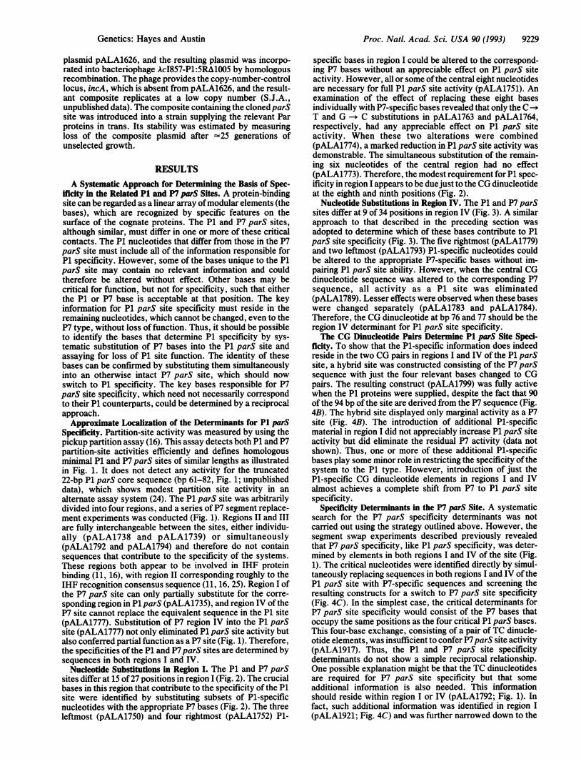
Nucleotide Substitutions in Region IV. The P1 and P7 parSsites differ at 9 of 34 positions in region IV (Fig. 3). A similarapproach to that described in the preceding section wasadopted to determine which of these bases contribute to P1parS site specificity (Fig. 3). The five rightmost (pALA1779)and two leftmost (pALA1793) P1-specific nucleotides couldbe altered to the appropriate P7-specific bases without im-pairing P1 parS site ability. However, when the central CGdinucleotide sequence was altered to the corresponding P7sequence, all activity as a P1 site was eliminated(pALA1789). Lesser effects were observed when these baseswere changed separately (pALA1783 and pALA1784).Therefore, the CG dinucleotide at bp 76 and 77 should be theregion IV determinant for P1 parS site specificity.The CG Dinucleotide Pairs Determine P1 parS Site Speci-
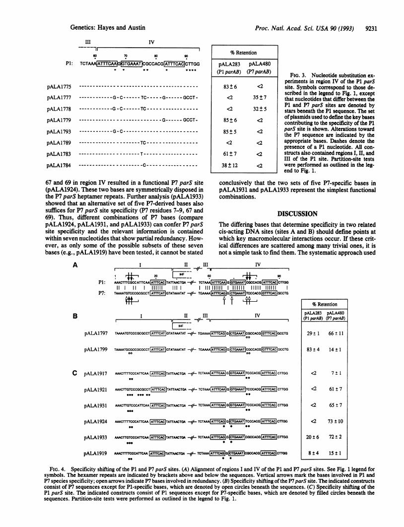
ficity. To show that the P1-specific information does indeedreside in the two CG pairs in regions I and IV of the P1 parSsite, a hybrid site was constructed consisting of the P7 parSsequence with just the four relevant bases changed to CGpairs. The resulting construct (pALA1799) was fully activewhen the P1 proteins were supplied, despite the fact that 90of the 94 bp of the site are derived from the P7 sequence (Fig.4B). The hybrid site displayed only marginal activity as a P7site (Fig. 4B). The introduction of additional P1-specificmaterial in region I did not appreciably increase P1 parS siteactivity but did eliminate the residual P7 activity (data notshown). Thus, one or more of these additional P1-specificbases play some minor role in restricting the specificity ofthesystem to the P1 type. However, introduction of just theP1-specific CG dinucleotide elements in regions I and IValmost achieves a complete shift from P7 to P1 parS sitespecificity.
Specificity Determinants in the P7 parS Site. A systematicsearch for the P7 parS specificity determinants was notcarried out using the strategy outlined above. However, thesegment swap experiments described previously revealedthat P7 parS specificity, like P1 parS specificity, was deter-mined by elements in both regions I and IV of the site (Fig.1). The critical nucleotides were identified directly by simul-taneously replacing sequences in both regions I and IV of theP1 parS site with P7-specific sequences and screening theresulting constructs for a switch to P7 parS site specificity(Fig. 4C). In the simplest case, the critical determinants forP7 parS site specificity would consist of the P7 bases thatoccupy the same positions as the four critical P1 parS bases.This four-base exchange, consisting of a pair of TC dinucle-otide elements, was insufficient to confer P7parS site activity(pALA1917). Thus, the P1 and P7 parS site specificitydeterminants do not show a simple reciprocal relationship.One possible explanation might be that the TC dinucleotidesare required for P7 parS site specificity but that someadditional information is also needed. This informationshould reside within region I or IV (pALA1792; Fig. 1). Infact, such additional information was identified in region I(pALA1921; Fig. 4C) and was further narrowed down to the
Genetics: Hayes and Austin

9230 Genetics: Hayes and Austin Po.Nt.Aa.Si S 0(93
11 HiI IVlr.. 11 lr..
20 IHF 60 80
AAACTTTCGCCATTCAA TATTAACTGACTG TTAAAGTAAATrACTCTAAA G CGCCACG CTTGG11 11 HIM III 11 III III HIM IIIII) 11 11 HIM
TAAAATGTCCCGCGCCTIATTTCAT GTATAAATATATGATATATATAGACATTCATGAAAA GTCCCACG GCCTG
..
... ......... ...... ... .... ..
.....
... ... .. .. .. ..
... .. ...
% Retention
pALA283 pALA48O(P1 parAB) (P7parAB)
83 ±6 <2
<2
36 ± 4
82 ± 7
81 ± 12
75 ± 10
4± 1
<2
<2) 35 ±7
77 ±1 <2
<2 74±t8
FiG. 1. Segment substitution experiments in the P1 and P7 parS sites. The sequence alignment of the two sites is shown with a commonnucleotide numbering system applying only to this study. Identical nucleotides are linked by the vertical lines. Heptamer repeat motifs recognizedby the ParB proteins (10, 16) and the consensus sequences for IHF binding (11, 16) are boxed. The bent arrows denote the approximate limitsof the P1 parS site (unpublished data) and the precise limits of the P7 parS site (16) in the pickup assay system. Reassortment of the four regions(I-IV) shown above the sequences gave the hybrid parS sites illustrated below. Open and stippled areas indicate P1- and P7-derived sequences,respectively. Retention frequencies in pickup partition-site assays (see Materials and Methods) were determined in the presence of pALA283and pALA480, which supply the P1 and P7 partition proteins, respectively. Averages of at least three individual experiments ± 1 SD are given.No retention was detected when the vector plasmid lacking Par protein production (pGB2) was present in trans.
single G nucleotide at position 7 (pALA1931; Fig. 4C). Thespecifi'city of the P7 parS site can therefore be determined byfive nucleotides (bp 7-9, 76, and 77), four of which constituteTC dinucleotide repeat sequences that map at correspondingpositions to the CG dinucleotide sequences responsible for P1parS site specificity.
I
Partial Redundancy of the P7parS Specificity Determinants.In the course of searching for the postulated additionalinformation required for P7 parS site activity, constructswere also made with additional P7-derived bases in region IV(Fig. 4C). When combined with the TC dinucleotide ele-ments, the introduction of two P7-speciftic bases at positions
Id
1 10 20
P1: MAACTTTCGC'CATTCAA jATTTCAC TATTAACTGA-
T--AA-GTC--GCG-CT-- --TGTA----
-GTC--GCG-CT ---------
~~~------- A-----
-G-~~-- - - -
- --T ----T---- --
% Retention
pALA283 pALA48O(P1 parAB) (P7parAB)
83±6
36±4
84 ± 2
22 ± 7
83± 7
80±2
63 ± 7
57±13
85±7
87± 3
85 5
89t±7
81±t8
80± 5
16±9
<2
4± 1
<2
4±3
<2
<2
<2
<2
<2
<2
<2
<2
<2
<2
<2
FIG. 2. Nucleotide substitution experimentsin region I of the P1 parS site. Symbols corre-
spond to those described in the legend to Fig. 1,
except that nucleotides that differ between the
P1 and P7 parS sites are denoted by stars
beneath the P1 sequence. The set of plasmidsused to define the key bases contributing to the
specificity of the P1 parS site is shown. Alter-
ations toward the P7 sequence are indicated bythe appropriate bases. Dashes denote the pres-
ence of a P1 nucleotide. All constructs also
contained regions II, III, and IV of the P1 site.
Partition-site tests were performed as outlined
in the legend to Fig. 1.
PI:
P7:
pALA1775
pALA 1791
pALA1735
pALA1738
pALAI739
pALAI777
pALA 1794
pALAI792
pALA1775
pALA1735
pALA175O
pALA17S1
pALA1752
pALA1762
pALA1763
pALA1764
pALA1765
pALA1766
pALA1767
pALA1768
pALA1769
pALA1773
pALA1774
Proc. Natl. Acad. Sci. USA 90 (1993)
I
Proc. Natl. Acad. Sci. USA 90 (1993) 9231
III IV
60 70 s0 90
P1: TCTAAAATTCAG TJ GCCACGIAMT CAC CTTGG* * * * * **
pALA1775
pALA1777-G-C----- TC-----G------ GCCT -
pALA1778 - G -C----- TC - - - - - - - - - - - - - - -
pALA1779 -- ------ GCCT-
pALA1793- G-C-
pALA1789 - - - - - - - - - - - - - - - - - - - - - TC - - - - - - - - - - - - - - - - -
pALA1783 - -- - - - - - - - - - - - - - - - - - - T - - - - - - - - - - - - - - - - - -
pALA1784 -C~~~~~~---C
% Retention
pALA283 pALA480(P1 parAB) (P7parAB)
83±6
<2
<2
85±6
85 ± 5
<2
61±7
38± 12
<2
35±7
32±5
<2
<2
<2
<2
<2
FIG. 3. Nucleotide substitution ex-periments in region IV of the P1 parSsite. Symbols correspond to those de-scribed in the legend to Fig. 1, exceptthat nucleotides that differ between theP1 and P7 parS sites are denoted bystars beneath the P1 sequence. The setofplasmids used to define the key basescontributing to the specificity of the P1parS site is shown. Alterations towardthe P7 sequence are indicated by theappropriate bases. Dashes denote thepresence of a P1 nucleotide. All con-structs also contained regions I, II, andIII of the P1 site. Partition-site testswere performed as outlined in the leg-end to Fig. 1.
67 and 69 in region IV resulted in a functional P7 parS site(pALA1924). These two bases are symmetrically disposed inthe P7 parS heptamer repeats. Further analysis (pALA1933)showed that an alternative set of five P7-derived bases alsosuffices for P7 parS site specificity (P7 residues 7-9, 67 and69). Thus, different combinations of P7 bases (comparepALA1924, pALA1931, and pALA1933) can confer P7 parSsite specificity and the relevant information is containedwithin seven nucleotides that show partial redundancy. How-ever, as only some of the possible subsets of these sevenbases (e.g., pALA1919) have been tested, it cannot be stated
A
P1:
P7:
conclusively that the two sets of five P7-specific bases inpALA1931 and pALA1933 represent the simplest functionalcombinations.
DISCUSSIONThe differing bases that determine specificity in two relatedcis-acting DNA sites (sites A and B) should define points atwhich key macromolecular interactions occur. If these crit-ical differences are scattered among many trivial ones, it isnot a simple task to find them. The systematic approach used
I II III IV
! rt 20 ! _o60 go
AACTTICGCCATTCAA ATCAC TATrAACTGA -e/- TCTAAA ATCAAGGTGAAT CACCACO A1CAC CTTGG
11 I 111111 1111 IIIIIIIII 1111111 11111 111111
TAAATGTCCCGCGCCT A1CAT GTATAAATAT -9/- TGAAA TOA G G CTGT|TCCCACG G1CAC GCCTG
B I__ II InI IV
_ZKL_1__eHIpALA1797 TAMATGTCCCGCGCCT ATCAT GTATAAATAT He TGAAAA ATCAG | CTGAAAT CGCCACG GCAC CTG
00
pALA1799 TAAATGCGCCGCGCCT AITTCAT GTATAAATAT 9/ TGAMAAA AGG CTGAMT CGCCACG GCAC CTG00 00
C pALA1917 AMCTIT CCCATTCAA ATCAC TA1TAACTGA w/ TCTAAAAmGcAAG OTOMAT TCCCACG CAC| CTTGG*- *9
pALA1921 AAACTTGTCCCGCGCCT ATCAC TATTAACTGA -e TCTAAA|ATCAA |G|TGAT|TCCCACG ATTCAC |CTGG
pALA193 1 AmCTTGTCCCATTCAA AmCAC TATTAACTGA -e TCTAAA A1TTCM G GTGAAT TCCCACG ATCAC CTTGG
pALA1924 Amc1T, CCCATrCAA A1CAC TATTAACTGA -/ TCTAAA A1TCAG 0 TGAMT TCCCACG ATTCAC CTrGG0* 0 0 0
pALA1933 AMCTTGTCCCATTCM ACAC TATTrCTGA -/-- TCTAM AAG|(3TGAMT CGCCACG A1TTCAC cTTGGS.. 00
pALA1919 AxMcrnrcccATTcA A1CAC TAT-r CT(M -/ TCTAAA TCGGCTT CGCCACGCATTCACCTTGGS. * S
% RetentionpALA283 pALA480(PI parAB) (P7 parAB)
29± 1 66 t 11
83±4 14±1
<2 7±1
<2 61±7
<2 65 ± 7
<2 73 ± 10
20 ± 6 72 ± 2
8±4 15±1
FIG. 4. Specificity shifting of the P1 and P7 parS sites. (A) Alignment of regions I and IV of the P1 and P7 parS sites. See Fig. 1 legend forsymbols. The hexamer repeats are indicated by brackets above and below the sequences. Vertical arrows mark the bases involved in P1 andP7 species specificity; open arrows indicate P7 bases involved in redundancy. (B) Specificity shifting ofthe P7parS site. The indicated constructsconsist of P7 sequences except for P1-specific bases, which are denoted by open circles beneath the sequences. (C) Specificity shifting of theP1 parS site. The indicated constructs consist of P1 sequences except for P7-specific bases, which are denoted by filled circles beneath thesequences. Partition-site tests were performed as outlined in the legend to Fig. 1.
Genetics: Hayes and Austin

9232 Genetics: Hayes and Austin
here may have some general application. The relevant site Abases are identified as those that cannot be changed to the siteB type without loss of site function. The choice of bases isrefined by using block substitutions prior to testing theremaining individual bases. The critical site A bases, thusidentified, are substituted together into an otherwise normalsite B. If the base identification is correct, the specificity thenswitches from the site B to the site A type, as was found withthe four bases identified in the P1 parS site. The approachassumes independence of information conferred by eachrelevant base and a lack of redundancy. Redundancy wouldhave prevented the approach from working for the P7 parSsite. For example, although the P7 TC dinucleotide (bp76-77) can contribute to P7 specificity (pALA1931; Fig. 4),its replacement by P1 bases does not lead to a significant lossof function as a P7 site (pALA1797; Fig. 4), due to redun-dancy. Fortunately, useful information about the relevant P7bases could be determined directly, based on the assumptionthat the critical P1 and P7 bases might be similarly placed.Two CG dinucleotide elements disposed at opposite ends
of the P1 parS site are necessary and sufficient to confercomplete P1 parS site activity to an otherwise P7 parS site.Similarly, two TC dinucleotide elements at identical positionsplay an important role in P7 specificity (Fig. 4). What is thefunction of these sequences? First, the roles of the dinucle-otides at each end of P1 parS are probably the same becausethey are nested within direct hexamer repeats: TCGCCA atbp 7-12 and bp 75-80 (Fig. 4). The integrity of the rightmostcopy of this repeat was previously shown to be important forpartition (24, 26). The equivalent P7 6-bp sequences thatcontain the TC dinucleotides can also be regarded as directhexamer repeats (TjICCM). In this instance, the significanceof the rightmost repeat is less clear, because this motif isinvolved in redundancy with region I and the last two basesof this repeat can be deleted without effect (16). Second, thedinucleotides are probably recognized by ParB. They areincluded in regions protected by the appropriate ParB pro-teins in both systems (10, 16) and are in close contact withParB, at least in the P1 parS site (26). However, the heptamerrepeat sequences are also required for ParB binding (10, 16),and a single heptamer unit acts as an individual ParB bindingsite (16). By comparing the ends of each parS site, it can beseen that the heptamer repeats and the dinucleotide-containing hexamer repeats show no simple relationship ofspacing or orientation to each other (Fig. 4). Thus, they arenot likely to be parts of an extended ParB binding consensussequence. Funnell and Gagnier (26) recently confirmed theimportance of both types of repeat in the P1 parS site andprovided evidence that both P1 motifs are recognized byParB. This probably occurs via separate sites on the ParBprotein. Presumably, ParB protein contacts the two recog-nition elements in some topologically complex fashion. Thiscan be envisioned most easily if each ParB protein joins twoseparate plasmids by a pairing reaction or bridges the ends ofa single parS site in a looped structure. The latter seemsplausible, because the ends of the parS site are thought to bealigned through IHF-mediated looping (11, 13). IHF proteinbridging of the parS site ends might explain the partialredundancy observed with the P7 specificity elements inwhich the absence of P7-specific nucleotides in region IV canbe compensated for by the presence of additional P7 infor-mation in region I (Fig. 4C).The specificity determinants of the P1 and P7parS sites are
not reciprocal, as the P7 determinants include extra bases anda degree of redundancy. An extra P7 base in region I (bp 7),adjacent to the leftmost P7 dinucleotide (bp 8-9), can com-pensate for the lack of the rightmost P7 dinucleotide (bp76-77) in region IV as long as P7 bases 67 and 69 are also
present. These latter bases conserve the perfect invertedheptamer repeats of the primary ParB binding sequence,converting it to its P7 form (Fig. 4). Presumably, the P7versions of the heptamer repeats are more favorable for P7ParB binding than the P1 equivalents, thereby contributing tospecificity. Preliminary in vitro binding studies appear toconfirm that the P7 ParB protein preferentially recognizes theheptamer repeats in P7 parS but that the P1 ParB proteininteracts equally well with either the P1 or P7 heptamer boxes(M. A. Davis and S.J.A., unpublished data). The presence ofredundant P7 information may explain why some of thehybrid parS sites confer significant partition activity withboth sets of Par proteins (e.g., pALA1797 and pALA1933;Fig. 4). Presumably this is due to the presence of both thecritical P1 region IV determinant at bp 76-77 and the P7heptamer box information at bp 67 and 69.Our results with the P7 determinants indicate that hep-
tamer box information can compensate for suboptimal dinu-cleotide-containing hexamer repeats and that region I infor-mation can compensate for suboptimal region IV informa-tion. The available assay is not precise enough to rule outsome minor advantage of having all these elements presenttogether as in the original P7 site. However, the elementsclearly show considerable redundancy. These effects aremost easily explained if the regions involved contact eachother in a cooperative complex, consistent with our sugges-tion that the two ends of such parS sites contact each otherby looping around a compact protein core (11).
This research was sponsored by the National Cancer Institute,Department of Health and Human Services, under contract numberNO1-CO-74101 with Advanced BioScience Laboratories.
1.2.3.4.5.
6.
7.8.
9.10.11.
12.13.14.
15.
16.
17.
18.
19.20.
21.
22.
23.
24.
25.
26.
Nordstrom, K. & Austin, S. J. (1989) Annu. Rev. Genet. 23, 37-69.Austin, S. J. (1988) Plasmid 20, 1-9.Hiraga, S. (1992) Annu. Rev. Biochem. 61, 283-306.Austin, S., Ziese, M. & Stemnberg, N. (1981) Cell 25, 729-736.Prentki, P., Chandler, M. & Caro, L. (1977) Mol. Gen. Genet. 152,71-76.Abeles, A. L., Friedman, S. A. & Austin, S. J. (1985) J. Mol. Biol.185, 261-272.Friedman, S. A. & Austin, S. J. (1988) Plasmid 19, 103-112.Martin, K. A., Friedman, S. A. & Austin, S. J. (1987) Proc. Natl.Acad. Sci. USA 84, 8544-8547.Austin, S. & Abeles, A. (1983) J. Mol. Biol. 169, 353-372.Davis, M. A. & Austin, S. J. (1988) EMBO J. 7, 1881-1888.Davis, M. A., Martin, K. A. & Austin, S. J. (1990) EMBO J. 9,991-998.Funnell, B. E. (1988) Proc. Natl. Acad. Sci. USA 85, 6657-6661.Funnell, B. E. (1991) J. Biol. Chem. 266, 14328-14337.Davis, M. A., Martin, K. A. & Austin, S. J. (1992) Mol. Microbiol.6, 1141-1147.Ludtke, D. L., Eichorn, B. G. & Austin, S. J. (1989) J. Mol. Biol.209, 393-406.Hayes, F., Davis, M. A. & Austin, S. J. (1993) J. Bacteriol. 175,3443-3451.Boyer, H. W. & Roulland-Dussoix, D. (1969) J. Mol. Biol. 41,459-472.Austin, S., Hart, F., Abeles, A. & Stemnberg, N. (1982) J. Bacteriol.152, 63-71.Dennert, A. & Henning, U. (1968) J. Mol. Biol. 33, 322-329.Sambrook, J., Fritsch, E. F. & Maniatis, T. (1989) MolecularCloning: A Laboratory Manual (Cold Spring Harbor Lab. Press,Plainview, NY), 2nd Ed.Stoker, N. G., Fairweather, N. F. & Spratt, B. G. (1982) Gene 18,335-341.Churchward, G., Belin, D. & Nagamine, Y. (1984) Gene 31,165-171.Abeles, A. L. & Austin, S. J. (1989) in Genetic Engineering, ed.Setlow, J. K. (Plenum, New York), Vol. 11, pp. 111-125.Martin, K. A., Davis, M. A. & Austin, S. (1991) J. Bacteriol. 173,3630-3634.Goodrich, J. A., Schwartz, M. L. & McClure, W. R. (1990) NucleicAcids Res. 18, 4993-5000.Funnell, B. E. & Gagnier, L. (1993) J. Biol. Chem. 268, 3616-3624.
Proc. Natl. Acad. Sci. USA 90 (1993)