special stains - sheba...the gms staining kit is used to demonstrate polysaccharides in the cell...
TRANSCRIPT
Special stains

Iron/Hemosiderin Prussian blue
Lipids Sudan stain (Sudan II, Sudan III, Sudan IV, Oil Red O, Sudan Black B)
Carbohydrates Periodic acid-Schiff stain
Amyloid Congo red
Bacteria Gram staining (Methyl violet/Gentian violet, Safranin) · Ziehl-Neelsen stain/acid-fast (Carbol fuchsin/Fuchsine, Methylene blue) · Auramine-rhodamine stain (Auramine O, Rhodamine B)
Connective tissue
trichrome stain: Masson's trichrome stain/Lillie's trichrome (Light Green SF yellowish, Biebrich scarlet, Phosphomolybdic acid, Fast Green FCF) Van Gieson's stain
Other H&E stain (Haematoxylin, Eosin Y) · Silver stain (Gömöri methenamine silver stain, Warthin–Starry stain) · Methyl blue · Wright's stain · Giemsa stain · Gömöri trichrome stain · Neutral red · Janus Green B

Hematoxylin + Eosin (H & E ) הצביעה השגרתית המבוצעת בחתכי רקמה
צבע בסיסי המתחבר לחומצות הגרעין – המטוקסילין לקצה הבסיסי של צבע חומצי המתחבר – אאוזין
החלבונים בציטופלסמה

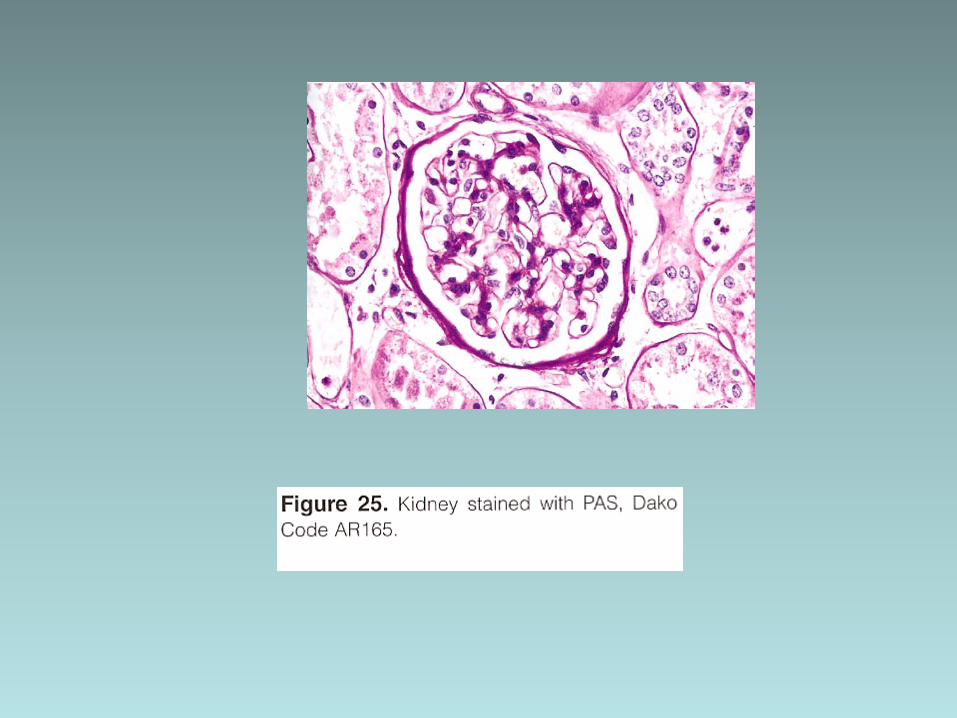
Pas stain
Demonstrate : • Glycogen • Basement membranes • Neutral mucosubstance


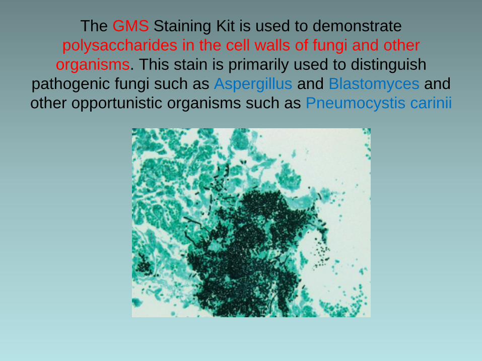
The GMS Staining Kit is used to demonstrate polysaccharides in the cell walls of fungi and other
organisms. This stain is primarily used to distinguish pathogenic fungi such as Aspergillus and Blastomyces and other opportunistic organisms such as Pneumocystis carinii
The Giemsa is used to differentiate leukocytes in bone marrow and other hematopoietic tissue (lymph nodes) as well as some microorganisms (Helicobacter pylori).
Giemsa Stain

The Elastic Staining Kit is used to demonstrate elastic fibers in tissue sections

The Mucicarmine Staining Kit is used to detect acid
mucopolysaccharides (mucin).


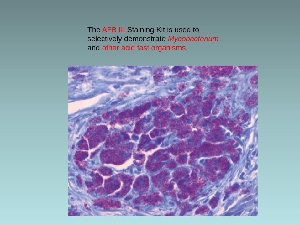
The AFB III Staining Kit is used to selectively demonstrate Mycobacterium and other acid fast organisms.

צביעה לחומרים הנאגרים ברקמות


Liver special stains
• Trichrom Stain • Reticulin Stain • Iron Stain • Pas diastas • Orcein Stain

Trichrom Stain
To assess the fibrosis in the liver stage and progression of disease:
• Hepatitis B and C • Fatty liver disease • Alcoholic liver disease • Chronic biliary diseases • Fibrous tissue including bridging fibrous
septa leading to the and stage- cirrhosis

Masson's trichrome Distinguishing cells from surrounding connective
tissue
keratin -red and muscle collagen and bone-blue or green cytoplasm - light red or pink cell nuclei - dark brown to black


Reticulin Stain
• Reticulin fibers are thin fibers composed of collagen lll which form a delicate stromal network
• The stain provide information about the architecture of the liver
• When hepatocytes are damaged and undergo necrosis , the reticulin fibers surrounding them


The Iron Stain used to detect ferric iron in bone marrow,
tissue with hemochromatosis and hemosiderosis


Bone marrow
• Trichrom Stain • Reticulin Stain • Iron Stain

Kidney special stains
•Pas •Met •Trichrom Stain

JONE'S METHENAMINE SILVER
For demonstrating the basement membrane of the
glomerulus in the kidney

The Jones H&E Staining Kit is used to demonstrate capillary basement membranes and is primarily used to distinguish pathological abnormalities in kidney diseases.
