special senses - western wyoming community college senses - western wyoming community college
TRANSCRIPT

The Nervous System
• Part 1: Neurons • Part 2: Neurophysiology – Postsynaptic Potentials and the Action Potential – The Axon Terminal
• Part 3: The Brain and Spinal Cord • Part 4: Special Senses
Receptors
• Thermoreceptors • Photoreceptors – sight
• Nociceptors • Chemoreceptors – smell/taste
• Mechanoreceptors – hearing

Eye Anatomy
• conjunctiva – thin connective tissue layer that
covers eye and inner eyelid • cornea
– transparent covering of the iris – course focus of images
• sclera – “white” of eye, dense connective
tissue
Eye Anatomy• choroid
– lines inner surface of sclera – helps nourish the retina – melanocytes give black appearance – absorbs excess light
• ciliary body – extends from edge of retina (ora
serrata) to junction of sclera and cornea
– ciliary processes • folds that connect to the suspensory ligaments to the lens
• cells secrete aqueous humor • ciliary muscles

Eye Anatomy• ciliary muscles
– contraction pulls choroid layer forward
– creates slack for the ciliary process and suspensory ligaments
– allows lens to become more round (curves light more)
• iris – colored part of eyeball – hole in center is pupil
• regulates amount of light that enters eye by reflex constriction or dilation of smooth muscles or iris
Eye Anatomy• retina
– beginning of visual pathway – detached retina: retina
separated from underlying epithelial layer • distorts shape of retina, making images blurry
• corrected by laser surgery • optic disc
– where optic nerve exits the eyeball
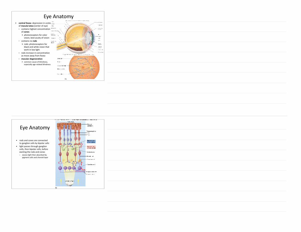
Eye Anatomy• central fovea: depression in center
of macula lutea (center of eye) – contains highest concentration
of cones • photoreceptors for color vision, best acuity of vision
– contains no rods • rods: photoreceptors for black and white vision that work in low light
– rods increase in concentration as move away from fovea
– macular degeneration • common cause of blindness,
especially age-‐related blindness
Eye Anatomy
• rods and cones are connected to ganglion cells by bipolar cells
• light passes through ganglion cells, then bipolar cells, before exciting the rods and cones – excess light then absorbed by
pigment cells and choroid layer
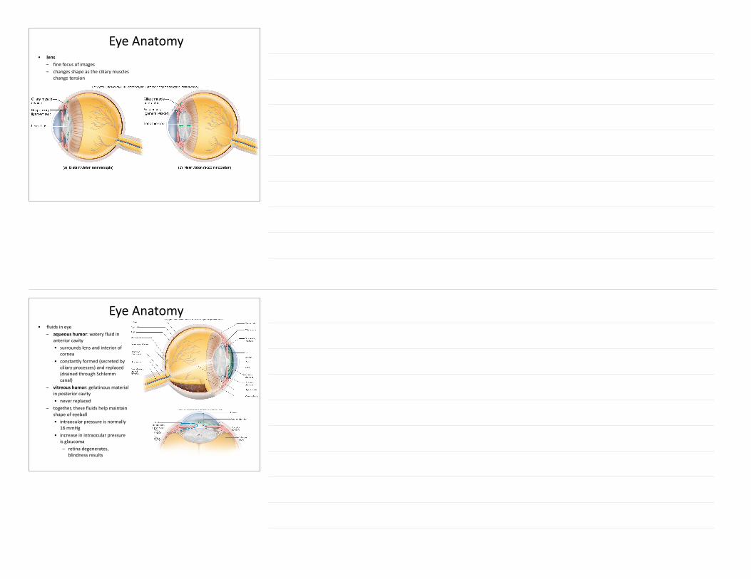
Eye Anatomy• lens
– fine focus of images – changes shape as the ciliary muscles
change tension
Eye Anatomy• fluids in eye
– aqueous humor: watery fluid in anterior cavity • surrounds lens and interior of
cornea • constantly formed (secreted by
ciliary processes) and replaced (drained through Schlemm canal)
– vitreous humor: gelatinous material in posterior cavity • never replaced
– together, these fluids help maintain shape of eyeball • intraocular pressure is normally
16 mmHg • increase in intraocular pressure
is glaucoma – retina degenerates,
blindness results

Image Formation• images formed on retina are upside-‐
down and inverted left to right, but brain compensates for this
• accommodation – increase in curvature of lens to allow for
near vision • presbyopia
– as we age, lens often loses elasticity and cannot accommodate
– near point of vision generally increases with age – corrected with “reading glasses” (magnifying
lens)
Abnormalities of Refraction (bending of light rays as they cross from one medium to another)
• normal (emmetropic) eye: image focused on retina
• nearsighted (myopic) eye: image focused in front of retina – corrected by scattering of image with
concave lens • farsighted (hypermetropic) eye: image
focused behind retina – corrected by helping cornea and lens
to focus with convex lens • astigmatism: irregular shape of cornea
or lens – causes blurring in part of field of
vision – corrected by irregularly shaped glass
lens or contact that compensates for defect

Convergence
• field of vision from left and right eye overlaps tremendously – creates single binocular vision
• brain perceives one image seen by two eyes
• convergence: eyes must rotate medially as object we are viewing moves closer to eyes – failure to do this is called “lazy eye” and is
common in children
Physiology of Vision• first step is bleaching of
photopigment – light strikes photopigment – photopigment (rhodopsin)
consists of opsin (glycoprotein) and retinal (vitamin A derivative) • retinal portion isomerizes (changes shape) from cis-‐retinal to trans-‐retinal
• trans-‐retinal separates from opsin
• isomerization causes previously OPEN Na+ channels to close

Physiology of Vision
• closing of Na+ channels inhibits release of neurotransmitter to bipolar cells
• bipolar cells are inhibited by neurotransmitter release when rods and cones are at rest
• when the cones and rods are stimulated, they cease to release neurotransmitter, allowing the bipolar neurons to fire
• retinal isomerase (enzyme) converts trans-‐retinal to cis, effectively resetting the rod or cone
Ear Anatomy• external ear
– pinna (auricle) – external auditory canal (meatus) – ceruminous glands secrete cerumen (earwax) – tympanic membrane (eardrum)
• middle ear – auditory (Eustachian) tube
• connects to throat • when opened by yawning or swallowing, it allows for equilibration of pressure
on both sides of tympanic membrane so that it may vibrate freely – auditory ossicles
• malleus (hammer) • incus (anvil) • stapes (stirrup) • transmit sound
waves from thetympanic membraneto the oval window

Ear Anatomy
• inner ear – bony labyrinth surrounding
membranous labyrinth – semicircular canals
• ampulla: enlarged end of each semicircular canal
– vestibule • utricle and saccule
Ear Anatomy
– cochlea • spiral shaped • contains spiral organ (organ of
Corti) for hearing – scala vestibuli: fluid filled
tunnel in contact with oval window
– scala tympani: fluid-‐filled tunnel in contact with round window
– cochlear duct (scala media) in between two scala
– helicotrema: area where scala vestibuli and scala tympani meet

Physiology of Hearing• sound waves cause tympanic membrane to vibrate
– slowly for low-‐pitch sounds – faster for high-‐pitch sounds – deeper for louder sounds
• ossicles moved by tympanic membrane • oval window moved by stapes
Physiology of Hearing• fluid waves within perilymph caused by movement of oval window
– causes walls of scala vestibuli and tympani to vibrate – this pushes on the endolymph in the cochlear duct, starting endolymph movement – endolymph movement causes the basilar membrane supporting the hair cell
supporting cells to vibrate

Physiology of Hearing– specific areas of the basilar membrane vibrate, depending on the characteristics of the fluid
wave • near the oval window, the membrane is narrow but stiff
– sensitive to high frequency vibrations • closer to the end of the cochlea, the membrane is wide and more flexible
– sensitive to low-‐frequency vibrations
Physiology of Hearing– as the basilar membrane vibrates, the hair cells move against the tectorial membrane,
bending the hair cells • the bending causes the hair cells to depolarize by opening K+ channels (unusual) in the
hair cell membrane • depolarization causes Ca+2 channels in the hair cell base to open, which causes exocytosis
of neurotransmitter

Equilibrium• utricle is oriented in a horizontal plane; saccule in a vertical plane • utricle helps determine when head deviates from its normal upright position with respect to
gravitational pull or linear acceleration • saccule probably helps orient us when our head is not in upright position
Equilibrium• utricle contains calcium carbonate crystals called
otoliths – if we bend over, for example, the otoliths are free
to move downward due to gravity – as they move down, the gelatinous material they
reside in moves the stereocilia of the hair cells, causing depolarization
– the utricle can also detect linear acceleration because the otoliths have great inertia • linear acceleration will cause the otoliths to lag
behind the hair cells, causing them to bend back and some of them to be excited
• hair cells are directional; they will only depolarize when the stereocilia move in the direction of the kinocilium

Semicircular Canals• detects angular acceleration in a specific plane
– anterior canal: touching head to shoulder – posterior canal: nodding head “yes” – lateral canal: shaking head “no”
• when movement occurs, the canal moves, but the fluid inside (endolymph) does not (no forces are causing it to move) – this gives the appearance that the endolymph moves
against the direction of motion • the relative movement of canal with respect to endolymph
causes the cupula (gelatinous material of semicircular canals) to bend – this bends the hair cells, depolarizing them
Semicircular Canals• semicircular canals adapt within 20 seconds
– it takes about a second for friction acting on the endolymph to overcome its inertia
– the endolymph then moves with the canal (no relative movement)
– it takes 15-‐20 seconds more for the cupula to return to their resting state
– therefore you must continue to accelerate to continually excite the semicircular canals
• by sensing acceleration, the semicircular canals allow you to predict that you are about to fall over (for example), allowing you to respond before you actually fall – the utricle cannot respond until you have already fallen