special section: ovarian cancer€¦ · · 2018-04-19origin, understanding of the ... especially...
TRANSCRIPT

28 Cancer Facts & Figures 2018
Special Section: Ovarian CancerIntroductionIn 2018, there will be approximately 22,240 new cases of ovarian cancer diagnosed and 14,070 ovarian cancer deaths in the US. Ovarian cancer accounts for just 2.5% of all female cancer cases, but 5% of cancer deaths because of the disease’s low survival. This is largely because 4 out of 5 ovarian cancer patients are diagnosed with advanced disease that has spread throughout the abdominal cavity. Improving the ability to detect ovarian cancer early is a research priority, given that women diagnosed with localized-stage disease have more than a 90% five-year survival rate. Although advancing knowledge about ovarian cancer has been hindered by substantial disease heterogeneity and uncertainties about tumor tissues of origin, understanding of the disease has evolved rapidly in recent years, especially for epithelial tumors, the most common subtype. This special section provides information about ovarian cancer risk factors, incidence and mortality rates and trends, early detection, and treatment that is primarily related to epithelial tumors.
What is ovarian cancer?The ovaries are a pair of reproductive glands, each about the size of a grape, located on either side of the uterus (Figure S1). They produce eggs that travel through the fallopian tubes into the uterus, where they are fertilized for reproduction. In premenopausal women, the ovaries
are the primary source of the hormones estrogen and progesterone, which maintain the health of the female reproductive system.
The three major types of ovarian cancer are epithelial, accounting for 90% of cases, germ cell (3%), and sex cord-stromal (2%) (Figure S2).1 Epithelial cancers are further subdivided into serous (52%), endometrioid (10%), mucinous (6%), and clear cell (6%) tumors.1 (See sidebar on opposite page for more information about non-epithelial cancers.) The process of epithelial ovarian tumor development has long perplexed researchers. As biological understanding has evolved, epithelial subtypes are increasingly characterized as distinct diseases with different molecular pathways, risk factors, and treatment.2-5 Serous tumors are mostly high-grade serous carcinomas, which are characterized by involvement of both ovaries, aggressive behavior, late-stage diagnosis, and low survival.2 Accumulating evidence suggests that these tumors actually originate in the epithelial cells of the fallopian tube as microscopic preliminary lesions that subsequently migrate to the ovaries and/or
Figure S1. Female Reproductive Anatomy
Fundusof uterusFallopian tube
Broad LigamentOvary
Vagina
Cervix
Fimbriae
Infundibulum
Ligamentof ovary
Cervical canal
Uterinecavity
Borderline malignant ovarian cancer Borderline malignant tumors, which most often affect younger women, are epithelial tumors with behavior characteristics in between benign and malignant tumors. They are also called tumors of low malignant potential because they do not usually grow into the stroma (the supportive tissue around the ovary), with 5-year survival rates greater than 98%.8 Although they are not included in ovarian cancer statistics because they are considered noninvasive, understanding and classification of these tumors continues to evolve.8, 9

Cancer Facts & Figures 2018 29
peritoneum (the lining of the pelvis and abdominal cavity), where they are diagnosed.2, 6, 7 In addition to their common origins, tumors of the fallopian tube and peritoneum are very similar to epithelial ovarian cancer in appearance and behavior, and are now often studied
jointly. In contrast, endometrioid and clear cell tumors are thought to originate in the endometrium (lining of the uterus), while mucinous tumors may originate in the ovaries or fallopian tube-peritoneal junction; these subtypes typically affect only one ovary.2
*Data are based on microscopically confirmed cases. Persons of Hispanic origin may be of any race; Asians/Pacific Islanders include those of Hispanic and non-Hispanic origin. American Indians and Alaska Natives are not shown due to <25 cases reported for certain subtypes.Source: North American Association of Central Cancer Registries (NAACCR), 2017. Data are collected by cancer registries participating in the National Cancer Institute's SEER program and the Centers for Disease Control and Prevention's National Program of Cancer Registries.
©2018, American Cancer Society, Inc., Surveillance Research
Figure S2. Distribution (%) of Major Types of Ovarian Cancer* by Race/Ethnicity, 2010-2014
Epithelial Germ cell Sex cord-stromal Other or unspecified
Percent
0 20 40 60 80 100
Hispanic
Asian/Pacific Islander
Non-Hispanicblack
Non-Hispanicwhite
All races
84
25 490
37 6
82 65 7
2 2 591
3 2 590
Non-epithelial ovarian cancerThe two main types of non-epithelial ovarian cancer, germ cell tumors and sex cord-stromal tumors, collectively account for only about 5% of ovarian cancer (Figure S2). Germ cell tumors arise from the germ (egg) cells of the ovary and primarily occur in adolescents and young women. Sex cord-stromal tumors form in the supportive tissue of the ovaries and can arise from different cells, including granulosa, Sertoli, and Leydig cells. Sex cord-stromal tumors generally occur most often in women in their 50s, although certain types are more common in adolescents and young women.1, 10 Due to their rarity, risk factors for non-epithelial tumors are poorly
understood, but may include hormone exposure11-13 and genetic mutations.14-16
Germ cell tumors are not easy to detect early, but symptoms include abdominal swelling and irregular vaginal bleeding.17 Sex cord-stromal tumors often produce sex hormones, which may affect menstruation and/or cause male physical characteristics, such as a deep voice or body hair.10 Treatment for both germ cell and sex cord-stromal tumors typically includes surgery, alone or in combination with chemotherapy and/or radiation.10, 17, 18
30 Cancer Facts & Figures 2018
How does ovarian cancer occurrence vary?The average lifetime risk of developing ovarian cancer is 1.3% (Table S1). Older women and non-Hispanic white (NHW) women have the highest risk, although the racial variation for ovarian cancer is smaller than for many other cancers. Similarly, rates vary only slightly among states due, in part, to the lack of advances in early detection, which often exacerbate disparities. Some of the variation in ovarian cancer risk is explained by differences in the prevalence of reproductive risk factors, such as number of childbirths, use of oral contraceptives, and tubal ligation, but the source of most of the variation remains unknown.19
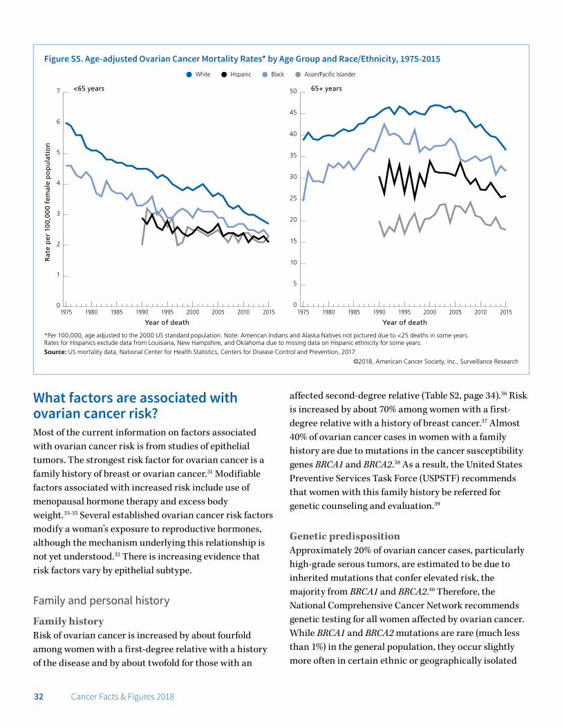
Race/EthnicityDuring 2010-2014, overall ovarian cancer incidence rates in NHW women (12.0 per 100,000 women) were 30% higher than those in non-Hispanic black (hereafter black; 9.4) and Asian American/Pacific Islander (API) women (9.2), who have the lowest rates (Figure S3). Despite this, black women have the second-highest mortality rates, likely due in part to later stage at diagnosis, a lower likelihood of receiving optimal treatment, and more comorbidities.20, 21 The distribution of ovarian cancer subtypes varies by race/ethnicity (Figure S2, page 29), although serous epithelial tumors are most common for all women.
AgeThe median age of diagnosis for ovarian cancer is 63 years, meaning that half of women are age 63 or younger at diagnosis. The age distribution of ovarian cancer varies by tumor type and race/ethnicity. For all women combined, incidence peaks in the late 70s for epithelial tumors, in the 50s for sex cord-stromal tumors, and in ages 15-19 years for germ cell tumors.1 Incidence of epithelial tumors is highest in NHW and API women until ages 50-54 years; however, from age 70, rates in NHWs are double those in APIs (Figure S4). In contrast, incidence of sex cord-stromal tumors is highest in black women from around age 30.1
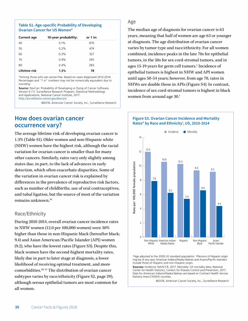
Table S1. Age-specific Probability of Developing Ovarian Cancer for US Women*
Current age 10-year probability: or 1 in:
40 0.1% 870
50 0.2% 474
60 0.3% 327
70 0.4% 265
80 0.4% 283
Lifetime risk 1.3% 78
*Among those who are cancer-free. Based on cases diagnosed 2012-2014.Percentages and “1 in” numbers may not be numerically equivalent due torounding.
Source: DevCan: Probability of Developing or Dying of Cancer Software, Version 6.7.5. Surveillance Research Program, Statistical Methodology and Applications, National Cancer Institute, 2017. http://surveillance.cancer.gov/devcan/
©2018, American Cancer Society, Inc., Surveillance Research
*Age adjusted to the 2000 US standard population. †Persons of Hispanic origin may be of any race; American Indians/Alaska Natives and Asians/Pacific Islanders include those of Hispanic and non-Hispanic origin.Sources: Incidence: NAACCR, 2017. Mortality: US mortality data, National Center for Health Statistics, Centers for Disease Control and Prevention, 2017. Data for American Indians/Alaska Natives are based on Contract Health Service Delivery Area (CHSDA) counties.
©2018, American Cancer Society, Inc., Surveillance Research
Rat
e p
er 1
00,
00
0 fe
mal
e p
op
ula
tio
n
Figure S3. Ovarian Cancer Incidence and Mortality Rates* by Race and Ethnicity†, US, 2010-2014
0
2
4
6
8
10
12
14
Asian/Pacific Islander
Non-HispanicBlack
HispanicAmerican Indian/Alaska Native
Non-HispanicWhite
Incidence Mortality
12.0
7.9
6.2
4.4
5.4
6.6
10.610.3
9.4 9.2

Cancer Facts & Figures 2018 31
How has the occurrence of ovarian cancer changed over time?Incidence trendsOverall ovarian cancer incidence rates have been decreasing since the mid-1980s, with the pace of the decline accelerating in the early 2000s.22 However, trends differ by age and race/ethnicity. From 2005 to 2014, incidence rates declined by 1.4% per year in NHWs, but were stable in blacks, Hispanics, American Indians and Alaska Natives (AIANs), and APIs, although rates appear to be decreasing modestly in the most recent years for Hispanics, blacks, and APIs. 23 Among women of all races ages 65 years and older, incidence rates increased from the late 1970s through the 1980s before beginning to decline around 1990, whereas rates among younger women have generally declined since at least 1975.24 The increase among older women prior to 1990 may be related to the decreasing birth rate in the US during the early- to mid-20th century. Subsequent declines in this age group, particularly among NHWs, may be partly due to decreased use of menopausal hormones, which
increases risk, following publication of a landmark report in 2002 linking them to elevated breast cancer risk.25 Use of oral contraceptives, which confers a substantial risk reduction, has also likely contributed to declines in incidence, especially among younger women.26
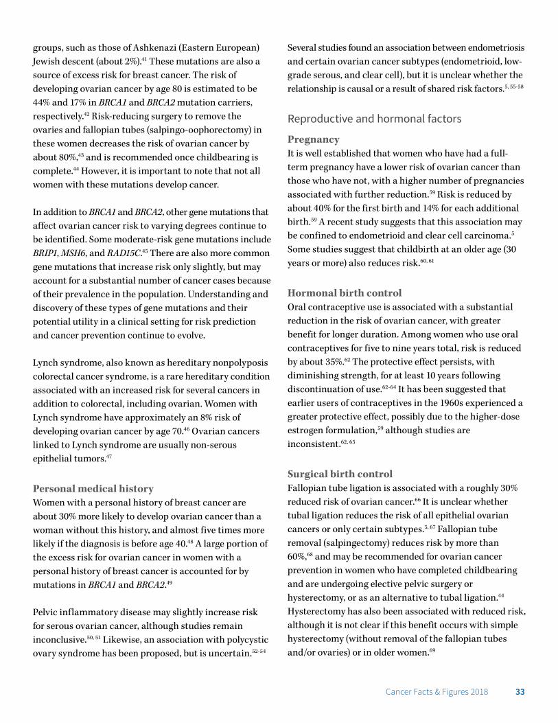
Mortality trendsMortality trends closely mirror those of incidence because of the low survival rate for ovarian cancer. Similar to incidence, mortality rates have decreased in women younger than 65 years since at least 1975, but only since the mid-2000s in women 65 years and older (Figure S5, page 32). Contrary to incidence trends by race/ethnicity, death rates declined during the most recent 10 years of data (2006 to 2015) in all racial/ethnic groups except AIANs, among whom rates were stable.27 Mortality rates decreased more rapidly among Hispanics and NHWs (by about 2% annually) than among blacks and APIs (1% annually).27 These declines are likely due to improvements in treatment, as well as reductions in incidence among NHWs.28-30
*Age adjusted to the 2000 US standard population. Persons of Hispanic origin may be of any race; Asians/Pacific Islanders include those of Hispanic and non-Hispanic origin. American Indians and Alaska Natives are not shown due to <25 cases reported for several age groups.Source: NAACCR, 2017.
©2018, American Cancer Society, Inc., Surveillance Research
Rat
e p
er 1
00,
00
0 fe
mal
e p
op
ula
tio
n
Figure S4. Epithelial Ovarian Cancer Incidence Rates* by Age and Race, US, 2010-2014
0
5
10
15
20
25
30
35
40
45
85+80-8475-7970-7465-6960-6455-5950-5445-4940-4435-3930-3425-2920-24
Non-Hispanic white Asian/Pacific Islander Non-Hispanic black Hispanic
Age at diagnosis (Years)
32 Cancer Facts & Figures 2018
What factors are associated with ovarian cancer risk?Most of the current information on factors associated with ovarian cancer risk is from studies of epithelial tumors. The strongest risk factor for ovarian cancer is a family history of breast or ovarian cancer.31 Modifiable factors associated with increased risk include use of menopausal hormone therapy and excess body weight.33-35 Several established ovarian cancer risk factors modify a woman’s exposure to reproductive hormones, although the mechanism underlying this relationship is not yet understood.32 There is increasing evidence that risk factors vary by epithelial subtype.
Family and personal history
Family historyRisk of ovarian cancer is increased by about fourfold among women with a first-degree relative with a history of the disease and by about twofold for those with an
affected second-degree relative (Table S2, page 34).36 Risk is increased by about 70% among women with a first-degree relative with a history of breast cancer.37 Almost 40% of ovarian cancer cases in women with a family history are due to mutations in the cancer susceptibility genes BRCA1 and BRCA2.38 As a result, the United States Preventive Services Task Force (USPSTF) recommends that women with this family history be referred for genetic counseling and evaluation.39
Genetic predispositionApproximately 20% of ovarian cancer cases, particularly high-grade serous tumors, are estimated to be due to inherited mutations that confer elevated risk, the majority from BRCA1 and BRCA2.40 Therefore, the National Comprehensive Cancer Network recommends genetic testing for all women affected by ovarian cancer. While BRCA1 and BRCA2 mutations are rare (much less than 1%) in the general population, they occur slightly more often in certain ethnic or geographically isolated
*Per 100,000, age adjusted to the 2000 US standard population. Note: American Indians and Alaska Natives not pictured due to <25 deaths in some years. Rates for Hispanics exclude data from Louisiana, New Hampshire, and Oklahoma due to missing data on Hispanic ethnicity for some years.Source: US mortality data, National Center for Health Statistics, Centers for Disease Control and Prevention, 2017.
©2018, American Cancer Society, Inc., Surveillance Research
Rat
e p
er 1
00,
00
0 fe
mal
e p
op
ula
tio
n
Figure S5. Age-adjusted Ovarian Cancer Mortality Rates* by Age Group and Race/Ethnicity, 1975-2015White Hispanic Black Asian/Pacific Islander
0
1
2
3
4
5
6
7
2015201020052000199519901985198019750
5
10
15
20
25
30
35
40
45
50
201520102005200019951990198519801975
<65 years 65+ years
Year of deathYear of death
Cancer Facts & Figures 2018 33
groups, such as those of Ashkenazi (Eastern European) Jewish descent (about 2%).41 These mutations are also a source of excess risk for breast cancer. The risk of developing ovarian cancer by age 80 is estimated to be 44% and 17% in BRCA1 and BRCA2 mutation carriers, respectively.42 Risk-reducing surgery to remove the ovaries and fallopian tubes (salpingo-oophorectomy) in these women decreases the risk of ovarian cancer by about 80%,43 and is recommended once childbearing is complete.44 However, it is important to note that not all women with these mutations develop cancer.
In addition to BRCA1 and BRCA2, other gene mutations that affect ovarian cancer risk to varying degrees continue to be identified. Some moderate-risk gene mutations include BRIP1, MSH6, and RAD15C.45 There are also more common gene mutations that increase risk only slightly, but may account for a substantial number of cancer cases because of their prevalence in the population. Understanding and discovery of these types of gene mutations and their potential utility in a clinical setting for risk prediction and cancer prevention continue to evolve.
Lynch syndrome, also known as hereditary nonpolyposis colorectal cancer syndrome, is a rare hereditary condition associated with an increased risk for several cancers in addition to colorectal, including ovarian. Women with Lynch syndrome have approximately an 8% risk of developing ovarian cancer by age 70.46 Ovarian cancers linked to Lynch syndrome are usually non-serous epithelial tumors.47
Personal medical historyWomen with a personal history of breast cancer are about 30% more likely to develop ovarian cancer than a woman without this history, and almost five times more likely if the diagnosis is before age 40.48 A large portion of the excess risk for ovarian cancer in women with a personal history of breast cancer is accounted for by mutations in BRCA1 and BRCA2.49
Pelvic inflammatory disease may slightly increase risk for serous ovarian cancer, although studies remain inconclusive.50, 51 Likewise, an association with polycystic ovary syndrome has been proposed, but is uncertain.52-54
Several studies found an association between endometriosis and certain ovarian cancer subtypes (endometrioid, low- grade serous, and clear cell), but it is unclear whether the relationship is causal or a result of shared risk factors.5, 55-58
Reproductive and hormonal factors
PregnancyIt is well established that women who have had a full-term pregnancy have a lower risk of ovarian cancer than those who have not, with a higher number of pregnancies associated with further reduction.59 Risk is reduced by about 40% for the first birth and 14% for each additional birth.59 A recent study suggests that this association may be confined to endometrioid and clear cell carcinoma.5 Some studies suggest that childbirth at an older age (30 years or more) also reduces risk.60, 61
Hormonal birth controlOral contraceptive use is associated with a substantial reduction in the risk of ovarian cancer, with greater benefit for longer duration. Among women who use oral contraceptives for five to nine years total, risk is reduced by about 35%.62 The protective effect persists, with diminishing strength, for at least 10 years following discontinuation of use.62-64 It has been suggested that earlier users of contraceptives in the 1960s experienced a greater protective effect, possibly due to the higher-dose estrogen formulation,59 although studies are inconsistent.62, 65
Surgical birth controlFallopian tube ligation is associated with a roughly 30% reduced risk of ovarian cancer.66 It is unclear whether tubal ligation reduces the risk of all epithelial ovarian cancers or only certain subtypes.5, 67 Fallopian tube removal (salpingectomy) reduces risk by more than 60%,68 and may be recommended for ovarian cancer prevention in women who have completed childbearing and are undergoing elective pelvic surgery or hysterectomy, or as an alternative to tubal ligation.44 Hysterectomy has also been associated with reduced risk, although it is not clear if this benefit occurs with simple hysterectomy (without removal of the fallopian tubes and/or ovaries) or in older women.69
34 Cancer Facts & Figures 2018
BreastfeedingSome studies have found a modest decrease in ovarian cancer risk among women who breastfed, with greater benefit for longer duration.5, 70-73
Menopausal hormonesWomen who ever used menopausal hormones (estrogen alone and estrogen combined with progesterone) have a 20% higher risk of developing ovarian cancer compared to never-users, with a stronger risk among recent users;34 among current or former (stopped within 5 years) users, risk of ovarian cancer is about 40% higher. Risk is increased even with short duration of hormone use and remains elevated for at least 10 years after discontinuation. The association appears to be confined to serous and endometrioid carcinoma, the two most common epithelial subtypes.
Fertility drugsThere is much interest in the long-term health effects of fertility drugs given their increasing use. While more studies examining the association between fertility drugs and ovarian cancer risk are needed, thus far there is little evidence of an association.74-76 This may be because women who have received fertility treatments have only just begun to enter the peak age for ovarian cancer, as well as the relative rarity of the disease.74
Excess body weightThe International Agency for Research on Cancer recently concluded that excess body weight modestly increases the risk of developing epithelial ovarian cancer, although the association is limited to women who have not used menopausal hormones.33, 35 Among women who have not used hormones, risk of ovarian cancer increases by about 10% for every 5 kg/m2 of body mass index. Results from studies evaluating whether this relationship varies by epithelial subtype are inconsistent.5, 33, 77
HeightHeight is associated with elevated risk for several cancers, including ovarian; risk increases by about 7% for each additional 5 centimeters of adult height relative to a height of less than 155 centimeters (about 5 feet).33 This association is unexplained, but may be related to genetic and environmental factors contributing to adult height, including growth hormone exposure during early life.78, 79
SmokingCigarette smoking increases the risk of mucinous ovarian cancer and decreases risk for endometrioid and clear cell carcinoma.80 In current smokers, risk of mucinous ovarian cancer is increased by about 80%, mainly for tumors of borderline malignancy.80
Diet and personal habitsA number of studies have evaluated the relationship between ovarian cancer risk and various foods and dietary patterns with inconsistent results.81, 82 Physical inactivity is associated with about a 30% higher risk of epithelial ovarian cancer,83 and likewise, sedentary behavior appears to increase risk.84-88 Some studies
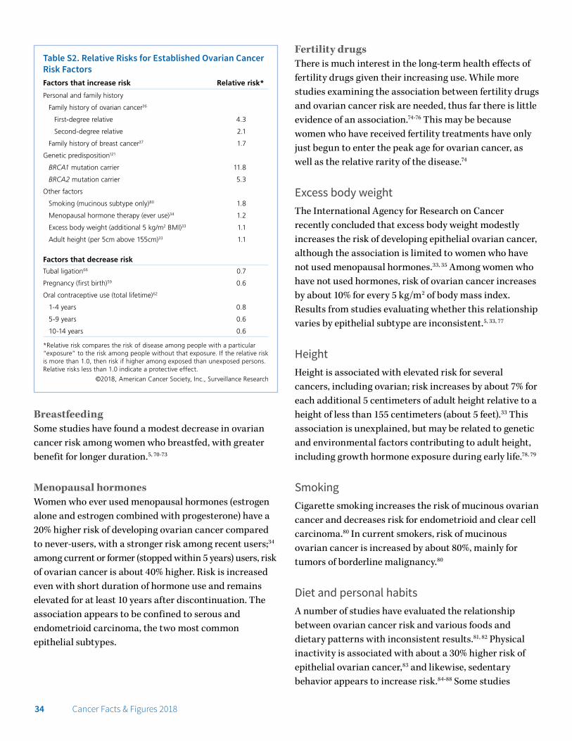
Table S2. Relative Risks for Established Ovarian Cancer Risk FactorsFactors that increase risk Relative risk*
Personal and family history
Family history of ovarian cancer36
First-degree relative 4.3
Second-degree relative 2.1
Family history of breast cancer37 1.7
Genetic predisposition121
BRCA1 mutation carrier 11.8
BRCA2 mutation carrier 5.3
Other factors
Smoking (mucinous subtype only)80 1.8
Menopausal hormone therapy (ever use)34 1.2
Excess body weight (additional 5 kg/m2 BMI)33 1.1
Adult height (per 5cm above 155cm)33 1.1
Factors that decrease risk
Tubal ligation66 0.7
Pregnancy (first birth)59 0.6
Oral contraceptive use (total lifetime)62
1-4 years 0.8
5-9 years 0.6
10-14 years 0.6
*Relative risk compares the risk of disease among people with a particular “exposure” to the risk among people without that exposure. If the relative risk is more than 1.0, then risk if higher among exposed than unexposed persons. Relative risks less than 1.0 indicate a protective effect.
©2018, American Cancer Society, Inc., Surveillance Research
Cancer Facts & Figures 2018 35
suggest that use of analgesics, such as aspirin or other nonsteroidal anti-inflammatory drugs, is associated with a decreased risk of ovarian cancer, although results have been mixed and more studies are needed to confirm this relationship.89-93
The International Agency for Research on Cancer has concluded there is limited evidence that perineal use of talc-based body powder increases the risk of ovarian cancer.94 Most of the current information on this relationship is based on a type of study design (case-control) that is particularly prone to bias. Of the large prospective studies, one found a slightly increased risk of invasive serous carcinoma among ever talc powder users,95 while the other found no relationship among perineal powder users.96 The study of this association is hindered by the difficulty in defining and measuring women’s exposure to body powder with or without talc and by the rarity of the disease.
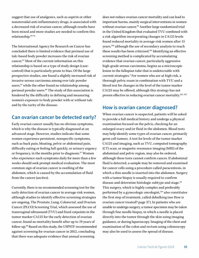
Can ovarian cancer be detected early?Early ovarian cancer usually has no obvious symptoms, which is why the disease is typically diagnosed at an advanced stage. However, studies indicate that some women experience persistent, nonspecific symptoms, such as back pain, bloating, pelvic or abdominal pain, difficulty eating or feeling full quickly, or urinary urgency or frequency, in the months prior to diagnosis.97 Women who experience such symptoms daily for more than a few weeks should seek prompt medical evaluation. The most common sign of ovarian cancer is swelling of the abdomen, which is caused by the accumulation of fluid from the cancer (ascites).
Currently, there is no recommended screening test for the early detection of ovarian cancer in average-risk women, although studies to identify effective screening strategies are ongoing. The Prostate, Lung, Colorectal, and Ovarian Cancer (PLCO) Screening Trial, which assessed the use of transvaginal ultrasound (TVU) and fixed cutpoints in the tumor marker CA125 for the early detection of ovarian cancer, found no mortality benefit after up to 19 years of follow-up.98 Based on this study, the USPSTF recommended against screening for ovarian cancer in 2012, concluding that there was adequate evidence that annual screening
does not reduce ovarian cancer mortality and can lead to important harms, mainly surgical interventions in women without ovarian cancer.99 Another large randomized trial in the United Kingdom that evaluated TVU combined with a risk algorithm incorporating changes in CA125 levels found reduced mortality in average-risk women after 15 years,100 although the use of secondary analysis to reach these results has been criticized.101 Identifying an effective screening method is complicated by accumulating evidence that ovarian cancer, particularly aggressive high-grade serous carcinoma, begins as a microscopic lesion in the fallopian tube that is undetectable with current strategies.2 For women who are at high risk, a thorough pelvic exam in combination with TVU and a blood test for changes in the level of the tumor marker CA125 may be offered, although this strategy has not proven effective in reducing ovarian cancer mortality.102, 103
How is ovarian cancer diagnosed? When ovarian cancer is suspected, patients will be asked to provide a full medical history and undergo a physical examination focused on the pelvis, checking for an enlarged ovary and/or fluid in the abdomen. Blood tests may help identify some types of ovarian cancer, primarily germ cell tumors. A test for levels of the tumor marker CA125 and imaging, such as TVU, computed tomography (CT) scan, or magnetic resonance imaging (MRI) of the abdominal and pelvic region, may also be ordered, although these tests cannot confirm cancer. If abdominal fluid is detected, a sample may be removed and examined for cancer cells using a procedure called paracentesis, in which a thin needle is inserted into the abdomen. Surgery with a tumor biopsy is usually required to confirm disease and determine histologic subtype and stage.28 This surgery, which is highly complex and preferably performed by a gynecologic oncologist,104 also constitutes the first step of treatment, called debulking (see How is ovarian cancer treated? page 37). In patients who are unable to undergo surgery, a tumor specimen may be taken through fine needle biopsy, in which a needle is placed directly into the tumor through the skin using imaging guidance, or during laparoscopy. Imaging of the chest and examination of the colon and rectum using colonoscopy may also be used to assess the spread of disease.
36 Cancer Facts & Figures 2018
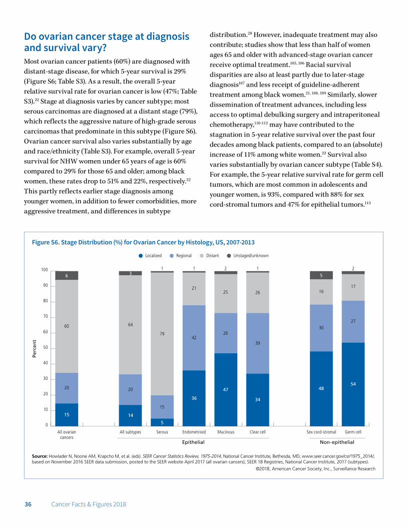
Do ovarian cancer stage at diagnosis and survival vary?Most ovarian cancer patients (60%) are diagnosed with distant-stage disease, for which 5-year survival is 29% (Figure S6; Table S3). As a result, the overall 5-year relative survival rate for ovarian cancer is low (47%; Table S3).22 Stage at diagnosis varies by cancer subtype; most serous carcinomas are diagnosed at a distant stage (79%), which reflects the aggressive nature of high-grade serous carcinomas that predominate in this subtype (Figure S6). Ovarian cancer survival also varies substantially by age and race/ethnicity (Table S3). For example, overall 5-year survival for NHW women under 65 years of age is 60% compared to 29% for those 65 and older; among black women, these rates drop to 51% and 22%, respectively.22 This partly reflects earlier stage diagnosis among younger women, in addition to fewer comorbidities, more aggressive treatment, and differences in subtype
distribution.28 However, inadequate treatment may also contribute; studies show that less than half of women ages 65 and older with advanced-stage ovarian cancer receive optimal treatment.105, 106 Racial survival disparities are also at least partly due to later-stage diagnosis107 and less receipt of guideline-adherent treatment among black women.21, 108, 109 Similarly, slower dissemination of treatment advances, including less access to optimal debulking surgery and intraperitoneal chemotherapy,110-112 may have contributed to the stagnation in 5-year relative survival over the past four decades among black patients, compared to an (absolute) increase of 11% among white women.22 Survival also varies substantially by ovarian cancer subtype (Table S4). For example, the 5-year relative survival rate for germ cell tumors, which are most common in adolescents and younger women, is 93%, compared with 88% for sex cord-stromal tumors and 47% for epithelial tumors.113
Source: Howlader N, Noone AM, Krapcho M, et al. (eds). SEER Cancer Statistics Review, 1975-2014, National Cancer Institute, Bethesda, MD, www.seer.cancer.gov/csr/1975_2014/, based on November 2016 SEER data submission, posted to the SEER website April 2017 (all ovarian cancers); SEER 18 Registries, National Cancer Institute, 2017 (subtypes).
©2018, American Cancer Society, Inc., Surveillance Research
Perc
ent
Figure S6. Stage Distribution (%) for Ovarian Cancer by Histology, US, 2007-2013
Localized Regional Distant Unstaged/unknown
0
10
20
30
40
50
60
70
80
90
100
Germ cellSex cord-stromalClear cellMucinousEndometrioidSerousAll subtypesAll ovariancancers
15
20
60
6
14
20
64
3
5
15
79
1
36
42
21
1
47
26
25
2
34
39
26
1
48
30
16
5
54
27
17
2
Epithelial Non-epithelial

Cancer Facts & Figures 2018 37
How is ovarian cancer treated?Treatment depends on the stage of the cancer; tumor characteristics and subtype; and the patient’s age, health, and preferences, but typically includes surgery and often chemotherapy (platinum- and taxane-based). Surgery, called debulking, optimally removes as much of the tumor as possible because patient prognosis is strongly linked to the amount of cancer remaining. Debulking surgery usually involves removal of both ovaries and fallopian tubes (bilateral salpingo-oophorectomy), the uterus (hysterectomy), and the omentum (fatty tissue attached to some of the organs in the belly), along with biopsies of the peritoneum (lining of the abdominal cavity). In younger women with very early-stage disease who want to preserve fertility, only the involved ovary and fallopian tube may be removed. Among patients with early ovarian cancer, more accurate surgical staging (microscopic examination of tissue from different parts of the pelvis and abdomen) has been associated with better outcomes.114 For women with advanced disease, debulking often involves removing parts or all of the other abdominal organs, with the goal of removing all visible disease or all tumors greater than
1 centimeter. For some women with advanced disease, chemotherapy may be administered prior to surgery to reduce tumor burden.28, 115 Chemotherapy administered directly into the abdomen (intraperitoneal chemotherapy) improves survival for advanced-stage epithelial disease; however, in 2012, less than half of eligible women received this treatment, perhaps because of the high risk for side effects.104, 116
Recurrence is very common in ovarian cancer, and eventual resistance to standard platinum-based chemotherapy also frequently occurs.117 There are an increasing number of treatment options for recurrence, including targeted therapies and treatments specifically for tumors associated with BRCA1 and BRCA2 mutations (PARP inhibitors).117 Clinical observation is the primary recommendation to monitor for recurrence of epithelial tumors. Use of CA125 tests to monitor for recurrence remains common, although it has not been shown to improve overall survival and may reduce patient quality of life.118 Imaging with CT , MRI, or positron emission tomography (PET)/CT when recurrence is suspected is being evaluated.119, 120
Table S3. Five-year Cause-specific Survival Rates* (%) for Ovarian Cancer by Stage at Diagnosis and Race/Ethnicity, US, 2007-2013
All racesNon-Hispanic
whiteNon-Hispanic
blackAmerican Indian/
Alaska NativeAsian/Pacific
Islander Hispanic
All stages 47 46 39 41 57 54
Localized 92 92 88 ^ 92 95
Regional 73 73 62 57 79 74
Distant 29 29 22 29 35 35
*See Sources of Statistics, page 68, for more information on the calculation of cause-specific survival. ^Statistic not shown due to fewer than 25 cases.
Source: SEER 18 Registries, National Cancer Institute, 2017.
©2018, American Cancer Society, Inc., Surveillance Research
Table S4. Five-year Relative Survival Rates* (%) for Ovarian Cancer by Stage at Diagnosis and Histology, US, 2007-2013Epithelial Non-epithelial
All subtypes Serous Endometrioid Mucinous Clear cellSex cord- stromal Germ cell
All stages 47 44 82 69 67 88 93
Localized 93 90 98 93 90 >99 98
Regional 74 75 87 81 74 89 93
Distant 30 35 48 18 26 53 77
*See Sources of Statistics, page 68, for more information on the calculation of relative survival.
Source: SEER 18 Registries, National Cancer Institute, 2017.
©2018, American Cancer Society, Inc., Surveillance Research

38 Cancer Facts & Figures 2018
What supportive care is available for ovarian cancer patients?Ovarian cancer patients cope with physical symptoms of the cancer, such as abdominal pain, bloating, cramping, and indigestion, in addition to side effects from treatment, some of which may last for years. Treatment side effects include pain, fatigue, numbness, hair loss, nausea, vomiting, and loss of appetite. Patients may also experience psychosocial effects, such as fear of recurrence or disease progression, depression, and anxiety. Younger women may cope with early menopause and loss of fertility.
Palliative care to improve patient quality of life by managing pain, relieving suffering, and addressing psychosocial needs optimally begins at diagnosis and continues through the end of life. Pain management strategies can be tailored to support quality-of-life goals and may include opioid analgesics. Chemotherapy or targeted drugs can be used as non-curative treatment to shrink tumors and diminish symptoms. Bowel obstruction caused by tumors can be relieved in select patients with surgery that may include an ostomy, which is an opening in the body for the elimination of waste. For patients with fluid accumulation in the abdomen, repeated drainage using paracentesis or continuous drainage using a semipermanent implanted tube may be used. Both bowel obstruction and fluid accumulation in the abdomen can cause lack of appetite; short-term nutrition can be provided through means other than eating, such as a feeding tube or intravenous delivery. A nutritionist can sometimes help patients with weight loss and loss of appetite.
At the end of life, palliative care services can help guide discussions about care options, including decisions to continue chemotherapy or receive invasive procedures, intensive care hospitalizations, and emergency department visits. In-home end-of-life care, or hospice, should be offered to all patients when treatment is palliative. Hospice services support individual quality-of-life goals, assist with pain control and comfort needs, and provide additional psychosocial services to patients and their families.
What is the American Cancer Society doing about ovarian cancer?Caring, trained American Cancer Society staff connect people to information about ovarian cancer, our clinical trials matching service, health insurance assistance, American Cancer Society programs and services, and referrals to other services at our 24-hour helpline at 1-800-227-2345. In addition, our website, cancer.org, offers comprehensive accurate information and news about ovarian cancer, including in-depth information on treatments and side effects, and programs and services in your area. For more information about American Cancer Society programs and services, see page 58.
The American Cancer Society also funds and conducts research to better understand ovarian cancer risk factors, prevention, treatment, and survivorship. Our Guideline Development Group, which issues cancer screening recommendations based on a comprehensive evaluation of evidence, closely monitors research on screening for ovarian cancer.
ResearchThe American Cancer Society, through our Extramural Grants program, funds individual investigators in medical schools, universities, research institutes, and hospitals throughout the United States. Currently, this program is funding $12,353,400 in ovarian cancer research through 46 research grants. Ongoing research topics include:
• Therapeutic targeting of ovarian cancer stem cells
• Developing effective nanomedicine strategies to improve the infiltration and function of immune cells in ovarian tumors
• Testing cell-based cancer immunotherapeutics in ovarian cancer to reactivate the immune response against the disease
• Mechanisms of carboplatin resistance in ovarian cancer
• Activatable nanoparticles for radiotherapy of metastatic ovarian cancer

Cancer Facts & Figures 2018 39
• Determining the impact of oophorectomy in BRCA mutation carriers on cardiac risk, bone health, and quality of life
The American Cancer Society Intramural Research program also conducts a wide range of research on ovarian cancer. For example, researchers from the Surveillance and Health Services Research program monitor trends in ovarian cancer occurrence and disparities in care. The group recently published a study showing that genetic testing for ovarian cancer risk increased from 2005 to 2015 among women with private or public insurance, but not among uninsured women.122 Using data collected in our Cancer Prevention Study II (CPS-II), American Cancer Society epidemiologists have examined the relationship between ovarian cancer and various factors, including recreational physical activity,84, 88 leisure time spent sitting,84 diabetes mellitus,123 menopausal hormone use,124 and circadian disruption.125 In addition, the CPS-II Nutrition Cohort is part of two large international research consortia. One of these, the Collaborative Group on Epidemiological Studies of Ovarian Cancer, helped to establish the links between ovarian cancer and excess body weight33 and oral contraceptive use.62 The other – Ovarian Cancer Cohort Consortium (OC3) – aims to identify lifestyle, hormonal, and genetic factors that contribute to the development of ovarian cancer subtypes. To date, OC3 researchers have reported that ovarian cancer associations with reproductive factors and tubal ligation are limited to endometrioid and clear cell carcinoma.5
AdvocacyACS CAN works to ensure that ovarian cancer patients and survivors have access to quality, affordable, and comprehensive health care coverage in both public and private insurance markets. This coverage must include the patient protections that are critical to cancer patients and survivors, like prohibitions on preexisting condition exclusions and lifetime and annual limits, and standards like essential health benefits. ACS CAN and its partners in One Voice Against Cancer (OVAC), a collaboration of national nonprofits with the goal of ensuring sufficient
funding for cancer, are leading the effort to advocate for necessary government funding for cancer research and programs to reduce the toll of ovarian cancer. In particular, ACS CAN advocates for dedicated funding at the CDC in support of Johanna’s Law, or The Gynecologic Education and Awareness Act, which provides for the education of women and medical professionals about the signs, symptoms, and early detection of ovarian and other gynecologic cancers. Federal funding for cancer-related initiatives have been under threat during the appropriations process. As the leading federal public health agency, the CDC must continue to receive the necessary funding to support lifesaving cancer education, prevention, and control initiatives.
What other resources are available?• National Cancer Institute
cancer.gov/types/ovarian
• Department of Defense – Congressionally Directed Medical Research Programs: Ovarian Cancer cdmrp.army.mil/ocrp
• Ovarian Cancer Research Fund Alliance ocrfa.org
• National Ovarian Cancer Coalition ovarian.org
References1. SEER*Stat Database: NAACCR Incidence Data – CiNA Analytic File, 1995-2014, for Expanded Races, Custom File With County, ACS Facts and Figures projection Project (which includes data from CDC’s National Program of Cancer Registries (NPCR), CCCR’s Provincial and Territorial Registries, and the NCI’s Surveillance, Epidemiology and End Results (SEER) Registries), certified by the North American Association of Central Cancer Registries (NAACCR) as meeting high-quality incidence data standards for the specified time periods, submitted December 2016.2. Kurman RJ, Shih Ie M. The Dualistic Model of Ovarian Carcinogenesis: Revisited, Revised, and Expanded. Am J Pathol. 2016;186: 733-747.3. Pearce CL, Rossing MA, Lee AW, et al. Combined and interactive effects of environmental and GWAS-identified risk factors in ovarian cancer. Cancer Epidemiol Biomarkers Prev. 2013;22: 880-890.4. Prat J. New insights into ovarian cancer pathology. Ann Oncol. 2012;23 Suppl 10: x111-117.

40 Cancer Facts & Figures 2018
5. Wentzensen N, Poole EM, Trabert B, et al. Ovarian Cancer Risk Factors by Histologic Subtype: An Analysis From the Ovarian Cancer Cohort Consortium. J Clin Oncol. 2016;34: 2888-2898.6. Kurman RJ. Origin and molecular pathogenesis of ovarian high-grade serous carcinoma. Ann Oncol. 2013;24 Suppl 10: x16-21.7. Meyn A, Lim B. A paradigm shift in the origin of ovarian cancer: the ovary is no longer to blame. BJOG. 2017;124: 859.8. Morice P, Uzan C, Fauvet R, Gouy S, Duvillard P, Darai E. Borderline ovarian tumour: pathological diagnostic dilemma and risk factors for invasive or lethal recurrence. Lancet Oncol. 2012;13: e103-115.9. Hauptmann S, Friedrich K, Redline R, Avril S. Ovarian borderline tumors in the 2014 WHO classification: evolving concepts and diagnostic criteria. Virchows Archiv. 2017;470: 125-142.10. Schultz KA, Harris AK, Schneider DT, et al. Ovarian Sex Cord-Stromal Tumors. J Oncol Pract. 2016;12: 940-946.11. Chen T, Surcel HM, Lundin E, et al. Circulating sex steroids during pregnancy and maternal risk of non-epithelial ovarian cancer. Cancer Epidemiol Biomarkers Prev. 2011;20: 324-336.12. Sieh W, Sundquist K, Sundquist J, Winkleby MA, Crump C. Intrauterine factors and risk of nonepithelial ovarian cancers. Gynecol Oncol. 2014;133: 293-297.13. Walker AH, Ross RK, Haile RW, Henderson BE. Hormonal factors and risk of ovarian germ cell cancer in young women. Br J Cancer. 1988;57: 418-422.14. Heravi-Moussavi A, Anglesio MS, Cheng SW, et al. Recurrent somatic DICER1 mutations in nonepithelial ovarian cancers. N Engl J Med. 2012;366: 234-242.15. Shah SP, Kobel M, Senz J, et al. Mutation of FOXL2 in granulosa-cell tumors of the ovary. N Engl J Med. 2009;360: 2719-2729.16. Van Nieuwenhuysen E, Lambrechts S, Lambrechts D, Leunen K, Amant F, Vergote I. Genetic changes in nonepithelial ovarian cancer. Expert Rev Anticancer Ther. 2013;13: 871-882.17. National Cancer Institute. Ovarian Germ Cell Tumors Treatment (PDQ) – Patient Version. Available from URL: https://www.cancer.gov/types/ovarian/patient/ovarian-germ-cell-treatment-pdq [accessed October 26, 2017].18. Horta M, Cunha TM. Sex cord-stromal tumors of the ovary: a comprehensive review and update for radiologists. Diagn Interv Radiol. 2015;21: 277-286.19. Wu AH, Pearce CL, Tseng CC, Pike MC. African Americans and Hispanics remain at lower risk of ovarian cancer than non-Hispanic whites after considering nongenetic risk factors and oophorectomy rates. Cancer Epidemiol Biomarkers Prev. 2015;24: 1094-1100.20. Long B, Chang J, Ziogas A, Tewari KS, Anton-Culver H, Bristow RE. Impact of race, socioeconomic status, and the health care system on the treatment of advanced-stage ovarian cancer in California. Am J Obstet Gynecol. 2015;212: 468 e461-469.21. Bristow RE, Powell MA, Al-Hammadi N, et al. Disparities in ovarian cancer care quality and survival according to race and socioeconomic status. J Natl Cancer Inst. 2013;105: 823-832.22. Howlader N, Noone AM, Krapcho M, Miller D, Bishop K, Kosary CL, Yu M, Ruhl J, Tatalovich Z, Mariotto A, Lewis DR, Chen HS, Feuer EJ, Cronin KA (eds). SEER Cancer Statistics Review, 1975-2014, National Cancer Institute. Bethesda, MD, https://seer.cancer.gov/csr/1975_2014/, based on November 2016 SEER data submission, posted to the SEER web site, April 2017.
23. Surveillance, Epidemiology, and End Results (SEER) Program (www.seer.cancer.gov) SEER*Stat Database: Incidence – SEER 13 Regs Research Data with Delay-Adjustment, Malignant Only, Nov 2016 Sub (1992-2014) <Katrina/Rita Population Adjustment> – Linked To County Attributes – Total U.S., 1969-2015 Counties, National Cancer Institute, DCCPS, Surveillance Research Program, released April 2017, based on the November 2016 submission.24. Surveillance, Epidemiology, and End Results (SEER) Program (www.seer.cancer.gov) SEER*Stat Database: Incidence – SEER 9 Regs Research Data with Delay-Adjustment, Malignant Only, Nov 2016 Sub (1975-2014) <Katrina/Rita Population Adjustment> – Linked To County Attributes – Total U.S., 1969-2015 Counties, National Cancer Institute, DCCPS, Surveillance Research Program, released April 2017, based on the November 2016 submission.25. Yang HP, Anderson WF, Rosenberg PS, et al. Ovarian cancer incidence trends in relation to changing patterns of menopausal hormone therapy use in the United States. J Clin Oncol. 2013;31: 2146-2151.26. Sopik V, Iqbal J, Rosen B, Narod SA. Why have ovarian cancer mortality rates declined? Part I. Incidence. Gynecol Oncol. 2015;138: 741-749.27. Surveillance, Epidemiology, and End Results (SEER) Program (www.seer.cancer.gov) SEER*Stat Database: Mortality – All COD, Total U.S. (1990-2015) <Early release with Vintage 2015 Katrina/Rita Population Adjustment> – Linked To County Attributes – Total U.S., 1969-2015 Counties, National Cancer Institute, DCCPS, Surveillance Research Program, released June 2017. Underlying mortality data provided by NCHS (www.cdc.gov/nchs).28. National Academies of Sciences, Engineering, and Medicine. Ovarian cancers: evolving paradigms in research and care. Washington, D.C.: The National Academies Press, 2016.29. Bray F, Loos AH, Tognazzo S, La Vecchia C. Ovarian cancer in Europe: Cross-sectional trends in incidence and mortality in 28 countries, 1953-2000. Int J Cancer. 2005;113: 977-990.30. Barnholtz-Sloan JS, Schwartz AG, Qureshi F, Jacques S, Malone J, Munkarah AR. Ovarian cancer: changes in patterns at diagnosis and relative survival over the last three decades. Am J Obstet Gynecol. 2003;189: 1120-1127.31. Jones MR, Kamara D, Karlan BY, Pharoah PDP, Gayther SA. Genetic epidemiology of ovarian cancer and prospects for polygenic risk prediction. Gynecol Oncol. 2017.32. Hunn J, Rodriguez GC. Ovarian cancer: etiology, risk factors, and epidemiology. Clin Obstet Gynecol. 2012;55: 3-23.33. Collaborative Group on Epidemiological Studies of Ovarian Cancer. Ovarian cancer and body size: individual participant meta-analysis including 25,157 women with ovarian cancer from 47 epidemiological studies. PLoS MEd. 2012;9: e1001200.34. Collaborative Group on Epidemiological Studies of Ovarian Cancer, Beral V, Gaitskell K, et al. Menopausal hormone use and ovarian cancer risk: individual participant meta-analysis of 52 epidemiological studies. Lancet. 2015;385: 1835-1842.35. Lauby-Secretan B, Scoccianti C, Loomis D, et al. Body fatness and cancer – viewpoint of the IARC Working Group. N Engl J Med. 2016;375: 794-798.36. Kerber RA, Slattery ML. The impact of family history on ovarian cancer risk. The Utah Population Database. Arch Intern Med. 1995;155: 905-912.

Cancer Facts & Figures 2018 41
37. Tung KH, Goodman MT, Wu AH, et al. Aggregation of ovarian cancer with breast, ovarian, colorectal, and prostate cancer in first-degree relatives. Am J Epidemiol. 2004;159: 750-758.38. Alsop K, Fereday S, Meldrum C, et al. BRCA mutation frequency and patterns of treatment response in BRCA mutation-positive women with ovarian cancer: a report from the Australian Ovarian Cancer Study Group. J Clin Oncol. 2012;30: 2654-2663.39. U. S. Preventive Services Task Force. Genetic risk assessment and BRCA mutation testing for breast and ovarian cancer susceptibility: recommendation statement. Ann Intern Med. 2005;143: 355-361.40. Norquist BM, Harrell MI, Brady MF, et al. Inherited mutations in women with ovarian carcinoma. JAMA Oncol. 2016;2: 482-490.41. Gabai-Kapara E, Lahad A, Kaufman B, et al. Population-based screening for breast and ovarian cancer risk due to BRCA1 and BRCA2. Proc Natl Acad Sci U S A. 2014;111: 14205-14210.42. Kuchenbaecker KB, Hopper JL, Barnes DR, et al. Risks of breast, ovarian, and contralateral breast cancer for BRCA1 and BRCA2 mutation carriers. JAMA. 2017;317: 2402-2416.43. Finch AP, Lubinski J, Moller P, et al. Impact of oophorectomy on cancer incidence and mortality in women with a BRCA1 or BRCA2 mutation. J Clin Oncol. 2014;32: 1547-1553.44. Walker JL, Powell CB, Chen LM, et al. Society of Gynecologic Oncology recommendations for the prevention of ovarian cancer. Cancer. 2015;121: 2108-2120.45. Ramus SJ, Song H, Dicks E, et al. Germline mutations in the BRIP1, BARD1, PALB2, and NBN genes in women with ovarian cancer. J Natl Cancer Inst. 2015;107.46. Bonadona V, Bonaiti B, Olschwang S, et al. Cancer risks associated with germline mutations in MLH1, MSH2, and MSH6 genes in Lynch syndrome. JAMA. 2011;305: 2304-2310.47. Ketabi Z, Bartuma K, Bernstein I, et al. Ovarian cancer linked to Lynch syndrome typically presents as early-onset, non-serous epithelial tumors. Gynecol Oncol. 2011;121: 462-465.48. American Cancer Society. Special Section: Multiple Primary Cancers. Cancer Facts & Figures 2009. Atlanta, GA: American Cancer Society, 2009.49. Walsh T, Casadei S, Lee MK, et al. Mutations in 12 genes for inherited ovarian, fallopian tube, and peritoneal carcinoma identified by massively parallel sequencing. Proc Natl Acad Sci U S A. 2011;108:18032-18037.50. Rasmussen CB, Jensen A, Albieri V, Andersen KK, Kjaer SK. Is pelvic inflammatory disease a risk factor for ovarian cancer? Cancer Epidemiol Biomarkers Prev. 2017;26: 104-109.51. Zhou Z, Zeng F, Yuan J, et al. Pelvic inflammatory disease and the risk of ovarian cancer: a meta-analysis. Cancer Causes Control. 2017;28: 415-428.52. Barry JA, Azizia MM, Hardiman PJ. Risk of endometrial, ovarian and breast cancer in women with polycystic ovary syndrome: a systematic review and meta-analysis. Hum Reprod Update. 2014;20: 748-758.53. Harris HR, Titus LJ, Cramer DW, Terry KL. Long and irregular menstrual cycles, polycystic ovary syndrome, and ovarian cancer risk in a population-based case-control study. Int J Cancer. 2017;140: 285-291.54. Schildkraut JM, Schwingl PJ, Bastos E, Evanoff A, Hughes C. Epithelial ovarian cancer risk among women with polycystic ovary syndrome. Obstet Gynecol. 1996;88: 554-559.
55. Guo SW, Zilberberg MD, Hummelshoj L. Endometriosis and ovarian cancer. Lancet Oncol. 2012;13: e189-190; author reply e190.56. Nezhat FR, Pejovic T, Reis FM, Guo SW. The link between endometriosis and ovarian cancer: clinical implications. Int J Gynecol Cancer. 2014;24: 623-628.57. Pearce CL, Templeman C, Rossing MA, et al. Association between endometriosis and risk of histological subtypes of ovarian cancer: a pooled analysis of case-control studies. Lancet Oncol. 2012;13: 385-394.58. Ness RB. Endometriosis and ovarian cancer: thoughts on shared pathophysiology. Am J Obstet Gynecol. 2003;189: 280-294.59. Whittemore AS, Harris R, Itnyre J. Characteristics relating to ovarian cancer risk: collaborative analysis of 12 US case-control studies. II. Invasive epithelial ovarian cancers in white women. Collaborative Ovarian Cancer Group. Am J Epidemiol. 1992;136: 1184-1203.60. Bevier M, Sundquist J, Hemminki K. Does the time interval between first and last birth influence the risk of endometrial and ovarian cancer? Eur J Cancer. 2011;47: 586-591.61. Wu AH, Pearce CL, Lee AW, et al. Timing of births and oral contraceptive use influences ovarian cancer risk. Int J Cancer. 2017;141: 2392-2399.62. Collaborative Group on Epidemiological Studies of Ovarian Cancer, Beral V, Doll R, Hermon C, Peto R, Reeves G. Ovarian cancer and oral contraceptives: collaborative reanalysis of data from 45 epidemiological studies including 23,257 women with ovarian cancer and 87,303 controls. Lancet. 2008;371: 303-314.63. Hankinson SE, Colditz GA, Hunter DJ, Spencer TL, Rosner B, Stampfer MJ. A quantitative assessment of oral contraceptive use and risk of ovarian cancer. Obstet Gynecol. 1992;80: 708-714.64. Tworoger SS, Fairfield KM, Colditz GA, Rosner BA, Hankinson SE. Association of oral contraceptive use, other contraceptive methods, and infertility with ovarian cancer risk. Am J Epidemiol. 2007;166: 894-901.65. Shafrir AL, Schock H, Poole EM, et al. A prospective cohort study of oral contraceptive use and ovarian cancer among women in the United States born from 1947 to 1964. Cancer Causes Control. 2017;28: 371-383.66. Cibula D, Widschwendter M, Majek O, Dusek L. Tubal ligation and the risk of ovarian cancer: review and meta-analysis. Hum Reprod Update. 2011;17: 55-67.67. Sieh W, Salvador S, McGuire V, et al. Tubal ligation and risk of ovarian cancer subtypes: a pooled analysis of case-control studies. Int J Epidemiol. 2013;42: 579-589.68. Falconer H, Yin L, Gronberg H, Altman D. Ovarian cancer risk after salpingectomy: a nationwide population-based study. J Natl Cancer Inst. 2015;107.69. Jordan SJ, Nagle CM, Coory MD, et al. Has the association between hysterectomy and ovarian cancer changed over time? A systematic review and meta-analysis. Eur J Cancer. 2013;49: 3638-3647.70. Gaitskell K, Green J, Pirie K, et al. Histological subtypes of ovarian cancer associated with parity and breastfeeding in the prospective Million Women Study. Int J Cancer. 2017.71. Danforth KN, Tworoger SS, Hecht JL, Rosner BA, Colditz GA, Hankinson SE. Breastfeeding and risk of ovarian cancer in two prospective cohorts. Cancer Causes Control. 2007;18: 517-523.72. Li DP, Du C, Zhang ZM, et al. Breastfeeding and ovarian cancer risk: a systematic review and meta-analysis of 40 epidemiological studies. Asian Pac J Cancer Prev. 2014;15: 4829-4837.
42 Cancer Facts & Figures 2018
73. Luan NN, Wu QJ, Gong TT, Vogtmann E, Wang YL, Lin B. Breastfeeding and ovarian cancer risk: a meta-analysis of epidemiologic studies. Am J Clin Nutr. 2013;98: 1020-1031.74. Diergaarde B, Kurta ML. Use of fertility drugs and risk of ovarian cancer. Curr Opin Obstet Gynecol. 2014;26: 125-129.75. Trabert B, Lamb EJ, Scoccia B, et al. Ovulation-inducing drugs and ovarian cancer risk: results from an extended follow-up of a large United States infertility cohort. Fertil Steril. 2013;100: 1660-1666.76. Rizzuto I, Behrens RF, Smith LA. Risk of ovarian cancer in women treated with ovarian stimulating drugs for infertility. Cochrane Database Syst Rev. 2013: CD008215.77. Dixon SC, Nagle CM, Thrift AP, et al. Adult body mass index and risk of ovarian cancer by subtype: a Mendelian randomization study. Int J Epidemiol. 2016;45: 884-895.78. Cairns BJ, Green J. Good news for “Alice”: height and sex differences in cancer risk. J Natl Cancer Inst. 2013;105: 841-843.79. Green J, Cairns BJ, Casabonne D, et al. Height and cancer incidence in the Million Women Study: prospective cohort, and meta-analysis of prospective studies of height and total cancer risk. Lancet Oncol. 2011;12: 785-794.80. Collaborative Group on Epidemiological Studies of Ovarian Cancer, Beral V, Gaitskell K, et al. Ovarian cancer and smoking: individual participant meta-analysis including 28,114 women with ovarian cancer from 51 epidemiological studies. Lancet Oncol. 2012;13: 946-956.81. World Cancer Research Fund / American Institute for Cancer Research. Continuous Update Project Report. Food, Nutrition, Physical Activity, and the Prevention of Ovarian Cancer 2014. Available at http://www.dietandcancerreport.org/cup/cup_resources.php.82. Crane TE, Khulpateea BR, Alberts DS, Basen-Engquist K, Thomson CA. Dietary intake and ovarian cancer risk: a systematic review. Cancer Epidemiol Biomarkers Prev. 2014;23: 255-273.83. Cannioto R, LaMonte MJ, Risch HA, et al. Chronic recreational physical inactivity and epithelial ovarian cancer risk: evidence from the Ovarian Cancer Association Consortium. Cancer Epidemiol Biomarkers Prev. 2016;25: 1114-1124.84. Patel AV, Hildebrand JS, Campbell PT, et al. Leisure-time spent sitting and site-specific cancer incidence in a large U.S. cohort. Cancer Epidemiol Biomarkers Prev. 2015;24: 1350-1359.85. Shen D, Mao W, Liu T, et al. Sedentary behavior and incident cancer: a meta-analysis of prospective studies. PloS one. 2014;9: e105709.86. Xiao Q, Yang HP, Wentzensen N, Hollenbeck A, Matthews CE. Physical activity in different periods of life, sedentary behavior, and the risk of ovarian cancer in the NIH-AARP diet and health study. Cancer Epidemiol Biomarkers Prev. 2013;22: 2000-2008.87. Zhang M, Xie X, Lee AH, Binns CW. Sedentary behaviours and epithelial ovarian cancer risk. Cancer Causes Control. 2004;15: 83-89.88. Hildebrand JS, Gapstur SM, Gaudet MM, Campbell PT, Patel AV. Moderate-to-vigorous physical activity and leisure-time sitting in relation to ovarian cancer risk in a large prospective US cohort. Cancer Causes Control. 2015;26: 1691-1697.89. Baandrup L, Faber MT, Christensen J, et al. Nonsteroidal anti-inflammatory drugs and risk of ovarian cancer: systematic review and meta-analysis of observational studies. Acta Obstet Gynecol Scand. 2013;92: 245-255.90. Brasky TM, Liu J, White E, et al. Non-steroidal anti-inflammatory drugs and cancer risk in women: results from the Women’s Health Initiative. Int J Cancer. 2014;135: 1869-1883.
91. Pinheiro SP, Tworoger SS, Cramer DW, Rosner BA, Hankinson SE. Use of nonsteroidal antiinflammatory agents and incidence of ovarian cancer in 2 large prospective cohorts. Am J Epidemiol. 2009;169: 1378-1387.92. Prizment AE, Folsom AR, Anderson KE. Nonsteroidal anti-inflammatory drugs and risk for ovarian and endometrial cancers in the Iowa Women’s Health Study. Cancer Epidemiol Biomarkers Prev. 2010;19: 435-442.93. Trabert B, Ness RB, Lo-Ciganic WH, et al. Aspirin, nonaspirin nonsteroidal anti-inflammatory drug, and acetaminophen use and risk of invasive epithelial ovarian cancer: a pooled analysis in the Ovarian Cancer Association Consortium. J Natl Cancer Inst. 2014;106: djt431.94. International Agency for Research on Cancer. IARC Monographs on the Evaluation of Carcinogenic Risks to Humans. Vol 93 Carbon Black, Titanium Dioxide, and Talc.2010.95. Gertig DM, Hunter DJ, Cramer DW, et al. Prospective study of talc use and ovarian cancer. J Natl Cancer Inst. 2000;92: 249-252.96. Houghton SC, Reeves KW, Hankinson SE, et al. Perineal powder use and risk of ovarian cancer. J Natl Cancer Inst. 2014;106.97. Goff BA, Mandel LS, Melancon CH, Muntz HG. Frequency of symptoms of ovarian cancer in women presenting to primary care clinics. JAMA. 2004;291: 2705-2712.98. Pinsky PF, Yu K, Kramer BS, et al. Extended mortality results for ovarian cancer screening in the PLCO trial with median 15years follow-up. Gynecol Oncol. 2016;143: 270-275.99. Moyer VA, U. S. Preventive Services Task Force. Screening for ovarian cancer: U.S. Preventive Services Task Force reaffirmation recommendation statement. Ann Intern Med. 2012;157: 900-904.100. Jacobs IJ, Menon U, Ryan A, et al. Ovarian cancer screening and mortality in the UK Collaborative Trial of Ovarian Cancer Screening (UKCTOCS): a randomised controlled trial. Lancet. 2016;387: 945-956.101. Narod SA, Sopik V, Giannakeas V. Should we screen for ovarian cancer? A commentary on the UK Collaborative Trial of Ovarian Cancer Screening (UKCTOCS) randomized trial. Gynecol Oncol. 2016;141: 191-194.102. Berchuck A, Havrilesky LJ, Kauff ND. Is there a role for ovarian cancer screening in high-risk women? J Clin Oncol. 2017;35: 1384-1386.103. Rosenthal AN, Fraser LSM, Philpott S, et al. Evidence of stage shift in women diagnosed with ovarian cancer during phase II of the United Kingdom Familial Ovarian Cancer Screening Study. J Clin Oncol. 2017;35: 1411-1420.104. Hennessy BT, Coleman RL, Markman M. Ovarian cancer. Lancet. 2009;374: 1371-1382.105. Lin JJ, Egorova N, Franco R, Prasad-Hayes M, Bickell NA. Ovarian cancer treatment and survival trends among women older than 65 years of age in the United States, 1995-2008. Obstet Gynecol. 2016;127: 81-89.106. Thrall MM, Gray HJ, Symons RG, Weiss NS, Flum DR, Goff BA. Trends in treatment of advanced epithelial ovarian cancer in the Medicare population. Gynecol Oncol. 2011;122: 100-106.107. Sakhuja S, Yun H, Pisu M, Akinyemiju T. Availability of healthcare resources and epithelial ovarian cancer stage of diagnosis and mortality among Blacks and Whites. J Ovarian Res. 2017;10: 57.108. Bandera EV, Lee VS, Rodriguez-Rodriguez L, Powell CB, Kushi LH. Racial/ethnic disparities in ovarian cancer treatment and survival. Clin Cancer Res. 2016;22: 5909-5914.

Cancer Facts & Figures 2018 43
109. Bristow RE, Chang J, Ziogas A, Campos B, Chavez LR, Anton-Culver H. Sociodemographic disparities in advanced ovarian cancer survival and adherence to treatment guidelines. Obstet Gynecol. 2015;125: 833-842.110. Aranda MA, McGory M, Sekeris E, Maggard M, Ko C, Zingmond DS. Do racial/ethnic disparities exist in the utilization of high-volume surgeons for women with ovarian cancer? Gynecol Oncol. 2008;111: 166-172.111. Bristow RE, Chang J, Ziogas A, Randall LM, Anton-Culver H. High-volume ovarian cancer care: survival impact and disparities in access for advanced-stage disease. Gynecol Oncol. 2014;132: 403-410.112. Fairfield KM, Murray K, LaChance JA, et al. Intraperitoneal chemotherapy among women in the Medicare population with epithelial ovarian cancer. Gynecol Oncol. 2014;134: 473-477.113. Surveillance, Epidemiology, and End Results (SEER) Program (www.seer.cancer.gov) SEER*Stat Database: Incidence – SEER 18 Regs Research Data + Hurricane Katrina Impacted Louisiana Cases, Nov 2016 Sub (2000-2014) <Katrina/Rita Population Adjustment> – Linked To County Attributes – Total U.S., 1969-2015 Counties, National Cancer Institute, DCCPS, Surveillance Research Program, released April 2017, based on the November 2016 submission.114. Trimbos JB. Surgical treatment of early-stage ovarian cancer. Best Pract Res Clin Obstet Gynaecol. 2017;41: 60-70.115. Wright AA, Bohlke K, Armstrong DK, et al. Neoadjuvant chemotherapy for newly diagnosed, advanced ovarian cancer: Society of Gynecologic Oncology and American Society of Clinical Oncology Clinical Practice Guideline. J Clin Oncol. 2016;34: 3460-3473.116. Wright AA, Cronin A, Milne DE, et al. Use and effectiveness of intraperitoneal chemotherapy for treatment of ovarian cancer. J Clin Oncol. 2015;33: 2841-2847.
117. Luvero D, Milani A, Ledermann JA. Treatment options in recurrent ovarian cancer: latest evidence and clinical potential. Ther Adv Med Oncol. 2014;6: 229-239.118. Esselen KM, Cronin AM, Bixel K, et al. Use of CA-125 Tests and Computed Tomographic Scans for Surveillance in Ovarian Cancer. JAMA Oncol. 2016;2: 1427-1433.119. Lee SI, Catalano OA, Dehdashti F. Evaluation of gynecologic cancer with MR imaging, 18F-FDG PET/CT, and PET/MR imaging. J Nucl Med. 2015;56: 436-443.120. Paik ES, Kim TJ, Lee YY, et al. Comparison of survival outcomes after recurrence detected by cancer antigen 125 elevation versus imaging study in epithelial ovarian cancer. J Gynecol Oncol. 2016;27: e46.121. Kurian AW, Hughes E, Handorf EA, et al. Breast and ovarian cancer penetrance estimates derived From germline multiple-gene sequencing results in women. JCO Precis Oncol. 2017:1-12.122. Han X, Jemal A. Recent patterns in genetic testing for breast and ovarian cancer risk in the U.S. Am J Prev Med. 2017;53:504-507.123. Gapstur SM, Patel AV, Diver WR, et al. Type II diabetes mellitus and the incidence of epithelial ovarian cancer in the cancer prevention study-II nutrition cohort. Cancer Epidemiol Biomarkers Prev. 2012;21:2000-2005.124. Hildebrand JS, Gapstur SM, Feigelson HS, Teras LR, Thun MJ, Patel AV. Postmenopausal hormone use and incident ovarian cancer: Associations differ by regimen. Int J Cancer. 2010;127:2928-2935.125. Carter BD, Diver WR, Hildebrand JS, Patel AV, Gapstur SM. Circadian disruption and fatal ovarian cancer. Am J Prev Med. 2014;46:S34-41.