spatial and cross-seasonal patterns of coral diseases in
TRANSCRIPT

DISEASES OF AQUATIC ORGANISMSDis Aquat Org
Vol. 146: 145–156, 2021https://doi.org/10.3354/dao03624
Published online October 21
1. INTRODUCTION
Coral diseases are a developing crisis thatthreaten the viability, structure, and function ofcoral reef ecosystems. These diseases contribute tocoral mortality and engender substantial losses inliving coral cover and reef biodiversity (Hughes1994, Porter & Tougas 2001, Lafferty et al. 2004).Caribbean reefs have been subject to repeated dis-ease outbreaks over the past 3 decades, and thedominant reef-building coral populations have
decreased considerably (Aronson & Precht 2001,Miller et al. 2009, Weil & Cróquer 2009). Reefs inthe Indo-Pacific region are less disturbed than thosein the Caribbean region; however, a number ofstudies have reported that diseases are highlyprevalent in this region (Willis et al. 2004, Ray-mundo et al. 2005, Myers & Raymundo 2009, Sato etal. 2009, Weil et al. 2012) and have the potential toseverely affect certain diverse ecosystems.
The prevalence of coral diseases is a crucial indica-tor of coral reef health, and the disease distribution
© The authors 2021. Open Access under Creative Commons byAttribution Licence. Use, distribution and reproduction are un -restricted. Authors and original publication must be credited.
Publisher: Inter-Research · www.int-res.com
*Corresponding author: [email protected]
Spatial and cross-seasonal patterns of coral diseases in reefs of Taiwan:
high prevalence and regional variation
Ching-Yun Huang1, Jiang-Shiou Hwang2,3,4, Hideyuki Yamashiro5, Sen-Lin Tang6,*
1Institute of Fisheries Science, National Taiwan University, Taipei 10617, Taiwan2Institute of Marine Biology, National Taiwan Ocean University, Keelung 20224, Taiwan
3Center of Excellence for Ocean Engineering, National Taiwan Ocean University, Keelung 20224, Taiwan4Center of Excellence for the Oceans, National Taiwan Ocean University, Keelung 20224, Taiwan
5Sesoko Station, Tropical Biosphere Research Center, University of the Ryukyus, Sesoko 3422, Motobu, Okinawa 905-0227, Japan6Biodiversity Research Center, Academia Sinica, Taipei 11529, Taiwan
ABSTRACT: Although research on coral diseases is increasing worldwide, it remains limited inTaiwan. Taiwan is located at the Tropic of Cancer and contains both tropical and subtropical reefs.We conducted spatial and cross-seasonal surveys in Taiwan in 2018 and identified 7 types of dis-ease and nondisease lesions and 6 potential factors influencing coral health. The overall meanprevalence of disease and nondisease lesions varied considerably across the reef regions, and hostsusceptibility differed among the coral taxa. The overall mean prevalence of disease and non -disease lesions was highest in Kenting (mean ± SEM: 8.58 ± 1.81%) and lowest on the SouthernIslands (2.12 ± 0.73%). Although the prevalence of diseases did not differ significantly betweenthe seasons, cyanobacteria-related diseases — including black band disease (BBD), BBD-like syn-drome, and other cyanobacterial syndromes — were slightly more prevalent in autumn than inspring. Furthermore, 3 of the potential factors influencing coral health (i.e. turf algae, bioerodingsponges, and coral bleaching) were strong predictors of disease and nondisease lesion preva-lence. These results advance our understanding of coral disease ecology in Taiwan and highlightthe need for further research on the correlations between diseases, hosts, and environment.
KEY WORDS: Coral disease · Taiwan · Distribution pattern · Host susceptibility
OPENPEN ACCESSCCESS

Dis Aquat Org 146: 145–156, 2021
patterns have been linked to both global and localfactors. On the global scale, seasonal fluctuations(e.g. sea surface temperature and light intensity) andclimate change-associated thermal stress are corre-lated with tissue-loss diseases (Boyett et al. 2007,Bruno et al. 2007, Brodnicke et al. 2019). In addition,localized human effects (e.g. eutrophication and sed-imentation) appear to promote disease transmissionby compromising host resistance or enhancing thevirulence of infectious pathogens (Haapkylä et al.2011, Kaczmarsky & Richardson 2011).
Despite global research efforts, studies on thepre valence of coral diseases in the Indo-Pacificregion remain limited, especially in the context ofmarginal reefs. Taiwan is a continental island lo -cated in the middle of the Philippines−Japan islandarc, which is near the northern limit of coral reefdevelopment (Beger et al. 2014) and includes bothtropical and subtropical reefs (Chen & Shas hank2009). To the west and north of Taiwan, the differ-ences in sea surface temperatures between seasonscan exceed 10°C because of monsoons and ChinaCoastal Waters (Jan et al. 2002). To the east andsouth, the Kuroshio Current brings warm water andstabilizes sea surface temperatures. The high vari-ability in hydrology creates a wide range of habitats,making coral reefs diverse and productive through-out Taiwan (Dai & Horng 2009a,b, Ribas-Deulofeuet al. 2016).
However, the dense population of Taiwan and itshighly developed industries threaten these reefs.Taiwan’s reefs are among the mostthreatened ecosystems in SoutheastAsia, due in part to unsustainable fish-ing, coastal development, and water-shed pollution (Burke et al. 2011). Theextent to which these factors affectcoral diseases remains unclear. A studyintegrating information on coral com-munities and disease prevalence in allregions of Taiwan is required to assessthe effects of coral diseases.
Accordingly, the present study (1)quantified the prevalence of coral dis-eases affecting coral taxa across reefs inTaiwan and (2) investigated the po -tential factors contributing to these dis-eases. Scleractinian coral reefs havebeen identified in 7 major regions inTaiwan: the northeast coast, the eastcoast, and Kenting on the main island,in addition to the nearby Green Island,Orchid Island, Penghu Islands, and
Liuqiu Island (Chen & Shashank 2009, Dai & Horng2009a). We investigated all of these reef regionsexcept for Liuqiu Island. Moreover, cross-seasonalinvestigations were conducted in 3 of the reefregions to determine the spatial and seasonal vari-abilities of disease prevalence. The correlation be -tween disease prevalence and potential contributingfactors was also examined.
2. MATERIALS AND METHODS
2.1. Survey sites and periods
To examine the spatial variability in the preva-lence of coral diseases, surveys were conducted in6 reef regions along the coast of Taiwan: the north-east coast, the east coast, Kenting, Green Island,Orchid Island, and the Southern Islands (located atthe southernmost margin of the Penghu Islands). Aspatial survey was conducted in the spring andsummer (i.e. from April to August) of 2018 by ag -gregating information from 3 to 5 sites in each reefregion, resulting in a total of 22 sites (Fig. 1). Toinvestigate the cross-seasonal variability in the pre -valence of coral diseases, additional surveys wereconducted in the autumn (October to November) of2018 in 3 of the reef regions — the northeast coast,Kenting, and Green Island — by aggregating infor-mation from 3 sites in each reef region, resulting ina total of 9 sites.
146
26°N
118°E 123°
21°
Fig. 1. Survey sites (crosses; n = 22) in 6 reef regions across Taiwan. Map datafrom the National Land Surveying and Mapping Center, Taiwan (http://
maps. nlsc.gov.tw/EN/)
Huang et al.: Patterns of coral disease in Taiwan 147
2.2. Survey method
At each site, depending on the reef configurations,a 75−100 m long belt transect was set up parallel tothe depth contour at a depth of 5 m and divided into3 to 4 sampling units. All coral colonies within 1 m oneither side of the transect were investigated. Eachsampling unit covered an area of 20 × 2 m2, and theunits were 5 m apart. The survey was conductedusing standard protocols (Raymundo et al. 2008).Every coral colony within each sampling unit wascounted, identified to the genus level, and inspectedfor disease and nondisease lesions and factors thatcould affect coral health. Colonies on the belt marginwere counted only when 50% or more of the colonylay within the belt.
Coral coverage was estimated using the pointintercept method, and the substrate type was re cor -ded at 50 cm intervals along the transect. The preva-lence of disease and nondisease lesions was cal -culated by dividing the number of affected coralco lonies by the total number of colonies. Moreover,taxon-specific prevalence was calculated for a subsetof coral hosts in cases that involved a particularlyhigh prevalence of disease and nondisease lesionsamong specific coral taxa. The influence of eachpotential factor was also estimated by dividing thenumber of affected coral colonies by the total numberof colonies. The mean value and standard error of themean (SEM) were calculated for each condition. Thefrequency of occurrence (percentage of transects sur-veyed with at least 1 lesion) was calculated for eachlesion type to estimate whether it was widespread inTaiwan’s reefs.
2.3. Statistical analysis
Statistical analyses were conducted using the ‘ve -gan’ community ecology package (Oksanan et al.2019) in R version 3.5.1 (R Core Team 2018). Permu-tational multivariate analysis of variance (PERM-ANOVA; Anderson 2001) with 999 random permu-tations was conducted to determine the degree ofspatial and cross-seasonal variance in the preva-lence of assemblages of disease and nondiseaselesions. The factor ‘reef region’ was used for thespatial survey data, and the factor ‘reef region ×season’ was used for the cross-seasonal survey data.For the cross-seasonal survey, only the data ob -tained from the cross-seasonally surveyed siteswere extracted for analysis. The data on diseaseand nondisease lesion prevalence were transformed
using , and resemblance matrices ofBray- Curtis dissimilarity were calculated betweenevery pair of observations. A nonparametric multi-dimensional scaling (NMDS) analysis (Agarwal etal. 2007) was conducted to visualize the diseaseassemblage distribution patterns, based on themean prevalence of lesions related to disease pro-cesses, in multivariate space across reef regions. Acanonical correspondence analysis (CCA; Legendre& Legendre 2012) was performed to examine thecovariances between the prevalence of disease andnondisease lesions and the potential factors affect-ing coral health.
3. RESULTS
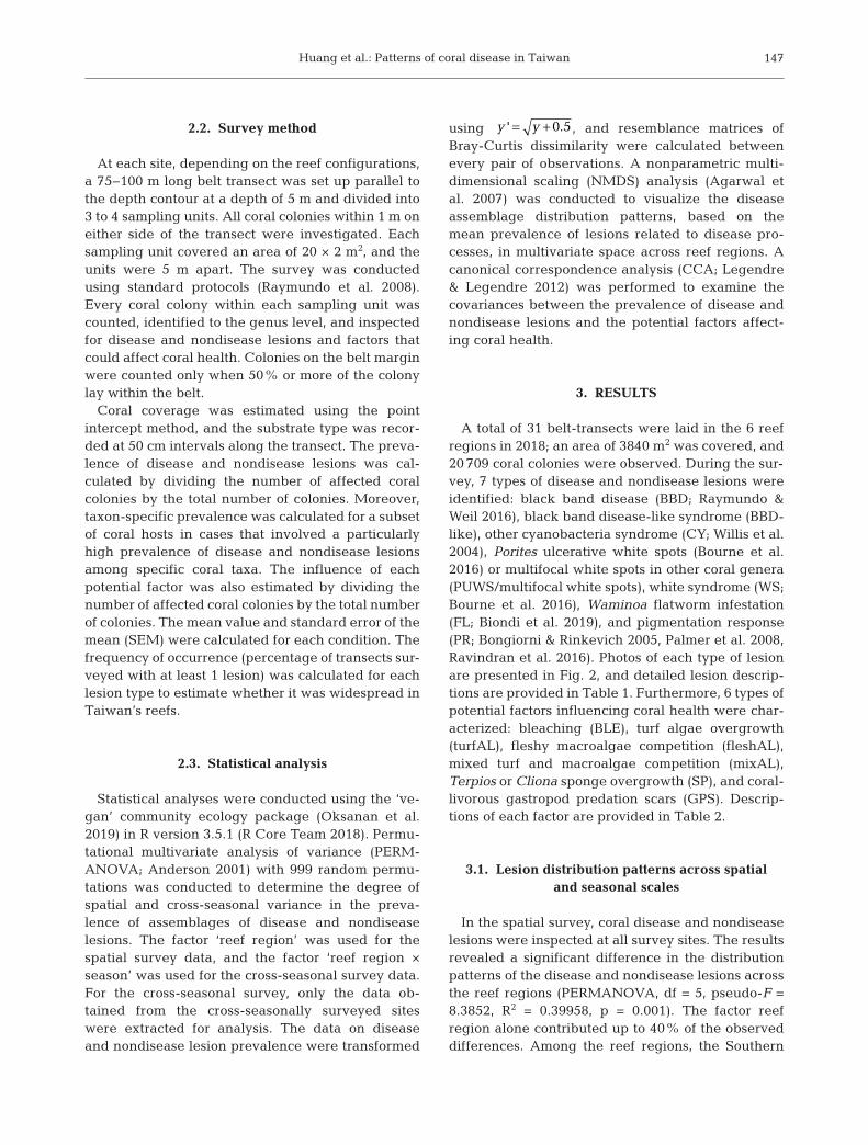
A total of 31 belt-transects were laid in the 6 reefregions in 2018; an area of 3840 m2 was covered, and20 709 coral colonies were observed. During the sur-vey, 7 types of disease and nondisease lesions wereidentified: black band disease (BBD; Raymundo &Weil 2016), black band disease-like syndrome (BBD-like), other cyanobacteria syndrome (CY; Willis et al.2004), Porites ulcerative white spots (Bourne et al.2016) or multifocal white spots in other coral genera(PUWS/multifocal white spots), white syndrome (WS;Bourne et al. 2016), Waminoa flatworm infestation(FL; Biondi et al. 2019), and pigmentation response(PR; Bongiorni & Rinkevich 2005, Palmer et al. 2008,Ravindran et al. 2016). Photos of each type of lesionare presented in Fig. 2, and detailed lesion descrip-tions are provided in Table 1. Furthermore, 6 types ofpotential factors influencing coral health were char-acterized: bleaching (BLE), turf algae overgrowth(turfAL), fleshy macroalgae competition (fleshAL),mixed turf and macroalgae competition (mixAL),Terpios or Cliona sponge overgrowth (SP), and coral-livorous gastropod predation scars (GPS). Descrip-tions of each factor are provided in Table 2.
3.1. Lesion distribution patterns across spatial and seasonal scales
In the spatial survey, coral disease and nondiseaselesions were inspected at all survey sites. The resultsrevealed a significant difference in the distributionpatterns of the disease and nondisease lesions acrossthe reef regions (PERMANOVA, df = 5, pseudo-F =8.3852, R2 = 0.39958, p = 0.001). The factor reefregion alone contributed up to 40% of the observeddifferences. Among the reef regions, the Southern
y y= +' 0.5
Dis Aquat Org 146: 145–156, 2021
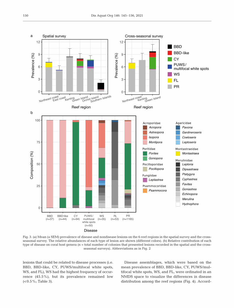
Islands presented the lowest overall mean preva-lence of disease and nondisease lesions; the eastcoast had an intermediate prevalence, and the otherreef regions had relatively high prevalence (Table 3,Fig. 3a).
Of the different types of disease and nondiseaselesions, the frequency of occurrence was different(Table 3). PR had the highest frequency of occur-rence (100%) and the highest prevalence amongall reef regions, except for the Southern Islands(Fig. 3a). Specifically, the Southern Islands had thelowest overall mean prevalence and the lowest
prevalence of PR but had the highest prevalence ofBBD. By contrast, Kenting presented the highestprevalence of PR and a low prevalence of BBD.Green Island also had a low prevalence of BBD, butnotably, it contained other diseases related to cyano-bacteria (BBD-like and CY). For Orchid Island, whichis near Green Island, of the cyanobacteria-relateddiseases observed, only CY was recorded. However,compared with other reef regions, Orchid Island hada higher prevalence of PUWS/multifocal white spots.FL was mostly recorded on the northeast coast, andonly 1 case was identified on the east coast. Of the
148
Fig. 2. Coral disease and nondisease lesions (arrowheads) observed on Taiwan’sreefs in 2018: (a) black band disease (BBD), (b) black band disease-like syndrome(BBD-like), (c) other cyanobacteria syndrome (CY), (d) Porites ulcerative whitespots (PUWS) and a close-up view of the lesion (inset), (e) white syndrome (WS), (f)Waminoa flatworm infestation (FL) and a close-up view of Waminoa flatworms(inset), (g) pigmentation response (PR) in Porites sp., (h) PR in Montipora sp., (i) PRin Dipsastraea sp., and (j) PR in Goniastrea sp. Detailed lesion descriptions are
given in Table 1

Huang et al.: Patterns of coral disease in Taiwan 149
Lesion Abbreviation Description
Black band disease BBD Dark band composed of filamentous cyanobacteria, adjacent toclinically healthy coral tissue and a zone of recently exposed whiteskeleton
Black band disease- BBD-like Lesions are similar to BBD, with cyanobacteria infections actively like syndrome spreading, No exposed white skeleton
Other cyanobacteria CY Other cyanobacterial infections that cannot be classified into BBD or syndrome BBD-like
Porites ulcerative white spots/ PUWS/ Multifocal tissue necrosis that exposes small circular areas of bare multifocal white spots multifocal white skeletonin other coral genera white spots
White syndrome WS White lesion that presents as a linear (or annular) band or an irregularpatch on the coral colony and is comprised of recently exposed coralskeleton adjacent to apparently healthy tissue
Flatworm infestation FL Surface of coral covered by high density of ovoid, brown flatworms,notably in the genus Waminoa
Pigmentation response PR Non-normally pigmented tissues in corals. The color can changedepending on the coral species, and is typically pink, purple, or blue
Table 1. Detailed descriptions of disease and non-disease lesions
Factor Abbreviation Description
Bleaching BLE Coral tissue discolorationTurf algae overgrow turfAL Colonization and overgrowth of living coral tissue by turf algae
(height <2 cm)Fleshy macroalgae competition fleshAL Macroalgae growing within 2 cm range of a coral colony and
able to generate an abrasion effectMixed turf and macroalgae competition mixAL Coral colony is affected by both turf algae overgrowth and
fleshy macroalgae competitionSponge overgrowth SP Terpios or Cliona sponge overgrowth on living coral tissueGastropod predation scars GPS Feeding scars generated by the corallivorous Drupella spp.
Table 2. Descriptions of potential factors influencing coral health
Northeast East coast Kenting Green Orchid Southern Frequency of coast Island Island Islands occurrence
Coral communityCoral coverage 24.84 ± 4.14 31.45 ± 4.33 31.67 ± 2.04 37.50 ± 6.33 54.72 ± 2.99 52.50 ± 7.24 Shannon diversity 2.16 ± 0.08 2.40 ± 0.09 2.44 ± 0.06 1.76 ± 0.16 1.93 ± 0.11 1.30 ± 0.09
Prevalence of disease/non-disease lesionsOverall mean prevalence 7.28 ± 0.80 5.25 ± 0.99 8.58 ± 1.81 8.03 ± 1.28 7.76 ± 0.94 2.12 ± 0.73 BBD 0 0 0.09 ± 0.06 0.14 ± 0.07 0 1.55 ± 0.51 27.3BBD-like 0 0 0 0.41 ± 0.21 0 0.04 ± 0.04 13.6CY 0 0 0 1.21 ± 0.61 0.15 ± 0.10 0 13.6PUWS/multifocal 0.08 ± 0.04 0.13 ± 0.13 0 0 1.48 ± 0.57 0 22.7
white spots WS 0.07 ± 0.05 0.04 ± 0.04 0.04 ± 0.04 0.44 ± 0.19 0.47 ± 0.16 0.08 ± 0.06 45.5FL 1.13 ± 0.30 0.05 ± 0.05 0 0 0 0 22.7PR 6.00 ± 0.86 5.03 ± 0.99 8.41 ± 1.84 5.78 ± 1.33 5.59 ± 1.02 0.44 ± 0.25 100.0
Table 3. Means ± SEM of coral coverage (%), Shannon diversity (calculated based on genus level), overall mean prevalence(%), and the prevalence (%) of each type of disease and nondisease lesion in different reef regions and its frequency of occur-
rence (% of transects surveyed with at least 1 lesion). Abbreviations as in Table 1
Dis Aquat Org 146: 145–156, 2021
lesions that could be related to disease processes (i.e.BBD, BBD-like, CY, PUWS/multifocal white spots,WS, and FL), WS had the highest frequency of occur-rence (45.5%), but its prevalence remained low(<0.5%; Table 3).
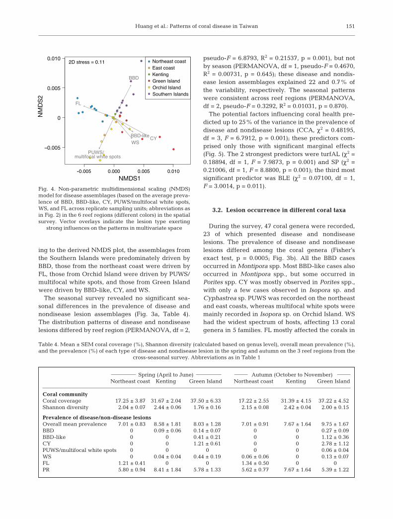
Disease assemblages, which were based on themean prevalence of BBD, BBD-like, CY, PUWS/mul-tifocal white spots, WS, and FL, were ordinated in anNMDS space to visualize the differences in diseasedistribution among the reef regions (Fig. 4). Accord-
150
a
b
Spatial survey
Reef region
Disease
Reef region
Cross-seasonal surveyPr
evale
nce
(%)
Com
posit
ion
(%)
Prev
alenc
e (%
)
Fig. 3. (a) Mean (± SEM) prevalence of disease and nondisease lesions on the 6 reef regions in the spatial survey and the cross-seasonal survey. The relative abundances of each type of lesion are shown (different colors). (b) Relative contribution of eachtype of disease on coral host genera (n = total number of colonies that presented lesions recorded in the spatial and the cross-
seasonal surveys). Abbreviations as in Fig. 2
Huang et al.: Patterns of coral disease in Taiwan
ing to the derived NMDS plot, the assemblages fromthe Southern Islands were predominately driven byBBD, those from the northeast coast were driven byFL, those from Orchid Island were driven by PUWS/multifocal white spots, and those from Green Islandwere driven by BBD-like, CY, and WS.
The seasonal survey revealed no significant sea-sonal differences in the prevalence of disease andnondisease lesion assemblages (Fig. 3a, Table 4).The distribution patterns of disease and nondiseaselesions differed by reef region (PERMANOVA, df = 2,
pseudo-F = 6.8793, R2 = 0.21537, p = 0.001), but notby season (PERMANOVA, df = 1, pseudo-F = 0.4670,R2 = 0.00731, p = 0.645); these disease and nondis-ease lesion assemblages explained 22 and 0.7% ofthe variability, respectively. The seasonal patternswere consistent across reef regions (PERMANOVA,df = 2, pseudo-F = 0.3292, R2 = 0.01031, p = 0.870).
The potential factors influencing coral health pre-dicted up to 25% of the variance in the prevalence ofdisease and nondisease lesions (CCA, χ2 = 0.48195,df = 3, F = 6.7912, p = 0.001); these predictors com-prised only those with significant marginal effects(Fig. 5). The 2 strongest predictors were turfAL (χ2 =0.18894, df = 1, F = 7.9873, p = 0.001) and SP (χ2 =0.21006, df = 1, F = 8.8800, p = 0.001); the third mostsignificant predictor was BLE (χ2 = 0.07100, df = 1,F = 3.0014, p = 0.011).
3.2. Lesion occurrence in different coral taxa
During the survey, 47 coral genera were recorded,23 of which presented disease and nondiseaselesions. The prevalence of disease and nondiseaselesions differed among the coral genera (Fisher’sexact test, p = 0.0005; Fig. 3b). All the BBD casesoccurred in Montipora spp. Most BBD-like cases alsooccurred in Montipora spp., but some occurred inPorites spp. CY was mostly observed in Porites spp.,with only a few cases observed in Isopora sp. andCyphastrea sp. PUWS was recorded on the northeastand east coasts, whereas multifocal white spots weremainly recorded in Isopora sp. on Orchid Island. WShad the widest spectrum of hosts, affecting 13 coralgenera in 5 families. FL mostly affected the corals in
151
Spring (April to June) Autumn (October to November)Northeast coast Kenting Green Island Northeast coast Kenting Green Island
Coral communityCoral coverage 17.25 ± 3.87 31.67 ± 2.04 37.50 ± 6.33 17.22 ± 2.55 31.39 ± 4.15 37.22 ± 4.52Shannon diversity 2.04 ± 0.07 2.44 ± 0.06 1.76 ± 0.16 2.15 ± 0.08 2.42 ± 0.04 2.00 ± 0.15
Prevalence of disease/non-disease lesionsOverall mean prevalence 7.01 ± 0.83 8.58 ± 1.81 8.03 ± 1.28 7.01 ± 0.91 7.67 ± 1.64 9.75 ± 1.67BBD 0 0.09 ± 0.06 0.14 ± 0.07 0 0 0.27 ± 0.09BBD-like 0 0 0.41 ± 0.21 0 0 1.12 ± 0.36CY 0 0 1.21 ± 0.61 0 0 2.78 ± 1.12PUWS/multifocal white spots 0 0 0 0 0 0.06 ± 0.04WS 0 0.04 ± 0.04 0.44 ± 0.19 0.06 ± 0.06 0 0.13 ± 0.07FL 1.21 ± 0.41 0 0 1.34 ± 0.50 0 0PR 5.80 ± 0.94 8.41 ± 1.84 5.78 ± 1.33 5.62 ± 0.77 7.67 ± 1.64 5.39 ± 1.22
Table 4. Mean ± SEM coral coverage (%), Shannon diversity (calculated based on genus level), overall mean prevalence (%),and the prevalence (%) of each type of disease and nondisease lesion in the spring and autumn on the 3 reef regions from the
cross-seasonal survey. Abbreviations as in Table 1
0.010
0.005
0
–0.005
NMDS1
NMDS
2
Fig. 4. Non-parametric multidimensional scaling (NMDS)model for disease assemblages (based on the average preva-lence of BBD, BBD-like, CY, PUWS/multifocal white spots,WS, and FL across replicate sampling units; abbreviations asin Fig. 2) in the 6 reef regions (different colors) in the spatialsurvey. Vector overlays indicate the lesion type exerting
strong influences on the patterns in multivariate space

Dis Aquat Org 146: 145–156, 2021
the family Merulinidae, and only 1 case occurred inPavona sp., which belongs to the family Agaricidae.PR was the most common condition among the reefregions (1165 cases in total). More than 50% of thecases occurred in Porites spp., 25% occurred in Mon-tipora spp., and another 10% occurred in the familyMerulinidae.
4. DISCUSSION
This is the first comprehensive and quantitativestudy to document the spatial and cross-seasonalvariations in coral diseases and coral health acrossreefs in Taiwan. Taiwan has been described as a‘stepping-stone’ connecting the tropical reefs in thePhilippines and the subtropical reefs in Japan (Chen& Shashank 2009). This study revealed that the over-all mean prevalence of disease and nondisease le -sions in Taiwan’s reef regions was high (2.12− 8.58%)and close to that in regions near Okinawa (3.6−9.7%;Weil et al. 2012) and the Philippines (5.09−11.6%;Raymundo et al. 2005). Despite the similarity in theoverall mean prevalence of disease and nondiseaselesions between the 3 regions, corals in Taiwan were
mainly affected by chronic and subacute lesion types(e.g. PR) rather than acute tissue loss diseases (e.g.BBD and WS).
4.1. Distribution patterns of lesion prevalence and host susceptibility to diseases
The prevalence of disease and nondisease lesionsvaried considerably across the reef regions. Thisfinding is consistent with studies on corals in otherIndo-Pacific regions (Myers & Raymundo 2009,Haapkylä et al. 2010, Couch et al. 2014). In additionto the NMDS results, this finding suggests that theprevalence of specific disease lesion types predomi-nantly influences coral health in different reefregions. This may be explained by host susceptibilityto diseases (Weil et al. 2006, Bruckner 2016) andabundance (Myers & Raymundo 2009). Moreover,host abundance is related to coral coverage (Bruno etal. 2007) and coral diversity (Aeby et al. 2011); there-fore, in regions with higher coral coverage and lowercoral diversity, colonies of susceptible coral speciesmay be closer to each other, thus potentially increas-ing the risk of pathogen transmission and infection(Caldwell et al. 2018). This phenomenon was ob -served for BBD on the Southern Islands, whichexhibited the highest coral coverage and the lowestShannon diversity among the reef regions examinedin this study (Table 3). In addition, the coral commu-nity was dominated by Acropora sp. and Montiporaaequituberculata. All BBD cases were recorded inMontipora spp., and the Southern Islands presentedthe highest BBD prevalence (Fig. 3a). Orchid Islandand Green Island had high coral coverage andmedium Shannon diversity. The coral community onOrchid Island was dominated by Isopora corals,which are susceptible to multifocal white spots andWS; the prevalence of both lesion types on OrchidIsland was higher than that in other reef regions.Green Island had a relatively high relative abun-dance of Montipora and Porites corals and relativelyhigher prevalence of BBD-like, CY, and WS (Table 3,Fig. 3).
Regarding the cross-seasonal survey data forGreen Island, no significant difference in prevalenceof disease and nondisease lesions was observedbetween the seasons; nevertheless, the prevalence ofBBD, BBD-like, and CY still increased slightly inautumn (see Fig. S1 in the Supplement at www.int-res.com/articles/suppl/ d146 p145_supp.pdf). In addi-tion, the prevalence increased by 2-fold at certainsites compared with other sites. Several seasonal
152
2
0
–2
–4
–6
–6 –4 –2 0 2 4CCA1
CCA2
Fig. 5. Canonical correspondence analysis (CCA) for preva-lence assemblages of disease and nondisease lesions acrossthe 6 reef regions (different colors) in the spatial survey,using the proportion of potential factors as predictors forthe prevalence data. The abbreviation for each lesion type(black text) is on the centroid of its prevalence data. Vectorsoverlayed (in red) indicate the potential factors exertingstrong influences on the patterns in multivariate space; onlythe potential factors with significant marginal effects areshown. BLE: bleaching; turfAL: turf algae overgrowth; SP:Terpios or Cliona sponge overgrowth; other abbreviations as
in Fig. 2
Huang et al.: Patterns of coral disease in Taiwan
monitoring programs have revealed the seasonalityof BBD (Rodríguez & Cróquer 2008, Sato et al. 2009,Zvuloni et al. 2009). Furthermore, BBD cases haveoften been found to be clustered during warmer sea-sons (Page & Willis 2006, Zvuloni et al. 2009). Boyettet al. (2007) and Sato et al. (2009) revealed that sea-sonally warmer water temperatures and increasedlight intensity can enhance both disease transmissionbetween colonies and rates of progress within co lo -nies, suggesting an increase in pathogen virulence orhost susceptibility (Sato et al. 2009). The latest Inter-governmental Panel on Climate Change (IPCC) mo -del predicts that climate change will continue to in -crease the temperature of the oceans (The CoreWriting Team IPCC 2014) and that the warmer seawater will likely exacerbate the impacts of BBDon Taiwan’s reefs by increasing the mortality ratesassociated with and the duration of coral diseases.Further research should be conducted on the season-ality of BBD and other cyanobacteria-related dis-eases (BBD-like and CY) and their effects on reefs in Taiwan.
In the 3 reef regions in which BBD was recorded(Kenting, Green Island, and Southern Islands), thecoral hosts were identified to the species level on thebasis of their morphologies. The affected specieswere M. aequituberculata on Green Island and theSouthern Islands and M. informis in Kenting. Ac -cording to the comprehensive list of species compiledby Raymundo & Weil (2016), at least 40 coral speciesin 12 genera and 7 families are susceptible to BBD inthe Indo-Pacific region. Although BBD appears tohave a broad range of hosts on a macro scale, it rarelyaffects more than 1 species on a given reef, even inthe presence of other documented host species (Suss-man et al. 2006, Myers & Raymundo 2009, Sato et al.2009). The present study also determined that BBD-like mostly shared the same host species as BBD onthe same reefs. Sato et al. (2010) described a similarlesion to BBD-like as ‘cyanobacterial patch(es)’ andindicated that it could be an early sign of, or a succes-sional change in, BBD. In the reef regions in whichBBD-like was recorded (Green Island and SouthernIslands), 89% of the cases were Montipora corals.
Of the other lesion types, WS had the highest fre-quency of occurrence (45.5%) among all types oflesions related to disease processes and the widestspectrum of susceptible coral species (Table 3). Thisfinding is attributable to the numerous types of WSthat cause tissue loss (Bourne et al. 2016). Despite itsgenerally low prevalence (<0.5%) in the 6 reefregions, WS occurred in 13 coral genera belonging to5 families and mainly affected Acroporidae, Poriti-
dae, and Merulinidae corals. WS was recently recor -ded in numerous reefs throughout the Indo-Pacificregion (Bourne et al. 2016), and some local outbreakshave been reported (Berkelmans et al. 2004, Roff etal. 2011, Aeby et al. 2016); therefore, the prevalenceof WS in Taiwan’s reefs cannot be ignored.
Flatworm infestation cases were mainly recordedon the northeast coast, and nearly all of the affectedcoral colonies belonged to Merulinidae. Waminoaspp. (acoels) live on the surface of anthozoans andare potential coral parasites, especially after reach-ing a high density on the coral surface. If Waminoaspp. cover the surface of corals, the resulting shadingmay reduce the photosynthetic production of thehosts’ own zooxanthellae (Haapkylä et al. 2009,Bion di et al. 2019). In addition, Waminoa spp. feed oncoral mucus, and the removal of the mucus mayreduce coral resistance to environmental stressorsand pathogens (Barneah et al. 2007, Naumann etal. 2010). Wijgerde et al. (2013) also observed thatWaminoa acoels impair the heterotrophic feedingefficiency of their hosts. Although Waminoa acoelshave not been confirmed to cause severe harm totheir hosts, they can weaken their hosts and shouldbe considered a chronic health problem.
PR, which indicates coral tissue inflammation, wasabundant across the surveyed sites in this study.Porites and Montipora were the most frequentlyaffected genera, typically displaying pink and blue-purple discoloration, respectively. Additionally, PRwas also observed in Dipsastraea and Goniastreacorals, presenting green-yellow and reddish discol-orations, respectively. PR is associated with a gener-alized innate immune response of corals to physicalor pathogenic challenges (Bongiorni & Rinkevich2005, Ravindran & Raghukumar 2006, Palmer et al.2008) and is linked to the production of physico-chemical barriers in weakened areas (Palmer et al.2008). PR leads to compromised health states in scle-ractinian corals and is often attributed to a variety oflocalized stressors (Bongiorni & Rinkevich 2005,Ravindran et al. 2016). In the present study, PR wasattributed to several mechanisms: (1) actions of coral-boring invertebrates (e.g. barnacles and gastropods),(2) aggressive overgrowth (e.g. algae, sponges, orthe competition between different coral colonies),(3) predation (e.g. corallivorous fish and gastropods),(4) sedimentation, (5) mechanical injuries, (6) dis-eases, and (7) unknown reasons. Although PR occur-rence is not necessarily correlated with pathogeninfections, these mechanisms produce open woundson coral surfaces and increase the possibility of coralsmaking contact with pathogens in the water, thus
153
Dis Aquat Org 146: 145–156, 2021154
raising the risk of infection. The prevalence of PR canbe an indicator of the stress level of a coral commu-nity. Furthermore, community composition requiresconsideration because the response of a coral to PR isspecies dependent.
4.2. Potential factors contributing to disease
We conducted a CCA to determine the strength ofpotential factors predicting the prevalence of diseaseand nondisease lesions. The 2 strongest predictorswere turfAL and SP, indicating that turf algae andbioeroding sponges play a decisive role in coralhealth. BLE was the third most significant predictor.These potential factors may not directly induce coraldiseases but can cause a certain level of stress incoral colonies and thus reduce their resistance to diseases.
The CCA results reveal that turfAL was associatedwith the prevalence of BBD, BBD-like, and CY. Aspotential competitors, turf algae seize space or over-grow corals (Barott et al. 2012, Jorissen et al. 2016).Moreover, some filamentous turf algae can produceallelopathic chemicals that cause bleaching andnecrosis of coral tissue, facilitating turf overgrowth(Barott et al. 2009, Rasher et al. 2011). Such algaealso exude photosynthate, which stimulates micro-bial activity (Smith et al. 2006), thus potentially in -creasing pathogen abundances (Ba rott et al. 2012).Consequently, dissolved oxygen de creases (Smithet al. 2006, Haas et al. 2011), and this engendershypoxia at night (Jorissen et al. 2016), thereby induc-ing partial or even complete mortality in the coralsand causing further turf overgrowth. This processcan easily become a vicious circle that creates adisease-susceptible microenvironment (Jo rissen etal. 2016).
SP was linked to the prevalence of WS and PUWS/multifocal white spots. Bioeroding sponges weremainly recorded on Green Island and Orchid Islandand overgrew on 3.08 ± 0.90 and 8.49 ± 1.64% ofcoral colonies, respectively. Among such bioerodingsponges, Terpios hoshinota is a very thin, en crus tingsponge (Rützler & Muzik 1993) that kills coral coloniesby overgrowing them (Plucer-Rosario 1987). Clionaspp. not only cover corals but also burrow into coralsby dissolving their limestone substratum and ex -tending through their complex network of reticu-late galleries and chambers (Rützler & Rieger 1973,Hatch 1980). The relationships between these spon -ges’ overgrowth and the occurrence of coral diseasesremain unclear; nevertheless, the sponges them-
selves can deteriorate coral health through the afore-mentioned mechanisms, causing extensive coral tis-sue loss (Rützler & Muzik 1993, Liao et al. 2007,Bautista-Guerrero et al. 2014). Coral susceptibility todiseases may increase under such circumstances.
BLE was not associated with a specific type oflesion. The proportion of bleached colonies in thesurveyed reef regions was generally low (0~4%), andmost coral colonies that were recorded as BLE werepartially bleached. We excluded corals with palegrowth margins or tips in the identification of BLE.Coral bleaching is mainly driven by seawater tem-perature anomalies, but such anomalies were notobserved during the survey period; therefore, corre-lating coral bleaching and coral disease may be inap-propriate in this study. However, several studieshave described the phenomenon of coral bleachingevents followed by disease outbreaks (Selig et al.2006, Bruno et al. 2007, Miller et al. 2009). Both BBDand WS outbreaks are driven by heat stress (Brandt& McManus 2009, Sato et al. 2009).
5. CONCLUSION
We conducted 1 spatial survey and 1 cross- seasonal survey across reefs in Taiwan to investigatethe prevalence of disease and nondisease lesions incorals. The overall mean prevalence of coral diseaseand nondisease lesions in these reefs are consistentwith those reported by similar studies on other Indo-Pacific reefs. In addition, the corals were mainlyaffected by chronic and subacute lesions rather thanacute tissue loss diseases. The mechanisms underly-ing the coral diseases could be attributed to variousfactors, including (1) host susceptibility to diseases,(2) host abundance, (3) coral coverage and diversity,and (4) potential factors that may induce certainstresses on the coral communities. These factors areconsistent with those observed by Weil & Cróquer(2009), who studied Caribbean reefs. The relativeimportance of each factor varied across regions,scales, seasons, and species. This study advances theunderstanding of coral health and its ecology in Taiwan’s reefs; highlights the correlation among dis-ease, host, and the inhabited environment; and pro-vides valuable information for future reef manage-ment strategies.
Acknowledgements. We thank the Biodiversity ResearchCenter, Academia Sinica, for support to make this study pos-sible. Thanks also to the Kuroshio Ocean Education Founda-tion for funding support and the Taiwan EnvironmentalInformation Association (TEIA) and their staff under the

Huang et al.: Patterns of coral disease in Taiwan
Reef Check Program for their assistance with fieldworkthroughout the study. Thanks to Shan-Hua Yang, NaohisaWada, and Ya-Fan Chan for their feedback on an earlier ver-sion of this manuscript
LITERATURE CITED
Aeby GS, Bourne DG, Wilson B, Work TM (2011) Coraldiversity and the severity of disease outbreaks: a cross-regional comparison of Acropora white syndrome in aspecies-rich region (American Samoa) with a species-poor region (northwestern Hawaiian Islands). J Mar Biol2011: 1−8
Aeby GS, Tribollet A, Lasne G, Work TM (2016) Assessingthreats from coral and crustose coralline algae disease onthe reefs of New Caledonia. Mar Freshw Res 67: 455−465
Agarwal S, Willis J, Cayton L, Lanckriet G, Kriegman D,Belongie S (2007) Generalized non-metric multidimen-sional scaling. In: Proceedings of the Eleventh Interna-tional Conference on Artificial Intelligence and Statistics.Proc Mach Learn Res 2:1–18
Anderson MJ (2001) A new method for non-parametric mul-tivariate analysis of variance. Austral Ecol 26: 32−46
Aronson RB, Precht WF (2001) White-band disease and thechanging face of Caribbean coral reefs. In: Porter JW(ed) The ecology and etiology of newly emerging marinediseases. Springer, Dordrecht, p 25−38
Barneah O, Brickner I, Hooge M, Weis VM, LaJeunesse TC,Benayahu Y (2007) Three party symbiosis: acoelomorphworms, corals and unicellular algal symbionts in Eilat(Red Sea). Mar Biol 151: 1215−1223
Barott K, Smith J, Dinsdale E, Hatay M, Sandin S, Rohwer F(2009) Hyperspectral and physiological analyses ofcoral−algal interactions. PLOS ONE 4: e8043
Barott KL, Williams GJ, Vermeij MJA, Harris J, Smith JE,Rohwer FL, Sandin SA (2012) Natural history of coral−algae competition across a gradient of human activity inthe Line Islands. Mar Ecol Prog Ser 460: 1−12
Bautista-Guerrero E, Carballo JL, Maldonado M (2014)Abundance and reproductive patterns of the excavatingsponge Cliona vermifera: a threat to Pacific coral reefs?Coral Reefs 33: 259−266
Beger M, Sommer B, Harrison PL, Smith SDA, Pandolfi JM(2014) Conserving potential coral reef refuges at highlatitudes. Divers Distrib 20: 245−257
Berkelmans R, De’ath G, Kininmonth S, Skirving WJ (2004)A comparison of the 1998 and 2002 coral bleachingevents on the Great Barrier Reef: spatial correlation, pat-terns, and predictions. Coral Reefs 23: 74−83
Biondi P, Masucci GD, Kunihiro S, Reimer JD (2019) The dis-tribution of reef-dwelling Waminoa flatworms in baysand on capes of Okinawa Island. Mar Biodivers 49: 405−413
Bongiorni L, Rinkevich B (2005) The pink-blue spot syn-drome in Acropora eurystoma (Eilat, Red Sea): a possiblemarker of stress? Zoology 108: 247−256
Bourne DG, Ainsworth TD, Willis BL (2016) White syn-dromes of Indo-Pacific corals. In: Woodley CM, DownsCA, Bruckner AW, Porter JW, Galloway SB (eds) Dis-eases of coral. John Wiley & Sons, Hoboken, NJ,p 300−315
Boyett HV, Bourne DG, Willis BL (2007) Elevated tempera-ture and light enhance progression and spread of blackband disease on staghorn corals of the Great Barrier
Reef. Mar Biol 151: 1711−1720Brandt ME, McManus JW (2009) Disease incidence is related
to bleaching extent in reef-building corals. Ecology 90: 2859−2867
Brodnicke OB, Bourne DG, Heron SF, Pears RJ, Stella JS,Smith HA, Willis BL (2019) Unravelling the linksbetween heat stress, bleaching and disease: fate of tabu-lar corals following a combined disease and bleachingevent. Coral Reefs 38: 591−603
Bruckner AW (2016) History of coral disease research. In: Woodley CM, Downs CA, Bruckner AW, Porter JW, Gal-loway SB (eds) Diseases of coral. John Wiley & Sons,Hoboken, NJ, p 52−84
Bruno JF, Selig ER, Casey KS, Page CA and others (2007)Thermal stress and coral cover as drivers of coral diseaseoutbreaks. PLOS Biol 5: e124
Burke L, Reytar K, Spalding M, Perry A (2011) Reefs at risk: revisited. World Resources Institute, Washington, DC
Caldwell JM, Donahue MJ, Harvell DC (2018) Host size andproximity to diseased neighbours drive the spread of acoral disease outbreak in Hawai’i. Proc R Soc B 285: 20172265
Chen CA, Shashank K (2009) Taiwan as a connective stepping-stone in the Kuroshio Traiangle [sic] and theconservation of coral ecosystems under the impacts ofclimate change. Kuroshio Sci 3: 15−22
Couch CS, Garriques JD, Barnett C, Preskitt L, Cotton S,Giddens J, Walsh W (2014) Spatial and temporal patternsof coral health and disease along leeward Hawai’i Island.Coral Reefs 33: 693−704
Dai CF, Horng S (2009a) Scleractinia fauna of Taiwan I. Thecomplex group. National Taiwan University, Taipei
Dai CF, Horng S (2009b) Scleractinia fauna of Taiwan II. Therobust group. National Taiwan University, Taipei
Haapkylä J, Seymour AS, Barneah O, Brickner I, Hennige S,Suggett D, Smith D (2009) Association of Waminoa sp.(Acoela) with corals in the Wakatobi Marine Park, South-East Sulawesi, Indonesia. Mar Biol 156: 1021−1027
Haapkylä J, Melbourne-Thomas J, Flavell M, Willis BL(2010) Spatiotemporal patterns of coral disease preva-lence on Heron Island, Great Barrier Reef, Australia.Coral Reefs 29: 1035−1045
Haapkylä J, Unsworth RKF, Flavell M, Bourne DG, Schaf-felke B, Willis BL (2011) Seasonal rainfall and runoff pro-mote coral disease on an inshore reef. PLOS ONE 6: e16893
Haas AF, Nelson CE, Kelly LW, Carlson CA and others(2011) Effects of coral reef benthic primary producers ondissolved organic carbon and microbial activity. PLOSONE 6: e27973
Hatch WI (1980) The implication of carbonic anhydrase inthe physiological mechanism of penetration of carbon-ate substrata by the marine burrowing sponge Clionacelata (Demospongiae). Biol Bull (Woods Hole) 159: 135−147
Hughes TP (1994) Catastrophes, phase shifts, and large-scale degradation of a Caribbean coral reef. Science 265: 1547−1551
Jan S, Wang J, Chern CS, Chao SY (2002) Seasonal varia-tion of the circulation in the Taiwan Strait. J Mar Syst 35: 249−268
Jorissen H, Skinner C, Osinga R, de Beer D, Nugues MM(2016) Evidence for water-mediated mechanisms incoral−algal interactions. Proc R Soc B 283: 20161137
Kaczmarsky LT, Richardson LL (2011) Do elevated nutrients
155
Dis Aquat Org 146: 145–156, 2021156
and organic carbon on Philippine reefs increase theprevalence of coral disease? Coral Reefs 30: 253−257
Lafferty KD, Porter JW, Ford SE (2004) Are diseases increas-ing in the ocean? Annu Rev Ecol Evol Syst 35: 31−54
Legendre P, Legendre L (2012) Numerical ecology, 3rd edn.Elsevier, Oxford
Liao MH, Tang SL, Hsu CM, Wen KC and others (2007) The‘black disease’ of reef-building corals at Green Island,Taiwan — outbreak of a cyanobacteriosponge, Terpioshoshinota (Suberitidae; Hadromerida). Zool Stud 46: 520
Miller J, Muller EM, Rogers C, Waara R and others (2009)Coral disease following massive bleaching in 2005causes 60% decline in coral cover on reefs in the US Vir-gin Islands. Coral Reefs 28: 925
Myers RL, Raymundo LJ (2009) Coral disease in Microne-sian reefs: a link between disease prevalence and hostabundance. Dis Aquat Org 87: 97−104
Naumann MS, Mayr C, Struck U, Wild C (2010) Coral mucusstable isotope composition and labeling: experimentalevidence for mucus uptake by epizoic acoelomorphworms. Mar Biol 157: 2521−2531
Oksanan J, Guillaume BF, Friendly M, Kindt R and others(2019) Vegan: community ecology package. R packageversion 2.5-6. http://CRAN.R-project.org/package=vegan
Page C, Willis B (2006) Distribution, host range and large-scalespatial variability in black band disease prevalence on theGreat Barrier Reef, Australia. Dis Aquat Org 69: 41−51
Palmer CV, Mydlarz LD, Willis BL (2008) Evidence of aninflammatory-like response in non-normally pigmentedtissues of two scleractinian corals. Proc R Soc B 275: 2687−2693
Plucer-Rosario G (1987) The effect of substratum on thegrowth of Terpios, an encrusting sponge which killscorals. Coral Reefs 5: 197−200
Porter JW, Tougas JI (2001) Reef ecosystems: threats to theirbiodiversity. In: Levin SA, Colwell R, Daily G, LubchencoJ, Mooney HA, Schulze ED, Tilman DG (eds) Encyclo -pedia of biodiversity. Academic Press, San Diego, CA,p 73−95
Rasher DB, Stout EP, Engel S, Kubanek J, Hay ME (2011)Macroalgal terpenes function as allelopathic agentsagainst reef corals. Proc Natl Acad Sci USA 108: 17726−17731
Ravindran J, Raghukumar C (2006) Pink-line syndrome, aphysiological crisis in the scleractinian coral Poriteslutea. Mar Biol 149: 347−356
Ravindran J, Raghukumar C, Manikandan B (2016) Pink-line syndrome. In: Woodley CM, Downs CA, BrucknerAW, Porter JW, Galloway SB (eds) Diseases of coral. JohnWiley & Sons, Hoboken, NJ, p 391−395
Raymundo LJ, Weil E (2016) Indo-Pacific colored-band dis-eases of corals. In: Woodley CM, Downs CA, BrucknerAW, Porter JW, Galloway SB (eds) Diseases of coral. JohnWiley & Sons, Hoboken, NJ, p 333−344
Raymundo LJ, Rosell KB, Reboton CT, Kaczmarsky L (2005)Coral diseases on Philippine reefs: genus Porites is adominant host. Dis Aquat Org 64: 181−191
Raymundo LJ, Couch CS, Harvell DC (eds) (2008) Coral dis-ease handbook guidelines for assessment, monitoring &management. Currie Communications, Melbourne
R CoreTeam (2018) R: a language and environment for sta-tistical computing. R Foundation for Statistical Comput-ing, Vienna
Ribas-Deulofeu L, Denis V, De Palmas S, Kuo CY, Heieh
HJ, Chen CA (2016) Structure of benthic communitiesalong the Taiwan latitudinal gradient. PLOS ONE 11: e0160601
Rodríguez S, Cróquer A (2008) Dynamics of black band dis-ease in a Diploria strigosa population subjected to annualupwelling on the northeastern coast of Venezuela. CoralReefs 27: 381−388
Roff G, Kvennefors ECE, Fine M, Ortiz J, Davy JE, Hoegh-Guldberg O (2011) The ecology of ‘Acroporid white syn-drome’, a coral disease from the southern Great BarrierReef. PLOS ONE 6: e26829
Rützler K, Muzik K (1993) Terpios hoshinota, a new cyano -bacteriosponge threatening Pacific reefs. Sci Mar 57: 395−403
Rützler K, Rieger G (1973) Sponge burrowing: fine structureof Cliona lampa penetrating calcareous substrata. MarBiol 21: 144−162
Sato Y, Bourne DG, Willis BL (2009) Dynamics of seasonaloutbreaks of black band disease in an assemblage ofMontipora species at Pelorus Island (Great Barrier Reef,Australia). Proc R Soc B 276: 2795−2803
Sato Y, Willis BL, Bourne DG (2010) Successional changes inbacterial communities during the development of blackband disease on the reef coral, Montipora hispida. ISMEJ 4: 203−214
Selig ER, Harvell CD, Bruno JF, Willis BL, Page CA, CaseyKS, Sweatman H (2006) Analyzing the relationshipbetween ocean temperature anomalies and coral diseaseoutbreaks at broad spatial scales. In: Phinney JT, Hoegh-Guldberg O, Kleypas J, Skirving W, Strong A (eds) Coralreefs and climate change: science and management.Coastal and estuarine studies Vol 61. American Geo-physical Union, Washington, DC, p 111−128
Smith JE, Shaw M, Edwards RA, Obura D and others (2006)Indirect effects of algae on coral: algae-mediated,microbe-induced coral mortality. Ecol Lett 9: 835−845
Sussman M, Bourne DG, Willis BL (2006) A single cyanobac-terial ribotype is associated with both red and blackbands on diseased corals from Palau. Dis Aquat Org 69: 111−118
The Core Writing Team IPCC (2014) Climate change 2014.Synthesis report. Contribution of Working Group I, II andIII to the Fifth Assessment Report of the Intergovernmen-tal Panel on Climate Change. IPCC, Geneva
Weil E, Cróquer A (2009) Spatial variability in distributionand prevalence of Caribbean scleractinian coral and octo - coral diseases. I. Community-level analysis. Dis AquatOrg 83: 195−208
Weil E, Smith G, Gil-Agudelo DL (2006) Status and progressin coral reef disease research. Dis Aquat Org 69: 1−7
Weil E, Irikawa A, Casareto B, Suzuki Y (2012) Extendedgeographic distribution of several Indo-Pacific coral reefdiseases. Dis Aquat Org 98: 163−170
Wijgerde T, Schots P, Van Onselen E, Janse M, Karruppan-nan E, Verreth JAJ, Osinga R (2013) Epizoic acoelomorphflatworms impair zooplankton feeding by the scleractiniancoral Galaxea fascicularis. Biol Open 2: 10−17
Willis BL, Page CA, Dinsdale EA (2004) Coral disease on theGreat Barrier Reef. In: Coral health and disease. Rosen-berg E, Loya Y (eds) Springer, Berlin p 69−104
Zvuloni A, Artzy-Randrup Y, Stone L, Kramarsky-Winter E,Barkan R, Loya Y (2009) Spatio-temporal transmissionpatterns of black-band disease in a coral community.PLOS ONE 4: e4993
Editorial responsibility: Esther Peters, Fairfax, Virginia, USA
Reviewed by: J. A Stoddart and 1 anonymous referee
Submitted: April 26, 2020Accepted: July 21, 2021Proofs received from author(s): October 14, 2021