sparassidae in china 2. species from the collection in · sparassidae in china 2. species from the...
TRANSCRIPT
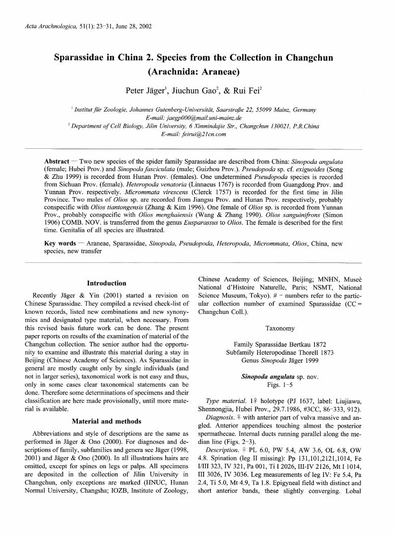
Acta Arachnologica, 51(1): 23-31, June 28, 2002
Sparassidae in China 2. Species from the Collection in
(Arachnida: Araneae)
Changchun
Peter Jagerl, Jiuchun Gaol, & Rui Fei2
' Institut fur Zoologie, Johannes Gutenberg-Universitat, Saarstraf3e 22, 55099 Mainz, Germany E-mail. [email protected]
2 Department of Cell Biology, Jilin University, 6 Xinmindajie Str., Changchun 130021, P.R. China
E-mail: feirui@21 cn.com
Abstract - Two new species of the spider family Sparassidae are described from China: Sinopoda angulata (female; Hubei Prov.) and Sinopoda fasciculata (male; Guizhou Prov.). Pseudopoda sp. cf. exiguoides (Song & Zhu 1999) is recorded from Hunan Prov. (females). One undetermined Pseudopoda species is recorded from Sichuan Prov. (female). Heteropoda venatoria (Linnaeus 1767) is recorded from Guangdong Prov. and Yunnan Prov. respectively. Micrommata virescens (Clerck 1757) is recorded for the first time in Jilin Province. Two males of Olios sp. are recorded from Jiangsu Prov. and Hunan Prov. respectively, probably conspecific with Olios tiantongensis (Zhang & Kim 1996). One female of Olios sp. is recorded from Yunnan Prov, probably conspecific with Olios menghaiensis (Wang & Zhang 1990). Olios sanguinifrons (Simon 1906) COMB. NOV. is transferred from the genus Eusparassus to Olios. The female is described for the first time. Genitalia of all species are illustrated.
Key words - Araneae, Sparassidae, Sinopoda, Pseudopoda, Heteropoda, Micrommata, Olios, China, new species, new transfer
Introduction
Recently Jager & Yin (2001) started a revision on Chinese Sparassidae. They compiled a revised check-list of known records, listed new combinations and new synony-mies and designated type material, when necessary. From this revised basis future work can be done. The present
paper reports on results of the examination of material of the Changchun collection. The senior author had the opportu-nity to examine and illustrate this material during a stay in Beijing (Chinese Academy of Sciences). As Sparassidae in general are mostly caught only by single individuals (and not in larger series), taxonomical work is not easy and thus, only in some cases clear taxonomical statements can be done. Therefore some determinations of specimens and their classification are here made provisionally, until more mate-rial is available.
Material and methods
Abbreviations and style of descriptions are the same as
performed in Jager & Ono (2000). For diagnoses and de-scriptions of family, subfamilies and genera see Jager (1998, 2001) and Jager & Ono (2000). In all illustrations hairs are omitted, except for spines on legs or palps. All specimens are deposited in the collection of Jilin University in Changchun, only exceptions are marked (HNUC, Hunan Normal University, Changsha; IOZB, Institute of Zoology,
Chinese Academy of Sciences, Beijing; MNHN, Musee National d'Histoire Naturelle, Paris; NSMT, National Science Museum, Tokyo). # - numbers refer to the partic-ular collection number of examined Sparassidae (CC = Changchun Coll.).
Taxonomy
Family Sparassidae Bertkau 1872
Subfamily Heteropodinae Thorell 1873
Genus Sinopoda Jager 1999
Sinopoda angulata sp. nov.
Figs. 1-5
Type material. 1- holotype (PJ 1637, label: Liujiawu, Shennongjia, Hubei Prov., 29.7.1986, #3CC, 86-333, 912).
Diagnosis. - with anterior part of vulva massive and an-
gled. Anterior appendices touching almost the posterior spermathecae. Internal ducts running parallel along the me-dian line (Figs. 2-3).
Description. - PL 6.0, PW 5.4, AW 3.6, OL 6.8, OW 4.8. Spination (leg II missing): Pp 131,101,2121,1014, Fe I/III 323, IV 321, Pa 001, Ti I 2026, III-IV 2126, Mt I 1014, III 3026, IV 3036. Leg measurements of leg IV: Fe 5.4, Pa 2.4, Ti 5.0, Mt 4.9, Ta 1.8. Epigyneal field with distinct and short anterior bands, these slightly converging. Lobal

24 P. Jager et al.
epigyneal pockets situated far from each other, connected by an anterior rim. Margins of lobal pockets running not
parallel to posterior margin of lateral lobes (Fig. 1). Anterior
vulval appendices extending laterally beyond posterior spermathecae (Fig. 2).
--~`---
Figs. 1-9. 1-5. Sinopoda angulata sp. nov., - holotype (#3CC, 86-333, 912) from Liujiawu, Shennongjia, Hubei Prov. 6-7. S. shennonga (Peng, Yin & Kim 1996), - holotype (HNUC #53) from Shennongjia, Hubei Prov. 8-9. S. wangi Song & Zhu 1999, syntype (HNUC #56) from Mt. Lu, Jiangxi Province. - 1,6,8, Epigyne, ventral view; 2-3,7,9, vulva (2,7,9, dorsal view; 3, anterior view); 4, left leg IV, prolateral view; 5,
prosoma, dorsal view. (Scales in mm)
Acta Arachnologica, 51(1), June 2002 OArachnological Society of Japan

Sparassidae from Changchun Collection 25
Color. Dark yellow-brown. Legs without pattern, becom-ing darker distally (Fig. 4). Chelicerae dark red-brown, with dark longitudinal bands frontally. PS with broad and bright median band (Fig. 5). Head region and marginal bands red-brown. Dorsal OS dark red-brown, with bright median patch above heart, which becoming darker posterior. OS in poste-rior half with indistinct bright patch. Lateral OS spotted with oblong patches. Ventral OS with some irregular
patches. unknown. Distribution. Only known from the type locality.
Etymology. The species name refers to the angled shape of anterior vulva in a dorsal view (Latin - angulatus: an-
gled); adjective. Note. This species can only be identified by preparing the
internal female genitalia. From the type locality another Sinopoda species is described: S. shennonga (Peng, Yin & Kim 1996) (Figs. 6-7). This shows several differences to the here described species: 1. Margins of lobal pockets par-allel to the posterior margin of lateral lobes, 2. anterior ap-
pendices of vulva do clearly not reaching posterior spermathecae, 3. anterior appendices do not extending be-
yond the posterior spermathecae, 4. anterior vulva without a massive and angled structure, 5. internal ducts running not
parallel along the median line, but diverging in their anterior and posterior part, 6. epigyneal field with indistinct anterior bands, 7. lobal pockets not connected by an anterior rim.
The new species can also be distinguished from other Sinopoda spp., e.g. S. wangi Song & Zhu 1999 (compare Figs. 8-9), by comparing carefully external and internal fe-male genitalia.
Sinopoda fasciculata sp. nov. Figs. 10-12
Type material. 1 holotype (PJ 1638, label: Fanjingshan, Guizhou Prov., 24.6.1985, #2CC, 85-295)
Diagnosis. Closely related to S. okinawana Jager & Ono 2000 and S. wangi Song & Zhu 1999 in Song et al. (1999)
(Figs. 8-9,13-15), but differs in the following characters: 1. Both, tip of embolus and well developed embolic apophysis distinctly bent, 2. tegulum covering proximal part of embo-lus (Figs. 10-11), 3. ventral RTA well developed, triangle-shaped (Fig. 12).
Figs. 10-15. 10-12. Sinopoda fasciculata sp. nov., holotype (#2CC, 85-295) from Fanjingshan, Guizhou Prov.; 13-15. S. wangi Song & Zhu 1999, syntype (HNUC #56) from Mt. Lu, Jiangxi Province. -10, left
palp, ventral view; l 1,13, tegulum, ventral view; 12,14-15 retrolateral tibial apophysis (12,14, retrolateral view; 15, ventral view). (Scales in mm)
Acta Arachnologica, 51(1), June 2002 ©Arachnological Society of Japan

26 P. Jager et al.
Description. PL 5.0, PW 4.7, AW 2.2, OL 5.7, OW 3.8. Spination: Pp 131,101,2101, Fe I 3(2)23, II-III 323, IV 331, Pa 101, Ti 2326, Mt I-II 1014, III 2014, IV 3036. Leg meas-urements of leg IV: Fe 7.1, Pa 2.3, Ti 7.0, Mt 8.0, Ta 2.6. Dorsal RTA very thin in ventral view, conical in lateral view. Sperm duct slightly curved (Figs. 10-11).
Color (based on the freshly moulted holotype). PS and legs yellowish with dark pattern and dark hairs. Head region darker. Posterior PS with dark transversal band behind fovea, followed by a bright transversal band. Posterior mar-
gin of PS dark. Chelicerae darker, with longitudinal bands frontally. Legs with distinct spine patches and small irregu-lar patches. OS dark-brown. Heart region brighter. Posterior OS with dark transversal patch. Ventral OS with two bright longitudinal lines. Spinnerets ventrally bright.
unknown.
Distribution. Only known from the type locality. Etymology. The specific epithet refers to the hair tuft
at the base of the retrolateral tibial apophysis (Latin -
fasciculatus: with a small tuft of hairs); adjective.
Genus Pseudopoda Jager 2000
Pseudopoda sp. cf. exiguoides (Song & Zhu Figs. 16-21
1999)
Material examined. 1 (PJ 1640, label: Hunan Prov, Huangshizhai, Zhangjiajie, June 1985, #5CC, 85-191). 2-
(PJ 1641-1642, without label, #9CC, 85-117). Diagnosis. - anterior margin of epigyneal field trilobate,
with distinct and short bands. Epigyneal field with characteristical lateral indentations. Posterior margins of lat-
Figs.16-26.16-21. Pseudopoda sp. cf. exiguoides (Song & Zhu 1999),16-19. l~ (#5CC, 85-191) from Zhangjiajie, Huangshizhai, Hunan Prov. 20-21.2- (#9CC, 85-117) without locality. 22-26. Pseudopoda sp., 1~ (#4CC, 75-673) from Qingshengshan, Sichuan Prov. - 16,20-22, Epigyne, ventral view; 17,23, vulva, dorsal view; 18,24, left leg IV, prolateral view; 19,25, prosoma, dorsal view; 26, opisthosoma, ventral view. (All scales 1 mm)
Acta Arachnologica, 51(1), June 2002 ©Arachnologica! Society of Japan
Sparassidae from Changchun Collection 27
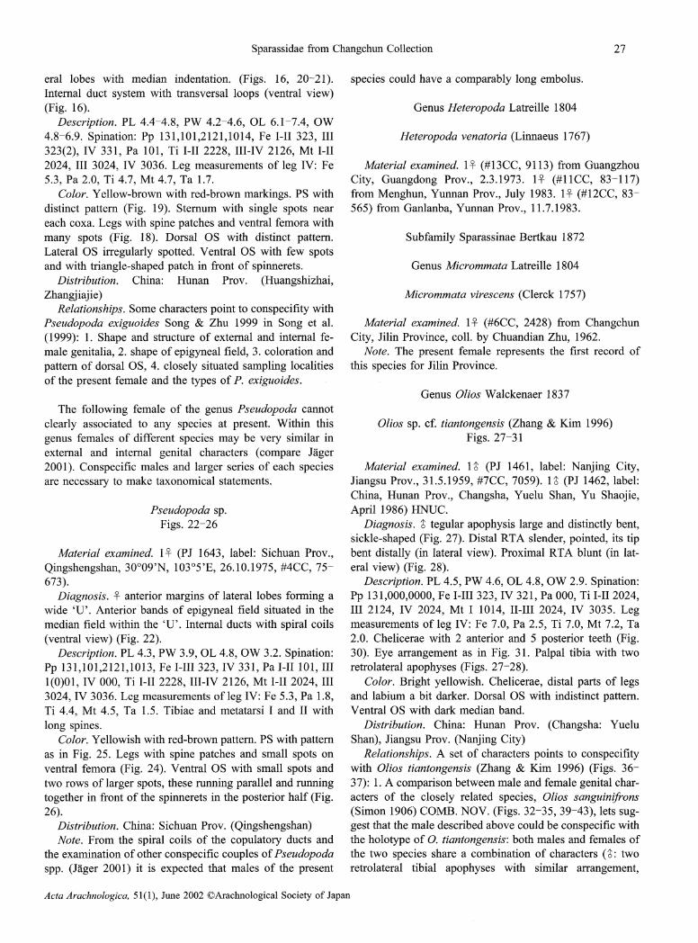
eral lobes with median indentation. (Figs. 16, 20-21). Internal duct system with transversal loops (ventral view)
(Fig. 16). Description. PL 4.4-4.8, PW 4.2-4.6, OL 6.1-7.4, OW
4.8-6.9. Spination: Pp 131,101,2121,1014, Fe I-II 323, III 323(2), IV 331, Pa 101, Ti I-II 2228, III-IV 2126, Mt I-II 2024, III 3024, IV 3036. Leg measurements of leg IV: Fe 5.3, Pa 2.0, Ti 4.7, Mt 4.7, Ta 1.7.
Color. Yellow-brown with red-brown markings. PS with distinct pattern (Fig. 19). Sternum with single spots near each coxa. Legs with spine patches and ventral femora with many spots (Fig. 18). Dorsal OS with distinct pattern. Lateral OS irregularly spotted. Ventral OS with few spots and with triangle-shaped patch in front of spinnerets.
Distribution. China: Hunan Prov. (Huangshizhai, Zhangjiajie) Relationships. Some characters point to conspecifity with Pseudopoda exiguoides Song & Zhu 1999 in Song et al.
(1999): 1. Shape and structure of external and internal fe-male genitalia, 2. shape of epigyneal field, 3. coloration and
pattern of dorsal OS, 4, closely situated sampling localities of the present female and the types of P. exiguoides.
The following female of the genus Pseudopoda cannot clearly associated to any species at present. Within this
genus females of different species may be very similar in external and internal genital characters (compare Jager 2001). Conspecific males and larger series of each species are necessary to make taxonomical statements.
Pseudopoda sp.
Figs. 22-26
Material examined. 1 (PJ 1643, label: Sichuan Prov.,
Qingshengshan, 30°09'N, 103°5'E, 26.10.1975, #4CC, 75-673). Diagnosis. - anterior margins of lateral lobes forming a wide `U'. Anterior bands of epigyneal field situated in the median field within the `U'. Internal ducts with spiral coils
(ventral view) (Fig. 22). Description. PL 4.3, PW 3.9, OL 4.8, OW 3.2. Spination:
Pp 131,101,2121,1013, Fe I-III 323, IV 331, Pa I-II 101, III 1(0)01, IV 000, Ti I-II 2228, III-IV 2126, Mt I-II 2024, III 3024, IV 3036. Leg measurements of leg IV: Fe 5.3, Pa 1.8, Ti 4.4, Mt 4.5, Ta 1.5. Tibiae and metatarsi I and IT with long spines.
Color. Yellowish with red-brown pattern. PS with pattern as in Fig. 25. Legs with spine patches and small spots on ventral femora (Fig. 24). Ventral OS with small spots and two rows of larger spots, these running parallel and running together in front of the spinnerets in the posterior half (Fig. 26). Distribution. China: Sichuan Prov. (Qingshengshan)
Note. From the spiral coils of the copulatory ducts and the examination of other conspecific couples of Pseudopoda spp. (lager 2001) it is expected that males of the present
species could have a comparably long embolus.
Genus Heteropoda Latreille 1804
Heteropoda venatoria (Linnaeus
Material examined. 1 (# 13 CC, City, Guangdong Prov., 2.3.1973. from Menghun, Yunnan Prov., July 565) from Ganlanba, Yunnan Prov.,
1767)
9113) from Guangzhou 1~ (#11CC, 83-117) 1983. 1~ (#12CC, 83-11.7.1983.
Subfamily Sparassinae Bertkau
Genus Micrommata Latreille
Micrommata virescens (Clerck
1872
1804
1757)
Material examined. 1 (#6CC, 2428) from Changchun City, Jilin Province, coll. by Chuandian Zhu, 1962.
Note. The present female represents the first record of this species for Jilin Province.
Olios sp.
Genus Olios Walckenaer 1837
cf. tiantongensis (Zhang & Kim Figs. 27-31
1996)
Material examined. 1 (PJ 1461, label: Nanjing City, Jiangsu Prov., 31.5.1959, #7CC, 7059). 1 (PJ 1462, label: China, Hunan Prov., Changsha, Yuelu Shan, Yu Shaoj ie, April 1986) HNUC.
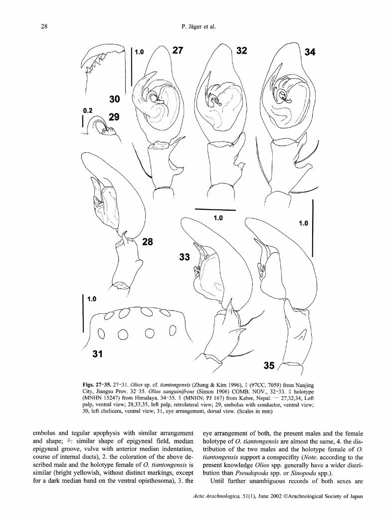
Diagnosis. ' tegular apophysis large and distinctly bent, sickle-shaped (Fig. 27). Distal RTA slender, pointed, its tip bent distally (in lateral view). Proximal RTA blunt (in lat-eral view) (Fig. 28).
Description. PL 4.5, PW 4.6, OL 4.8, OW 2.9. Spination: Pp 131,000,0000, Fe I-III 323, IV 321, Pa 000, Ti I-II 2024, III 2124, IV 2024, Mt I 1014, II-III 2024, IV 3035. Leg measurements of leg IV: Fe 7.0, Pa 2.5, Ti 7.0, Mt 7.2, Ta 2.0. Chelicerae with 2 anterior and 5 posterior teeth (Fig. 30). Eye arrangement as in Fig. 31. Palpal tibia with two retrolateral apophyses (Figs. 27-28).
Color. Bright yellowish. Chelicerae, distal parts of legs and labium a bit darker. Dorsal OS with indistinct pattern. Ventral OS with dark median band.
Distribution. China: Hunan Prov. (Changsha: Yuelu Shan), Jiangsu Prov. (Nanj ing City)
Relationships. A set of characters points to conspecifity with Olios tiantongensis (Zhang & Kim 1996) (Figs. 36-37): 1. A comparison between male and female genital char-acters of the closely related species, Olios sanguinifrons
(Simon 1906) COMB. NOV. (Figs. 32-35, 39-43), lets sug-gest that the male described above could be conspecific with the holotype of 0. tiantongensis: both males and females of the two species share a combination of characters (~ : two retrolateral tibial apophyses with similar arrangement,
Acta Arachnologica, 51(1), June 2002 OArachnological Society of Japan
28 P. Jager et al.
embolus and tegular apophysis with similar arrangement and shape; -: similar shape of epigyneal field, median epigyneal groove, vulva with anterior median indentation, course of internal ducts), 2, the coloration of the above de-scribed male and the holotype female of 0. tiantongensis is similar (bright yellowish, without distinct markings, except for a dark median band on the ventral opisthosoma), 3. the
eye arrangement of both, the present males and the female holotype of 0. tiantongensis are almost the same, 4. the dis-tribution of the two males and the holotype female of 0. tiantongensis support a conspecifty (Note. according to the
present knowledge Olios spp, generally have a wider distri-bution than Pseudopoda spp. or Sinopoda spp.).
Until further unambiguous records of both sexes are
Acta Arachnologica, 51(1), June 2002 OArachnological Society of Japan
Figs. 27-35. 27-31. Olios sp. cf. tiantongensis (Zhang & Kim 1996), a (#7CC, 7059) from Nanjing City, Jiangsu Prov. 32-35. Olios sanguinifrons (Simon 1906) COMB. NOV., 32-33. holotype (MNHN 15247) from Himalaya. 34-35. ' (MNHN; PJ 167) from Kabre, Nepal. - 27,32,34, Left palp, ventral view; 28,33,35, left paip, retrolateral view; 29, embolus with conductor, ventral view; 30, left chelicera, ventral view; 31, eye arrangement, dorsal view. (Scales in mm)

Sparassidae from Changchun Collection 29
available, no final statement on the taxonomical status of the
two males can be made.
Olios sp. cf. menghaiensis (Wang & Zhang 1990) Figs. 44-47
Material examined. 1 (PJ 1639, label: Menghai, Yunnan Prov., 15.7.1983, #1CC, 83-299).
Diagnosis. epigyneal field almost rectangular (Fig. 44). Internal duct system with a bit more than three regular coils of each, copulatory duct and fertilisation duct. Posterior
parts of fertilisation ducts close together (Figs. 45-46). Description. PL 7.3, PW 7.4, AW 4.9, OL 9.5, OW 6.1.
Spination: Pp 131,001,0011,1013, Fe I-III 323, IV 321, Pa 000, Ti 2024, Mt I-III 2024, IV 3026. Leg measurements of leg IV: Fe 8.0, Pa 3.5, Ti 7.4, Mt 6.7, Ta 2.2.
Color. Red-brown. PS with distinct pattern (Fig. 47) and bright hairs at its margins. Chelicerae dark red-brown.
Gnathocoxae and labium dark with their inner margins dis-tinctly brighter. Legs with bright oblong patches dorsally. Ventral tibiae with indistinct annulation, consisting of bright and dark hairs. Tarsi and metatarsi with thick scopulae. Dorsal OS dark with angle-shaped patches and brighter heart region. Ventral OS dark brown.
Distribution. China: Yunnan Prov. (Menghai) Relationships. Some characters points to conspecifity
with Olios menghaiensis (Wang & Zhang 1990) (Fig. 48), a species described from a single male from Menghai, Yunnan Prov.: 1. type locality of 0. menghaiensis and the locality of the above described female are identical, 2. the screw-like course of the internal ductsystem of the above described female and the screw-shaped embolus of 0. menghaiensis would fit together, thinking of copulatory me-chanics.
Acta Arachnologica, 51(1), June 2002 ©Arachnological Society of Japan
Figs. 36-43.36-38.Olios tiantongensis (Zhang & Kim 1996), - holotype (HNUC #39) from Tiantong, Ningbo City, Zhejiang Province. 39-43. Olios sanguinifrons (Simon 1906) COMB. NOV., - (MNHN 15247) from Himalaya. -36,39,41, Epigyne, (36,39, ventral view; 41, posterior view); 37,40, vulva, dorsal view, 38,42, schematical course of internal duct system, dorsal view; 43, left chelicera, ventral view. (scales in mm)

30 P. Jager et al.
Material examined for comparison. Sinopoda wangi Song & Zhu 1999 (Figs. 8-9, 13-15):
2a', 5~~ syntypes (#56) from Mt. Lu, Jiangxi Province, China, 7.8.1987, leg. by Jia-fu Wang; 10~ ' syntypes (#57) from Mt. Lu, Jiangxi Province, China, 15.6.1987, leg. b Jia-fu Wang; 4 3 syntypes (#1 1) from Mt. Lu, Jiangxi Province, China, August 1987, leg. by Jia-fu Wang; all HNUC.
Sinopoda okinawana Jager & Ono 2000: 1 a holotype
(4220) from Takasato, Okinawajima Is., Okinawa Pref,. Japan, 1-IV-1997, Takeshi Sasaki leg.; 1~ paratype (4221)
from Yona, Okinawajima Is., Okinawa Pref,. Japan, 30-III- 1997, Akio Tanikawa leg.; 1 paratype (4219) from
y Iheyajima Is., Okinawa Pref., Japan, 27-X-1993, M. Kimura; all NSMT.
Sinopoda shennonga (Peng, Yin & Kim 1996) (Figs. 6- 7): 1 holotype (#53) from Shennongjia, Xiangyang
County, 32.1 °N 112.1°E, Hubei Prov., China, 10.1990, leg.
Acta Arachnologica, 51(1), June 2002 ©Arachnological Society of Japan
Figs. 44-48.44-47.Olios sp. cf. menghaiensis (Wang & Zhang 1990), l - (# 1 CC, 83-299) from Menghai, Yunnan Prov. 48. Olios menghaiensis (Wang & Zhang 1990) ' holotype (according to Wang & Zhang 1990) from Menghai, Yunnan Prov. - 44, Epigyne, ventral view; 45, vulva, dorsal view; 46, schematical course of internal duct system, dorsal view; 47, prosoma, dorsal view; 48, left palp, ventral view. (Scales in mm)

Sparassidae from Changchun Collection 31
by Jia-fu Wang; HNUC.
Pseudopoda exiguoides (Song & Zhu 1999) in Song et al.
(1999): 1 lectotype, 1 paralectotype (#41) from Mt. Yuelu, Changsha City, Hunan Prov., China, 10.8.1980, by Jia-fu Wang; HNUC.
Olios tiantongensis (Zhang & Kim 1996) (Figs. 36-38): l~ holotype (#39): from Tiantong, Ningbo City, 29.9°N, 121.5°E, Zhejiang Province, China, 20.11.1994, leg. by Yong jin Zhang. HNUC.
Olios sanguinifrons (Simon 1906) COMB. NOV. (Figs. 32-35, 39-43): 1 holotype, PJ 675, label: Eusparassus sanguiniceps E.S., bas pl. du 1'Himalaya, {Type}. MNHN 15247; 1 ~, PJ 676, with same data as holotype male. 1 (PJ 167) from Kabre, Nepal, 17.1.1967, M. Hubert leg.; all MNHN.
Notes. The species, described by Simon (1906) sub Eusparassus is explicitely transferred to the genus Olios. Both specimens clearly possess characters, which are also shared by other Olios spp. e.g. 0. tener (Thorell 1891): typi-cal, bent embolus, with reduced conductor, two tibial apophyses and synapomorphic characters of Sparassinae: two anterior cheliceral teeth and a characteristic eye ar-rangement. Simon wrote on the label and in the text of his original
publication (1906: 312) a different name (sanguiniceps) than he published in the original description in the heading
(sanguinifrons). From comparison of the male specimen (Figs. 32-33) and the original description (Simon 1906) it is clear that this specimen represents the holotype male. `san-
guinifrons' is considered the valid name. The female, which was stored together in the vial with
the holotype male was subadult. Under the skin, which was almost coming off, a developed epigyne was present. Although a more sclerotized epigyne of an mature individ-ual may show a different shape or coloration, the female
genitalia are illustrated here for the first time (Figs. 39-41). A full description will be given later in a revisional paper on the genus Olios. It is not clear, whether the female was added later by Simon or a subsequent curator or whether it was already in the type series and Simon did not mention it, because it was not mature. However, both specimens are
considered conspecific from their size and coloration.
Acknowledgments
The first author wish to thank Prof. Dr. Changmin Yin, Prof. Dr. Songping Liang, Prof. Dr. Liangbi Cheng, Ms. You-hui Bao, Ms. Xiang Xu, Dr. Xianjin Peng and Mr. Guo Tang (all HNUC) for sup-port during the stay in Changsha and Dr. Hirotsugu Ono (NSMT, Tokyo) for helpful support in loan concerns and information on Japanese Sparassidae. Thanks are also due to Dr. Christine Rollard for her help during the stay in Paris and Dr. Peter Schwendinger for send-ing an important paper. The present paper resulted from a research travel of the first author to Beijing (Institute of Zoology, CAS) and Changsha (Hunan Normal University). This travel was partly spon-sored by a travel grant of the Deutscher Akademischer Austausch-dienst (DAAD, Germany) and by the Chinese National Science Foundation. The first author wish also to thank Dr. Shuqiang Li (IOZB) for his helpful efforts in Beijing. A research travel to Paris was supported by the European Community (Access to Research Infrastructure action of the Improving Human Potential Programme).
References
Jager, P. 1998. First results of a taxonomic revision of the SE Asian Sparassidae (Araneae). pp. 53-59. In: P. A. Selden (ed.), Proceed-
ings of the 17th European Colloquium of Arachnology, Edinburgh, 1997. Burnham Beeches, Bucks: Bull. British Arach. Soc. 350 pp.
Jager, P. 2001. Diversitat der Riesenkrabbenspinnen im Himalaya. Uber eine Radiation zweier Gattungen in den Schneetropen.
(Araneae: Sparassidae: Heteropodinae). Courier Forschungsinstitut Senckenberg, 232: 1-136.
Jager, P. & H. Ono. 2000. The Sparassidae of Japan. I. New species of Olios, Heteropoda and Sinopoda with remarks on known species
(Arachnida: Araneae). Acta Arachnol., 49: 41-60. Jager, P. & Yin, C.-M. 2001. Sparassidae in China. 1. Revised list of
known species with new transfers, new synonymies and type desig- nations (Arachnida: Araneae). Acta Arachnol. 50: 123-134.
Peng, X.-J., Yin, C.-M. & Kim, J.-P. 1996. One species of the genus Heteropoda and a description of the female Heteropoda minschana
Schenkel, 1936 (Araneae: Heteropodidae). Korean Arachnol., 12: 57-61.
Simon, E. 1906. Voyage de M. Maurice Maindron Bans 1'Inde meridionale (mai a november 1901). 8e memoire. Arachnides (2e
partie). Ann. Soc. ent. France, 75: 279-314. Song, D.-X., Zhu, M.-S. & Chen, J. 1999. The spiders of China. Hebei
Sci. Technol. Publ. House, Shijiazhuang, 640 pp. Wang, J.-F. & Zhang, Z. 1990. [A new species of the genus
Heteropoda from Yunnan Province, China]. J. Yunnan Norm. Univ., 10 (3/4): 9-10. (In Chinese, with English abstract)
Zhang, Y.-J. & Kim, J.-P. 1996. Three new species of the family Heteropodidae from China (Arachnida: Araneae). Korean
Arachnol., 12: 77-85.
Received January 31, 2002 / Accepted March 23, 2002
Acta Arachnologica, 51(1), June 2002 OArachnological Society of Japan