sorbitol dehydrogenase from bacillus subtilis
TRANSCRIPT
THE JOURNAL OF BIOLOGICAL CHEMISTRY 0 1992 by The American Society for Biochemistry and Molecular Biology, Inc.
Vol. 267, No. 35, Issue of December 15, pp. 24989-24994,1992 Printed in U.S.A.
Sorbitol Dehydrogenase from Bacillus subtilis PURIFICATION, CHARACTERIZATION, AND GENE CLONING*
(Received for publication, August 5, 1992)
Kenneth Ng, Ruiqiong Ye, Xu-Chu Wu, and Sui-Lam WongS From the Department of Biological Sciences, Division of Biochemistry, University of Calgary, Calgary, Alberta T2N IN4, Canada
Cloning of the sorbitol dehydrogenase gene (gutB) from Bacillus subtilis offers an excellent system for studying zinc binding, substrate specificity, and cata- lytic mechanism of this enzyme through protein engi- neering. As a first step to clone gutB, B. subtilis sor- bitol dehydrogenase has been purified to homogeneity and characterized. It is a tetrameric enzyme with a molecular mass of 38 kDa for each subunit. Atomic absorption analysis shows the presence of 1 mol of zinc atom/subunit. Substrate specificity and stereospecific- ity of the enzyme toward C-2 and C-4 of hexitols were established. Sequence of the first 31 amino acids was determined, and a set of oligonucleotide probes was designed for gene cloning. A positive clone carrying a 5-kilobase pair Hind111 insert was isolated and se- quenced. Sequence alignment indicated that the de- duced amino acid sequence of B. subtilis sorbitol de- hydrogenase shows 36% identity in sequence with the liver sorbitol dehydrogenase from sheep, rat, and hu- man. In reference to the sequence of alcohol dehydro- genase, two potential zinc binding sites were identi- fied. Sequence information related to the structure- function relationships of the enzyme is discussed.
Sorbitol dehydrogenase, also known as glucitol, polyol, or L-iditol dehydrogenase (EC 1.1.1.14), catalyzes the oxidation of sorbitol to fructose with NAD as a cofactor. It has been detected in a wide variety of animals, plants, and microorga- nisms (1). Liver sorbitol dehydrogenase has been purified to homogeneity from sheep and human (2,3). The entire protein sequence of these two enzymes and a cDNA sequence of the rat sorbitol dehydrogenase have also been determined (4, 5). Sequence alignment demonstrates that sorbitol dehydrogen- ase shares homology with the mammalian alcohol dehydro- genase family (6-8). However, these mammalian sorbitol de- hydrogenases differ from the alcohol dehydrogenase family in two aspects: 1) sorbitol dehydrogenase is tetrameric rather than dimeric; 2) it has one instead of two zinc atoms per subunit and only the binding site for the catalytic zinc atom
* This work was supported by an operating grant from the Natural Sciences and Engineering Research Council of Canada. The costs of publication of this article were defrayed in part by the payment of page charges. This article must therefore be hereby marked “aduer- tisement” in accordance with 18 U.S.C. Section 1734 solely to indicate this fact.
The nucleotide sequence(s) reported in thispaper has been submitted
M9694 7. to the GenBankTM/EMBL Data Bank with accession number(s)
$ Medical scholar of the Alberta Heritage Foundation for Medical Research. To whom correspondence should be addressed Dept. of Biological Sciences, Division of Biochemistry, University of Calgary, 2500 University Dr. N.W., Calgary, Alberta T2N 1N4, Canada. Tel.: 403-220-5721; Fax: 403-289-9311.
is conserved. For horse liver alcohol dehydrogenase, the three- dimensional structye of the protein has been determined a t a resolution of 2.4 A (9). Protein ligands that coordinate the binding of the catalytic zinc atom are His-67, Cys-46, and Cys-174. The fourth ligand is water or substrate during the reaction process. Through homology modeling, the three- dimensional structure of the sheep sorbitol dehydrogenase has been predicted (7). The resulting model forms a basis to explain the substrate specificity of this enzyme. For the cat- alytic zinc binding site, it has 1 histidine and 1 cysteine in the corresponding positions that are equivalent to those found in alcohol dehydrogenase. However, the third protein ligand in zinc binding is predicted to be a glutamic acid instead of a cysteine. To critically evaluate this model, it would be essen- tial to have a sorbitol dehydrogenase gene cloned so that large quantities of sorbitol dehydrogenase can be available for x- ray crystallographic study. Site-directed mutagenesis can also be performed to identify critical residues involved in zinc or substrate binding.
In Bacillus subtilis, sorbitol dehydrogenase activity was detected and partially purified in 1964 (10). However, the purity, molecular weight, subunit structure, and fine substrate specificity of the enzyme were not determined. Furthermore, the structural relationship between procaryotic and mamma- lian sorbitol dehydrogenases is unknown. To address these questions and to establish B. subtilis sorbitol dehydrogenase as a model system to study the structure-function relation- ships through modeling and protein engineering, we report the purification and characterization of B. subtilis sorbitol dehydrogenase. The cloning and characterization of the sor- bitol dehydrogenase gene (gutB)’ are also described.
EXPERIMENTAL PROCEDURES
Materials
Sorbitol, nitro blue tetrazolium, phenazine methosulfate, thiogly- collate, and glutathione were from Sigma; NAD was from Boehringer Mannheim. Electrophoretic reagents were from Bio-Rad. Restriction enzymes, modification enzymes, and a random primer labeling kit were from Bethesda Research Laboratories, Boehringer Mannheim, New England Biolabs, Inc., and Pharmacia LKB Biotechnology Inc. DE-52 was from Whatman. A T7 sequencing kit, Sepharose-400, and two FPLC columns (Superose 12 HR 10/30 and Mono Q HR 5/5) were obtained from Pharmacia. [w3’S]ATP (1,000 Ci/mmol) was from Amersham Corp. Immobilon PVDF membrane was from Milli- pore Corp.
cloning gutB. DB104 (his nprR2 nprEI8 aprA3) (11) and PG668 Chromosomal DNA was prepared from B. subtilis 168 (trpC2) for
(gutB2 l e d 8 trpCg; obtained from the Bacillus Genetic Stock Center, Columbus, OH) (12), were used for routine subcloning and expression studies. If these cells carry the cloning vector pUB18 or its derivatives,
The abbreviations used are: gutB, structural gene encoding sor- bitol dehydrogenase; DTT, dithiothreitol; kb, kilobase pair(s); PVDF, polyvinyldifluoride; PAGE, polyacrylamide gel electrophoresis; FPLC, fast protein liquid chromatography.
24989
24990 B. subtilis Sorbitol Dehydrogenase
kanamycin was added to the medium at a final concentration of 10 pg/ml. C-medium (12) and super-rich medium (13) were used for the cultivation of E. subtilis. Required amino acids were added to C- medium at a final concentration of 50 mg/liter. For induction studies, C-medium was supplemented with 0.5% sorbitol instead of glucose. Escherichia coli DH5a (@30dlacZAM15)[endAI recAl hsdR17 (r- m-) supE44 thi-1 X- gyrA relAl F- A(lacZYA-argF)U169]) was used as a host for routine subcloning. Bluescribe plasmid (pBS) was obtained from Stratagene.
Methods Enzyme Assay-Sorbitol dehydrogenase activity was determined
spectrophotometrically by following the absorbance of NADH at 340 nm ( 6 = 6220 M” cm-’) at 30 ‘C. The absorbance was monitored by using a Beckman DU-65 spectrophotometer equipped with a constant temperature cuvette chamber. Each assay was performed with 1 ml of reaction mixture containing 0.1 M Tris-HC1 (pH 9.0), 50 mM sorbitol, 2 mM NAD, and 10-100 pl of enzyme extract. One unit is defined as the amount of enzyme needed to generate 1 pmol of NADH/min. For determining the kinetic parameters, 0.78 pg of the purified enzyme was used in each assay. The substrate concentration ranged from 1.5 to 12.5 mM for sorbitol, xylitol, and L-iditol and 30 mM to 0.5 M for ribitol, galactitol, and D-mannitol.
Enzyme Purification-B. subtilis DB104 was cultivated at 37 “C in 2-liter super-rich medium containing 2% sorbitol. When the cell density reached 600 klett units, cells were harvested by centrifugation (7,000 X g, 5 min) and resuspended in 50 ml of cell breakage buffer (50 mM Tris-HC1, pH 7.5,l mM phenylmethylsulfonyl fluoride). Cells were broken by passing through a French press at 10,000 psi. Cell debris was removed by centrifugation (27,000 X g, 15 min at 4 “C). The supernatant was adjusted to a final volume of 85 ml with the cell breakage buffer and loaded to a DE-52 column (2.5 X 10 cm) pree- quilibrated with the cell breakage buffer. After washing the column with 3 bed volumes of buffer, proteins were eluted with a salt gradient (0-0.5 N NaCl) in the same buffer. Sorbitol dehydrogenase was eluted off from the column when the salt concentration reached 0.15 M NaC1. These fractions were pooled together and passed through an Amicon 8050 filtration unit with a Diaflo XM 50 membrane that retained proteins with molecular weight larger than 50,000. The sample was concentrated from 100 down to 9 ml through ultrafiltra- tion and loaded to a Sephacryl S-400 (2.5 X 80-cm) column, which was preequibrated with 50 mM sodium phosphate buffer, pH 7.5, containing 1 mM DTT. Fractions with activity (about 50 ml) were collected and combined after eluting proteins from the column with the phosphate buffer. Solid ammonium sulfate was added to the sample to a final concentration of 1 M. The sample was loaded to a phenyl-Sepharose CL-4B column (1 X 10 cm) preequilibrated with 100 ml of 50 mM sodium phosphate buffer, pH 7.5, containing 1 mM DTT and 1.7 M ammonium sulfate. Elution was performed by low- ering the ammonium sulfate concentration from 1.7 to 0 M. Sorbitol dehydrogenase activity was detected in fractions with 0.085 M am- monium sulfate. Active fractions were pooled and concentrated to 2 ml by using an Amicon Centriprep 10 filtration unit (3,640 X g for 30 min at 4 ‘C). Further purification of the enzyme was achieved by using the FPLC system. The sample was applied to a Superose 12 B column equilibrated with 50 mM Tris buffer containing 1 mM DTT (pH 7.5). The active fractions were pooled and loaded to a Mono Q column equilibrated with the same buffer. After washing the column with 4 bed volumes of buffer, proteins were eluted with a sodium chloride gradient (0-0.3 M) in the same buffer. The enzyme was detected in fractions with 0.15 M NaCl. Purity of the enzyme was determined with a 12% SDS-polyacrylamide gel.
Metal Analysis-The zinc content of the purified sorbitol dehydro- genase was determined by the graphite furnace atomic absorption method (14) with a Perkin Elmer model 3030B atomic absorption spectrophotometer equipped with a model HG600 graphite furnace. This analysis was performed by Dr. Paul C. Leavis at Analytical Biotechnology Services (Boston, MA). Two (10 pg of purified enzyme per sample) samples were used in the analysis.
Molecular Weight Determination-The subunit molecular weight of the enzyme was determined on a 12% SDS-polyacrylamide gel. For determining the molecular weight of the native sorbitol dehydrogen- ase, the purified enzyme was loaded to a Sephacryl S-400 column (2.5 X 70 cm). The following molecular weight markers are used in the analysis: blue dextran, catalase (240,0000), aldolase (158,0001, bovine serum albumin (68,000), ovalbumin (45,000) and chymotrypsinogen (24,000). K., is defined as V. - Vo divided by VT - VO. The bed
volume ( VT) of the gel matrix was 344 ml. The void volume ( Vo) was determined to be 96 ml. V. is the elution volume for individual protein.
Determination of Amino Acid Composition and N-terminal Se- quence of Sorbitol Dehydrogenase-Sorbitol dehydrogenase was ana- lyzed on a 12% SDS-polyacrylamide gel and electroblotted to PVDF membrane (Immobilon P) as described by Matsudaira (15). After a brief staining of the membrane with Coomassie Blue, the protein band was sliced and used for both protein sequence and composition determination by Sandra L. Kielland at the Protein Microchemistry Centre of the University of Victoria. Sequence of the first 31 amino acids from the N terminus of this protein was determined with an AB1 model 470 gas-phase sequenator equipped with an on-line phenylthiohydantoin analyzer and 900A system controller/data ana- lyzer. For composition analysis, sample was gas-phase-hydrolyzed and analyzed by the method of Dupont (16) with an AB1 model 420A derivatizer/amino acid analyzer.
Design of Oligonucleotide Probes-Based on the codon degeneracy of amino acids and a table of codon usage generated from 53 vegetative B. subtilis genes (17), protein sequence from residues 6 to 17 was selected to design oligonucleotide probes. This region allows the design of a set of degenerate oligonucleotide probes with the least redundancy (i.e. a set of probes containing 64 combinations with the incorporation of two deoxyinosines). The probes are 35 nucleotides long and have the following sequence: 5’-CC(G,T)CA(A,G)AA (T,C)ATGAAAGCIGCIGT(A,T,G,C)ATGCATAA(T,C)AC-3’. They were synthesized at the Regional DNA Synthesis Laboratory of the University of Calgary.
Other Methods-The preparation of bacterial chromosomal and plasmid DNA, Southern and colony hybridization, and DNA sequenc- ing were performed as described previously (18). PC Gene (version 6.6) from IntelliGenetics was used for the analysis of protein and nucleotide sequences. Antibodies specific to E. subtilis sorbitol dehy- drogenase were prepared from two male BALB/c mice as described previously (19). Protein concentration was estimated by the Coo- massie Blue dye binding assay (20) with bovine serum albumin as a standard.
RESULTS
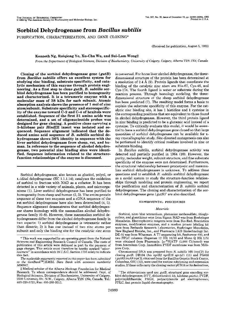
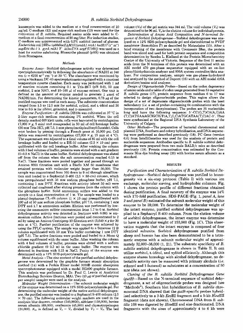
Purification and Characterization of B. subtilis Sorbitol De- hydrogenase-Sorbitol dehydrogenase was purified to homo- geneity via a six-step protocol (Table I) with various ion exchange, molecular sieving, and hydrophobic columns. Fig. 1 shows the protein profile of different fractions obtained during purification. A final recovery of the enzyme was 14% with 170-fold purification. SDS-PAGE (Fig. 1, panel A, lane 2 and panel B ) estimated the subunit molecular weight of this enzyme to be 39,000. To determine the molecular weight of the intact enzyme, purified sorbitol dehydrogenase was ap- plied to a Sephacryl S-400 column. From the elution volume of sorbitol dehydrogenase, the intact enzyme was determine to have a molecular weight of 153,000 (Fig. 1C). This obser- vation suggests that the intact enzyme is composed of four identical subunits. Sorbitol dehydrogenase purified from sheep and human has also been demonstrated to be a tetra- meric enzyme with a subunit molecular weight of approxi- mately 32,000-38,000 (1, 21). The substrate specificity of B. subtilis sorbitol dehydrogenase is shown in Table 11. It can utilize sorbitol, L-iditol, and xylitol efficiently. Although this enzyme shares homology with alcohol dehydrogenase, no de- tectable activity can be measured with primary alcohols (i.e. ethanol and 1-butanol) as substrates at a concentration of 50 mM (data not shown).
Cloning of the B. subtilis Sorbitol Dehydrogenase Gene (gutB)-Based on the N-terminal sequence of sorbitol dehy- drogenase, a set of oligonucleotide probes was designed (see “Methods”). Southern blot hybridization of B. subtilis chro- mosomal DNA showed that the gutB-specific probes hybrid- ized selectively to a 3-kb EcoRI fragment and a 5-kb HindIII fragment (data not shown). Chromosomal DNA from B. sub- tilis 168 was digested by HindIII and size-fractionated. DNA fragments with the sizes of approximately 4 to 6 kb were
B. subtilis Sorbitol Dehydrogenase 24991
TABLE I Purification of Bacillus subtilis sorbitol dehydrogenase
SteD Volume Protein Activity Specific activity Purification Recovery ml
Crude extract 85 DEAE-cellulose 100 Ultrafiltration 9 Sephacryl S-400 50 Phenyl-Sepharose 25 Superose 12 25 Mono Q 2
mg 1253.80 224.60 218.40 34.30 10.50 2.24 1.04
unitsjml 1.18 0.86 9.34 1.04 1.61 0.82 7.10
unitsjmg 0.080 0.383 0.385 1.520 3.830 9.200
13.600
-fold 1.0 4.7 4.8
19.0 47.8
115.0 170.0
5% 100 86 84 52 40 21 14
1 2 3 4 5 6 7 8
92 68
45
31
21
14
Mobilily Kaw
FIG. 1. Purification and molecular weight determination of sorbitol dehydrogenase. A, protein fractions obtained during dif- ferent stages of purification were analyzed on a 12% SDS-polyacryl- amide gel. The gel was stained with Coomassie Blue. Lane 1, molec- ular size markers (in kilodaltons); lanes 8-2 are samples (10 pl) from crude extract, DE-52, ultrafiltration, Sephacryl 400, phenyl-Sepha- rose CL-4B, Superose 12, and Mono Q fractions. B, subunit molecular weight determination of sorbitol dehydrogenase by SDS-PAGE. The mobility of molecular size markers and sorbitol dehydrogenase was calculated from panel A (lanes 1 and 2) . The arrow indicates the subunit molecular weight of sorbitol dehydrogenase (39,000). C, mo- lecular weight determination of intact sorbitol dehydrogenase by Sephacryl S-400. The arrow indicates the molecular weight of intact sorbitol dehydrogenase (153,000).
TABLE I1 Substrate specificity of B. subtilis sorbitol dehydrogenase
Substrate KM Vmax V , J K M R" mM p ~ m i n " min"
D-Sorbitol 11 6.25 5.68 X lo-' 100.00 Xylitol 14 6.30 4.50 X lo" 79.22 L-Iditol 18 8.02 4.45 X lo" 78.35 Ribitol (adonitol) 125 4.08 3.26 X 5.74 Galactitol (dulcitol) 500 1.67 3.34 X 5.88 D-Mannitol 555 0.40 7.21 X 0.13
~~~ ~ ~ ~
a R is defined as a ratio of the V,,,.JKM values between a substrate and sorbitol multiplied by 100.
200 bp FIG. 2. Restriction map and sequencing strategy of gutB.
Restriction sites used for subcloning are indicated. The open reading frame of gutB is boxed. Nucleotide sequence was determined by sequencing both strands. The direction and extent of sequence deter- mined from each subclone was shown by a solid arrow. Dotted arrows indicate the sequence determined by using synthetic primers P1, P2, and P3.
1 0 2 0 30 4 0 50 60 L L l M C C A G A T I I M T G G G G G C A G T M C A G A G I I M ~ T G T A T G C A ~ A C A ~
A ~ C T ~ G ~ M ~ C C ~ T G A C T C A C A C A G T A C ~ ~ C A T G ~ G C G G
E T G 1 T A T G C A C M C 1 C M G A G A G A T ~ 1 T G ~ C A 1 T G C E T G ~ C E T G A T A T C M ~ A V M H N T R E I K I E T L P V P D I N
A T G A T C M G T G 1 T G A T T M G G ~ A ~ C T G T C G G M ~ C G G A ~ A ~ C A T T A C T
:.?.r.?..v..p..?.r.IL.>..A.. ........................................... H D E V L I K V M A V G I C G S D L H Y
A T A C A M T G G C C G M T A G G C M C ~ T G G ~ C C A ~ A ~ - G C A T G M T Y T N G R I G N Y V V E K P F I L C H E
CTGCGU;TG~1TGCCGETGTCGGATCATETGTffiATCM1TCMGG~AGACffiffi C A G E I A A V G S S V D Q F K V C D R
T C O E T G T A G A G C C G G G ~ A ~ ~ ~ T G C G G A ~ ~ ~ ~ T G A G G C G T G C A M G M G G A ~ ~ ~ ~ A T A V A V E P G V T C G R C E A C K E C R Y
AT-CCCGGATGTACAG~CTACACffiCffiGTAGAEGGTGCG~~CMT N L C P D V Q F L A T P P V D G A F V Q
A T A T T I I M T G C G T C A G G A C T F F G ~ M T C C C A G A ~ C A ~ ~ A T G M G M G Y I K M R Q D F V F L I P D S L S Y E E
E T G r r P K . A ~ A C C f f i ~ C T C G G T A T C C A T G ~ C G G C C A G M f f i M G ~ A ~ G C A A L I E P F S V G I H A A A R T K L Q
C C G G A T h T C M C G A 1 T G f f i A 1 T A ~ A T G G G C C C K . T K i O G P G S T I A T M C M G P V G L W A V A A
CTLMGCA1ITKiGGGCACGCACMTU~TCACffiA~AGAGCCGETG~AGMG A K A F G A G T I I V T D L E P L R L E
E T G C G ~ ~ A G C G A C C A T T A 1 T M T A T A C G T G M C A G G A T G C A ~ M G A A K K M G A T H I I N I R E Q D A L E
A G A ~ I I M C G A ~ A C C M T G A T A G A G G C ~ A T G 1 T G ~ ~ C A G C A ~ M T C E I K T I T N D R G V D V A W E T A G N
C A G E G G C A 1 T G C M T C f f i C A C K . G C 1 T C T C T C C G C E G G C G C G P A A L Q S A L A S V R R G G K L A l V
GTFFGCC?TCACAGMCGAGA1TCff i~CMCGTGCff i~A1TGCGGATMTGAGA1TG G L P S Q N E I P L N V P F I A D N E I
A T A m A C G G G A T C T P C C G T T A T C h T C M T A f f i T A T C C ~ G G G M T C G M ~ ~ D I Y G I F R Y A N T Y P K G I E F L A
CAGGCA1TGTGGACACGMCCAT~AGTMCGGACCMTATTCGCG~GCAGACGCMG S G I V D T K H L V T D Q Y S L E Q T Q
A T G C G A T G G A G C G G G C G ~ C M ~ M G M T G M T G ~ I I M C T G A ~ ~ T A T C C M D A M E R A L Q F K N E C L K V M V Y P
A T C G C T G A C T G M C A G G G A C G A T ~ f f i T A T T C T C C C T C C N R
60
1 2 0
180
240
300
360
4 2 0
480
540
600
660
7 2 0
780
840
900
960
1020
1080
1140
1 2 0 0
FIG. 3. Nucleotide sequence of gutB. The open reading frame of gutB extends from nucleotide 87 to nucleotide 1148, including the TGA termination codon. Amino acid sequence marked by a dotted line indicates the sequence had been determined experimentally by N-terminal sequence analysis from the purified sorbitol dehydrogen- ase. A putative ribosome-binding site is underlined.
24992 B. subtilis Sorbitol Dehydrogenase
TABLE 111 Amino acid composition of B. subtilis sorbitol dehydrogenase
Amino acid Subunit composition predicted from
Amino acid analysis DNA sequence
Ala 41 38 Arg 15 15 Asx 34 33 ASP 18 Asn 15 CYS ND" 7 Glx 40 37 Gln 13 Glu 24 GlY 32 28 His 8 8 Ile 28 29 Leu 28 25 LY s 18 17 Met 4 11 Phe 12 12 Pro 22 19 Ser 14 12 Thr 18 19 Trp ND 1 TY r 10 11 Val 33 31
No. of residues 353 M, 38,390
ND, not determined.
applied to construct a subgenomic library with pBS as the cloning vector. One positive clone (SDH1) was detected from 15,000 transformants through colony hybridization. Restric- tion digestion demonstrated this clone carried two HindIII inserts with the sizes of 3 and 5 kb, respectively. The 5-kb HindIII fragment was subcloned to a B. subtilis vector pUB18 (22) to generate pSDH2. This plasmid was transformed to PG668, a strain deficient in sorbitol dehydrogenase. The transformants showed a relatively high sorbitol dehydrogen- ase activity under the induction condition (specific activity of sorbitol dehydrogenase in PG668[pSDH2] is 5.1 units/mg while no detectable activity can be observed from PG668[pUB18]) and grew much better than the parental mutant in minimal medium containing sorbitol as the sole carbon source (data not shown).
Nucleotide Sequence of gutB-The restriction map of the cloned 5-kb HindIII fragment was determined and is shown in Fig. 2. A 500-base pair EcoRV fragment was found to carry the 5' end of gutB since this fragment was hybridized with the gutB-specific probe. An open reading frame from nucleo- tides 87 to 1148 (including a translation stop codon TGA) was found (Fig. 3). The deduced N-terminal sequence from this open reading frame matches exactly with the sequence of the first 31 amino acids determined by automated Edman degradation from the purified sorbitol dehydrogenase. From the nucleotide sequence, the deduced subunit molecular weight of sorbitol dehydrogenase is 38,390, which agrees nicely with the experimental determination (Fig. 1B). A putative ribosome binding site (AAAGGA) was located 9 base pairs upstream from the initiation codon.
Amino Acid Composition Determination-The amino acid composition of this enzyme is given in Table 111. The experi- mental result was in good agreement with the deduced com- position from the open reading frame of sorbitol dehydrogen- ase except for the following 3 amino acids: tryptophan, cys- teine, and methionine. It is well established that both tryptophan and cysteine residues would be destroyed under the hydrolysis conditions (16) used in this study, and the
HSDH
RSDH SSDH
BSSDH ECTDH
HADHE BSTADH
HSDH SSDH RSDH BSSDH ECTDH BSTADH HADHE
HSDH
RSDH SSDH
BSSDH ECTDH BSTADH HADHE
HSDH SSDH RSDH BSSDH ECTDH BSTADH HADHE
HSDH
RSDH SSDH
BSSDH ECTDH
HADHE BSTADH
HSDH SSDH RSDH BSSDH ECTDH BSTADH HADHE
HSDH
RSDH SSDH
BSSDH ECTDH
HADHE BSTADH
HSDH SSDH RSDH BSSDH ECTDH BSTADH HADHE
. . . . . ..* .*
GAPRENDEFCKMGRYNLSPSIF--FCATPPDDGN----------------- GAPRQTDEFCKIGRYNLSPTIF--FCATPPDDGN---------------- GVPREIDEFCKIGRYNLTPSIF--FCATPPDDGN----------------- GVTOXCEACKEGRYNLCPDVQ--FLATPPWGA"---------------- HITCGHCRNCRGGRTHLCI---GVGVNRPGC---------- - - - - - - YSAffiHCDYCLSGQETLCERQ€--NAGYSWGG"---------------- TPQGKCRV~PEGNFCLKNDLSMPRGTMQC$TSRF'ICRGKPIHHFLGT
. . . . . . . * . .
.. . .. ... ...
. . . . . . . * . .
355 355 356 353 341 337 374
47 47 48 49 42 42 50
94 94
95 96 89 88 93
126 126 127 128 120 119 143
172 172 173 174 165 166 193
222 222 223 224 215 215 242
270 270 271 270 2 61 259 291
315 315 316 316 3 07 305 341
. . . .... . . . . . . FIG. 4. Sequence alignment of B. subtilis sorbitol dehydro-
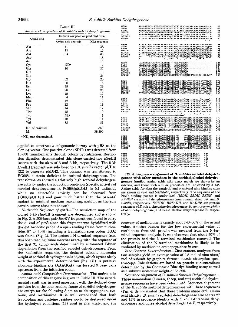
genase with other members in the sorbitol/alcohol dehydro- genase family. Amino acids with exact match are shown by an asterisk, and those with similar properties are indicated by a dot. Amino acids forming the catalytic and structural zinc binding sites are shown in bold and boldlitalic, respectively. The sequence of the NAD binding pocket is underlined. HSDH, SSDH, RSDH, and BSSDH are sorbitol dehydrogenases from human, sheep, rat, and B. subtilis, respectively. ECTDH, BSTADH, and HADHE are protein sequences of E. coli L-threonine dehydrogenase, B. stearothermophilus alcohol dehydrogenase, and horse alcohol dehydrogenase E, respec- tively.
recovery of methionine is usually about 40-60% of the actual value. Another reason for the low experimental value of methionine from this protein was revealed from the N-ter- minal sequence analysis. I t was observed that about 50% of the protein had the N-terminal methionine removed. The elimination of the N-terminal methionine is likely to be mediated by methionine aminopeptidase in uiuo.
Zinc Content Determination-Zinc content analyses from two samples yield an average value of 0.8 mol of zinc atom/ mol of subunit by graphite furnace atomic absorption spec- troscopy. Calculations are based on protein concentrations determined by the Coomassie Blue dye-binding assay as well as a subunit molecular weight of 38,390.
Sequence Alignment of B. subtilis Sorbitol Dehydrogenase- Three mammalian (human, sheep, and rat) sorbitol dehydro- genase sequences have been determined. Sequence alignment of the B. subtilis sorbitol dehydrogenase with these sequences (Fig. 4) demonstrated that these proteins share 36% amino acid identity. B. subtilis sorbitol dehydrogenase also shares 27 and 15% in sequence identity with E. coli L-threonine dehy- drogenase and horse alcohol dehydrogenase E, respectively.

B. subtilis Sorbitol Dehydrogenase 24993
DISCUSSION
Biochemical characterization of the purified B. subtilis sor- bitol dehydrogenase and cloning of gutB provide vital infor- mation regarding various properties of this enzyme including molecular size of subunit and intact enzyme, substrate speci- ficity, metal content, zinc binding site, primary structure, and sequence homology with other members in the alcohol/polyol dehydrogenase family. SDS-PAGE, gel filtration, and gutB sequence determination demonstrated that sorbitol dehydro- genase is a tetrameric enzyme with a subunit molecular weight of 38,390. The overall structure of this enzyme is similar to tha t of the mammalian sorbitol dehydrogenases.
Sorbitol dehydrogenases prepared from many different sources are reported to have a broad substrate specificity (1). However, specificity is difficult to assess since most of the enzymes were only partially purified. This includes the one from E . subtilis. In addition to sorbitol and L-iditol, B. subtilis sorbitol dehydrogenase has been reported to utilize xylitol, a pentitol, efficiently (10). In the present study, we confirm this observation. This enzyme has a strict specificity for the sec- ondary alcohol group, since it exerts no enzymatic activity with ethanol and 1-butanol as substrates. To evaluate the stereospecificity of the enzyme toward C-2, C-3, and C-4 of hexitols and pentitols, D-mannitol (C-2 epimer of sorbitol), ribitol (C-3 epimer of xylitol), and galactitol (C-4 epimer of sorbitol) were tested as substrates. As shown in Table 11, the high KIM of the enzyme with D-mannitol and galactitol as substrates suggests the stereochemical requirement at posi- tions C-2 and C-4. This property is similar to that of the mammalian sorbitol dehydrogenases (7, 21).
Mammalian sorbitol dehydrogenases are known to be mem- bers of the alcohol/polyol dehydrogenase family. Other mem- bers of this family include E. coli L-threonine dehydrogenase (23) and {"crystallin (24). Since B. subtilis sorbitol dehydro- genase shares only 14-35% of sequence identity with other members in this family, identification of highly conserved residues through homology alignment would provide vital information concerning active sites and cofactor binding site. As shown in Fig. 4, 28 residues are strictly conserved. Within the boundary defined by these 28 residues, two functional domains can be assigned. They are the zinc and cofactor (NAD) binding sites. The number of zinc binding site in this family varies from 0 to 2. The long chain alcohol dehydrogen- ase has two zinc binding sites. One of them involves the catalytic reaction and is named catalytic zinc binding site. The other is the non-catalytic or structural zinc binding site. Mammalian sorbitol dehydrogenases have only one zinc bind- ing site, which corresponds to the catalytic one. {-Crystallin has lost both sites during evolution (24). In the horse liver alcohol dehydrogenase, the three ligand residues forming the catalytic zinc binding site are Cys-46, His-67, and Cys-174. Sequence alignment indicated that the corresponding cysteine and histidine residues (Fig. 4, shown in bold type) could be found from other homologous enzymes including B. subtilis sorbitol dehydrogenase. However, Cys-174 could be aligned only with other alcohol dehydrogenases but not with sorbitol dehydrogenase. All sorbitol dehydrogenases and E. coli L- threonine dehydrogenase have glutamic acid and aspartic acid, respectively, at the corresponding position in optimal sequence alignments. Since nearby conserved cysteine resi- dues can be located in all the mammalian sorbitol dehydro- genases, it has been suggested that Cys-138 and Cys-163 (numbered according to sheep sorbitol dehydrogenase), may act as zinc ligands instead. This suggestion has been countered by modeling of sheep sorbitol dehydrogenase based on the crystal structure of horse alcohol dehydrogenase-E. Glu-156
could fit into the structural model to act as a zinc ligand. Neither Cys-138 nor Cys-163 could fit the model. Experimen- tal analysis of sheep sorbitol dehydrogenase with the extended x-ray absorption fine structure is also consistent with gluta- mate as the third zinc ligand (25). Furthermore, glutamic acid has been reported to serve as a zinc ligand in carboxypeptidase A (26) and thermolysin (27). The B. subtilis sorbitol dehydro- genase sequence presented here further strengthens the ar- guments favoring glutamic acid as the third zinc ligand, because the 2 nearest cysteines, at position 138 and 163 in sheep sorbitol dehydrogenase, are clearly replaced by valine and alanine in B. subtilis sorbitol dehydrogenase and no cysteines are even nearby. With the cloned gutB available, site-directed mutagenesis can be performed to determine the functional role of this residue in zinc binding.
The second zinc binding site of alcohol dehydrogenase is located in a short segment from residues 97-111. Four cysteine residues (Cys-97, Cys-100, Cys-103, and Cys-111) (Fig. 4, residues are shown in bold and italic characters) serve as the zinc ligands. Sequence alignment indicates that these four cysteine residues are conserved in alcohol dehydrogenases. They can also be found in both E. coli L-threonine dehydro- genase (23) and B. subtilis sorbitol dehydrogenase. It has been demonstrated that E. coli L-threonine dehydrogenase has 1 zinc atom/subunit as determined by atomic absorption and neutron activation analyses (14). However, by using the metal ion exchange experiment with 65Zn2+, this enzyme is capable of binding up to 2 zinc atoms/subunit. The binding of the second zinc atom is not essential for enzymatic activity, since full activity can be obtained with only 1 zinc atom/enzyme subunit. It is likely that B. subtilis sorbitol dehydrogenase behaves in the same manner. The significance for the presence of the second zinc binding site in this enzyme is unknown, since the second zinc binding site is absent in all the mam- malian sorbitol dehydrogenases with known sequences.
The second relatively conserved region is from residues 174 to 205 (numbered according to B. subtilis sorbitol dehydrogen- ase) (Fig. 4, underlined region). These residues form the NAD binding pocket. Secondary structure prediction using two algorithms developed by Gamier et al. (28) and Novotny (as), respectively, suggests that this region exists as a /?-T(turn)- a-/? structure, which is characteristic of the NAD binding fold. A segment with a consensus G-X-G-X-(V,I,L)-G sequence can be found in the dinucleotide binding fold in most of the dehydrogenases (8). This segment corresponds to the G-M- G-P-V-G sequence from residues 180-185 in B. subtilis sor- bitol dehydrogenase. The three highly conserved glycine res- idues are required to make sharp turns without causing steric hindrance. In fact, many glycine and proline residues to be important to the overall structure of the enzyme are con- served. With the cloned gutB, site-directed mutagenesis can be applied to determine structure-function relationships in detail. Overproduction of this enzyme would also be possible.
Acknowledgment-We thank the Bacillus Genetic Stock Center for strain 1A443 (PG668).
REFERENCES 1. Jeffery, J., and Jornvall, H. (1988) Ado. Enzymol. 61,47-106 2. Jeffery, J., Cederlund, E., and Jornvall, H. (1984) Eur. J . Biochem. 140,
3. Karlsson, C., Maret, W., Auld, D. S., Hoog, J.-0.. and Jornvall, H. (1989)
4. Jornvall, H. (1970) Eur. J . Biochem. 1 6 , 25-40 5. Karlsson, C., Jornvall, H., and Hoog, J.-0. (1991) Eur. J. Biochem. 198 ,
6. Jornvall, H., von Bahr-Lindstrom, H., and Jeffery, J. (1984) Eur. J .
7. Eklund, H., Hojales, E., Jornvall, H., Branden, C.-I., and Jeffery, J. (1985)
8. Jornvall, H., Persson, B., and Jeffery, J. (1987) Eur. J . Biochem. 167, 195-
7-16
Eur. J. Biochem. 186 , 543-550
761-765
Biochem. 140,17-23
Biochemistry 24,8005-8012
201
24994 B. subtilis Sorbitol Dehydrogenase 9. Eklund, H., Nordstrom, B., Zeppezauer, E., Soderlund, G., Ohlsson, I., 19. Wu, X.-C Nathoo S. Pang, A. S.-H., Carne, T., and Wong, S.-L. (1990)
Boiwe, T., Soderberg, B.-O., Tapia, O., Branden, C.-I., and Akeson, A. (1976) J. Mol. Bid . 102,27-59 20. Bradford M. M. (1976) Anal. Biochem. 72, 248-254
J. Bid. h e m . 265, '6845-6850
11. Kawamura, F., and Doi, R. H. (1984) J. Bacteriol. 160,442-444 10. Horwitz, S. B., and Kaplan, N . 0. (1964) J. Biol. Chem. 239,830-838 21. Maret, W., and Auld, D. S. (1988) Biochemistry 27, 1622-1628
22. Wong, S.-L., Wang, L.-F., and Doi, R. H. (1988) J. Gen. Microbiol. 134, 12. Chalumeau, H., Delobbe, A., and Gay, P. (1978) J. Bacteriol. 134,921-928 13. Hailing, s. M., Sanchez-Anzaldo, F. J., Fukuda, R., ~ ~ i , R. H., and Meares, 23. Aronsqn, B. D., Somemik R. L., Epperly, B. R., and Dekker, E. E. (1989)
3269-3276
J. B~ol. Chem. 264,5226-5232
6139 25. Feiters, M. C., and Jeffer , J (1989) Biochemistry 28, 7257-7262
K. J. (1989) in Techniques in Protein Chemistry (Hugli, T. E., ed) pp. 284-294, Academic Press, San Diego
K.'B. ( l k ) Prdc. datl. Acad. hci. U. 's. x 78,3408-3412 (1988) Nucleic Acids Res. 16, (suppl.), r315-r402 97-120
C. F. (1977) Biochemistry 16,2880-2884 14. Epperly, B, R., and &&er, E. E. (1991) J, Bioi. Chm, 266, 6086-6092 Z4. Borras9 T., Persson, B.7 and Jornvall, H. (lga9) Biochemistry Z8, 6133- 15. Matsudaira, P. (1987) J. Biol. Chen. 262, 10035-10038 16. D w n t , D. R., Keim, P. s., Chui, A. H., Bello, R., Bozzini, M., and Wilson, 26. Rees D. c Lewis M, &-,&o R. B, Li womb, w. w., and Hardman,
17. Aota, S.-L Gohobori, T., Tshibashi, F., Mawama, T., and Ikemura, T. 28. Garnier, J., Osguthorpe, D. J., and Robson, B. (1978) J( Mol. Biol. 120,
18. Tran, L., Wu, X.-C., and Wong, S.-L. (1991) J. Bacteriol. 173,6364-6372 29. Novotny, J., and Auffray, C. (1984) Nucleic Acids Res. 12,243-255
27. Holmes, M. A., and Matthews, B. W. (1981) Blochernut 20,6912-6920