somatosensory system, limbic system, and reticular...
TRANSCRIPT

Somatosensory System,Limbic System,
and Reticular Formation
Dr. Hugo Bergen, Ph.D.
Dept. Human Anatomy & Cell Science
Faculty of Medicine
E-mail: [email protected]

Objectives:
1. Describe the basic organization of the somatosensory system.
2. Describe the concepts of receptor coding and receptive fields .
3. Describe the organization of the dorsal column and spinothalamic pathways.
4. Describe the organization and function of the limbic system
5. Describe the organization and function of the reticular formation

Sensory Information
Sensory information flows from PNS to CNS via:• Cranial nerves (Head)• Spinal nerves (Body)
CNS receives info. from a variety of receptors (transduce signals into action potentials):
• Retina: vision• Inner ear: hearing and balance• Nose: smell (olfaction)• Tongue: taste• Skin: pain, temp., discriminative touch, pressure,
vibration, itch• Joints & muscles: proprioception (i.e., position,
movement, stretch/tension)
Somatosensation

Somatosensation: CN V & Spinal Nerves
• Trigeminal n. has 3 branches: supply forehead, maxilla (upper jaw), and mandible (lower jaw)
• Body supplied by 31 pairs of spinal nerves (8 cervical, 12 thoracic, 5 lumbar, 5 sacral and 1 coccygeal)
• Dermatome: area of skin innervated by the sensory fibres of a spinal nerve; accounts for the segmental innervation of the body
• Injury to 1 spinal nerve rarely causes the loss of sensitivity in a dermatome (much overlap between dermatomes)

From: Essential Clinical Anatomy 3rd
ed. by Moore & Agur (2007). LWW.

From: Essential Clinical Anatomy 3rd ed. by Moore & Agur (2007). LWW. Fig. I.22
From: Neuroanatomy: An Illustrated Colour Text (5th
Ed.) by Crossman & Neary. © 2015, Elsevier Limited.
Trigeminal N (CN V)

Tendon
Muscle
Sensory Fibres
Motor Fibres
Sensory ending
MotorEndplate
Dorsal RootGanglion

Spinal Cord Video #1
• http://aclandanatomy.com.uml.idm.oclc.org/MultimediaPlayer.aspx?multimediaId=10528251

Spinal Cord Video #2
• http://aclandanatomy.com.uml.idm.oclc.org/MultimediaPlayer.aspx?multimediaid=10528253

Somatosensory System
• Sensation is dependent on the type of receptor on the nerve ending
• The ability to discriminate the quality and type of stimulus is dependent on the specificity of the receptor
• Skin & muscle contain a number of receptor types activated by mechanical stimuli (touch, vibration, muscle length, muscle tension)
• Also contain receptors that detect temperature, chemical irritation, ‘painful’ stimuli

Somatosensory System (cont.)
Initiation of the sensation depends on the stimulus being converted into action potentials Transmission of the signal travels from the i. Receptors, which are at the end of the ii. Peripheral nerves, which are continuous with the iii. Spinal nerves, which enter the iv. Spinal cord containing thev. Ascending sensory tracts, that are relayed to thevi. Thalamus. Thalamic neurons project tovii. Sensory regions of the brain (i.e., 10 sensory cortex)

Somatosensory System (cont.)
Somatosensation involves:1. Sensory info.
o Nerve impulses generated by the original stimuli2. Sensation
o Awareness of stimuli by the senses3. Perception
o Interpretation of the sensation into meaningful formso Interaction between brain & environmento e.g., perceive a stimulus as ‘painful’ and respond to
decrease the pain
• What regions of the CNS do these functions occur in?

Sensory Coding: Place & Pattern
1. Place: stimulus is only detected if the place where the receptor specific to a certain stimuli is found, is stimulated.
2. Pattern: perceived sensations result from the activation of more then 1 receptor type in more then 1 place, with the relay of info to the brain occurring along more then 1 pathway.

Rate of Coding & Receptor Adaptation
Rate coding: stimulus intensity increases (beyond threshold for initiation of an AP) and this is coded as an increased rate of firing of the afferent fiber; i.e. receptors detect stimulus & intensity
Receptor Adaptation: Receptors are able to adapt to a maintained stimulus (eg. presence of clothing).
1. Slowly adapting (tonic)– continue firing for a prolonged period during the maintained stimulus
2. Rapidly adapting (phasic) – receptors cease firing soon after stimulus is applied.

Sensory Receptor Classification
Divide receptors on the basis of the stimulus to which they are most sensitive (‘adequate stimulus’)
1. Chemoreceptors: smell, taste, and internal stimuli like pH, O2, CO2 , metabolites
2. Photoreceptors: light (retina)
3. Thermoreceptors: temperature & temp. changes
4. Mechanoreceptors: many types including auditory and vestibular receptors, touch, muscle tension
5. Nociceptors: ‘pain’ receptors (mechanical & chemical)

Sensory Receptors: Receptive Fields
• Receptors differ in their receptive field, which is the area where adequate stimulus produces activation; tested by 2-point discrimination,
• Also depends on anatomical arrangement

Sensory Receptors: Receptive Fields
• Area of skin specific to each individual receptor
• Sensitivity relies on # of sensory endings
• 2-pt. discrimination depends on anatomical arrangement

Afferent Nerve Characteristics
For somatosensation, there are different fibres types; a 100x range in axon diameter and conduction velocity (Note that larger fibres = faster conduction; myelinated> unmyelinated):
1. Touch, ,Vibration and Proprioception: larger myelinated fibres (conduction velocity in the 90 -120 m/sec range)
2. Pain and Temperature: small myelinated fibres and unmyelinated fibres (conduction velocity in the 0.5 – 5 m/sec range)

Axon Diameter and Conduction
1. Large myelinated fibres
o Muscles, tendons and joints
2. Medium:
o Joint capsules, Muscle spindles, Cutaneous touch & stretch, Pressure
3. Small
o Pain, crude touch / itch, temperature

Somatosensation: Proprioception
• Provides info on position of joints, tension on tendons, stretch on muscles (can be static or dynamic).
• Self regulation of posture & movement

Muscles Spindles
• Stretch receptors contained within muscle and may be stretched passively (reflex) or actively
• Intrafusal fibres innervated by large (Ia) fibres
• Surrounded by extrafusalfibres (‘ordinary’ muscle fibres)
From: Fitzgerald’s Clinical Neuroanatomy and Neuroscience (7th Ed.) by Mtui, Gruener, & Dockery. © 2016, Elsevier Limited.
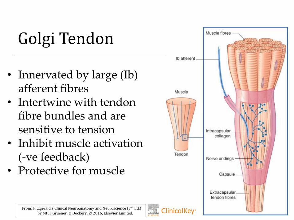
Golgi Tendon
• Innervated by large (Ib) afferent fibres
• Intertwine with tendon fibre bundles and are sensitive to tension
• Inhibit muscle activation (-ve feedback)
• Protective for muscle
From: Fitzgerald’s Clinical Neuroanatomy and Neuroscience (7th Ed.) by Mtui, Gruener, & Dockery. © 2016, Elsevier Limited.

Joint Receptors
Ligament Receptors (Ib):• sensitive to tensionRuffini Endings:• Sensitive to changes in range of
motionPaciniform Corpuscles:• Activated by extremes in range of
motion and vibrationFree Nerve Endings (Ad & C):• responsive to inflammation

Somatosensation:
Skin Receptors - Encapsulated Endings
Consists of nerve ending within a connective tissue capsule:
1. Meissner’s corpuscles – light touch; textured surfaces (5 mm elevation is detectable!). Located at dermis epidermis interface they have a small receptive field (rapidly adapting)
2. Ruffini corpuscles – sensitive to stretch of skin, with a large receptive field; deeper in the dermis (slowly adapting)
3. Pacinian corpuscles – sensitive to vibration (& touch), with a large receptive field; deep within dermis (rapidly adapting)

Skin Receptors: Nerve Endings
Consists of nerve ending without a connective tissue capsule:
1. Follicular Nerve Ending– nerve endings closely associated with hair follicles. Sensitive to bending of hair. Have a small receptive field (rapidly adapting).
2. Merkel cells – sensitive to sustained pressure on skin, with a small receptive field; Nerve ending closely associated with a Merkel cell in base of epidermis. (slowly adapting)
3. Free Nerve endings – some are thermoreceptors (sensitive to warm or cold); some are nociceptors, sensitive to pain a large receptive field; also deep in dermis. Also sense mechanical deformation and chemical irritants

Skin showing free nerve endings (B), Merkel cells (C), and Follicular endings (D)
From: Fitzgerald’s Clinical Neuroanatomy and Neuroscience (7th Ed.) by Mtui, Gruener, & Dockery. © 2016, Elsevier Limited.

Skin showing Meissner’s corpuscle (C), Ruffini endings (D), and Pacinian endings (E)
From: Fitzgerald’s Clinical Neuroanatomy and Neuroscience (7th Ed.) by Mtui, Gruener, & Dockery. © 2016, Elsevier Limited.

Anatomical Organization of Somatosensory System

Somatosensory System
• Conscious somatosensation uses a relay of 3 neurons from periphery to the 10 somatosensory cortex in the parietal lobe (conscious perception)
• Uses specific pathways/tracts to get to cortex
3 major pathways:
1. Dorsal Column: touch, vibration & consciousproprioception
2. Spinothalamic Tract: pain and temp.
3. Trigeminal System: somatosensation from the face

1. Dorsal Column / Medial lemniscus
• The 1st order neuron is in the dorsal root ganglion. It extends axon out to periphery and ends in receptor.
• Also extends axon into spinal cord where it ascends in the dorsal part of the spinal cord to the medulla (fasciculus gracilis [leg] & fasciculus cuneatus [arm])
• Synapses onto 2nd order neuron in medulla that projects to contralateral thalamus as the medial lemniscus. 3rd order neuron from thalamus to cortex

3rd order neuron
2nd order neuron
1st order neuron
Dorsal Column / Medial Lemniscus System

2. Spinothalamic Pathway
• The 1st order neuron is in the dorsal root ganglion. It extends axon out to periphery and ends in receptor.
• Also extends axon into spinal cord where it synapses onto 2nd order neuron in the gray matter of spinal cord
• 2nd order neuron in cord sends axon across the midline and projects to contralateral thalamus.3rd order neuron from thalamus to cortex

3rd order neuron
2nd order neuron
1st order neuron
Spinothalamic / Anterolateral System
1st order neuron
2nd order neuron

3. Trigeminal System
• The 1st order neuron is in the trigeminal ganglion. It extends axon out to periphery and ends in receptor.
• Also extends axon into brainstem where it synapses onto 2nd order neuron
• 2nd order neuron in brainstem nucleus sends axon across the midline and projects to contralateral thalamus. 3rd order neuron from thalamus to cortex

Key Features
• The cell body of 1st order neuron is in a dorsal root ganglion or trigeminal ganglion.
• 2nd order neuron is in the CNS and its axon crosses the midline to project to the thalamus
• 3rd order neuron is in the thalamus and its axon projects (via internal capsule) to the 10
somatosensory cortex in the parietal lobe in a topographic manner (somatosensory homunculus)

Cerebral Cortex
• The cortex has been mapped out according to microscopic features by Brodmann in 1909
• Each colour represents an area
and is numbered 1-52.

Cortical Somatotopy (Maps of the body
on the cortex)
• Wilder Penfield (McGill/MNI) neurosurgeon
• Stimulated the cortex of awake patients and developed the motor and sensory homunculi

Primary Sensory Cortex
• In addition to somatosensory cortex, also have 10
auditory cortex (temporal lobe), 10 visual cortex (occipital lobe), and 10 olfactory cortex (temporal lobe)
• 10 cortex discriminates intensities & qualities of the sensory information
• 10 auditory ctx discriminates loudness and pitch
• 10 visual ctx discriminates shapes, sizes, location
• 10 somatosensory ctx discriminates, texture, shape and size

1. Primary Sensory Cortex

Sensory Association Cortex
• Association cortex carries out higher order interpretation, analysis and processing of sensory information
• Receives input from 10 sensory cortex
• Auditory association ctx important for understanding and producing language
• Visual association ctx interprets visual images, recognition of faces etc.
• Somatosensory association ctx recognition and identification of objects by touch

2. Sensory Association Areas

Association Cortex
• Areas of cortex not involved with sensation or movement (heteromodal association cortex)
• Houses very complex abilities.o Personality and goal oriented behaviour
o integration & interpretation of sensations
o processing of memory
o generations of emotions
o Connects to many different areas of cortex

Limbic System
• Provides the basis for giving subjective properties (feelings/ emotions) to a wide variety stimuli (e.g., music, photographs, effect of colors on emotions)
• Aspects of memory (emotional & declarative)
• Transmits signals via somatic, reticular, hormonal & autonomic pathways.
• The limbic system is the source of emotions and instincts (organizes inputs into complex behavior)
• Regulates feeding, drinking, defensive, reproductive behaviors, fear, aggression, as well as visceral & hormonal functions

Limbic System
What does it do? A number of things:
Market Researcher: “..approximately 80% of all decision making is done at the level of the limbic system -- our…emotional level.”
• Is intimately tied to decision making
• Regulates emotional behavior and visceral responses to emotion (think “road rage”)
• Sometimes referred to as the ‘emotional brain’

Limbic System: Components
• Several evolutionarily older cortical and sub-cortical structures
Includes:
• Hippocampus
• Cingulate gyrus
• Olfactory system
• And others
Connects to hypothalamus (controls Autonomic NS and Endocrine System)

Location of Limbic System

Limbic System
• Provides the feelings of love, generosity, altruism….and hate, greed, resentment, & rage; also involved in motivation and drug addiction
• A number of psychiatric diseases (e.g., depression, anxiety, PTSD, & other affective disorders) may involve the Limbic System
• Alzheimer’s disease affects the hippocampus
• Drug addiction involves limbic system
Limbic System: Stress Response
• Emotions influence function of all organs because of a bi-directional connection between nervous & immune systems
• Reaction to experiences may trigger a stress response, which disrupts the body’s homeostasiso Increase in muscle tension (somatic system)
o Increase blood flow to muscles, decrease blood flowtoorgans (autonomic)
o Increase in HR and BP (hormone release - epinephrine)
o Mediated by Autonomic NS and Endocrine system

Short Term Response to Emotional Stress

Long Term Response to Emotional Stress

Reticular Formation (RF)
• Complex network of connected nuclei located in the brainstem
Its functions include regulation of:
o Consciousness and sleep
o Attention and arousal
o Pain
o Movement
o Cardiovascular activity
o Respiratory activity

Basic Organization of RF
The nuclei are arranged in 3 zones:
1) Median/Midline Zone: these are aka the raphe nuclei and have serotonin as the neurotransmitter
2) Medial Zone (aka paramedian zone): contains the nuclei that have dopamine, noradrenaline, and adrenaline as their neurotransmitter
3) Lateral Zone: receive sensory information and are largely involved in visceral function

From: Fitzgerald’s Clinical Neuroanatomy and Neuroscience (7th
Ed.) by Mtui, Gruener, & Dockery. © 2016, Elsevier Limited.
Reticular Nuclei

Serotonin
From: Fitzgerald’s Clinical Neuroanatomy and Neuroscience (7th
Ed.) by Mtui, Gruener, & Dockery. © 2016, Elsevier Limited.

Serotonergic Neurons: Functions
• Activity of the neurons that project to the forebrain fluctuates with sleep and wakefulness
• These neurons are thought to determine the overall level of arousal and receive input from numerous sensory systems
• Enhancing serotonin: treatment of depression
• Neurons that project to the brainstem (PAG) and spinal cord (dorsal horn) decrease pain transmission

Noradrenaline
From: Fitzgerald’s Clinical Neuroanatomy and Neuroscience (7th
Ed.) by Mtui, Gruener, & Dockery. © 2016, Elsevier Limited.

Noradrenergic Neurons: Functions
• Changes in activity of the neurons is associated changes in sleep and wakefulness
• Are largely silent during REM sleep, lightly active during non-REM sleep, and increase in activity during wakefulness
• These neurons are thought to be especially important in promoting attention and vigilance

Dopamine
From: Fitzgerald’s Clinical Neuroanatomy and Neuroscience (7th
Ed.) by Mtui, Gruener, & Dockery. © 2016, Elsevier Limited.

Dopaminergic Neurons: Functions
• Nigrostriatal projections are important in initiating motor activity
• DA projections are considered to be important in cognition and motivation
• Many drugs of abuse are thought to target the DA system which increases the sensation of pleasure (‘reward circuit’)
• Imbalance in DA transmission may underlysome forms of mental illness (schizophrenia)

From: Nolte’s The Human Brain (7th Ed.) by Vanderah & Gould. © 2016, Elsevier Limited. Fig. 11-28.
Acetylcholine

Cholinergic Neurons: Functions
• Activation of cholinergic neurons that project to cortex are involved in heightened arousal
• Modulate the sleep - wakefulness cycle; REM sleep is associated with the activation of the pontine cholinergic neurons (decreased activity of the Noradrenergic neurons)
• May be involved in learning and memory
• Alzheimer’s disease is associated with loss of cholinergic neurons in the basal forebrain