solvent-dependent conformational behaviour of lipochitoligosaccharides related to nod factors
TRANSCRIPT

www.elsevier.nl/locate/carres
Carbohydrate Research 318 (1999) 10–19
Solvent-dependent conformational behaviour oflipochitoligosaccharides related to Nod factors
Leandro Gonzalez a, Manuel Bernabe a, Juan Felix Espinosa a, Pilar Tejero-Mateo b,Antonio Gil-Serrano b, Natacha Mantegazza c, Anne Imberty c, Hugues Driguez c,
Jesus Jimenez-Barbero a,*a Instituto Quimica Organica, CSIC, Juan de la Cier6a 3, E-28006 Madrid, Spain
b Departamento de Quımica Organica, Facultad de Quımica, Uni6ersidad de Se6illa, E-41071 Se6ille, Spainc Centre de Recherches sur les Macromolecules Vegetales, CERMAV-CNRS 1, BP 53, F-38041 Grenoble, France
Received 17 November 1998; accepted 29 March 1999
Abstract
The solution conformation of two lipooligosaccharides related to Nod factors or lipochitoligosaccharides havebeen analysed by 1D and 2D 1H and 13C NMR spectroscopy, molecular mechanics and dynamics calculations. Theobtained data indicate that the glycosidic torsion angles have restricted fluctuations, but may adopt a variety ofshapes. Remarkably, the relative orientation of the fatty acid chain towards the oligosaccharide backbone is solventdependent. In water solution, the acyl residue and the oligosaccharide adopt a quasi-parallel orientation for asignificant amount of time. © 1999 Elsevier Science Ltd. All rights reserved.
Keywords: Conformational analysis; Nuclear magnetic resonance; Molecular dynamics; Nodulation factors
(Nod) factors or lipochitoligosaccharides(LCOs).
The Nod factors are oligosaccharides con-sisting of a backbone of three to five GlcNAcresidues, bearing an amide-bound fatty acylresidue (saturated or unsaturated) on the non-reducing terminal glucosamine residue. Thisbasic skeleton has structural variations thatdetermine the host-specificity. Compoundswith O-sulfate, O-carbamoylate, and N-methyl have been identified, and differentstudies have indicated a close relationship be-tween host specificity and Nod factor struc-ture, regardless of the taxonomy of thebacterial symbiont. Tetra- or pentasaccharideswhose reducing ends are glycosylated at posi-tion O-6 by either a-fucosyl or 2-O-methyl-a-fucosyl residue have also been described.Recently, a structure that bears a fucosylresidue a-(1�3)-linked to the N-acetylglu-
1. Introduction
Rhizobia are nitrogen-fixing bacteria thatare able to invade the roots of leguminousplants and trigger the formation of nodulesthat contain the nitrogen-fixing microsym-biont. Infection and nodule development arehighly specific and depend on both partners,the bacterium and the plant, to be effective.During the symbiotic process, the host plantroots secrete flavonoids that induce the rhizo-bial nodulation genes (nod and nol genes) [1–3]. These genes have been shown to beinvolved in the synthesis and excretion ofbacterial nodulation signals called nodulation
* Corresponding author. Tel.: +34-91-562-2900; fax: +34-91-564-4853.
1 Affiliated with J. Fourier University.
0008-6215/99/$ - see front matter © 1999 Elsevier Science Ltd. All rights reserved.
PII: S 0 0 0 8 -6215 (99 )00082 -8

L. Gonzalez et al. / Carbohydrate Research 318 (1999) 10–19 11
cosaminyl residue proximal to the nonreducingterminal N-acetylglucosaminyl residue and anovel minimal structure having only a N,N II-di-acetyl-chitobiosyl backbone structure havebeen identified [4].
The amino group of the nonreducing endglucosaminyl residue is acylated with C-16 orC-18 fatty acid chains with one to four doublebonds such as those of cis-vaccenic (C18:1). Itis also usually N-methylated.
Thus, the host-specificities of Rhizobiumleguminosarum bv. 6iciae and trifolii are deter-mined by the lipid moiety [5]. The sulfate groupon O-6 of the reducing glucosaminyl residue isnecessary for nodulation by Sinorhizobiummeliloti of alfalfa plants [6]. Finally, bacteriathat are symbionts of soybean (Bradirhizobiumjaponicun and Sinorhizobium fredii ) and thebroad host-range Rhizobium sp. NGR234 pro-duce nodulation factors that are 6-O-substi-tuted on the reducing glucosaminyl residue witha 2-O-methyl fucosyl residue [1,7–10].
The structures of the LCOs produced bySinorhizobium fredii HH103 have been reported[10] and shown to consist of a backbone ofchitin oligomers ranging from trimer to pen-tamers, substituted by fucosyl (minor compo-nents) and 2-O-methyl fucosyl (majorcomponents) at the reducing terminal N-acetyl-glucosamine residue and N-acylated at thenon-reducing terminal residue with octade-canoic acid, cis-octadec-11-enoic acid, cis-hex-adec-9-enoic acid or hexadecanoic acid.Further studies have shown that the presence of2-O-methyl-fucosyl in the LCOs produced byS. fredii strain HH103 is important for theefficiency of nodulation on some legumes asCajanus cajan and for the bacterial competitivecapacity to nodulate soybean.
The biological relevance of these molecules
has encouraged several groups to performstructural studies of different Nod factors.Several reports on the synthesis of several LCOsand analogues have also been described [11]. Inmany examples, biological properties ofmolecules strongly depend on their conforma-tion and their configuration. Amide Z/E iso-merism due to hindered rotation has beenstudied in simple derivatives [12], but, to thebest of our knowledge, no detailed conforma-tional analysis on these molecules has beenreported so far. The conformation of chi-tooligosaccharides has been studied in severalinstances, since chitobiose is a key componentof the glycoprotein oligosaccharide chains.However, the relative orientation of theoligosaccharide and lipid chains is not preciselyknown, along with the extent of flexibilityaround the glycosidic linkages of the backboneand the pendant groups.
We report here on the conformational studiesof two of these LCOs in solution, namely 1 and2, by using NMR and molecular mechanics anddynamics calculations (Fig. 1). These com-pounds have been isolated [10] and synthesized[13] in Seville and Grenoble, respectively. It isshown that the backbone displays a majorconformation, similar to that described forregular chitooligosaccharides, but that the rela-tive orientation of the fatty acid chain withrespect to the backbone is solvent dependent.In particular, in water solution, the acyl chainspends a significant time adopting a quasi-par-allel orientation to the oligosaccharide chain. In1, the 6-O-linked 2-O-methyl fucosyl residuemay adopt a variety of conformations.
2. Experimental
Molecular mechanics and dynamics calcula-tions.—Molecular mechanics and dynamicscalculations were performed using both theCVFF force field [14] within the INSIGHT II/DIS-COVER programs of BIOSYM technologies(San Diego, CA, USA) and the MM3* forcefield as implemented in MACROMODEL 4.5 [15].F is defined as H1%�C1%�O�C4 and C asC1%�O�C4�H4. For the 6-O-linked 2-O-methylfucose residue, F is defined as H1¦�C1¦�O6�C6and C as C1¦�O6�C6�C5. Only the gg orienta-Fig. 1. Schematic view of LCOs 1 and 2.
L. Gonzalez et al. / Carbohydrate Research 318 (1999) 10–1912
tion of the lateral chain was used for theb-GlcNAc moieties of the backbone, exceptfor the 6-O-substituted one. For the 1�6linkage, both gg and gt rotamers [16] wereconsidered for the b-glucosaminyl moiety. Sep-arate calculations for a dielectric constant o=80 (with CVFF and MM3* for compound 1) andfor the continuum GB/SA solvent model (withMM3* for compound 2) were performed [17].First, potential-energy maps were calculatedfor the constituent disaccharide (N,N %-diacetylchitobiose): relaxed (F,C) potential-energymaps were calculated as described [18]. Oneintial geometry of the secondary hydroxylgroups of the pyranosyl moieties was consid-ered, r (reverse clockwise). The previous stepinvolved the generation of the correspondingrigid residue maps by using a grid step of 18°.Then, every F,C point of this map was opti-mised using 200 steepest descent steps, fol-lowed by 1000 conjugate gradient iterations.From these relaxed maps, the probability dis-tributions were calculated for each f, c pointaccording to a Boltzmann function at 303 K.
Starting structures of both oligosaccharides1 and 2 were built by combining the morestable conformers of the different glycosidiclinkages and subjecting them to extensive en-ergy minimization with conjugate gradients.An extended structure of the acyl chain wasused as input geometry. The sulfate group wasemployed in the MM3* simulations using theparameters developed by Perez [19]. Then,these structures were used as starting ge-ometries for molecular dynamics (MD) simu-lations [20] at 300 K. The CVFF (o=80,compound 1) and the MM3* force field (o=80,compound 1, GB/SA, compound 2) [17] wereemployed with a time step of 1 fs. The equili-bration period was 100 ps. After this period,structures were saved every 0.5 ps. The totalsimulation time was between 2 and 3 ns forevery run. Average distances between intra-residue and inter-residue proton pairs werecalculated from the dynamics simulations.
NMR spectroscopy.—NMR experimentswere recorded on a Varian Unity 500 spec-trometer, using an approximately 3 mg mL−1
solution of the lipooligosaccharides at differ-ent temperatures. The spectra for lipopen-tasaccharide 1 were recorded in a 49:1
DMSO-d6–D2O solvent mixture, due to solu-bility problems in pure water. Those spectrafor sulfated lipotetrasaccharide 2 were ac-quired in D2O. Chemical shifts are reported inppm, using the residual HDO signal (4.71ppm) and external TMS (0 ppm) as references.The double quantum filtered COSY spectrumwas performed with a data matrix of 256×1K to digitize a spectral width of 2000 Hz.Sixteen scans were used with a relaxation delayof 1 s. The 2D TOCSY experiment was per-formed using a data matrix of 256×2 K todigitize a spectral width of 2000 Hz. Fourscans were used per increment with a relax-ation delay of 2 s. MLEV 17 was used for the100 ms isotropic mixing time. The one-bondproton–carbon correlation experiment wascollected using the gradient-enhanced HMQCsequence [21]. A data matrix of 256×2 K wasused to digitize a spectral width of 2000 Hz inF2 and 10,000 Hz in F1. Four scans were usedper increment with a relaxation delay of 1 sand a delay corresponding to a J value of 145Hz. 13C decoupling was achieved by theWALTZ scheme. The 2D-HMQC-TOCSY ex-periment was conducted with 80 ms of mixingtime (MLEV 17). The same conditions as forthe HMQC were employed. HMBC experi-ments were performed using the gradient en-hanced sequence with a data matrix of 256×2K to digitize a spectral width of 2000×15,000Hz. Eight scans were acquired per incrementwith a delay of 65 ms for evolution of longrange couplings.
2D NOESY, 2D ROESY, and 2D T-ROESY experiments were performed usingfour different mixing times, namely150, 300, 450, and 600 ms, with 256×2 Kmatrices. Good linearity was observed up to200 ms (NOESY) and 300 ms (ROESY). Esti-mated errors in the NOE intensities are smallerthan 20%.
3. Results and discussion
Conformational analysis: molecular dynamicsstudies.—In a first step to determine the over-all three-dimensional structure of the lipo-oligosaccharides, molecular mechanics and dy-namics calculations were performed. Infor-

L. Gonzalez et al. / Carbohydrate Research 318 (1999) 10–19 13
Table 1Torsional angle values (F,C) of the predicted minima andMM3* (GB/SA solvent model) and CVFF (o=80) populationsof the low-energy regions of 1 and 2 a
b(1�4) a(1�6)linkagelinkage
B C AMM3* A48/5Torsion −40/173 −52/−175175/−6
angle(F/C)
96.2 3.7Population 0.1 100.0(%)
B CCVFF AATorsion 55/2 178/4−52/ −58/−179
angle−172
(F/C)98.7 1.3 B0.1Population 100.0
(%)
a The regions around F extend ca. 25° and around C ca.35°.
dependence of the results on the dielectricconstant used [22f]. First, relaxed potential-en-ergy maps (F/C) were calculated for N,N II-di-acetyl-chitobiose [22e]. The glycosidic linkagepresents well-defined low-energy regions,which cover less than 20% of the completepotential energy surface. All the energy re-gions show F values (Table 1) which arecentred around those expected for the exo-anomeric effect [23], although the expectedprobability distributions occupy a wide sur-face. Then, two models of the pentasaccharide1 were built, composed of all the differentglycosidic linkages of the polysaccharide andthe fatty acid chain, and submitted to different2 or 3 ns simulations again using both forcefields. Both gg and gt conformations of thelateral chain of the 2-O-methyl Fucp-(1�6)-b-D-GlcNAc linkage were considered. For alllinkages, the glycosidic torsion angles cover awell-defined part of the complete F/C map,which is fairly independent of the rotamerpresent at the hydroxymethyl group of thereducing b-D-GlcNAc moiety. Independentlyof the force fields used, the glycosidic torsionangles F and C show oscillations centred at60 and 0° (Fig. 2). In all cases, these torsionaloscillations are more pronounced around C,as expected by the operativity of the exo-anomeric effect around F. Some excursions tothe anti regions are evident. In terms of theavailable space for the interglycosidic torsionangles, the calculated results are fairly similarfor the different b-(1�4)-linkages, almost in-dependently of the particular structure of the
mation on the accessible amount of conforma-tional space was obtained through moleculardynamics simulations [20] of 1 and 2. Simula-tions with the DISCOVER-CVFF and MM3* pro-grammes were performed, since they haveprovided satisfactory results in the study ofthe conformation of a variety of oligosaccha-ride molecules [22]. For uncharged compound1, both force fields were shown to providesimilar results as also described for smallerdisaccharides [22f]. Simulations at a highdielectric constant were performed, accordingto our previous studies which demonstratedthat for these two force fields there is a minor
Fig. 2. Trajectory plots of the MD simulation (1 ns) of 1 by using MM3* (o=80). The trajectories of the simulation in F/C spacefor two of the b(1�4) glycosidic linkages are shown. The trajectories of the b(1�4) glycosidic linkages during the CVFF (o=80)simulation of 1 and the MM3* (GB/SA) simulation of 2 are basically identical.
L. Gonzalez et al. / Carbohydrate Research 318 (1999) 10–1914
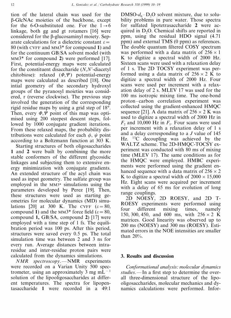
Fig. 3. Trajectory plots of the MD simulation (1 ns) of 1 by using MM3* (o=80). The relevant information for the fucoseglycoside linkage is presented. The trajectory of this a(1�6) glycosidic linkage during the CVFF (o=80) simulation is basicallyidentical.
N-acyl moiety. For the F/C values (Table 1)sampled during these trajectories, no impor-tant interaction of the Fuc moiety with thecontiguous GlcNAc residue takes place. Infact, with respect to the accessible conforma-tional space for the (1�6)-linkage (Fig. 3),the observed results are also independent onthe conformation (gg or gt) of the exocyclicchain and indicate a larger accessible area ofconformational space. In most of the cases,several transitions between the rotamers of thehydroxymethyl group were observed. Never-theless, the hydroxymethyl groups displayedeither the gg or the gt conformations for mostof the simulation time (\90%) Finally, aver-age expected interproton distances from thedifferent MD simulations (Table 2) were esti-mated and compared to those observed exper-imentally. No close proton–proton distanceswere deduced from the MD models which
Table 2Average relevant proton–proton inter-residue distances fromthe MM3* (o=80) MD simulations for 1 (starting from ggand gt conformers for the a(1�6) linkage of 1) and experi-mentally observed NOEs (strong (s), medium (m), weak (w)) a
NOE intensitygg gt
AH1/BH6S m2.86 3.12s2.37AH1/BH6R 2.47
2.36 2.38 sCH1/BH4CH1/BH6S m2.533.07
2.90 m2.53CH1/BH6RsDH1/CH4 2.43 2.36
2.93DH1/CH6S 2.80 mDH1/CH6R 3.36 3.00 m
2.45 2.39EH1/DH4 s2.91 2.51EH1/DH6S m
EH1/DH6S 3.35 2.92 m
a The average simulation results for both compounds arebasically identical independently of the force field (MM3* orCVFF) and conditions (o=80 or GB/SA) used.

L. Gonzalez et al. / Carbohydrate Research 318 (1999) 10–19 15
Fig. 4. Superimposition of several snapshots taken from the MD simulations of 1 (MM3*, o=80, bottom) and 2 (MM3*, GB/SA,top). The existence of an important amount of flexibility around the glycocidic linkages is evident.
were not apparent in the experimental NOEdata.
A superimposition of different conformersfound in the MD simulation is shown in Fig.4. It is shown that despite the relatively nar-row variation of the glycosidic torsion angles,the conformational space accessible to thelipid chain may be fairly large. Regarding theorientation of the lipid chain, it is observedthat it may display a variety of orientationwith respect to the sugar residues. In fact, forsome time, it may remain in close contact withthe sugar residues, adopting a quasi-paralleldisposition with regard to the tetrasaccharidebackbone, making van der Waals contactsbelow 3.7 A, .
Regarding the sulfated tetrasaccharide 2,GB/SA simulations were carried out with theMM3* programme. Similar results were ob-served to those described above for 1, bothregarding the oligosaccharide backbone (Fig.5) and the lateral chain. Thus, the obtainedtrajectories do not depend either on the size ofthe oligosaccharide or the nature of the acylchain. A large variation on the orientation ofthe O-sulfate chain were observed. Neverthe-
less, for the F values (Table 1) sampled duringthe trajectory, which are those expected forthe exo-anomeric effect, no important interac-tion of the sulfate group with the contiguousGlcNAc residue takes place. A superimposi-tion of different conformers found in the MDsimulation is shown in Fig. 6.
1H NMR data.—Since NMR parametersare essentially time averaged, the informationthat it is possible to deduce from these experi-ments corresponds to the time-averaged con-formation in solution. The validity of thetheoretical results has been tested using NMRmeasurements of vicinal coupling constantsand NOEs [24,25]. 1H NMR and 13C NMRspectra were completely assigned by a combi-nation of homonuclear COSY, TOCSY, andheteronuclear HMQC, HMBC, and HMQC-TOCSY techniques. The corresponding 1Hand 13C NMR chemical shifts are listed inTables 3 and 4. In any case, severe overlap-ping for the central GlcNAc units was ob-served and the conclusions should be regardedas semiquantitative. For both oligosaccha-rides, the pyranoid rings can be described asessentially monoconformational: 4C1, as de-

L. Gonzalez et al. / Carbohydrate Research 318 (1999) 10–1916
duced from the vicinal proton proton cou-plings (data not shown). Then, NOESY andROESY experiments were used to qualita-tively estimate proton/proton inter-residuedistances [25]. For 1, all NOESY cross peakswere negative at 500 MHz and 299 K. Thedistance results estimated by using the isolatedspin pair approximation are given in Table 2.Although only approximated, the Fuc H-1/H-2 intra-residue signals were used as reference
(2.5 A, ). Since overlapping is present, thesedata should be regarded as merely qualitative.Thus, the NOEs were assigned as strong (s),medium (m), and weak (w). The MD-calcu-lated distances are shown in Table 2. It can beobserved that a good matching is observedbetween the distances found experimentallyand those estimated through molecular me-chanics and dynamics simulations. The ob-tained results may indicate that, as least for
Fig. 5. Trajectory plots of the MD simulation (1 ns) of 2 by using MM3* (GB/SA). The F/C histories of the simulation for oneof the b(1�4) glycosidic linkage and the history of v, NH, and lipid chain torsion angles are shown.

L. Gonzalez et al. / Carbohydrate Research 318 (1999) 10–19 17
Fig. 6. Superimposition of several snapshots taken from the MD simulations of 2 (MM3*, GB/SA). The possibility of aquasi-parallel orientation of the acyl chain with respect to the oligosaccharide backbone has been emphasised.
Table 31H NMR data (d ppm) of 1 in DMSO-d6 and of 2 in D2O a
H1Ring H2 H3 H4 H5 H6S H6R
3.28 3.66 3.46A 3.794.91 1.04 3.373.58/3.74 3.76/3.94 3.46/3.70 3.60/4.09 3.46/4.25 3.59/4.12Ba 4.85/5.193.44/3.73 3.44/3.72 3.36/3.724.41/4.72 3.36/3.74Bb 3.28/4.17 3.35/4.12
4.35/4.56C 3.48/3.78 3.23/3.58 3.22/3.61 3.29/3.71 3.28/3.88 3.59/3.713.48/3.78 3.23/3.52D 3.22/3.614.35/4.56 3.29/3.71 3.36/3.86 3.61/3.683.43/3.82 3.24/3.58 3.01/3.49 3.15/3.50 3.69/3.94 3.35/3.774.32/4.60E
a 1H NMR signals for the lipid chain: (1) double bond, 5.31. CH3 group, 0.84 ppm, amide linked �CH2� 2.05 ppm, double bondlinked �CH2� 1.97 ppm, rest of the lipid chain, 1.16–1.30 ppm. NHAc groups, 1.76–1.82 ppm; (2) double bond, 5.44. CH3 group,0.88 ppm, amide linked �CH2�, 2.30 ppm, double bond linked �CH2�, 2.06 ppm, rest of the lipid chain, 1.28–1.35 ppm. NHAcgroups, 2.03–2.08 ppm.
Table 413C NMR data (d ppm) of 1 in DMSO-d6 and of 2 in D2O a
78.0 68.6A 72.496.5 65.6 16.0 58.0Ba 91.0, 92.0 56.3, 54.4 68.1, 72.8 71.6, 79.2 68.6, 69.2 65.4, 66.8 –Bb 101.5, 98.6 56.4, 55.6 72.4, 72.8 80.4, 79.2 80.4, 69.2 62.7, 66.8 –
54.2, 56.0 74.4, 75.6 81.4, 80.0101.5, 103.7 81.0, 80.4C 59.9, 60.8 –54.2, 56.0 74.4, 75.6 81.4, 80.0 81.0, 80.4D 59.9, 60.8101.5, 103.7 –54.8, 55.4 74.4, 74.4 70.4, 71.2 76.5, 76.8103.0, 103.7 60.7, 61.6E –
a 13C NMR signals for the lipid chain: (1) double bond: 130.0 ppm. CH3 groups: 13.8 ppm, amide linked �CH2�: 35.6 ppm,double bond linked �CH2�: 26.5 ppm, rest of the lipid chain: 25.0, 29.0, 21.8, 28.3, 31.2 ppm. NHAc groups: 22.6 ppm. (2) doublebond: 132.0 ppm. CH3 groups: 14.8 ppm, amide linked �CH2�: 36.8 ppm, double bond linked �CH2�: 28.0 ppm, rest of the lipidchain: 26.4–29.6 ppm. NHAc groups: 24.3 ppm. CO groups: 173.2–173.8.
this particular case, these unrestrained MDsimulations [26] provide a fair description of themotion around the different glycosidic linkagesof this molecule. Regarding the acyl chain, noNOE contacts between its protons and those ofthe oligosaccharide, apart from the trivial oneswith H-2 of the non-reducing end, were ob-served, probably indicating that this chain mayadopt a variety of conformations.
Therefore summarizing the theoretical and
experimental results, it seems that there is animportant amount of conformational freedomfor the glycosidic and exocyclic torsion anglesof the lipopentasaccharide, although a repre-sentative major conformer may be deducedfrom Fig. 4.
For 2, spectra were recorded in pure heavywater. NOESY cross peaks were again neg-ative (Fig. 7) at 500 MHz and 299 K. Theestimated interproton distances are given in
L. Gonzalez et al. / Carbohydrate Research 318 (1999) 10–1918
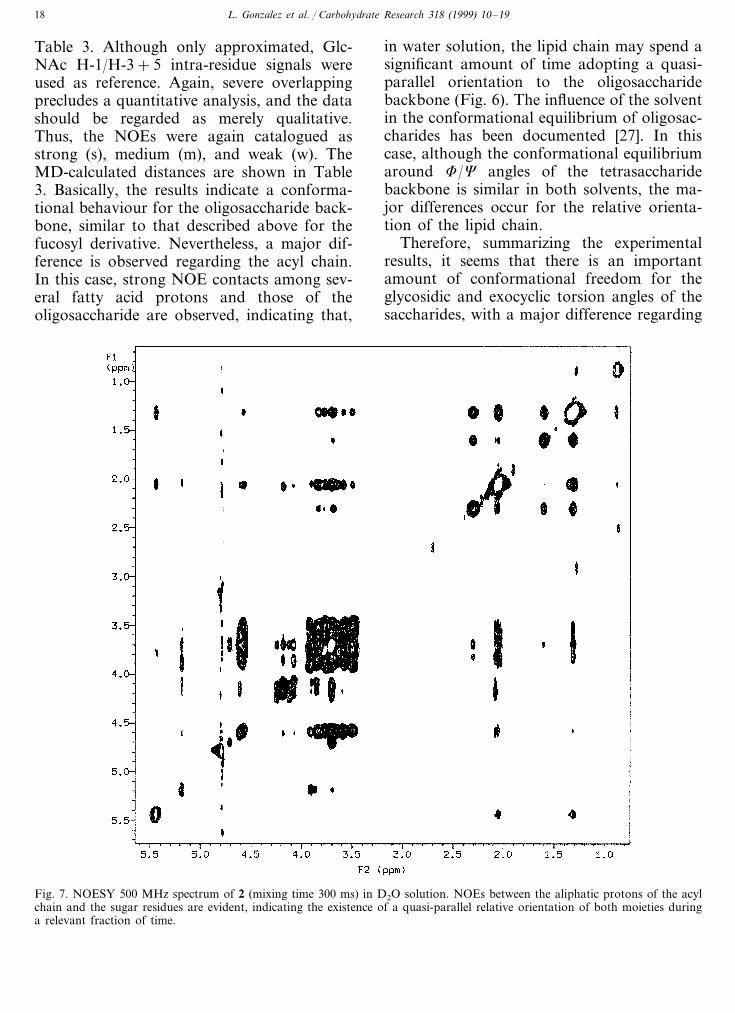
Table 3. Although only approximated, Glc-NAc H-1/H-3+5 intra-residue signals wereused as reference. Again, severe overlappingprecludes a quantitative analysis, and the datashould be regarded as merely qualitative.Thus, the NOEs were again catalogued asstrong (s), medium (m), and weak (w). TheMD-calculated distances are shown in Table3. Basically, the results indicate a conforma-tional behaviour for the oligosaccharide back-bone, similar to that described above for thefucosyl derivative. Nevertheless, a major dif-ference is observed regarding the acyl chain.In this case, strong NOE contacts among sev-eral fatty acid protons and those of theoligosaccharide are observed, indicating that,
in water solution, the lipid chain may spend asignificant amount of time adopting a quasi-parallel orientation to the oligosaccharidebackbone (Fig. 6). The influence of the solventin the conformational equilibrium of oligosac-charides has been documented [27]. In thiscase, although the conformational equilibriumaround F/C angles of the tetrasaccharidebackbone is similar in both solvents, the ma-jor differences occur for the relative orienta-tion of the lipid chain.
Therefore, summarizing the experimentalresults, it seems that there is an importantamount of conformational freedom for theglycosidic and exocyclic torsion angles of thesaccharides, with a major difference regarding
Fig. 7. NOESY 500 MHz spectrum of 2 (mixing time 300 ms) in D2O solution. NOEs between the aliphatic protons of the acylchain and the sugar residues are evident, indicating the existence of a quasi-parallel relative orientation of both moieties duringa relevant fraction of time.
L. Gonzalez et al. / Carbohydrate Research 318 (1999) 10–19 19
the orientation of the lipid chain. In watersolution, there is a stabilization of conformerswhich present hydrophobic contacts betweenthe sugar units and the fatty acid, while in arelatively less polar solvent such as DMSO,these contacts do not take place. It is clearfrom these data that the nature of the receptorbinding site may modulate the conformationalbehaviour of the Nod factor, giving rise todifferent biological responses.
Acknowledgements
Financial support by DGICYT (GrantsPB96-0833 and BIO96-1469-C03) is gratefullyacknowledged. The authors also thank Dr J.E.Ruiz-Sainz (Departamento de Microbiologıa,Facultad de Biologıa, Universidad de Sevilla)for supplying bacterial cultures. L.G. wouldlike to thank Ministerio de Educacion y Cien-cia for a postdoctoral fellowship.
References
[1] J. Denarie, F. Debelle, J.-C. Prome, Ann. Re6. Biochem.,65 (1996) 503–535.
[2] R.W. Carlson, N.P.J. Price, G. Stacey, Mol. Plant Mi-crobe Interact., 7 (1995) 684–695.
[3] J.L. Firmin, K.E. Wilson, L. Rossen, A.W.B. Johnson,Nature, 324 (1986) 90–94.
[4] M.M.A. Olsthoorn, I.M. Lopez-Lara, B.O. Petersen, K.Bock, J. Haverkamp, H.P. Spaink, J.E. Thomas-Oates,Biochemistry, 37 (1998) 9024–9032.
[5] H.P. Spaink, G.V. Bloemberg, A.A.N. van Brussel, B.J.J.Lugtenberg, K.M.G.M. van der Drift, J. Haverkamp,J.E. Thomas-Oates, Mol. Plant Microbe Interact., 8(1995) 155–164.
[6] P. Roche, F. Debelle, F. Maillet, P. Lerouge, C. Faucher,G. Truchet, J. Denarie, J.-C. Prome, Cell, 67 (1991)1131–1143.
[7] N.P.J. Price, F. Talmont, J.-M. Wieruszeski, D. Prome,J.-C. Prome, Carbohydr. Res., 289 (1996) 115–136.
[8] R.W. Carlson, J. Sanjuan, U.R. Bath, J. Glushka, H.P.Spaink, A.H.M. Wijfjer, A.A.N. van Brussel, T.J.W.Stokkermans, K. Peters, G. Stacey, J. Biol. Chem., 268(1993) 18372–18381.
[9] M.P. Bec-Ferte, H.B. Krishnan, D. Prome, A. Savagnac,S.G. Pueppke, J.-C. Prome, Biochemistry, 33 (1994)11782–11788.
[10] A.M. Gil-Serrano, G. Franco-Rodrıguez, P. Tejero-Ma-teo, J. Thomas-Oates, H.P. Spaink, J.E. Ruiz-Sainz, M.Megıas, Y. Lamrabet, Carbohydr. Res., 303 (1997) 435–443.
[11] (a) K.C. Nicolaou, N.J. Bockovich, D.R. Carcanague,C.W. Hummel, L.F. Even, J. Am. Chem. Soc., 114 (1992)8701–8702. (b) L.X. Wang, C. Li, Q. Wang, Y.Z. Hui,
Tetrahedron Lett., 34 (1993) 7763–7766. (c) S. Ikeshita,A. Sakamoto, Y. Nakahara, Y. Kakahara, T. Ogawa,Tetrahedron Lett., 35 (1994) 3123–3126. (d) D. Tailler,J.C. Jacquinet, J.M. Beau, J. Chem. Soc., Chem. Com-mun., (1994) 1827–1828. (e) I. Robina, E. Lopez-Barba,J. Jimenez-Barbero, M. Martın-Pastor, J. Fuentes Tetra-hedron Asymm., 8 (1997) 1207–1224. (f) L.X. Wang, C.Li, Q. Wang, Y.Z. Hui, J. Chem. Soc., Perkin Trans 1,(1994) 621–628. (g) J.S. Debenham, R. Rdebaugh, B.Fraser-Reid, J. Org. Chem., 61 (1996) 6478–6479.
[12] I. Robina, E. Lopez-Barba, J. Fuentes, Tetrahedron, 52(1996) 10771–10784.
[13] N. Mantegazza, E. Samain, H. Driguez (to be pub-lished).
[14] A.T. Hagler, S. Lifson, P. Dauber, J. Am. Chem. Soc.,101 (1979) 512.
[15] F. Mohamadi, N.G.I. Richards, W.C. Guida, R.Liskamp, C. Canfield, G. Chang, T. Hendrickson, W.C.Still, J. Comput. Chem., 11 (1990) 440–467.
[16] K. Bock, J. Duus, J. Carbohydr. Chem., 13 (1994) 513–543.
[17] W.C. Still, A. Tempczyk, R.C. Hawley, T. Hendrickson,J. Am. Chem. Soc., 112 (1990) 6127–6128.
[18] A.D. French, J.D. Brady (Eds.). Computer Modelling ofCarbohydrate Molecules, ACS Symp. Ser., 430, 1990.
[19] D. Lamba, S. Glover, W. Mackie, A. Rashid, B.Sheldrick, S. Perez, Glycobiology, 4 (1994) 151–163.
[20] (a) S.W. Homans, Biochemistry, 29 (1990) 9110–18. (b)B.J. Hardy, W. Egan, G. Widmalm, Int. J. Biol. Macro-mol., 17–18 (1995) 149–60. (c) C.J. Edge, U.C. Singh, R.Bazzo, G.L. Taylor, R.A. Dwek, T.W. Rademacher,Biochemistry, 29 (1990) 1971–74. (d) P.J. Hajduk, D.A.Horita, L. Lerner, J. Am. Chem. Soc., 115 (1993) 9196–201. (e) T.J. Rutherford, D.G. Spackman, P.J. Simpson,S.W. Homans, Glycobiology, 4 (1994) 59–68. (f) A.Poveda, J.L. Asensio, M. Martin-Pastor, J. Jimenez-Bar-bero, J. Chem. Soc., Chem. Commun., (1996) 421–422.
[21] For a revision on the use of pulse field gradients inNMR, see J.T. Keeler, R.T. Clowes, A.L. Davies, E.D.Laue, Methods Enzymol., 239 (1994) 145–207.
[22] (a) H.C. Siebert, G. Reuter, R. Schauer, C.W. von derLieth, J. Dabrowski, Biochemistry, 31 (1992) 6962–6971.(b) J.L. Asensio, M. Martin–Pastor, J. Jimenez-Barbero,Int. J. Biol. Macromol., 17 (1995) 52–55. (c) J.M.Coteron, K. Singh, J.L. Asensio, M.D. Dalda, A. Fer-nandez-Mayoralas, J. Jimenez-Barbero, M. Martın-Lo-mas, J. Org. Chem., 60 (1995) 1502–1513. (d) M.K.Dowd, P.J. Reilly, A.D. French, J. Comput. Chem., 13(1992) 102–114. (e) J.F. Espinosa, J.L. Asensio, M.Bruix, J. Jimenez-Barbero, Ann. Quim. Int. Ed., 96 (1996)320–324. (f) J.L. Asensio, M. Martin-Pastor, J. Jimenez-Barbero, J. Mol. Struct., 395–396 (1997) 245–270.
[23] R.U. Lemieux, K. Bock, L.T.J. Delbaere, S. Koto, V.S.Rao, Can. J. Chem., 58 (1980) 631–653.
[24] (a) J.P. Carver, Pure Appl. Chem., 65 (1993) 763–770. (b)K. Bock, Pure Appl. Chem., 55 (1983) 605–622.
[25] D. Neuhaus, M.P. Williamson, The Nuclear O6erhauserEffect in Structural and Conformational Analysis, VCH,New York, 1989.
[26] (a) T.J. Rutherford, S.W. Homans, Biochemistry, 33(1994) 9606–9614. (b) T.J. Rutherford, D.C.A. Neville,S.W. Homans, Biochemistry, 34 (1995) 14131–14137.
[27] (a) C.A. Bush, Z.-Y. Yan, B.N.N. Rao, J. Am. Chem.Soc., 108 (1986) 6168–6173. (b) L. Poppe, C.W. von derLieth, J. Dabrowski, J. Am. Chem. Soc., 112 (1990)7762–7771.
.