solutions to daily challenges in radiation...
TRANSCRIPT

Solutions to Daily Challenges in Radiation OncologyPrecision, Accuracy, and Patient Care

Aromatherapy Elequil aromatabs® ................................................ 1-2
CT Treatment PlanningCT-SPOT® Crosshair ............................................... 3-4
CT-SPOT® Line ....................................................... 5-6 CT-SPOT® Pellet ..................................................... 7-8
Conventional Simulation SPOT® Metallic Markers ....................................... 9-10
Temporary Marks PointGuards® Temporary Mark Covers ................. 11-12
Table of Contents

Aromatherapy as integrative care in radiation oncology
Treatments, regular testing, coping with symptoms of the disease, and side effects from medication can drain patients physically and emotionally.
Radiation oncology services are keenly aware of this and strive to help reduce the stress, anxiety, and nervousness their patients’ experience during treatment and beyond.
Patient counseling, therapy, and fi nancial advisory programs are not uncommon. Neither is adding integrative care services such as hydrotherapy, massage, or aromatherapy.
What is aromatherapy and how does it help?
According to the National Cancer Institute, “aromatherapy is the therapeutic use of essential oils (also known as volatile oils) from fl owers, herbs, or trees for the improvement of physical, emotional, and spiritual well-being.”1
International studies have been published regarding aromatherapy’s ability in reducing anxiety2 in patients in preoperative surgery and also in reducing nausea and vomiting.3
Cancer centers using aromatherapy do so as a means to promote relaxation for their patients and soothe queasiness.
What are the challenges with aromatherapy oils?
There are several challenges with using aromatherapy oils in the clinical setting.
The fi rst challenge is obtaining high quality 100% pure essential oils. Some essential oils can be quite costly and not every retailer sells premium grades.
The second challenge is in the delivery method. It’s very easy to have inconsistency in the amount of oils, or blend of oils, used when pouring oils into a diffuser or applying to a cotton ball. As a result, the desired impact can vary from day to day and patient to patient.
The third challenge is that aromatherapy oils can be messy if spilled or applied too liberally.
However, despite these challenges, aromatherapy remains an easy-to-implement integrative therapy that helps to improve the patient experience.
Aromatherapy designed for the clinical setting
Elequil aromatabs® are 100% pure essential oils in a unique controlled delivery system.
Patients can wear Elequil® during simulation, waiting for treatment, and even at home after treatment.
Lavender and Lavender-Sandalwood scents promote relaxation, comfort, and sleep. Orange-Peppermint helps to uplift, energize, and soothe queasiness.
1 “Aromatherapy and Essential Oils.” National Cancer Institute. NIH, Sept. 2015. Web. 22 Dec. 2015.2 Fayazi S, Babashahi M, Rezaei M. The effect of inhalation aromatherapy on anxiety level of the patients in preoperative period. Iranian Journal of Nursing and Midwifery Research. 2011;16(4):278-283.3 Abdel Ghani, Rania Mahmoud, and Adlia Tawfi k Ahmed Ibrahim. “The Effect of Aromatherapy Inhalation on Nausea and Vomiting in Early Pregnancy: A Pilot Randomized Controlled Trial.” Journal of
Natural Sciences Research 5 (2013): 192-206.
Aromatherapy
1Visit www.beekley.com for additional Product Safety Related Information.
Back to Table of Contents

We typically use the orange-peppermint Elequil aromatabs® for our patients in the oncology setting. Elequil aromatabs® is wonderful in helping control queasiness. Currently, we send them home with our patients to use between treatments, and then they have them on hand in a time of need. Great product.
~ Director of Oncology Services, Clinch Valley Medical Center, Richlands, VA
Elequil aromatabs® Lavender-SandalwoodLavandula angustifolia-Santalum Album
370 50 / Box
• 100% pure essential oils• scented tab on self-adhesive label• allows for minimum or maximum
scent exposure• promotes relaxation, comfort,
and sleep
Elequil aromatabs® LavenderLavandula angustifolia
372 50 / Box
• 100% pure essential oils• scented tab on self-adhesive label• allows for minimum or maximum
scent exposure• promotes relaxation, comfort,
and sleep
Elequil aromatabs® Orange-PeppermintCitrus sinensis-Mentha piperita
371 50 / Box
• 100% pure essential oils• scented tab on self-adhesive label• allows for minimum or maximum
scent exposure• uplifts, energizes, and can soothe
queasiness
Certifi cate of Analysis available upon request.
Elequil aromatabs®
Elequil aromatabs®
Aromatherapy you can wear. Easy application method and no training necessary. Allows minimum or maximum scent exposure. Lasts 8 hours or more.
Call 1-800-233-5539 • Fax 1-800-735-1234 • Visit www.beekley.com • Email [email protected] outside the U.S. – Contact your local distributor for pricing and product availability.
To locate a distributor call +1-860-583-4700 or email [email protected]
Back to Table of Contents
Finding the zero slice
The goal of the radiation therapist is to accurately locate the central axis or zero slice on the tumor fi eld for treatment planning.
A marker with a distinct shape that clearly defi nes the area of focus as you are scrolling through the images can save you time and improve accuracy.
A marker with a distinct shape
Using the CT-SPOT® crosshair in the formation of an “X” during treatment planning creates a unique shape that helps radiation oncology teams determine the zero slice.
Gayle Crowley, Chief Therapist at the Harold Leever Cancer Center (Waterbury, CT), describes how she utilizes this technique:
“We all know what it’s like scrolling through CT images looking for those magical marks or the 3 points on the patient,” Gayle told us in an interview.
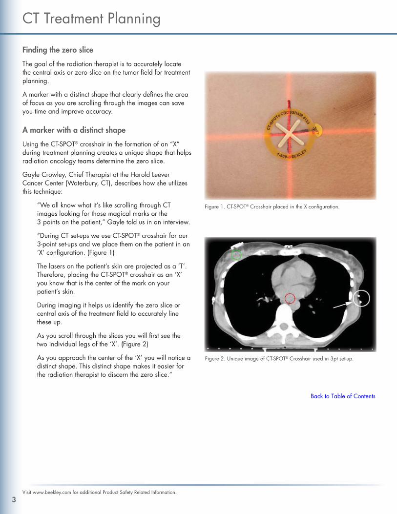
“During CT set-ups we use CT-SPOT® crosshair for our 3-point set-ups and we place them on the patient in an‘X’ confi guration. (Figure 1)
The lasers on the patient’s skin are projected as a ‘T’. Therefore, placing the CT-SPOT® crosshair as an ‘X’ you know that is the center of the mark on your patient’s skin.
During imaging it helps us identify the zero slice or central axis of the treatment fi eld to accurately line these up.
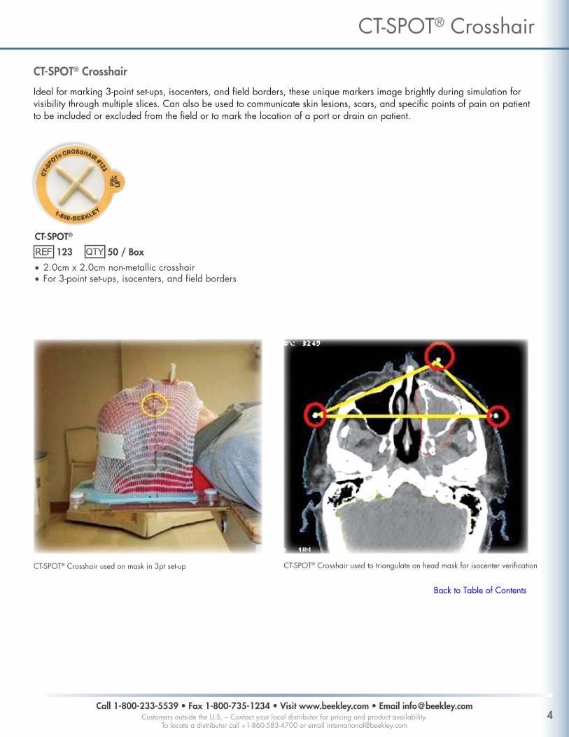
As you scroll through the slices you will fi rst see the two individual legs of the ‘X’. (Figure 2)
As you approach the center of the ‘X’ you will notice a distinct shape. This distinct shape makes it easier for the radiation therapist to discern the zero slice.”
CT Treatment Planning
Figure 1. CT-SPOT® Crosshair placed in the X confi guration.
Figure 2. Unique image of CT-SPOT® Crosshair used in 3pt set-up.
3Visit www.beekley.com for additional Product Safety Related Information.
Back to Table of Contents
CT-SPOT®
123 50 / Box
• 2.0cm x 2.0cm non-metallic crosshair• For 3-point set-ups, isocenters, and fi eld borders
CT-SPOT® Crosshair
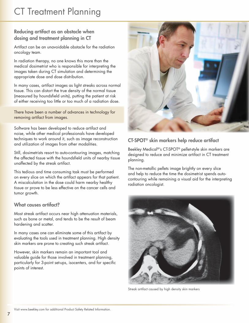
Ideal for marking 3-point set-ups, isocenters, and fi eld borders, these unique markers image brightly during simulation for visibility through multiple slices. Can also be used to communicate skin lesions, scars, and specifi c points of pain on patient to be included or excluded from the fi eld or to mark the location of a port or drain on patient.
Call 1-800-233-5539 • Fax 1-800-735-1234 • Visit www.beekley.com • Email [email protected] outside the U.S. – Contact your local distributor for pricing and product availability.
To locate a distributor call +1-860-583-4700 or email [email protected]
CT-SPOT® Crosshair
CT-SPOT® Crosshair used to triangulate on head mask for isocenter verifi cationCT-SPOT® Crosshair used on mask in 3pt set-up
Back to Table of Contents

Accuracy in treatment planning affects the effi cacy of the treatment
The challenge every radiation oncology department faces is to destroy cancer cells and stop the growth of the tumor while limiting harm to nearby healthy tissue.
Accuracy is measured in millimeters and the precision of the CT simulation will affect subsequent treatments. Consequently, the quality of each tool the radiation therapist uses in set-ups and treatment planning will make a difference.
Linear markers are such tools.
Used to mark fi eld borders, tangents, scars, sarcomas, and breast tissue, one should expect their linear marker to be fl exible, contour to the skin, and clearly denote the area of concern in imaging and CT simulation without lifting or coming off.
Linear markers without these key attributes can negatively affect the accuracy and effi cacy of the treatment.
Infl exible wires are often cut into small strips for better handling. Not only is this process of cutting several small strips time consuming and wasteful, it can also result in unintended inaccuracies when marking fi eld borders and treatment areas or over/under radiation of the target area.
Using tape and wire to defi ne fi eld borders does not provide the desired fl exibility nor the appropriate accuracy. Tape sticks to the skin with the wire caught underneath.
More targeted placement, more fi rst time accuracy
Beekley Medical®’s CT-SPOT® lines are fl exible and contour easily around corners. With a medical-grade, latex-free adhesive, it provides the “just right” stick that radiation therapists value.
The non-metallic line images brightly on every slice that it appears on during simulation while also reducing artifact and streaking.
This makes it easier for the therapist or radiation oncologist to measure and align the marker with the desired area of treatment.
CT Treatment Planning
5Visit www.beekley.com for additional Product Safety Related Information.
Non-metallic CT-SPOT® Line images without artifact
CT-SPOT® Line marking medial and lateral breast tangents

CT-SPOT®
118 320cm cut to measure roll / Box
• 2.0mm diameter non-metallic line• For marking scars and fi eld borders
S-SPOT®
607 Florals® 363cm cut to measure roll / Box
• .33mm diameter radiopaque line• Burnout-resistant brighter image• For marking scars and fi eld borders
CT-SPOT® Line
Continuous linear markers designed to delineate fi elds for accurate CT treatment planning calculations. Flexible line contours easily around corners. Ideal for marking fi eld borders, tangents, scars, sarcomas and larger treatment areas.
Call 1-800-233-5539 • Fax 1-800-735-1234 • Visit www.beekley.com • Email [email protected] outside the U.S. – Contact your local distributor for pricing and product availability.
To locate a distributor call +1-860-583-4700 or email [email protected]
CT-SPOT® Line
Strips of CT-SPOT® line used for tangents and fi eld borders. Scar is marked with S-SPOT® .33mm metallic line
Continuous strip of CT-SPOT® Line identifi es treatment area by encircling entire breast
• Burnout-resistant brighter image• For marking scars and fi eld borders
Back to Table of Contents
Reducing artifact as an obstacle when dosing and treatment planning in CT
Artifact can be an unavoidable obstacle for the radiation oncology team.
In radiation therapy, no one knows this more than the medical dosimetrist who is responsible for interpreting the images taken during CT simulation and determining the appropriate dose and dose distribution.
In many cases, artifact images as light streaks across normal tissue. This can distort the true density of the normal tissue (measured by houndsfi eld units), putting the patient at risk of either receiving too little or too much of a radiation dose.
There have been a number of advances in technology for removing artifact from images.
Software has been developed to reduce artifact and noise, while other medical professionals have developed techniques to work around it, such as image reconstruction and utilization of images from other modalities.
Still, dosimetrists resort to auto-contouring images, matching the affected tissue with the houndsfi eld units of nearby tissue unaffected by the streak artifact.
This tedious and time consuming task must be performed on every slice on which the artifact appears for that patient. A miscalculation in the dose could harm nearby healthy tissue or prove to be less effective on the cancer cells and tumor growth.
What causes artifact?
Most streak artifact occurs near high attenuation materials, such as bone or metal, and tends to be the result of beam hardening and scatter.
In many cases one can eliminate some of this artifact by evaluating the tools used in treatment planning. High density skin markers are prone to creating such streak artifact.
However, skin markers remain an important tool and valuable guide for those involved in treatment planning, particularly for 3-point set-ups, isocenters, and for specifi c points of interest.
CT-SPOT® skin markers help reduce artifact
Beekley Medical®’s CT-SPOT® pellet-style skin markers are designed to reduce and minimize artifact in CT treatment planning.
The non-metallic pellets image brightly on every slice and help to reduce the time the dosimetrist spends auto-contouring while remaining a visual aid for the interpreting radiation oncologist.
CT Treatment Planning
7Visit www.beekley.com for additional Product Safety Related Information.
Streak artifact caused by high density skin markers
CT-SPOT®
119 85 / Box
• 2.3mm non-metallic pellet• For 3-point set-ups and isocenters
CT-SPOT®
120 85 / Box
• 4.0mm non-metallic pellet• For 3-point set-ups and isocenters
CT-SPOT® Pellet
Unique non-metallic markers image brightly during simulation without streaking or artifact.
Call 1-800-233-5539 • Fax 1-800-735-1234 • Visit www.beekley.com • Email [email protected] outside the U.S. – Contact your local distributor for pricing and product availability.
To locate a distributor call +1-860-583-4700 or email [email protected]
CT-SPOT® Pellet
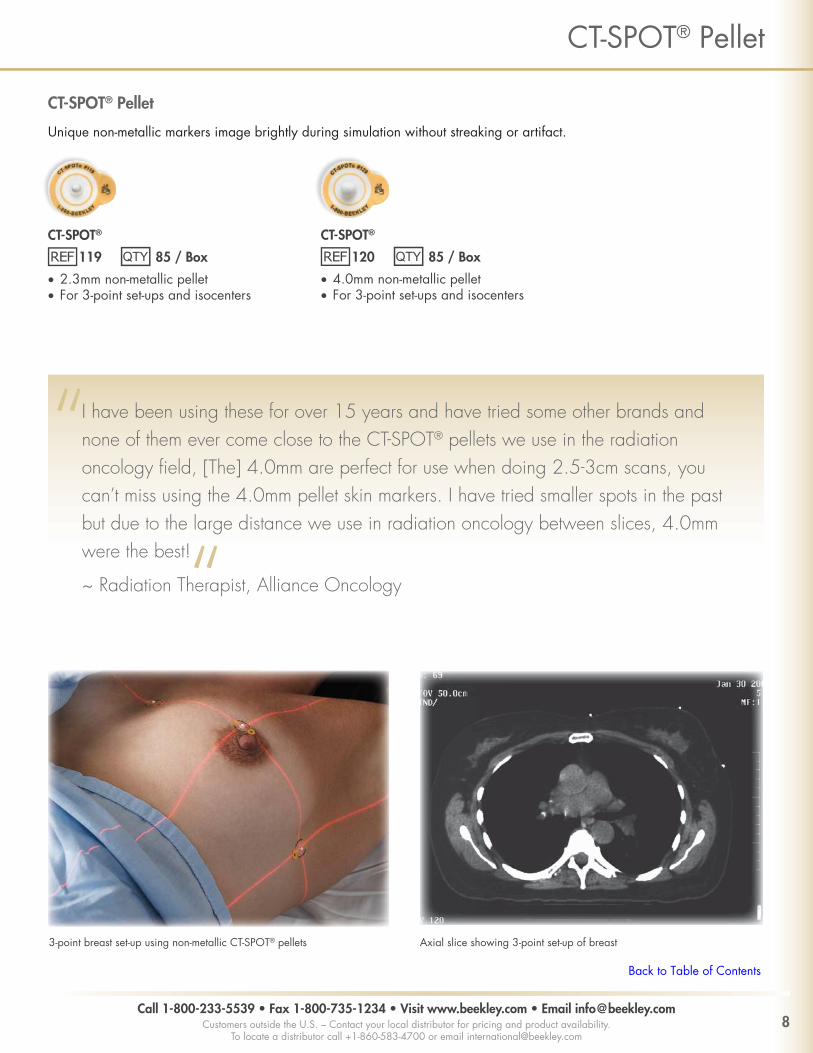
I have been using these for over 15 years and have tried some other brands and none of them ever come close to the CT-SPOT® pellets we use in the radiation oncology fi eld, [The] 4.0mm are perfect for use when doing 2.5-3cm scans, you can’t miss using the 4.0mm pellet skin markers. I have tried smaller spots in the past but due to the large distance we use in radiation oncology between slices, 4.0mm were the best!
~ Radiation Therapist, Alliance Oncology
3-point breast set-up using non-metallic CT-SPOT® pellets Axial slice showing 3-point set-up of breast
Back to Table of Contents
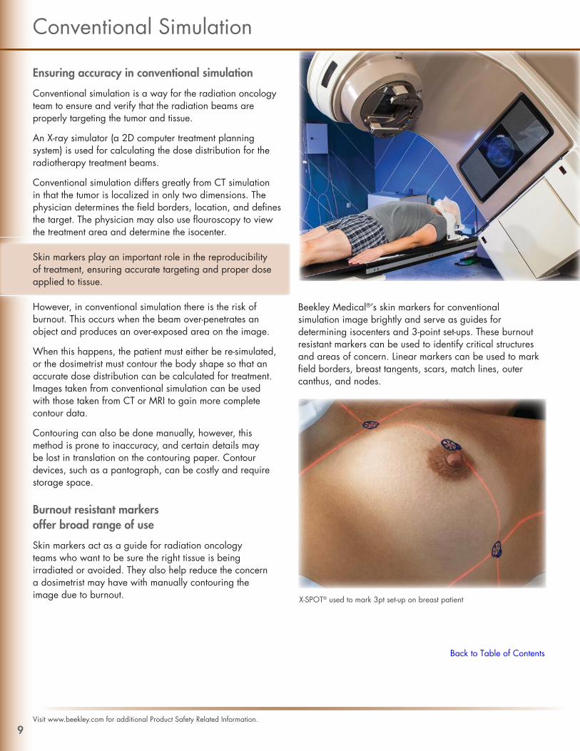
Ensuring accuracy in conventional simulation
Conventional simulation is a way for the radiation oncology team to ensure and verify that the radiation beams are properly targeting the tumor and tissue.
An X-ray simulator (a 2D computer treatment planning system) is used for calculating the dose distribution for the radiotherapy treatment beams.
Conventional simulation differs greatly from CT simulation in that the tumor is localized in only two dimensions. The physician determines the fi eld borders, location, and defi nes the target. The physician may also use fl ouroscopy to view the treatment area and determine the isocenter.
Skin markers play an important role in the reproducibility of treatment, ensuring accurate targeting and proper dose applied to tissue.
However, in conventional simulation there is the risk of burnout. This occurs when the beam over-penetrates an object and produces an over-exposed area on the image.
When this happens, the patient must either be re-simulated, or the dosimetrist must contour the body shape so that an accurate dose distribution can be calculated for treatment. Images taken from conventional simulation can be used with those taken from CT or MRI to gain more complete contour data.
Contouring can also be done manually, however, this method is prone to inaccuracy, and certain details may be lost in translation on the contouring paper. Contour devices, such as a pantograph, can be costly and require storage space.
Burnout resistant markers offer broad range of use
Skin markers act as a guide for radiation oncology teams who want to be sure the right tissue is being irradiated or avoided. They also help reduce the concern a dosimetrist may have with manually contouring the image due to burnout.
Beekley Medical®’s skin markers for conventional simulation image brightly and serve as guides for determining isocenters and 3-point set-ups. These burnout resistant markers can be used to identify critical structures and areas of concern. Linear markers can be used to mark fi eld borders, breast tangents, scars, match lines, outer canthus, and nodes.
Conventional Simulation
9Visit www.beekley.com for additional Product Safety Related Information.
X-SPOT® used to mark 3pt set-up on breast patient
Back to Table of Contents

X-SPOT®
101 150 / Box
• 1.5mm lead-free pellet• For marking 3-point set-ups, isocenters,
outer canthus and nodes
Y-SPOT®
102 132 / Box
• 2.0mm lead-free pellet• For marking 3-point set-ups, isocenters,
outer canthus and nodes
D-SPOT®
104 99 / Box
• lead-free arrow• For marking nodes and
outer canthus
T-SPOT®
111 275cm cut to measure roll / Box
• .8mm diameter radiopaque line• For marking scars, nodes, fi eld borders,
match lines and breast tangents
Z-SPOT®
114 40 / Box
• 4.3mm lead-free pellet• For marking port fi lms
V-SPOT®
603 Florals® 130 / Box
• 2.5mm lead-free pellet• For marking 3-point set-ups
and isocenters
SPOT® Metallic Markers
Metallic pellets, lines, and arrows help differentiate specifi c points of reference in conventional simulation and port fi lms.
Call 1-800-233-5539 • Fax 1-800-735-1234 • Visit www.beekley.com • Email [email protected] outside the U.S. – Contact your local distributor for pricing and product availability.
To locate a distributor call +1-860-583-4700 or email [email protected]
SPOT® Metallic Markers
T-SPOT® used to identify size and location of abdominal tumor Y-SPOT® used for 3-point set-up on prostate
Back to Table of Contents

Ensuring accurate daily reproducibility for treatment setups
Temporary marks are an important part of CT simulation, allowing for accurate and reproducible set-ups for treatment.
However, temporary marks are only helpful if they remain on the patient. Otherwise, the patient will need to be re-simulated before receiving or continuing treatment.
According to Phyllis Burch, Administrative Director, City of Hope Radiation Oncology (Duarte, CA), “If we can reduce the times that we are re-simulating patients due to either lost marks, or even the surgical tape coming off and really having to re-identify those markings to ensure accuracy, I feel that in itself is a really high cost savings to the department.”
Covering temporary marks is a common way to avoid re-simulating patients
Some departments use surgical tape or wound dressing products to cover temporary marks.
However, as Ms. Burch pointed out, this method of cutting strips of tape can be time consuming and produce waste. In addition, surgical tape rarely stays on the skin for more than a couple days and is prone to causing skin irritation.
A low-cost insurance plan against re-simulating the patient
PointGuards® temporary mark covers are a fast and convenient way to protect the patient’s temporary marks from disappearing. The watertight seal stays securely in place for up to 4 weeks. Patients do not have to alter their routine or their daily living:
“I do my normal routine,” says Elaine, a breast cancer patient from New London, CT. “I shower, go out, take my walks, sweat in the sun, and I don’t even know they’re there. They feel like part of my skin.”
Temporary Marks
11Visit www.beekley.com for additional Product Safety Related Information.
Back to Table of Contents
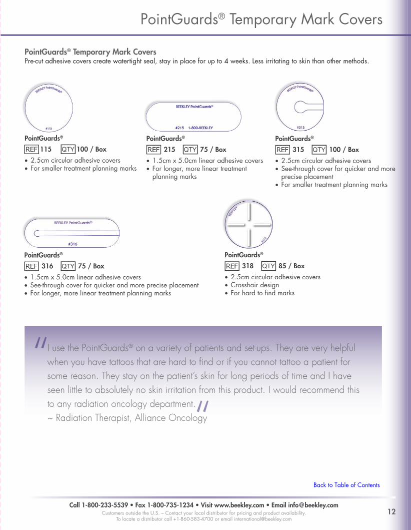
PointGuards®
115 100 / Box
• 2.5cm circular adhesive covers• For smaller treatment planning marks
PointGuards®
215 75 / Box
• 1.5cm x 5.0cm linear adhesive covers• For longer, more linear treatment
planning marks
PointGuards®
315 100 / Box
• 2.5cm circular adhesive covers• See-through cover for quicker and more
precise placement• For smaller treatment planning marks
PointGuards®
316 75 / Box
• 1.5cm x 5.0cm linear adhesive covers• See-through cover for quicker and more precise placement• For longer, more linear treatment planning marks
PointGuards®
318 85 / Box
• 2.5cm circular adhesive covers• Crosshair design• For hard to fi nd marks
PointGuards® Temporary Mark CoversPre-cut adhesive covers create watertight seal, stay in place for up to 4 weeks. Less irritating to skin than other methods.
Call 1-800-233-5539 • Fax 1-800-735-1234 • Visit www.beekley.com • Email [email protected] outside the U.S. – Contact your local distributor for pricing and product availability.
To locate a distributor call +1-860-583-4700 or email [email protected]
PointGuards® Temporary Mark Covers
I use the PointGuards® on a variety of patients and set-ups. They are very helpful when you have tattoos that are hard to fi nd or if you cannot tattoo a patient for some reason. They stay on the patient’s skin for long periods of time and I have seen little to absolutely no skin irritation from this product. I would recommend this to any radiation oncology department.~ Radiation Therapist, Alliance Oncology
Back to Table of Contents

Manufactured by Beekley CorporationOne Prestige Lane, Bristol, CT 06010 USA Tel: 1-800-233-5539 or +1-860-583-4700 Fax: 1-800-735-1234 www.beekley.com Made in the USABEEKLEY, BEEKLEY MEDICAL, ELEQUIL, ELEQUIL AROMATABS, AROMATABS, FLORALS, POINTGUARDS, SPOT, X-SPOT, WHEN YOUR DIAGNOSIS MUST BE RIGHT, and the color pink are Reg. U.S. Pat. & Tm. Off. BEEKLEY, AROMATABS, and SPOT are Registered Community Trademarks. BEEKLEY and AROMATABS are registered trademarks in Canada.
© 2016 Beekley Corporation. All rights reserved. REV: RT_CT_0616