snake venom l-amino acid oxidases - … snake venom l-amino acid oxidases ... 222 b. reconstitution...
TRANSCRIPT
219
10 Snake Venom L-Amino Acid Oxidases
Nget-Hong Tan and Shin-Yee Fung
L-amino acid oxidase (LAAO) occurs widely in snake venoms. The enzyme is highly specific for L-amino acids, and generally hydrophobic amino acids are the best substrates. LAAO is a flavo-protein and consists of two identical subunits, each with a molecular weight of 57–68 kDa. The purified enzymes are glycoproteins with 3–4% carbohydrate. Deglycosylation of the enzyme does not alter the enzymatic activity but appears to alter its pharmacological activities. The amino acid
Contents
I. Introduction ..........................................................................................................................220 II. Assay Methods .....................................................................................................................220 III. Occurrence in Snake Venoms .............................................................................................. 221 IV. Purification of Snake Venom LAAOs .................................................................................. 221 V. Cloning and Expression of Snake Venom LAAOs .............................................................. 222 VI. Physical Properties of Snake Venom LAAOs ...................................................................... 222 A. General Physical Properties of Snake Venom LAAOs .................................................. 222 B. Reconstitution of LAAO ................................................................................................ 223 VII. The Chemical Structure of LAAOs ..................................................................................... 223 A. N-terminal Sequences .................................................................................................... 223 B. Amino Acid Sequences .................................................................................................. 223 C. The Glycan Structure .....................................................................................................224 VIII. Three-dimensional Structure of LAAOs .............................................................................225 A. X-ray Structure of C. Rhodostoma LAAO.....................................................................225 B. Molecular Modeling of B. Jararacussu and B. Moojeni LAAOs ..................................225 IX. The Enzymatic Properties of LAAOs ..................................................................................226 A. General Enzymatic Properties .......................................................................................226 B. Substrate Specificity .......................................................................................................226 C. Mechanism of Catalysis .................................................................................................226 X. Immunological Properties of LAAOs .................................................................................. 227 XI. The Pharmacological Activities of LAAOs ......................................................................... 227 A. Edema-inducing and Hemorrhagic Activities ................................................................228 B. Anticoagulant Effects .....................................................................................................228 C. Effects on Platelet Aggregation ......................................................................................228 D. Apoptosis-inducing Effect .............................................................................................. 229 E. Antibacterial Activity ..................................................................................................... 229 F. Leishmanicidal Activity .................................................................................................230 G. Anti-HIV Activity ..........................................................................................................230 XII. Conclusions ..........................................................................................................................230 Acknowledgment ........................................................................................................................... 231References ...................................................................................................................................... 231
9165_C010.indd 219 3/12/09 2:02:04 PM
220 Handbook of Venoms and Toxins of Reptiles
sequences of snake venom LAAOs show a high degree of homology. X-ray structural analysis of LAAO revealed a dynamic active site and the presence of three domains: an FAD-binding domain, a substrate-binding domain, and a helical domain. LAAOs were reported to exhibit moderate lethal toxicity. Recent studies showed that LAAOs are multifunctional enzymes exhibiting edema- inducing, platelet aggregation-inducing or -inhibiting, apoptosis-inducing, as well as antibacterial, anticoagulant, and anti-HIV effects. These effects are mostly mediated by the H2O2 liberated in the oxidation process, but direct interactions between LAAO and the target cells may play an important role. High-resolution x-ray structural analysis of the enzyme revealed the presence of a channel that would direct the H2O2 product to the exterior surface of the protein, near the glycan moiety at Asn 172, which is thought to be involved with LAAO–target cell interaction. This may explain the abil-ity of LAAO to localize H2O2 to the targeted cells. A better understanding of the pharmacological actions of LAAOs will facilitate the application of snake venom LAAOs in the design of anticancer and anti-HIV drugs, as well as drugs for the treatment of infectious diseases caused by parasites such as Leishmania.
I. IntroduCtIon
L-amino acid oxidase (L-amino acid: O2 oxidoreductase, EC 1.4.3.2) is a flavoenzyme that cata-lyzes the oxidative deamination of an L-amino acid to form the corresponding α-ketoacid and ammonia:
RCH(NH3+)COO– + O2 + H2O → RCOCOO– + NH4
+ + H2O2
L-amino acid oxidase (LAAO) occurs widely in nature (Iwanaga and Suzuki, 1979), and snake venoms are perhaps the richest sources of this enzyme. Snake venom LAAOs are generally very active and have been used widely in preparation of α-keto acids because of their chemo- and stereo-specificity (Szwajcer et al., 1982; Findrik et al., 2006). α-Keto acids of essential amino acids are useful nutraceuticals as well as therapeutic agents for certain diseases. Recently, snake venom LAAO has become an interesting object for biomedical studies because of its antimicrobial, anti-HIV, anticoagulant, platelet aggregation-inducing and -inhibiting, apoptosis-inducing, as well as anticancer activities. Snake venom LAAO is recognized as a multifunctional protein with prom-ising biomedical application. Several reviews on snake venom L-amino acid oxidases have been published (Meister and Wellner, 1963; Bright and Porter, 1975; Tu, 1977; Iwanaga and Suzuki, 1979; Curti et al., 1992; Tan, 1998; Du and Clemetson, 2002).
II. AssAy Methods
Many methods of L-amino acid oxidase assay are available (Iwanaga and Suzuki, 1979). The O2 electrode technique has been widely used, particularly in kinetic studies. A commonly used spec-trophotometric method was described by Bergmeyer (1983) that followed the rate of oxidation by measuring the rate of formation of color complex between the hydrogen peroxide produced and o-dianisidine. Based on the same principle, a spectrophotometric microplate assay has been devel-oped suitable for processing large numbers of samples (Kishimoto and Takahashi, 2001).
Assay Procedure
The reaction mixture consists of 0.05 ml of enzyme, 0.05 ml of 0.0075% horseradish peroxidase (100 purpurogalin units/mg), 67.5 μg of o-dianisidine, and 10 µM L-leucine in 0.9 ml of 0.1 M Tris-HCl, pH 8.5, and the initial rate was measured as the increase in absorbance at 436 nm. The molar absorption coefficient in this assay system is 8.31 × 10³ M–1cm–1. One unit of enzyme activity was defined as the oxidation of 1 µmole of L-leucine per minute.
9165_C010.indd 220 3/12/09 2:02:04 PM
Snake Venom L-Amino Acid Oxidases 221
III. oCCurrenCe In snAke VenoMs
L-amino acid oxidase can be found in venoms from most genera of snakes (Tan and Ponnudurai, 1992). The richest sources of L-amino acid oxidase are crotaline venoms. The enzyme usually constitutes 1–4% of the venom by weight, but in Calloselasma rhodostoma (Malayan pit viper) it constitutes up to 30% by weight of the dried venom (Tan, 1998). Venoms from mambas and sea snakes contain either no or only trace amounts of L-amino acid activity. LAAO activity is typically absent from colubrid venoms (Mackessy, 2002).
IV. PurIfICAtIon of snAke VenoM LAAos
Since the 1990s, many authors have reported the purification and characterization of L-amino acid oxidases from various snake venoms (Table 10.1). In some snake venoms, the enzymes present in many isoforms. Hayes and Wellner (1969), for example, reported that there were at least eighteen isoforms of the L-amino acid oxidase in Crotalus adamanteus venom, and that glycosylation con-tributes to the microheterogeneity for the enzyme. However, microheterogeneity was not observed for L-amino acid oxidases isolated from most other venoms. In general, it is relatively easy to obtain homogenous LAAO from snake venom. For example, the LAAO from C. rhodostoma venom can be obtained using a simple two-step procedure: Sephadex gel filtration chromatography followed by Mono-Q high-performance ion exchange chromatography (Ponnudurai et al., 1994).
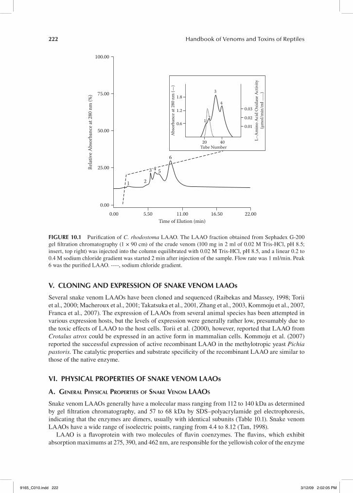
Crude venom was dissolved in the appropriate buffer and fractionated by Sephadex G-200 gel filtration chromatography. Fractions exhibiting high LAAO activity were pooled and further frac-tionated by Mono-Q HR 5/5 high-performance anion exchange chromatography to yield the puri-fied enzyme (Figure 10.1).
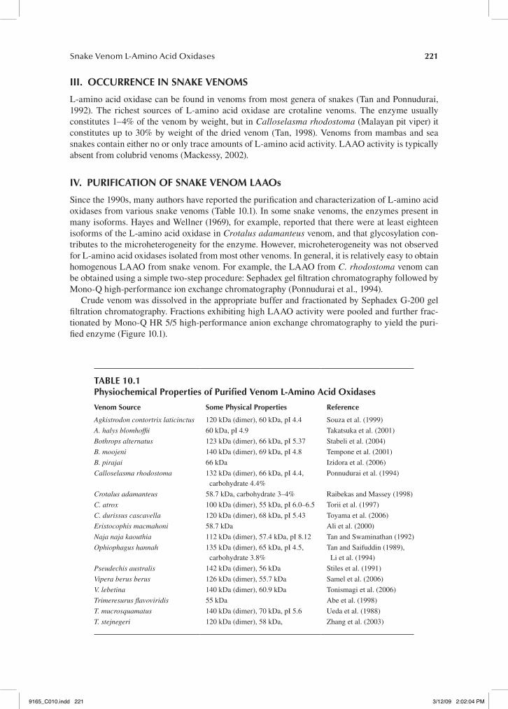
tAbLe 10.1Physiochemical Properties of Purified Venom L-Amino Acid oxidases
Venom source some Physical Properties reference
Agkistrodon contortrix laticinctus 120 kDa (dimer), 60 kDa, pI 4.4 Souza et al. (1999)
A. halys blomhoffii 60 kDa, pI 4.9 Takatsuka et al. (2001)
Bothrops alternatus 123 kDa (dimer), 66 kDa, pI 5.37 Stabeli et al. (2004)
B. moojeni 140 kDa (dimer), 69 kDa, pI 4.8 Tempone et al. (2001)
B. pirajai 66 kDa Izidora et al. (2006)
Calloselasma rhodostoma 132 kDa (dimer), 66 kDa, pI 4.4, carbohydrate 4.4%
Ponnudurai et al. (1994)
Crotalus adamanteus 58.7 kDa, carbohydrate 3–4% Raibekas and Massey (1998)
C. atrox 100 kDa (dimer), 55 kDa, pI 6.0–6.5 Torii et al. (1997)
C. durissus cascavella 120 kDa (dimer), 68 kDa, pI 5.43 Toyama et al. (2006)
Eristocophis macmahoni 58.7 kDa Ali et al. (2000)
Naja naja kaouthia 112 kDa (dimer), 57.4 kDa, pI 8.12 Tan and Swaminathan (1992)
Ophiophagus hannah 135 kDa (dimer), 65 kDa, pI 4.5, carbohydrate 3.8%
Tan and Saifuddin (1989), Li et al. (1994)
Pseudechis australis 142 kDa (dimer), 56 kDa Stiles et al. (1991)
Vipera berus berus 126 kDa (dimer), 55.7 kDa Samel et al. (2006)
V. lebetina 140 kDa (dimer), 60.9 kDa Tonismagi et al. (2006)
Trimeresurus flavoviridis 55 kDa Abe et al. (1998)
T. mucrosquamatus 140 kDa (dimer), 70 kDa, pI 5.6 Ueda et al. (1988)
T. stejnegeri 120 kDa (dimer), 58 kDa, Zhang et al. (2003)
9165_C010.indd 221 3/12/09 2:02:04 PM
222 Handbook of Venoms and Toxins of Reptiles
V. CLonIng And exPressIon of snAke VenoM LAAos
Several snake venom LAAOs have been cloned and sequenced (Raibekas and Massey, 1998; Torii et al., 2000; Macheroux et al., 2001; Takatsuka et al., 2001, Zhang et al., 2003, Kommoju et al., 2007, Franca et al., 2007). The expression of LAAOs from several animal species has been attempted in various expression hosts, but the levels of expression were generally rather low, presumably due to the toxic effects of LAAO to the host cells. Torii et al. (2000), however, reported that LAAO from Crotalus atrox could be expressed in an active form in mammalian cells. Kommoju et al. (2007) reported the successful expression of active recombinant LAAO in the methylotropic yeast Pichia pastoris. The catalytic properties and substrate specificity of the recombinant LAAO are similar to those of the native enzyme.
VI. PhysICAL ProPertIes of snAke VenoM LAAos
A. GenerAl PhysicAl ProPerties of snAke Venom lAAos
Snake venom LAAOs generally have a molecular mass ranging from 112 to 140 kDa as determined by gel filtration chromatography, and 57 to 68 kDa by SDS–polyacrylamide gel electrophoresis, indicating that the enzymes are dimers, usually with identical subunits (Table 10.1). Snake venom LAAOs have a wide range of isoelectric points, ranging from 4.4 to 8.12 (Tan, 1998).
LAAO is a flavoprotein with two molecules of flavin coenzymes. The flavins, which exhibit absorption maximums at 275, 390, and 462 nm, are responsible for the yellowish color of the enzyme
100.00
75.00
50.00
25.00Rela
tive A
bsor
banc
e at 2
80 n
m (%
)
0.000.00
1 2
5
6
0.6
20
1
3
4
40Tube Number
0.01
0.02
0.03
Abs
orba
nce a
t 280
nm
(—)
L–A
min
o Ac
id O
xida
se A
ctiv
ity(µ
mol
/min
/ml .
......)
1.2
1.8
5.50 11.00Time of Elution (min)
16.50 22.00
2
3 4
fIgure 10.1 Purification of C. rhodostoma LAAO. The LAAO fraction obtained from Sephadex G-200 gel filtration chromatography (1 × 90 cm) of the crude venom (100 mg in 2 ml of 0.02 M Tris-HCl, pH 8.5; insert, top right) was injected into the column equilibrated with 0.02 M Tris-HCl, pH 8.5, and a linear 0.2 to 0.4 M sodium chloride gradient was started 2 min after injection of the sample. Flow rate was 1 ml/min. Peak 6 was the purified LAAO. ----, sodium chloride gradient.
9165_C010.indd 222 3/12/09 2:02:05 PM
Snake Venom L-Amino Acid Oxidases 223
as well as for the venoms. Most authors reported that the flavin coenzymes are both FAD, though some earlier reports suggested FMN as the coenzymes in some LAAOs (Tan, 1998).
Snake venom LAAOs are stable at room temperature and at 4°C (Tan 1998). Ophiophagus hannah LAAO, for example, retained 100% and 80% of activity after incubating at 37°C for 5 days or 14 days at pH 7.4, respectively. Many LAAOs, however, are unstable under alkaline conditions.
Some snake venom LAAOs have a highly sensitive active site. For example, Crotalus adaman-teus LAAO undergoes reversible pH or temperature-dependent inactivation, accompanied by struc-tural changes in the flavin-binding site, though retaining its overall secondary structure (Coles et al., 1977). Earlier, Curti et al. (1968) reported that C. adamanteus LAAO was inactivated by storage at –5°C and –60°C, and by freeze-drying. Many other snake venom LAAOs are also inactivated by freezing. Generally, the inactivated enzyme can be reactivated completely by heating at pH 5. The inactivation was accompanied by shifts in the absorption spectrum and optical rotary dispersion spectrum, and reactivation reverses the spectra changes completely. The inactivation was believed to be due to a limited conformational change of the enzyme structure, presumably also in the vicin-ity of the flavin-binding site (Soltysik et al., 1987). This has been substantiated by x-ray structural studies (see below). Some snake venom LAAOs (for example, LAAOs from O. hannah and C. rho-dostoma), however, are not inactivated by freezing (Tan, 1998).
B. reconstitution of lAAo
As a result of the high sensitivity of many snake venom LAAOs to their microenvironment, it was not possible to prepare reconstitutable apoprotein, as reconstitution with the FAD coenzyme often resulted in an inactive protein, with a perturbed conformation of the flavin-binding site. However, Raibekas and Massey (1996) reported near complete activation of the reconstituted apoprotein and the restoration of its native flavin-binding site in the presence of 50% glycerol. Glycerol as a cosol-vent plays a special role in this restorative process by induction of rearrangement in the protein structure. The authors suggested that hydrophobic effect appears to be the dominating force in this in vitro–assisted restorative process.
VII. the CheMICAL struCture of LAAos
A. n-terminAl sequences
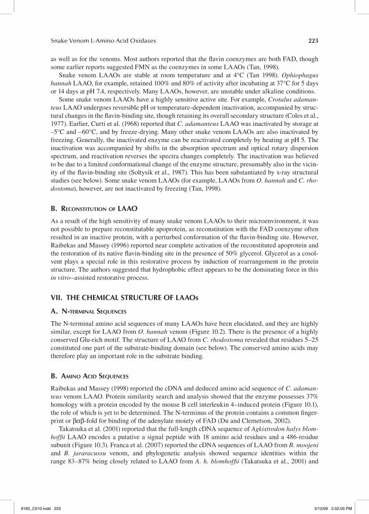
The N-terminal amino acid sequences of many LAAOs have been elucidated, and they are highly similar, except for LAAO from O. hannah venom (Figure 10.2). There is the presence of a highly conserved Glu-rich motif. The structure of LAAO from C. rhodostoma revealed that residues 5–25 constituted one part of the substrate-binding domain (see below). The conserved amino acids may therefore play an important role in the substrate binding.
B. Amino Acid sequences
Raibekas and Massey (1998) reported the cDNA and deduced amino acid sequence of C. adaman-teus venom LAAO. Protein similarity search and analysis showed that the enzyme possesses 37% homology with a protein encoded by the mouse B cell interleukin 4–induced protein (Figure 10.1), the role of which is yet to be determined. The N-terminus of the protein contains a common finger-print or βαβ-fold for binding of the adenylate moiety of FAD (Du and Clemetson, 2002).
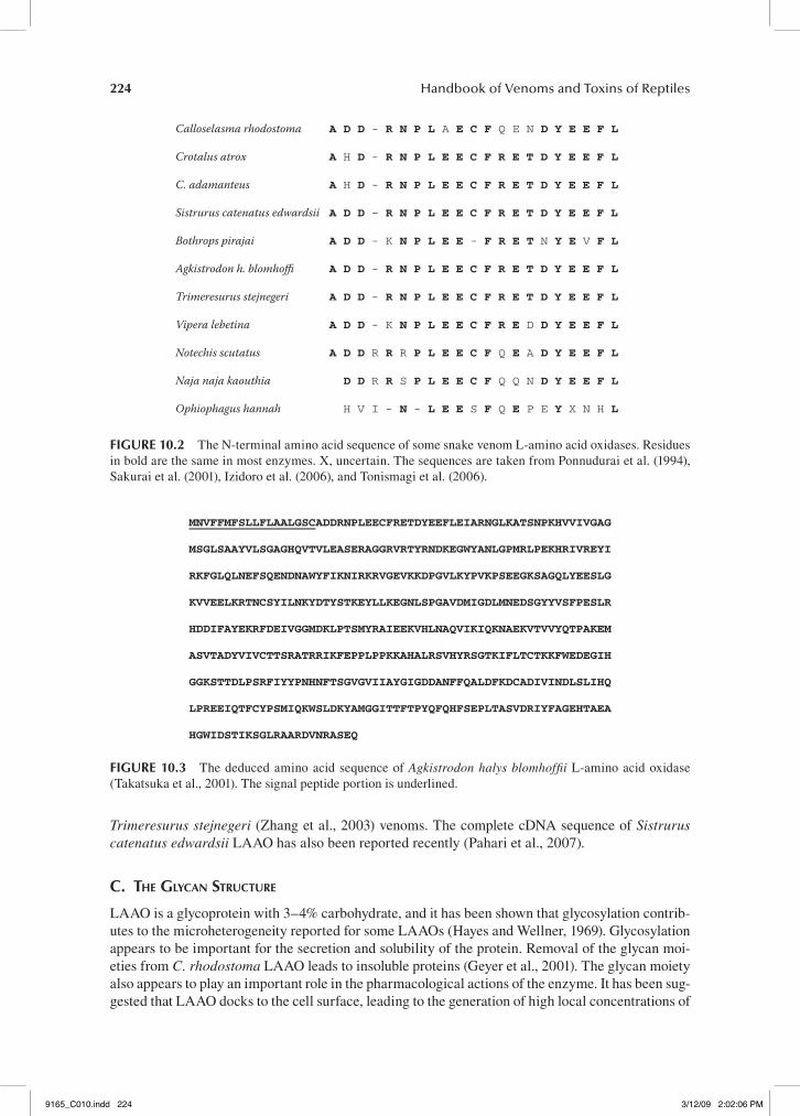
Takatsuka et al. (2001) reported that the full-length cDNA sequence of Agkistrodon halys blom-hoffii LAAO encodes a putative a signal peptide with 18 amino acid residues and a 486-residue subunit (Figure 10.3). Franca et al. (2007) reported the cDNA sequences of LAAO from B. moojeni and B. jararacussu venom, and phylogenetic analysis showed sequence identities within the range 83–87% being closely related to LAAO from A. h. blomhoffii (Takatsuka et al., 2001) and
9165_C010.indd 223 3/12/09 2:02:05 PM
224 Handbook of Venoms and Toxins of Reptiles
Trimeresurus stejnegeri (Zhang et al., 2003) venoms. The complete cDNA sequence of Sistrurus catenatus edwardsii LAAO has also been reported recently (Pahari et al., 2007).
c. the GlycAn structure
LAAO is a glycoprotein with 3–4% carbohydrate, and it has been shown that glycosylation contrib-utes to the microheterogeneity reported for some LAAOs (Hayes and Wellner, 1969). Glycosylation appears to be important for the secretion and solubility of the protein. Removal of the glycan moi-eties from C. rhodostoma LAAO leads to insoluble proteins (Geyer et al., 2001). The glycan moiety also appears to play an important role in the pharmacological actions of the enzyme. It has been sug-gested that LAAO docks to the cell surface, leading to the generation of high local concentrations of
Calloselasma rhodostoma A D D - R N P L A E C F Q E N D Y E E F L
Crotalus atrox A H D - R N P L E E C F R E T D Y E E F L
C. adamanteus A H D - R N P L E E C F R E T D Y E E F L
Sistrurus catenatus edwardsii A D D – R N P L E E C F R E T D Y E E F L
Bothrops pirajai A D D - K N P L E E - F R E T N Y E V F L
Agkistrodon h. blomhoffi A D D - R N P L E E C F R E T D Y E E F L
Trimeresurus stejnegeri A D D - R N P L E E C F R E T D Y E E F L
Vipera lebetina A D D - K N P L E E C F R E D D Y E E F L
Notechis scutatus A D D R R R P L E E C F Q E A D Y E E F L
Naja naja kaouthia D D R R S P L E E C F Q Q N D Y E E F L
Ophiophagus hannah H V I - N - L E E S F Q E P E Y X N H L
fIgure 10.2 The N-terminal amino acid sequence of some snake venom L-amino acid oxidases. Residues in bold are the same in most enzymes. X, uncertain. The sequences are taken from Ponnudurai et al. (1994), Sakurai et al. (2001), Izidoro et al. (2006), and Tonismagi et al. (2006).
MNVFFMFSLLFLAALGSCADDRNPLEECFRETDYEEFLEIARNGLKATSNPKHVVIVGAG
MSGLSAAYVLSGAGHQVTVLEASERAGGRVRTYRNDKEGWYANLGPMRLPEKHRIVREYI
RKFGLQLNEFSQENDNAWYFIKNIRKRVGEVKKDPGVLKYPVKPSEEGKSAGQLYEESLG
KVVEELKRTNCSYILNKYDTYSTKEYLLKEGNLSPGAVDMIGDLMNEDSGYYVSFPESLR
HDDIFAYEKRFDEIVGGMDKLPTSMYRAIEEKVHLNAQVIKIQKNAEKVTVVYQTPAKEM
ASVTADYVIVCTTSRATRRIKFEPPLPPKKAHALRSVHYRSGTKIFLTCTKKFWEDEGIH
GGKSTTDLPSRFIYYPNHNFTSGVGVIIAYGIGDDANFFQALDFKDCADIVINDLSLIHQ
LPREEIQTFCYPSMIQKWSLDKYAMGGITTFTPYQFQHFSEPLTASVDRIYFAGEHTAEA
HGWIDSTIKSGLRAARDVNRASEQ
fIgure 10.3 The deduced amino acid sequence of Agkistrodon halys blomhoffii L-amino acid oxidase (Takatsuka et al., 2001). The signal peptide portion is underlined.
9165_C010.indd 224 3/12/09 2:02:06 PM
Snake Venom L-Amino Acid Oxidases 225
H2O2 (Suhr and Kim, 1999), which mediates many pharmacological actions of LAAOs (see below). Deglycosylation of the enzyme, however, did not alter the enzyme activity (Stabeli et al., 2004; Izidoro et al, 2006).
Geyer et al. (2001) reported on the glycosylation of LAAO from C. rhodostoma. Its glycosylation is remarkably homogeneous, with the major oligosaccharide accounting for approximately 90% of the total sugar content. The glycan is identified as a bis-sialylated, biantennary, core-fucosylated dodecasaccharide. An interesting possibility with regard to the homogeneity of the glycan moiety is that it is a functional requirement connected with the biological activities ascribed to LAAO.
VIII. three-dIMensIonAL struCture of LAAos
A. X-rAy structure of C. Rhodostoma lAAo
The x-ray structure of C. rhodostoma LAAO has been elucidated (Pawelek et al., 2000), and the topography of the enzyme most closely resembles that of polyamine oxidase. The data indicate that it is functionally a dimer consisting of two 55 kDa monomers. Each monomer consists of fifteen α-helices and twenty-two β-strands that fold into three well-defined domains: an FAD-binding domain, a substrate-binding domain, and a helical domain. The main structural feature of the FAD-binding domain (consists of residues 35–64, 242–318, and 446–486) is a six-stranded β-pleated sheet sandwiched between three α-helices and a four-stranded β-pleated sheet. This motif makes up the classical nucleotide-binding fold seen in many FAD-binding enzymes. The substrate-binding domain is made up of residues 5–25, 73–129, 233–236, and 323–420. The helical domain consists of residues 130–230 and comprises one side of a funnel-shaped entrance to the active site of the enzyme. The interface between the substrate-binding and helical domains forms a 25 Å long funnel, which provides access to the active site. Comparison of the LAAO with the structure of mammalian D-amino acid oxidase reveals significant differences in their modes of substrate entry.
Moustafa et al. (2006) reported a high-resolution (1.8 Å) x-ray structure of C. rhodostoma LAAO with its substrate L-phenylalanine. The data reveal a dynamic active site, as conformational changes are apparent for the isoalloxazine ring. There is a Y-shaped channel system, extending from the external surface of the protein to the active site. The authors suggested that one portion of this chan-nel may serve as the entry path for O2 during the oxidative half-reaction. On the other hand, the sec-ond region, which is separated from the proposed O2 channel by the N terminus (residues 8–16) of the protein, may play a role in H2O2 release. Presumably, the channel would direct the H2O2 product to the exterior surface of the protein, near the glycan moiety at Asn172, which was thought to anchor the enzyme to the host cell. This channel location may explain the ability of the enzyme to localize H2O2 to the targeted cell, thus inducing the apoptotic effect as well as other pharmacological activities (see below). The x-ray structure confirmed that the carbohydrate moieties are linked to Asn172 and Asn361. The authors speculated that the disialylated oligosaccharides at Asn172, which is located in the vicinity of the channel leading to the active site of the enzyme, may bind to siglecs (sialic acid-binding immunoglobulin superfamily lectins) of the target cells via its sialylated glycan moiety, resulting in production of a locally high concentration of H2O2 in or near the binding interface. This, in turn, could lead to oxidative damage to the siglec or another adjacent cell structural element.
B. moleculAr modelinG of B. JaRaRaCussu And B. mooJeni lAAos
Molecular modeling experiments with overlapping of B. jararacussu and B. moojeni LAAO models demonstrated that these proteins are almost identical (Franca et al., 2007). In addition, the overall fold of the two models is very similar to that of LAAO from C. rhodostoma venom. All the essential residues in C. rhodostoma LAAO are conserved in the B. moojeni and B. jararacussu LAAO models, demonstrating the putative functional similarity between the models and the C. rhodostoma LAAO structure. It is possible that most snake venom LAAOs have a similar three-dimensional structure.
9165_C010.indd 225 3/12/09 2:02:06 PM
226 Handbook of Venoms and Toxins of Reptiles
Ix. the enzyMAtIC ProPertIes of LAAos
A. GenerAl enzymAtic ProPerties
LAAO required Mg2+ and was inhibited by Ca2+, phosphate, or p-chloromercuribenzoate. Certain amino acids stabilize the enzyme, while at high concentration they become inhibitors. The enzyme is also competitively inhibited by various aliphatic and aromatic acids and had a pH optimum from 7 to 8.5. (Tan, 1998). LAAOs from different sources differ substantially in their specific activities. When L-leucine was used as the substrate, at pH 8.5, the specific activities of the enzyme isolated from C. rhodostoma, N. kaouthia, and O. hannah were 0.54, 4.59, and 20.9 µmole/min/mg, respec-tively. Substrate inhibition occurs at high substrate concentrations.
B. suBstrAte sPecificity
Many authors have investigated substrate specificity of snake venom LAAO (Tan, 1998). LAAO did not oxidize any D-amino acid and was highly specific for the L-enantiomer of amino acids. Effective oxidation of L-amino acid by the enzyme requires the presence of a free primary α-amino group. Generally, the best substrates are L-Leu, L-Met, L-Phe, L-Tyr, and L-Trp, whereas L-Lys, L-Ser, L-Thr, L-Asp, and L-Glu were generally hydrolyzed slowly or not at all (Ponnudurai et al., 1994; Souza et al., 1999; Stabeli et al., 2004; Izidoro et al., 2006; Samel et al., 2006; Tonismagi et al., 2006). One exception is O. hannah LAAO, for which L-Lys is the best substrate (Tan and Saifuddin, 1991). It is interesting to note that the N-terminal sequence of the O. hannah (king cobra) enzyme is also quite different from the other snake venom LAAOs.
Examination of the substrate specificity data of snake venom LAAOs suggested the presence of an alkyl side chain–binding site that comprises at least four subsites, each accommodating a methyl/methylene carbon. Ophiophagus hannah LAAO, on the other hand, appears to have an additional amino-binding subsite (Tan, 1998). A similar alkyl-binding site was suggested to be present in D-amino acid oxidase (Dixon and Kleppe, 1965).
c. mechAnism of cAtAlysis
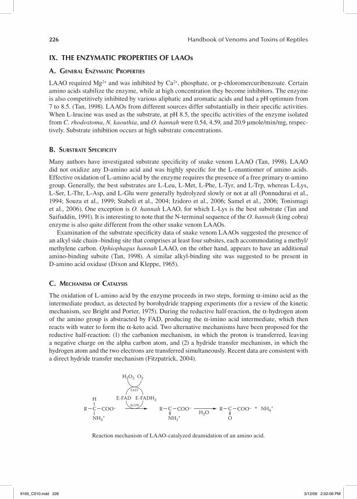
The oxidation of L-amino acid by the enzyme proceeds in two steps, forming α-imino acid as the intermediate product, as detected by borohydride trapping experiments (for a review of the kinetic mechanism, see Bright and Porter, 1975). During the reductive half-reaction, the α-hydrogen atom of the amino group is abstracted by FAD, producing the α-imino acid intermediate, which then reacts with water to form the α-keto acid. Two alternative mechanisms have been proposed for the reductive half-reaction: (1) the carbanion mechanism, in which the proton is transferred, leaving a negative charge on the alpha carbon atom, and (2) a hydride transfer mechanism, in which the hydrogen atom and the two electrons are transferred simultaneously. Recent data are consistent with a direct hydride transfer mechanism (Fitzpatrick, 2004).
R COO–
E-FAD E-FADH2
FAST
SLOWH
C
NH3+
H2O2
H2O
O2
R COO–C
NH2+
NH4++R COO–C
O
Reaction mechanism of LAAO-catalyzed deamidation of an amino acid.
9165_C010.indd 226 3/12/09 2:02:06 PM
Snake Venom L-Amino Acid Oxidases 227
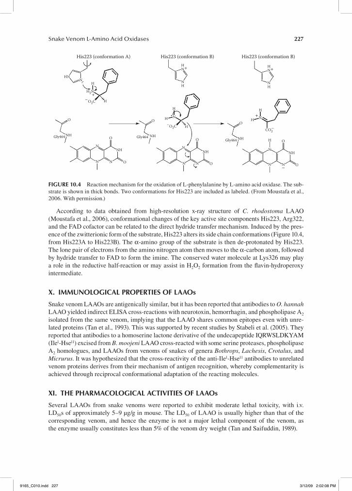
According to data obtained from high-resolution x-ray structure of C. rhodostoma LAAO (Moustafa et al., 2006), conformational changes of the key active site components His223, Arg322, and the FAD cofactor can be related to the direct hydride transfer mechanism. Induced by the pres-ence of the zwitterionic form of the substrate, His223 alters its side chain conformations (Figure 10.4, from His223A to His223B). The α-amino group of the substrate is then de-protonated by His223. The lone pair of electrons from the amino nitrogen atom then moves to the α-carbon atom, followed by hydride transfer to FAD to form the imine. The conserved water molecule at Lys326 may play a role in the reductive half-reaction or may assist in H2O2 formation from the flavin-hydroperoxy intermediate.
x. IMMunoLogICAL ProPertIes of LAAos
Snake venom LAAOs are antigenically similar, but it has been reported that antibodies to O. hannah LAAO yielded indirect ELISA cross-reactions with neurotoxin, hemorrhagin, and phospholipase A2 isolated from the same venom, implying that the LAAO shares common epitopes even with unre-lated proteins (Tan et al., 1993). This was supported by recent studies by Stabeli et al. (2005). They reported that antibodies to a homoserine lactone derivative of the undecapeptide IQRWSLDKYAM (Ile1-Hse11) excised from B. moojeni LAAO cross-reacted with some serine proteases, phospholipase A2 homologues, and LAAOs from venoms of snakes of genera Bothrops, Lachesis, Crotalus, and Micrurus. It was hypothesized that the cross-reactivity of the anti-Ile1-Hse11 antibodies to unrelated venom proteins derives from their mechanism of antigen recognition, whereby complementarity is achieved through reciprocal conformational adaptation of the reacting molecules.
xI. the PhArMACoLogICAL ACtIVItIes of LAAos
Several LAAOs from snake venoms were reported to exhibit moderate lethal toxicity, with i.v. LD50s of approximately 5–9 µg/g in mouse. The LD50 of LAAO is usually higher than that of the corresponding venom, and hence the enzyme is not a major lethal component of the venom, as the enzyme usually constitutes less than 5% of the venom dry weight (Tan and Saifuddin, 1989).
HNN
H
HN
NH
NH NH
NHNH
H
H H
H
N
N N
HH
N
N
NN
N
NH
H2N
H
O O
O
O
O
O
O2CCO2
O2C
O
O
O
NNH
N
NHGly464 Gly464Gly464
+
+
+
–
–
HN+
His223 (conformation A) His223 (conformation B) His223 (conformation B)
–
–
–
N
fIgure 10.4 Reaction mechanism for the oxidation of L-phenylalanine by L-amino acid oxidase. The sub-strate is shown in thick bonds. Two conformations for His223 are included as labeled. (From Moustafa et al., 2006. With permission.)
9165_C010.indd 227 3/12/09 2:02:08 PM
228 Handbook of Venoms and Toxins of Reptiles
Over the last 15 years, LAAO has become an interesting object for biomedical studies because of its apoptotic, cytotoxic, platelet aggregation, anticoagulant, and other physiological effects. These effects are thought to be mediated by the chemically reactive H2O2 generated in the oxida-tion process, because H2O2 scavengers such as catalase neutralize the effects. Sometimes the toxic effects cannot be attributed to H2O2 liberated alone, and direct interactions between LAAO and the target cells may play an important role (Zhang et al., 2003).
A. edemA-inducinG And hemorrhAGic ActiVities
Several authors reported that venom LAAO was able to induce extensive edema in the mouse paw, some with slight hemorrhage (Tan and Choy, 1993; Du and Clemetson, 2002; Stabeli et al., 2004; Izidoro et al., 2006). Tan and Choy (1993) reported that O. hannah LAAO exhibited strong edema-inducing activity, and the enzyme elicited a delayed-type time course of edema formation, indicating that the edema formation caused by LAAO was not mediated through release of amines subsequent to mast cell degradation, which usually elicited a rapid type of edema formation. The edema-inducing activity of the enzyme was not inhibited by diphenhydramine or dexamethasone. Izidoro et al. (2006) suggested that edema formation is due to activation of the inflammatory response by the H2O2 generated, as administration of glutathione to the mouse paw inhibited the edema-inducing activity of the enzyme. The hemorrhagic effect of LAAO results from complex effects, and may involve apoptosis of endothelial and other vascular cells.
B. AnticoAGulAnt effects
Sakurai et al. (2003) reported that LAAO purified from A. h. blomhoffii venom possesses antico-agulant activity. The enzyme significantly delayed the onset and progress of blood coagulation, pro-longed the activated partial thromboplastin time, but had little effect on the prothrombin time. The results indicated that LAAO interferes primarily with the intrinsic blood coagulation pathway, and further studies by the authors indicated that the anticoagulant effect of LAAO is due to its inhibitory action on clotting factor IX.
c. effects on PlAtelet AGGreGAtion
Reports on the effects of snake venom LAAOs on platelet aggregation seem to be contradictory: some authors reported that LAAOs induce platelet aggregation, whereas other authors reported that LAAOs have an inhibitory action on platelet aggregation (Du and Clemetson, 2002). Catalase, an H2O2 scavenger, inhibited both platelet aggregation-inducing and -inhibiting effects, indicating that both effects are due primarily to the action of H2O2 produced by the enzyme during the oxidation.
LAAOs from venoms of C. durissus cascavella, E. macmahoni, B. alternatus, B. pirajai, and O. hannah induce platelet aggregation (Li et al., 1994; Ali et al., 2000; Stabeli et al., 2004; Toyama et al., 2006, Izidoro et al., 2006). Toyama et al. (2006) reported that the platelet aggregation- inducing activity was inhibited by cyclooxygenase pathway inhibitors such as aspirin and indomethacin, sug-gesting that the H2O2 liberated leads to activation of inflammatory enzymes. Du and Clemetson (2002) suggested that H2O2 production promoted a rapid increase of thromboxane A2 synthesis and consequently the platelet aggregation. Conversely, LAAOs from A. h. blomhoffi, V. lebetina, and N. naja kaouthia dose-dependently inhibited both agonist-induced platelet aggregation and shear-induced platelet aggregation (Sakurai et al., 2001; Takatsuka et al., 2001; Tonismagi et al., 2006). One mechanism for platelet aggregation inhibition may be connected with a reduced bind-ing of ADP in platelets exposed to H2O2, or the interference of the peroxide in the interaction between the activated platelet integrin GPIIb/IIIa and fibrinogen (Takatsuka et al., 2001; Samel et al., 2006).
9165_C010.indd 228 3/12/09 2:02:08 PM
Snake Venom L-Amino Acid Oxidases 229
It is still not clear why some LAAOs induce and others inhibit platelet aggregation. Sakurai et al. (2001) suggested that the controversies may be connected with differences in the experimental pro-cedure or preparation of blood samples. Other possibilities include the difference in specific activity of the enzyme, or the involvement of mechanisms other than H2O2 liberation that are present only in certain LAAOs. Takatsuka et al. (2001), for example, suggested that only LAAOs with very high specific activities would induce platelet aggregation, as initiation of aggregation requires relatively high peroxide concentration (millimolar range). However, since LAAOs generally liberate H2O2 in only micromolar concentrations, it has been further suggested that these enzymes may bind to the platelet and be able to generate high concentrations of H2O2 locally to induce platelet aggregation.
d. APoPtosis-inducinG effect
Many snake venoms are known to exhibit apoptosis-inducing effects. Apoptosis is the programmed cell death characterized by a distinct pattern of cellular events, including cleavage of nuclear DNA into fragments that produce a typical nucleosomal DNA ladder upon agarose gel electrophoresis. Suhr and Kim (1996) and Torii et al. (1997) reported that the snake venom component that induced apoptosis was an LAAO, and that LAAO induced apoptosis in human umbilical vein endothelial, human promyelocytic leukemia HL-60, human ovarian carcinoma A2789, and mouse endothelial KN-3 cells. Since then, many snake venom LAAOs were reported to also exhibit apoptosis-inducing activity (Souza et al., 1999; Takatsuka et al., 2001; Sun et al., 2003; Ande et al., 2006; Izidoro et al., 2006; Samel et al., 2006), and apoptosis was usually demonstrated by the DNA fragmentation gel pattern. The apoptosis-inducing activity was abolished by catalase and other H2O2 scavengers, indi-cating that the H2O2 generated by LAAO action plays an important role in the apoptosis. Tempone et al. (2001) suggested that cells exposed to oxidative stress induced by LAAO generated H2O2 that could activate heat shock proteins and initiate cell membrane disorganization, DNA fragmentation, apoptosis, and therefore cell death. Sun et al. (2003) suggested that the generated peroxide activates the transcription of such factors as the nuclear factor B, the activator protein 1, Fas/Apo-1, and p53.
Suhr and Kim (1999), however, demonstrated that the LAAO-induced apoptotic mechanism was clearly distinguishable from the one stimulated directly by exogenous H2O2, suggesting that the LAAO-induced apoptosis was not solely triggered by the peroxide produced by oxidation reactions. Takatsuka et al. (2001) demonstrated that venom LAAOs directly bind to cell surface, thereby increasing the local peroxide concentration. On the other hand, Torii et al. (2000) reported that the venom LAAO did not associate with human embryonic kidney cells. The reason for these discrep-ancies is not clear.
Ande et al. (2006) and Samel et al. (2006), using Jurkat and K562 (human chronic myeloid leu-kemia) cells, respectively, reported that at low concentration LAAO induced apoptosis, but caused necrosis of the cells at higher concentrations. According to Ande et al. (2006), the factors contribut-ing to apoptosis are: (1) generation of toxic intermediates from fetal calf serum and (2) binding and internalization of LAAO, which appears to be mediated by the glycan moiety of the enzyme, as desialylation of the enzyme reduces cytotoxicity. D-amino acid oxidase, which lacks glycosylation, also triggers necrosis by the H2O2 liberated, but it does not cause apoptosis. Thus, just like the effect on platelet aggregation, induction of cell death by LAAO also appears to involve both the genera-tion of H2O2 and the molecular interaction of the glycan moiety of the enzyme with structures at the cell surface.
e. AntiBActeriAl ActiVity
Stiles et al. (1991) reported that two LAAOs from the venom of Pseudechis australis (mulga snake) have a powerful antibacterial effect against Gram-positive and Gram-negative bacteria. Compared to tetracycline, the in vitro antibacterial effects of the enzymes were 18–70 times more effective, on
9165_C010.indd 229 3/12/09 2:02:08 PM
230 Handbook of Venoms and Toxins of Reptiles
a molar basis. Recently, many authors reported LAAO from other snake venoms also exhibited sim-ilar antibacterial activity (Izidoro et al., 2006; Stabeli et al., 2004; Tonismagi et al., 2006; Toyama et al., 2006). It is believed that the antibacterial effect of LAAO is also due to the H2O2 liberated, as addition of catalase completely suppressed the antibacterial activity. Electron microscopic studies suggested that the H2O2 generated in the oxidation process induced bacterial membrane rupture and then cell death (Toyama et al., 2006). Zhang et al. (2004) reported that the A. halys LAAO was able to bind to the surfaces of bacteria and generate high concentrations of H2O2 locally, which enables the enzyme to inhibit bacterial growth at low concentrations. It is not clear whether this happens with other snake venom LAAOs.
f. leishmAnicidAl ActiVity
Leishmaniasis, caused by several protists in the genus Leishmania, includes a spectrum of human infectious disease ranging from self-healing cutaneous ulceration to a progressive and lethal visceral infection. It is a disease that affects approximately 12 million people and is prevalent in eighty-eight nations throughout the world. Tempone et al. (2001) and Toyama et al. (2006) reported that snake venom LAAO possesses strong leishmanicidal activity, as the H2O2 generated by the enzyme was a strong inducer of apoptosis in promastigotes of Leishmania ssp. cells.
At present, few drugs are available for treatment of leishmaniasis. The understanding of the mode of action of LAAO upon parasites may lead to the design of new drugs or therapeutic approaches for leishmaniasis. For example, if one were able to target a H2O2 generator (such as snake venom LAAO) toward the intracellular parasitophorous vacuole occupied by Leishmania, this would repre-sent a highly specific treatment not only for leishmaniasis but also for other intracellular parasites.
G. Anti-hiV ActiVity
Zhang et al. (2003) reported that LAAO isolated from T. stejnegeri venom possesses antiviral activ-ity. The enzyme exhibited dose-dependent inhibition of HIV-I infection and replication at con-centrations that showed little effect on cell viability. Under the same experimental conditions, no anti-HIV-1 activity was observed by exogenous addition of H2O2. Furthermore, the presence of cat-alase caused a decrease in its antivirus activity but resulted in an increase of its antiviral selectivity. The authors suggested that while liberated H2O2 is involved in the anti-HIV-1 activity of the LAAO, the dosages of H2O2 and relative molecular pathways mediating suppression in virus infection and replication are independent of or different from those causing cell death. Presumably, the mecha-nism of the anti-HIV-1 effect of LAAO involves specific binding of the enzyme to cell membrane, which helps to generate high local concentrations of H2O2 and trigger specific signal reactions and activation of host cells, resulting in the inhibition of HIV infection or replication.
xII. ConCLusIons
Prior to the 1990s, studies of snake venom LAAO dealt mainly with their enzymatic properties and industrial applications. In the past 15 years, there has been considerable progress in the studies of the structure and mechanism of the enzyme, but the focus has shifted to the investigations of the phar-macological actions of the enzyme and its potential biotechnological and medical applications.
Snake venom LAAOs are interesting multifunctional enzymes exhibiting edema-inducing, plate-let aggregation-inhibiting or -inducing, apoptotic-inducing, and anti-HIV-1 activities, as well as anticoagulation effect. Their toxicological actions are due mainly, but not entirely, to the H2O2 liberated during the oxidation. The exact mechanism of the toxicological actions of snake venom LAAO awaits further studies.
Sun et al. (2003) suggested that LAAO may be applied clinically in glioma therapy by cloning the cDNA of the enzyme and transfecting the tumor cells of patients to induce apoptosis in the
9165_C010.indd 230 3/12/09 2:02:08 PM
Snake Venom L-Amino Acid Oxidases 231
target tumor cells. Many authors have demonstrated the apoptotic effect of snake venom LAAO on various malignant cells. There is therefore potential in the application of LAAO in cancer therapy. The understanding of the LAAO mode of action upon parasites may also lead to the design of new drugs or therapeutic approaches for leishmaniasis as well as other intracellular parasites. In addi-tion, investigation on the anti-HIV activity of LAAO would also provide valuable information on the therapeutic development of new generations of anti-HIV drugs.
ACknowLedgMent
This work was supported by research grant ScienceFund 02-01-03-SF0153 from the Ministry of Science, Technology and Innovation (MOSTI), Government of Malaysia.
referenCes
Abe, Y., Y. Shimoyama, H. Munakata, J. Ito, N. Nagata, and K. Obtsuki. 1998. Characterization of an apoptosis-inducing factor in habu snake venom as a glycyrrihizin (GL)-binding protein potently inhibited by GL in vitro. Biol. Pharm. Bull. 21:924–27.
Ali, S. A., S. Stoeva, A. Abbasi, J. M. Alam, R. Kayed, M. Faigle, B. Neumeister, and W. Voelter. 2000. Isolation, structural and functional characterization of an apoptosis-inducing L-amino acid oxi-dase from leaf-nosed viper (Eristocophis macmahoni) snake venom. Arch. Biochem. Biophys. 384: 216–26.
Ande, S. R., P. R. Kommoju, S. Draxyl, M. Murkovic, P. Macheroux, S. Ghisla, and E. Ferrando-May. 2006. Mechanisms of cell death induction by L-amino acid oxidase, a major component of ophidian venom. Apoptosis 11:1439–51.
Bergmeyer, H. V. 1983. L-amino acid oxidase. In Methods in enzymatic analysis, 149–50. Vol. 2. Weinheim: Verlag Chimie.
Bright, D. J., and D. J. T. Porter. 1975. Flavoprotein oxidases. In The enzymes, ed. P. D. Boyer, 421–505. 3rd ed., Vol. XIIB. New York: Academic Press.
Coles, C. J., D. E. Edmondson, and T. P. Singer. 1977. Reversible inactivation of L-amino acid oxidase. Properties of the three conformational forms. J. Biol. Chem. 252:8035–39.
Curti, B., V. Massey, and M. Zmudha. 1968. Inactivation of snake venom L-amino acid oxidase by freezing. J. Biol. Chem. 243:2306–14.
Curti, B., S. Ronchi, and P. M. Simonetta. 1992. D- and L-amino acid oxidases. In Chemistry and biochemistry of flavoenzyme, ed. F. Mueller, 69–94. Vol. 3. Boca Raton, FL: CRC Press.
Dixon, M., and K. Kleppe. 1965. D-amino acid oxidase II. Specificity, competitive inhibition and reaction sequence. Biochim. Biophys. Acta 96:368–82.
Du, X. Y., and K. J. Clemetson. 2002. Snake venom L-amino acid oxidase. Toxicon 40: 659–65.Findrik, Z., B. Geueke, W. Hummel, and D. Vasic-Racki. 2006. Modelling of L-DOPA enzymatic oxidation
catalyzed by L-amino acid oxidase from Crotalus adamanteus and Rhodococcus opacus. Biochem. Eng. J. 27:275–86.
Fitzpatrick, P. F. 2004. Carbanion versus hydride transfer mechanisms in flavoprotein-catalyzed dehydrogena-tions. Bioorg. Chem. 32:125–39.
Franca, S. C., S. Kashima, P. G. Roberto, M. Marins, F. K. Ticli, J. O. Pereira, S. Astolfi-Filho, R. G. Stabeli, A. J. Magro, M. R. M. Fontes, S. V. Sampaio, and A. M. Soares. 2007. Molecular approaches for struc-tural characterization of Bothrops L-amino acid oxidases with antiprotozoal activity: cDNA cloning, comparative sequence analysis, and molecular modeling. Biochem. Biophys. Res. Commun. 355:302–6.
Geyer, A., T. B. Fitzpatrick, P. D. Pawelek, K. Kitzing, A. Vrielink, S. Ghisla, and P. Macheroux. 2001. Structure and characterization of the glycan moiety of L-amino acid oxidase from the Malayan pit viper Calloselasma rhodostoma. Eur. J. Biochem. 268:4044–53.
Hayes, M. B., and D. Wellner. 1969. Microheterogeneity of L-amino acid oxidase. J. Biol. Chem. 244:6636–44.Iwanaga, S., and T. Suzuki. 1979. L-amino acid oxidase. In Handbook of experimental pharmacology, ed. C. Y.
Lee, 75–84. Vol. 52. Berlin: Springer.Izidoro, L. F. M., M. C. Ribeiro, G. R. L. Souza, C. D. Sant’Ana, A. Hamaguchi, M. I. Homsi-Brandeburgo,
L. R. Goulart, R. O. Beleboni, A. Nomizo, S. V. Sampaio, A. M. Soares, and V. M. Rodrigues. 2006. Biochemical and functional characterization of an L-amino acid oxidase isolated from Bothrops pirajai snake venom. Bioorg. Med. Chem. 14:7034–43.
9165_C010.indd 231 3/12/09 2:02:08 PM

232 Handbook of Venoms and Toxins of Reptiles
Kishimoto, M., and T. Takahashi. 2001. A spectrophotometric microplate assay for L-amino acid oxidase. Anal. Biochem. 298:136–39.
Kommoju, P. R., P. Macheroux, and S. Ghisla. 2007. Molecular cloning, expression and purification of L-amino acid oxidase from Malayan pit viper Calloselasma rhodostoma. Protein Exp. Purif. 52:89–95.
Li, Z. Y., T. F. Yu, and E. C. Lian. 1994. Purification and characterization of L-amino acid oxidase from king cobra (Ophiophagus Hannah) venom and its effects on human platelet aggregation. Toxicon 32:1349–58.
Macheroux, R., O. Seth, C. Bollschweiler, M. Schwarz, M. Kurfurst, L. C. Au, and S. Ghisla. 2001. L-amino acid oxidase from the Malayan pit viper Calloselasma rhodostoma. Comparative sequence analysis and characterization of active and inactive forms of the enzyme. Eur. J. Biochem. 268:1679–86.
Mackessy, S. P. 2002. Biochemistry and pharmacology of colubrid snake venoms. J. Toxicol.-Toxin Rev. 21:43–83.
Meister, A., and D. Wellner. 1963. Flavoprotein amino acid oxidases. In The enzymes, ed. P. D. Boyer, H. Lardy, and K. Myrback, 609–48. 2nd ed., Vol. VII. New York: Academic Press.
Moustafa, I. M., S. Foster, A. Y. Lyubimov, and A. Vrielink. 2006. Crystal structure of LAAO from Callose-lasma rhodostoma with an L-phenylalanine substrate: Insights into structure and mechanism. J. Mol. Biol. 364:991–1002.
Pahari, S., S. P. Mackessy, and R. M. Kini. 2007. The venom gland transcriptome of the desert massasauga rattlesnake (Sistrurus catenatus edwardsii): Towards an understanding of venom composition among advanced snakes (superfamily Colubroidea). BMC Mol. Biol. 8:115.
Pawelek, P. D., J. Cheah., R. Coulombe, P. Macheroux, S. Ghisla, and A. Vrielink. 2000. The structure of L-amino acid oxidase reveals the substrate trajectory into an enantiometrically conservative active site. EMBO J. 19:4204–15.
Ponnudurai, G., M. C. M. Chung, and N. H. Tan. 1994. Purification and properties of the L-amino acid oxidase from Malayan pit viper (Calloselasma rhodostoma) venom. Arch. Biochem. Biophys. 313:373–78.
Raibekas, A. A., and V. Massey. 1996. Glycerol-induced development of catalytically active conformation of Crotalus adamanteus L-amino acid oxidase in vitro. Proc. Natl. Acad. Sci. USA 93:7546–51.
Raibekas, A. A., and V. Massey. 1998. Primary structure of the snake venom L-amino acid oxidase shows high homology with the mouse B cell interleukin 4-induced protein. Biochem. Biophys. Res. Commun. 248:476–78.
Sakurai, Y., M. Shima, T. Matsumoto, H. Takatsuka, K. Nishiya, S. Kasuda, Y. Fujimura, and A. Yoshioka. 2003. Anticoagulant activity of M-LAO, L-amino acid oxidase purified from Agkistrodon halys blom-hoffii, through selective inhibition of factor IX. Biochim. Biophys. Acta 1649:51–57.
Sakurai, Y., H. Takatsuka, A. Yoshioka, T. Matsui, M. Suzuki, K. Titani, and Y. Fujimura. 2001. Inhibition of human platelet aggregation by L-amino acid oxidase purified from Naja naja kaouthia venom. Toxicon 39:1827–33.
Samel, M., H. Vija, G. Ronnholm, J. Sugiir, N. Kalkkinen, and E. Sugiir. 2006. Isolation and characterization of an apoptotic and platelet aggregation inhibiting L-amino acid oxidase from Vipera berus berus (common viper) venom. Biochim. Biophys. Acta 1764:707–14.
Soltysik, S., C. M. Byron, G. H. Einarsdottir, and M. T. Stankovich. 1987. The effects of reversible freezing inactivation and inhibitor binding on redox properties of L-amino acid oxidase. Biochim. Biophys. Acta 911:201–8.
Souza, D. H. F., L. M. Eugenio, J. E. Fletcher, M. S. Jiang, R. C. Garratt, G. Oliva, and H. S. Selistre-de-Araujo. 1999. Isolation and structural characterization of a cytotoxic L-amino acid oxidase from Agkistrodon contortrix laticinctus snake venom: Preliminary crystallographic data. Arch. Biochem. Biophys. 368:285–90.
Stabeli, R. G., L. M. P. Magalhaes, H. S. Selistre-de-Araujo, and E. B. Oliveira. 2005. Antibodies to a fragment of the Bothrops moojeni L-amino acid oxidase cross-react with snake venom components unrelated to the parent protein. Toxicon 46:308–31.
Stabeli, R. G., S. Marcussi, G. B. Carlos, R. C. L. R. Pietro, H. S. Selistre-de-Araujo, J. R. Giglio, E. B. Oliveira, and A. M. Soares. 2004. Platelet aggregation and antibacterial effects of an L-amino acid oxi-dase purified from Bothrops alternatus snake venom. Bioorg. Med. Chem. 12:2881–86.
Stiles, B. G., F. W. Sexton, and S. A. Weinstein. 1991. Antibacterial effect in different snake venoms: Purification and characterization of antibacterial protein from Pseudechis australis (Australian king or mulga snake) venom. Toxicon 29:1129–41.
Suhr, S. M., and D. S. Kim. 1996. Identification of the snake venom substance that induces apoptosis. Biochim. Biophys. Res. Commun. 224:134–39.
Suhr, S. M., and D. S. Kim. 1999. Comparison of the apoptotic pathways induced by L-amino acid oxidase and hydrogen peroxide. J. Biochem. (Tokyo) 125:305–9.
9165_C010.indd 232 3/12/09 2:02:08 PM
Snake Venom L-Amino Acid Oxidases 233
Sun, L. K., Y. Yoshii, A. Hyodo, H. Tsurushima, A. Saito, T. Harakuni, Y. P. Li, K. Kariya, M. Nozaki, and N. Morine. 2003. Apoptotic effect in the glioma cells induced by specific protein extracted from Okinawa habu (Trimeresurus flavoviridis) venom in relation to oxidative stress. Toxicol. In Vitro 17:169–77.
Szwajcer, E., P. Brodelius, and K. Mosbach. 1982. Production of α-keto acids. Part 2. Immobilized whole cells of Providencia sp. PCM 1298 containing L-amino acid oxidase. Enzyme Microb. Technol. 4:409–13.
Takatsuka, H., Y. Sakurai, A. Yoshioka, T. Kokubo, Y. Usami, M. Suzuki, T. Matsui, K. Titani, H. Yagi, M. Matsumoto, and Y. Fujimura. 2001. Molecular characterization of L-amino acid oxidase from Agkistrodon halys blomhoffii with special reference to platelet aggregation. Biochim. Biophys. Acta 1544:267–77.
Tan, N. H. 1998. L-amino acid oxidases and lactate dehydrogenases. In Enzymes from snake venom, ed. G. S. Bailey, chap. 19, 579–98. Fort Collins, CO: Alaken.
Tan, N. H., and S. K. Choy. 1993. The edema-inducing activity of Ophiophagus hannah (king cobra) venom L-amino acid oxidase. In Advances in venom and toxin research, ed. N. H. Tan, S. L. Oo, V. Thambyrajah, and N. Azila, 268–73. Kuala Lumpur: Malaysian Society on Toxinology.
Tan, N. H., K. K. Lim, and M. I. N. Jaafar. 1993. An investigation into the antigenic cross-reactivity of Ophiophagus hannah (king cobra) venom and neurotoxin, phospholipase A2, hemorrhagin and L-amino acid oxidase using enzyme-linked immunosorbent assay. Toxicon 31:865–72.
Tan, N. H., and G. Ponnudurai. 1992. Biochemical characterization of snake venoms. In Recent advances in toxinology research, ed. P. Gopalakrishnakone and C. K. Tan, 210–58. Singapore: National University of Singapore.
Tan, N. H., and M. N. Saifuddin. 1989. Isolation and characterization of an unusual form of L-amino acid oxi-dase from king cobra (Ophiophagus hannah) venom. Biochem. Int. 19:937–44.
Tan, N. H., and M. N. Saifuddin. 1991. Substrate specificity of king cobra (Ophiophagus hannah) venom L-amino acid oxidase. Int. J. Biochem. 23:323–27.
Tan, N. H., and S. Swaminathan. 1992. Purification and properties of the L-amino acid oxidase from monocel-late cobra (Naja naja kaouthia) venom. Int. J. Biochem. 24:967–73.
Tempone, A. G., H. F. Andrade, Jr., P. J. Spencer, C. O. Lourenco, J. R. Rogero, and N. Nascimento. 2001. Bothrops moojeni venom kills Leishmania spp. with hydrogen peroxide generated by its L-amino acid oxidase. Biochem. Biophys. Res. Commun. 280:620–24.
Tonismagi, K., M. Samel, K. Trummal, G. Ronnholm, J. Sugiir, N. Kalkkinen, and E. Sugiir. 2006. L-amino acid oxidase from Vipera lebetina venom: Isolation, characterization, effects on platelets and bacteria. Toxicon 48:227–37.
Torii, S., M. Naito, and T. Tsuruo. 1997. Apoxin I, a novel apoptosis-inducing factor with L-amino acid oxidase activity purified from western diamondback rattlesnake venom. J. Biol. Chem. 272:9539–42.
Torii, S., K. Yamane, T. Mashima, N. Haga, K. Yamamoto, J. W. Fox, M. Naito, and T. Tsuruo. 2000. Molecular cloning and functional analysis of apoxin I, a snake venom-derived apoptosis-inducing factor with L-amino acid oxidase activity. Biochemistry 39:3197–205.
Toyama, M. H., D. de O. Toyama, L. F. D. Passero, M. D. Laurenti, C. E. Corbett, T. Y. Tomokane, F. V. Fonseca, E. Antunes, P. P. Joazeiro, L. O. S. Beriam, M. A. C. Martins, H. S. A. Monteiro, and M. C. Fonteles. 2006. Isolation of a new L-amino acid oxidase from Crotalus durissus cascavella venom. Toxicon 47:47–57.
Tu, A. T. 1977. Venoms: Chemistry and molecular biology. New York: John Wiley.Ueda, M., C. C. Chang, and M. Ohno. 1988. Purification and characterization of L-amino acid oxidase from the
venom of Trimeresurus mucrosquamatus (Taiwan habu snake). Toxicon 26:695–706.Zhang, H., Q. Yang, M. Sun, M. Teng, and L. Niu. 2004. Hydrogen peroxide produced by two amino acid oxi-
dases mediates antibacterial actions. J. Microbiol. 42:336–39.Zhang, Y. I., J. H. Wang, W. H. Lee, Q. Wang, H. Liu, Y. T. Zheng, and Y. Zhang. 2003. Molecular characteriza-
tion of Trimeresurus stejnegeri venom L-amino acid oxidase with potential anti-HIV activity. Biochim. Biophys. Res. Commun. 309:598–604.
9165_C010.indd 233 3/12/09 2:02:09 PM

9165_C010.indd 234 3/12/09 2:02:09 PM