small-molecule modulation of lipid-dependent cellular
TRANSCRIPT
Review ArticleSmall-Molecule Modulation of Lipid-Dependent CellularProcesses against Cancer: Fats on the Gunpoint
Aswin T. Srivatsav , Manjari Mishra, and Shobhna Kapoor
Membrane Biophysics and Chemical Biology Lab, Department of Chemistry, Indian Institute of Technology Bombay,Powai 400076, India
Correspondence should be addressed to Shobhna Kapoor; [email protected]
Received 18 March 2018; Accepted 22 July 2018; Published 15 August 2018
Academic Editor: Min He
Copyright © 2018 Aswin T. Srivatsav et al. This is an open access article distributed under the Creative Commons AttributionLicense, which permits unrestricted use, distribution, and reproduction in any medium, provided the original work is properlycited.
Lipid cell membrane composed of various distinct lipids and proteins act as a platform to assemble various signaling complexesregulating innumerous cellular processes which are strongly downregulated or altered in cancer cells emphasizing the still-underestimated critical function of lipid biomolecules in cancer initiation and progression. In this review, we outline the currentunderstanding of how membrane lipids act as signaling hot spots by generating distinct membrane microdomains called rafts toinitiate various cellular processes and their modulation in cancer phenotypes. We elucidate tangible drug targets and pathways allamenable to small-molecule perturbation. Ranging from targeting membrane rafts organization/reorganization to rewiring lipidmetabolism and lipid sorting in cancer, the work summarized here represents critical intervention points being attempted for lipid-based anticancer therapy and future directions.
1. Introduction
Lewis Thomas in the Lives of a Cell [1] underscored theramifications rendered by the variety of lipids and theirstructural platforms. Lipid assemblies are noncovalently self-assembling biological constituents that create lipid bilayers,within which lipid molecules can relocate in innumer-ous ways. Polar lipids, consisting of a hydrophobic and ahydrophilic portion, majorly form the matrix of cellularmembranes. The propensity of the hydrophobic moieties toself-associate is entropically driven by water and togetherwith the tendency of the hydrophilic moieties to interact withaqueous environments forms the physical basis of the sponta-neous formation of lipid membranes.With advances in lipid-based analytical techniques, lipidomics, we are only begin-ning to appreciate the astounding diversity of lipids in cells.Eukaryotic cell membranes house a wide repertoire of struc-tural lipids, including glycerophospholipids such as phos-phatidylcholine (PC), phosphatidylethanolamine (PE), phos-phatidylserine (PS), phosphatidylinositol (PI), and phospha-tidic acid (PA) [2]. Sphingolipids constitute another classof structural lipids with ceramide unit as their hydrophobic
backbone. The major sphingolipids in mammalian cells aresphingomyelin and glycosphingolipids and sterols representthe major class of nonpolar lipids attributed to their annealedstructures that embody a highly condensed hydrophobicarea. Various permutations and combinations of the lipid’sheadgroups and hydrophobic acyl chains add a high degree ofcomplexity to the existing vast pool of known lipids. Asmuchas∼5% of our genes are devoted to continuously synthesizingand regulating this complex array of lipids, bringing toforefront some exciting questions such as the following: Whyis such a complex diversity of lipids required in a cell? Arecells continuously trying to create structural heterogeneityguided by compositional heterogeneity? Is phase coexistencemanifested as the existence of domains of coexisting phase(s)functionally relevant? In this lieu, every kind of lipid mem-brane ranging from eukaryotic to prokaryotic or within thesame cell possess unique lipid composition that plays crucialrole in not only functional organization but also regulatinga plethora of cellular processes. Additionally, steric andelectrostatic interactions and hydrophobic mismatch inducedistinct domain formation within the bilayer plane providinga platform for organization and assembling of signaling
HindawiBioMed Research InternationalVolume 2018, Article ID 6437371, 17 pageshttps://doi.org/10.1155/2018/6437371
2 BioMed Research International
molecules [3–5]. Lipids exist in a multitude of phases eachmarked by distinct spatial arrangements, molecular struc-ture, and motional freedom of the hydrophobic chains, andbeing susceptible to environmental conditions like pH, ionicstrength, water content, temperature, and pressure is alreadyredefining membrane functionality and offering significantinsights to their functional roles in addition to their long heldstructural roles [6].
Lipids form a considerable part of the dry weight ofmammalian cells. A substantial supply of lipids is requiredfor cell proliferation [7]. Usually, during in vitro growth ofcancer cells, there are abundant nutrients and these cellssynthesize fatty acids de novo. But, under conditions of stress,the cancer cells usually adapt to the cell growth by scavengingextracellular lipids [8], This scavenging spares the cells theneed to supply reducing powers and sources of carbon whichare energy demanding. Lysophospholipids are supplied to thelipid pool for growth by K- or H-Ras that stimulate the lipidutilization and uptake. It is well known that mitochondrialfatty acid oxidation produces twice the ATP than that ofcarbohydrate (glucose) oxidation. Mitochondrial fatty acidoxidation enables cancer cells to survive [9]. Cancer cellsacquire a lipogenic phenotype due to high expression levels ofthe enzyme monoacylglycerol lipase (MAGL). MAGL alongwith hormone sensitive lipase hydrolyzes triglycerides, storedin adipocytes and other cells to free fatty acids, which nowact as a source of energy [10]. Due to this reprogrammedmetabolic circuits, various T cells that infiltrate the tumorexperience the modulated tumor microenvironment and thealtered metabolic network of the growing tumors. Activationofmanypathways needs to be accomplished for their function[11]. But the tumor puts several limitations to dampen theT cell activity due to its reprogrammed metabolism likeupregulated glucose consumption due to Warburg Effect, asa result the extracellular tumor microenvironment showsreduced glucose levels. Since the T cell for its effectivefunctioning requires energy from its environment in termsof glucose or glutamine [12], due to the tumor metabolism,these sources are rapidly depleted which hence puts the Tcell in a dormant state. Therefore, finding ways to modulatethesemetabolic changes that give cancer cells an advantage tosucceed would prove to be useful targets. Some of these havebeen detailed ahead in this review.
Lipids fulfill many critical requirements in the cell includ-ing composing membrane bilayer, storing energy due totheir reduced state, acting as first and second messengersin signal transduction, providing functional implementa-tions of membrane-proteins structure and function, andfinally recognition processes. Advanced mass-spectroscopyand analytical techniques have allowed sensitive and highlyselective analysis of lipids of diverse chemical structureswithin complex biological samples and testified to theirintimate involvement and their aberrations in many diseasessuch as neurodegenerative disorders, infectious diseases, andcancer. Interestingly, lipidomics have revealed cancer type-specific alteration in the lipidome of cells implying criticalroles of lipids in cancer progression and initiation [13–15]. Admittingly, most of the drugs in the market againstcancer (also diabetes and inflammation) elicit their effects via
binding to their target proteins and regulating the underlyingcancer-related cellular process. However in line with theincreasing evidence elucidating the role of membrane lipidsin regulating numerous cellular functions, they have emergedas attractive molecular targets wherein therapies modulat-ing membrane lipids structures and localization could bedeveloped to control molecular events including changes incell signaling, membrane protein function, localization, andgene expression related to various pathological states—theso-called “membrane-lipid therapy” [16, 17].
2. Membrane-Lipid Microdomains as theCellular Signaling Hot Spots
High compositional complexity modulates interactions andlocalizations of lipids in membrane and befittingly influencesthe diverse phases lipid can form [37]. The same are theauthors of spatially constrained distinct regions enrichedin certain lipids within the membrane bilayer plane calledmicrodomains [38, 39]. One such microdomain, lipid rafts,is dynamic assemblies of cholesterol and sphingolipids pre-dominantly in the exoplasmic leaflet of the lipid bilayer.Underpinning this concept is the propensity, in vitro, ofthe saturated hydrocarbon chains intercalating strongly withcholesterol molecules inducing liquid ordered lo phases [40–42]. The membrane surrounding lipid rafts is fluid-like dueto the presence of unsaturated phospholipids forming theliquid disordered ld phase. Thus, the raft domains can beimagined as platforms of lo phases dispersed in the ld matrixof unsaturated glycerolipids. These raft-like assemblies areordered and tightly packed but are still fluid due to lipidacyl chain packing differences. This, in turn, is governedby the saturation level of the hydrocarbon chains in therafts compared with the unsaturated state of fatty acids ofphospholipids in the ld phase. These rafts act as membranescaffolds to house various proteins, organize receptors andtheir downstream molecules, and hence regulate a numberof membrane-associated signaling pathways [43, 44]. Inthis regard, posttranslational modification of proteins suchas addition of GPI anchor, acylation, etc. indispensablymodulates the membrane affinities of proteins includingGPI anchored proteins, epidermal growth factor receptors,estrogen receptors, etc. Membrane rafts are implicated inregulation of cell differentiation proliferation, apoptosis, andnecrosis as well as in cancer initiation and progression [45].Their involvement in numerous tumor models such as colon,prostrate, lung, and breast has been identified [46–49] buttheir structure, function, and associated signaling pathwaysare still under intense scrutiny and subject of immensetherapeutic interest.
On the mechanistic level, a cellular signaling eventoriginates from the raft domains at the membranes withtransmission of signals (chemical, biological, and physical innature) through membrane-bound receptors, e.g., receptortyrosine kinases (RTKs). Stimulation by the diverse signalspromotes receptor dimerization enabling their phospho-rylation mostly via their intrinsic tyrosine kinase activ-ity. The resulting phosphorylated residues exposed to thecytoplasm act as docking sites for effector proteins that
BioMed Research International 3
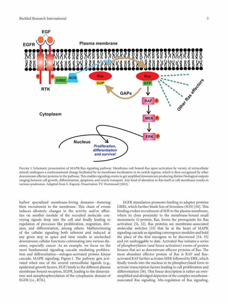
Figure 1: Schematic presentation of MAPK/Ras signaling pathway. Membrane-raft bound-Ras upon activation by variety of extracellularstimuli undergoes a conformational change facilitated by its membrane localization in its switch regions, which is then recognized by otherdownstream effector proteins in the pathway.This enables signaling events to get amplified downstream producing distinct biological outputsranging between cell growth, differentiation, apoptosis, and vesicle transport. Any kind of alteration in Ras itself or raft membrane results invarious syndromes. Adapted from S. Kapoor, Dissertation TU Dortmund (2012).
harbor specialized membrane-loving domains—fosteringtheir recruitment to the membrane. This chain of eventsinduces allosteric changes in the activity and/or affini-ties on another module of the recruited molecule con-veying signals deep into the cell and finally leading toregulation of processes like proliferation, migration, divi-sion, and differentiation, among others. Malfunctioningof the cellular signaling both inherent and induced atany given step in space and time results in uncheckeddownstream cellular functions culminating into various dis-eases, especially cancer. As an example, we focus on themost fundamental signaling cascade mediating prolifera-tion and differentiation—mitogen-activated protein kinasecascade, MAPK signaling, Figure 1. The pathway gets acti-vated when one of the several extracellular ligands (e.g.,epidermal growth factors, EGF) binds to the different plasmamembrane-bound receptors, EGFR, leading to the dimeriza-tion and autophosphorylation of the cytoplasmic domain ofEGFR (i.e., RTK).
EGFR stimulation promotes binding to adaptor proteinsGRB2, which further binds Son of Sevenless (SOS) [50]. Thisbinding evokes recruitment of SOS to the plasma membrane,where its close proximity to the membrane-bound smallmonomeric G-protein, Ras, forms the prerequisite for Rasactivation [51, 52]. Ras proteins are membrane-associatedmolecular switches [53] that lie at the heart of MAPKsignaling cascade as signaling convergencemodules and holdthe place of the first oncogene to be discovered [54, 55]and yet undruggable to date. Activated Ras initiates a seriesof phosphorylation (and hence activation) events of proteinkinases that act as downstream effector proteins of Ras. Themost abundant effector protein of Ras is RAF and Ras-activated RAF further activatesMEK followed by ERK, whichfinally travels into the nucleus in its phosphorylated form toactivate transcription factors leading to cell proliferation anddifferentiation [56]. This linear description is rather an over-simplified and abridged depiction of the complexmembrane-associated Ras signaling. Mis-regulation of Ras signaling,
4 BioMed Research International
e.g., by virtue of failed phosphorylation events, impropermembrane recruitment of effector proteins, and improperlocalization with raft domains or mutations account for 50and 80 % of colon and pancreatic cancers, thus emphasizingthe essential role of Ras in normal cell development [57].
3. Aberrations of Lipids and Lipid Domains inCancers: Tangible Targets
Pathological, pharmacological, and nutritional situationsstrongly regulate lipids in cell with profound biologicalimplications. With advances in high-throughput lipidomics,precise characterization of lipid structures is revealing crit-ical lipid alterations in composition and abundance amongvarious cell types and cancers and surprisingly during variedcellular processes as well [13, 58]. For instance, Eggert et al.[59] demonstrated nice correlation of lipidome changes withcell cycle, with up to eleven different lipid families (chemi-cally distinct structures) accumulating in the dividing cellsconcluding that cells actively modulate the lipid compositionand localization to specific membrane locations requiredfor a particular cellular event. Cellular lipidome remodelingin cancer is manifold and occurs at transcriptomic andlipidomic levels with intriguing complexities [60].
Lipid membranes of cancer cells possess relatively highernegative charge due to increased abundance of PS and PEphospholipids on the exoplasmic membrane surface [61,62]. On a different note, this contributes to attenuation ofrepulsion between polar head groups leading to denser lipidpacking and concomitantly higher rigidity and poor drugpenetration. Thus, exposed PE on the outer membrane ofcancer cell represents a suitable molecular target to developnovel cancer therapeutics aimed at specific binding to orselective sorting of PE leading to cancer cell membranedisruption, permeabilization, and finally cell death [63–65]. Cholesterol is another significantly altered moleculewithin lipid rafts during cancer [66], wherein the levelsare strikingly increased compared with normal cells [67].Higher cholesterol leads to a more rigid and hence lesspermeable cell membrane [68, 69]. In addition to cholesterol,other phospholipids such as PC and PI are also found inincreased abundance in cancer cells. The most foremosteffect of elevated cholesterol is higher raft formation andmomentous enrichment of specific proteins and receptorssuch as EGFR, IGF-1, CD44, and CD24 involved in cellularsignaling mediating tumor progression and invasion [48, 70,71]. Thus strategies involved in modulation of lipid rafts areincreasingly becoming enticing candidates for cancer therapy[72–74]. Downregulation of ceramide metabolism is anotherstrategy found in cancer cells [75] leading to formation ofspecialized membrane domains that recruit specific proteinsinvolved in apoptosis highlighting proteins and kinasesinvolved in ceramide metabolism as potential cancer targets.In addition, a wide variety of tumors also show upregulatedtranscripts involved in lipogenesis and cholesterol synthesispathway, essential for their development and cancer progres-sion. Lipogenic enzymes such as acetyl-CoA carboxylase andfatty acid synthase display a universal increased expressioncoupled with specific alterations in lipid messengers (PIs),
lipid mediators (leukotrienes), and structural lipids (GSL)in most tumors [76]. In this review we will mainly focuson potential anticancer strategies using small molecules thatalter raft assembly, lipid metabolism, regulate lipid sorting,and modulate lipoprotein trafficking inherent in oncogenicsignaling. These strategies are presented along with potentialtargets illustrated with several examples (Figure 4, Table 1).
4. Small-Molecule Chemical Biology Tools
Small molecules targeting specific biomolecules and modu-lating their structure and activity in vivo have transformedthe field of eukaryotic cell biology. Small-molecule-mediatedinhibition of the function of specific proteins has enabledcell biologists to query their functional roles. Most classicexample in this regard is of colchicine and paclitaxel astubulin depolymerizes and stabilizers, respectively, whichhave provided unprecedented insights into the function ofthis cytoskeletal protein [18, 19]. Development of a toolboxof small-molecule inhibitors against cytoskeletal proteinsand many more has enabled regulation of their structure,function, and localization in such ways that were difficult toachieve solely by genetic approaches. The use of chemicalbiology tools specifically to study lipid organization offerskey advantages. (a) They act fast and their activity can bemodulated as a function of dose. (b) They may be reversibleor not (covalent binders). (c)They require nomanipulation ofthe chromosome. (d) Inhibitors targeting conserved cellularprocesses may be applicable across a broad range of species.Due to such salient features, they have a great potential instudying the lipid domain organization in live cells, thuspermitting insights into the functional role of membraneorganization in cancers and other diseases [20, 77].
5. Membrane-Raft ModulatingAgents in Cancer
Membrane rafts regulate key signaling molecules and pro-teins implicated in cancer by modulating their associationwith and localization with lipid membranes including inter-actions with other membrane-bound proteins [43, 45, 72, 78,79]. Thus small-molecule approaches aimed at interruptingthe association of such molecules with membrane rafts byinterfering with association steps directly or modulating therafts themselves represent innovative therapeutic ways forprevention and treatment of cancer.
6. Small Molecules Acting viaMembrane-Raft Disruption
Central roles in the initiation and progression of many tumortypes responsible for the alteration of cell cycle, cell adhesion,cell migration, and programmed cell death are regulatedby various factors. Lipid rafts and membrane microdomainor compartments play an active role in each of these cellprocesses by mainly regulating the downstream intracellularsignaling pathways [21, 22, 43]. Involvement of cholesterol inmaintaining the stability, integrity, and functions aspects ofsuch rafts is indispensable as pharmacological depletion of
BioMed Research International 5
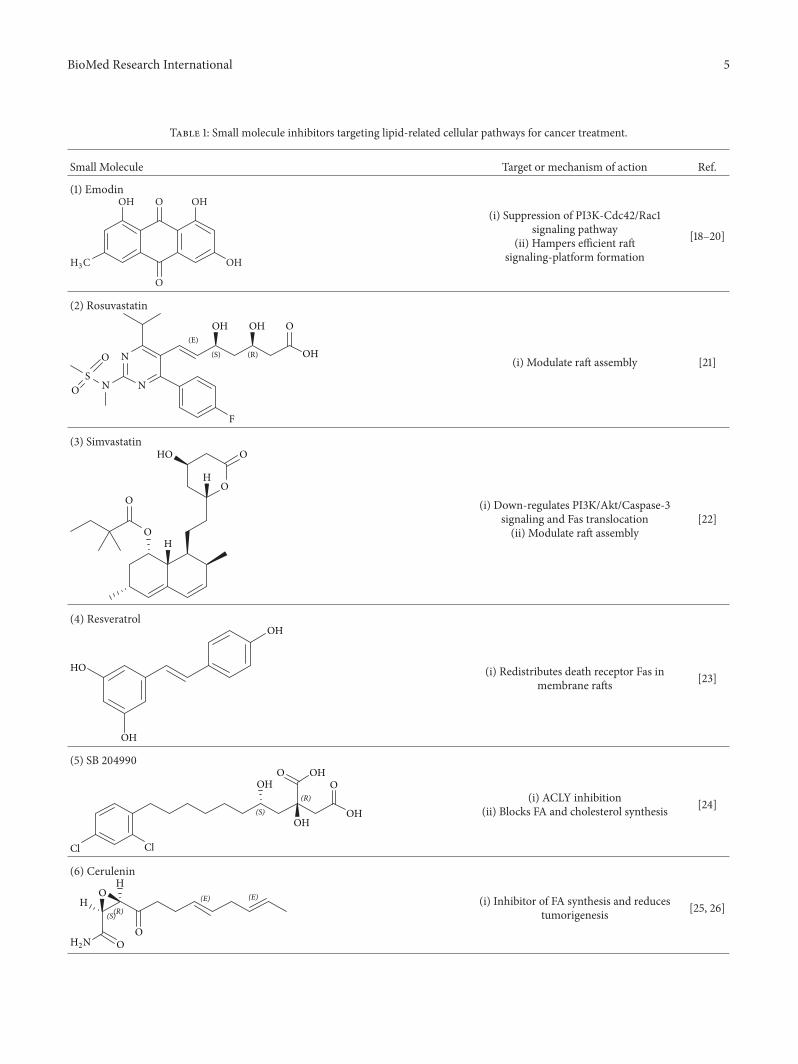
Table 1: Small molecule inhibitors targeting lipid-related cellular pathways for cancer treatment.
Small Molecule Target or mechanism of action Ref.
(1) Emodin
(3#
OH O
O
OH
OH
(i) Suppression of PI3K-Cdc42/Rac1signaling pathway
(ii) Hampers efficient raftsignaling-platform formation
[18–20]
(2) Rosuvastatin
(E)
(S) (R) OH
OOHOH
N
F
N
N
S
O
O
(i) Modulate raft assembly [21]
(3) SimvastatinO
OO
O
H
H
HO
(i) Down-regulates PI3K/Akt/Caspase-3signaling and Fas translocation(ii) Modulate raft assembly
[22]
(4) Resveratrol
HO
OH
OH
(i) Redistributes death receptor Fas inmembrane rafts [23]
(5) SB 204990
Cl Cl
OHOH
OHOH
OO
(S)
(R) (i) ACLY inhibition(ii) Blocks FA and cholesterol synthesis [24]
(6) CeruleninH
HO
OO
(2.
(S)(R)(E) (E) (i) Inhibitor of FA synthesis and reduces
tumorigenesis [25, 26]
6 BioMed Research International
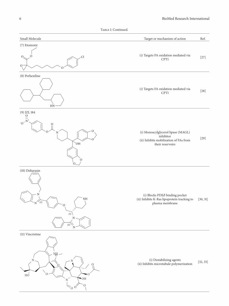
Table 1: Continued.
Small Molecule Target or mechanism of action Ref.
(7) Etomoxir
O
O
ClO
O
(i) Targets FA oxidation mediated viaCPT1 [27]
(8) Perhexiline
HN
(i) Targets FA oxidation mediated viaCPT1 [28]
(9) JZL 184
-/
/
.+
.
OHO
OO
O
/
/ (i) Monoacylglycerol lipase (MAGL)inhibitor
(ii) Inhibits mobilization of FAs fromtheir reservoirs
[29]
(10) Deltarasin
N
O
N
N
NHN (Z)
(S)
(Z)
(i) Blocks PDE𝛿 binding pocket(ii) Inhibits K-Ras lipoprotein tracking to
plasma membrane[30, 31]
(11) Vincristine
N
NH
N
N
O O O
OHO
O
O
O
O
HO
(i) Destabilizing agents(ii) Inhibits microtubule polymerization [32, 33]
BioMed Research International 7
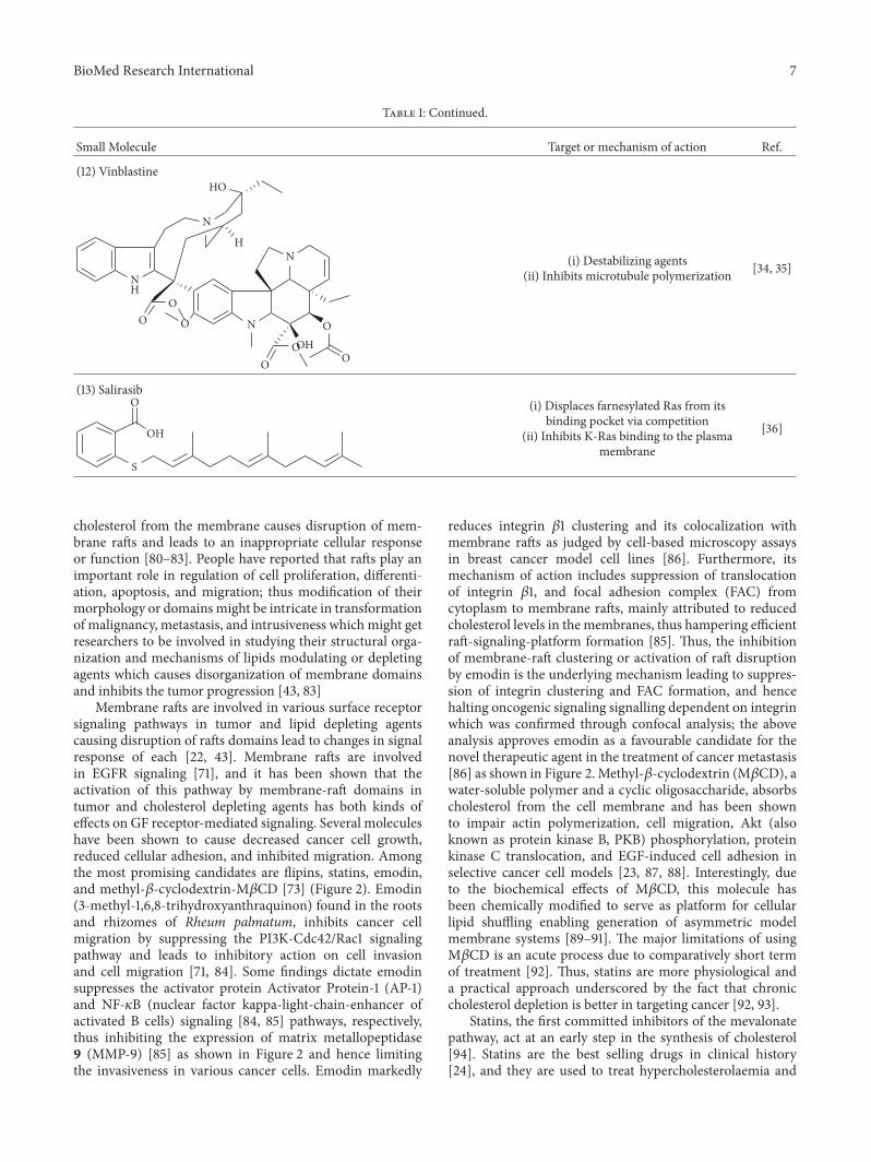
Table 1: Continued.
Small Molecule Target or mechanism of action Ref.
(12) Vinblastine
NH
N
HO
H
N
N
OOH
O
OO
O
OO
(i) Destabilizing agents(ii) Inhibits microtubule polymerization [34, 35]
(13) Salirasib
OH
S
O (i) Displaces farnesylated Ras from itsbinding pocket via competition
(ii) Inhibits K-Ras binding to the plasmamembrane
[36]
cholesterol from the membrane causes disruption of mem-brane rafts and leads to an inappropriate cellular responseor function [80–83]. People have reported that rafts play animportant role in regulation of cell proliferation, differenti-ation, apoptosis, and migration; thus modification of theirmorphology or domains might be intricate in transformationof malignancy, metastasis, and intrusiveness which might getresearchers to be involved in studying their structural orga-nization and mechanisms of lipids modulating or depletingagents which causes disorganization of membrane domainsand inhibits the tumor progression [43, 83]
Membrane rafts are involved in various surface receptorsignaling pathways in tumor and lipid depleting agentscausing disruption of rafts domains lead to changes in signalresponse of each [22, 43]. Membrane rafts are involvedin EGFR signaling [71], and it has been shown that theactivation of this pathway by membrane-raft domains intumor and cholesterol depleting agents has both kinds ofeffects on GF receptor-mediated signaling. Several moleculeshave been shown to cause decreased cancer cell growth,reduced cellular adhesion, and inhibited migration. Amongthe most promising candidates are flipins, statins, emodin,and methyl-𝛽-cyclodextrin-M𝛽CD [73] (Figure 2). Emodin(3-methyl-1,6,8-trihydroxyanthraquinon) found in the rootsand rhizomes of Rheum palmatum, inhibits cancer cellmigration by suppressing the PI3K-Cdc42/Rac1 signalingpathway and leads to inhibitory action on cell invasionand cell migration [71, 84]. Some findings dictate emodinsuppresses the activator protein Activator Protein-1 (AP-1)and NF-𝜅B (nuclear factor kappa-light-chain-enhancer ofactivated B cells) signaling [84, 85] pathways, respectively,thus inhibiting the expression of matrix metallopeptidase9 (MMP-9) [85] as shown in Figure 2 and hence limitingthe invasiveness in various cancer cells. Emodin markedly
reduces integrin 𝛽1 clustering and its colocalization withmembrane rafts as judged by cell-based microscopy assaysin breast cancer model cell lines [86]. Furthermore, itsmechanism of action includes suppression of translocationof integrin 𝛽1, and focal adhesion complex (FAC) fromcytoplasm to membrane rafts, mainly attributed to reducedcholesterol levels in the membranes, thus hampering efficientraft-signaling-platform formation [85]. Thus, the inhibitionof membrane-raft clustering or activation of raft disruptionby emodin is the underlying mechanism leading to suppres-sion of integrin clustering and FAC formation, and hencehalting oncogenic signaling signalling dependent on integrinwhich was confirmed through confocal analysis; the aboveanalysis approves emodin as a favourable candidate for thenovel therapeutic agent in the treatment of cancer metastasis[86] as shown in Figure 2. Methyl-𝛽-cyclodextrin (M𝛽CD), awater-soluble polymer and a cyclic oligosaccharide, absorbscholesterol from the cell membrane and has been shownto impair actin polymerization, cell migration, Akt (alsoknown as protein kinase B, PKB) phosphorylation, proteinkinase C translocation, and EGF-induced cell adhesion inselective cancer cell models [23, 87, 88]. Interestingly, dueto the biochemical effects of M𝛽CD, this molecule hasbeen chemically modified to serve as platform for cellularlipid shuffling enabling generation of asymmetric modelmembrane systems [89–91]. The major limitations of usingM𝛽CD is an acute process due to comparatively short termof treatment [92]. Thus, statins are more physiological anda practical approach underscored by the fact that chroniccholesterol depletion is better in targeting cancer [92, 93].
Statins, the first committed inhibitors of the mevalonatepathway, act at an early step in the synthesis of cholesterol[94]. Statins are the best selling drugs in clinical history[24], and they are used to treat hypercholesterolaemia and

8 BioMed Research International
RAS
PP P
RAF
MEK
ERK
PI3K/AKTP
m-TOR
MPAK
Angiogenesis
Nutrients, GHEmodin,MCD
EGFR
cMET
IGF-1
Non-Raft domain Organized Raft domain
Raft disruption
Raft organization throughrecruitment of receptors
MCD, Filipins, Statinsand Emodin
No Signal leads to apoptosis
VEGF
MMP-2
MMP-9 Survivin
Caspase-3
Bax-L
Apoptosome
Releases Cyt C
Cell Survival signal, metastasis
Adhesion, migration and tumorangiogenesis
Emodin, M-CD,Filipins
Various possible anti-tumour mechanisms of other cholesterol depleting agents (Emodin, Methyl-eta cyclodextrins and filipins)
Apoptosis
Figure 2: Schematic overview of the targets of action of various cholesterol depleting agents as antitumor drugs.
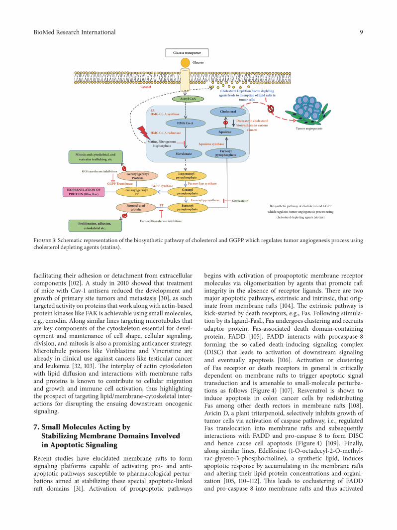
dyslipidaemia. They reversibly inhibit the HMG-CoA reduc-tase enzyme function, the rate limiting step in cholesterolbiosynthetic pathway. Statins also inhibit the isoprenoids pro-duction such as farnesyl pyrophosphate (FP) and geranylger-anylpyrophosphate (GGPP) [94] as shown in Figure 3; theyactivate the cellular signals by releasing or expressing variousproteins with GGPP (RhoA and Rac1) and FP(Ras) lipidmodifications.The primarymechanism ofHMG-CoA reduc-tase enzyme is to convert HMG-CoA into L-mevalonate andcoenzyme A [95], Figure 3. Statins by inhibiting HMG CoA reductase activity reduce cholesterol concentration andalso control the expression of LDL receptor [85]. They alsoreduce the synthesis of Apolipoprotein B100, hence inhibitingthe secretion and synthesis of triglycerides. The statin familyconsists of several drugs—synthetically derived (fluvastatin,cerivastatin, atorvastatin, rosuvastatin, and pitavastatin) andnaturally isolated from fungi (lovastatin, pravastatin, andsimvastatin)—that are notoriously known for inhibitingoncogenic signaling, inhibiting cell invasion and metastasisin cancer cells through disintegration of membrane raftsresulting from reduced cholesterol levels [25, 95]. Rosu-vastatin inhibits prostate cancer cell growth and inhibitsangiogenesis [26] and Simvastatin, another drug belonging tothe statin family, acts by downregulating PI3K/Akt/Caspase-3 signaling and Fas translocation mainly by modulation ofraft assembly [96]. Furthermore, simvastatin blocks Ras-membrane localization and downmodulates H-Ras protein atthe posttranslational level [27]. It also selectively dissociatedlatent membrane protein 1 (LMP1) from membrane raftsand reduces activation of NK-𝜅B signaling culminating toapoptosis [28, 29] and helps in survival of severe combined
immunodeficiency (SCID) mice with lymphonomas [29].Lipid rafts with its arsenal of lipid and protein componentsinvolved in many signaling pathways increase the possibilityof potential targets for cancer treatments. On a different note,artificial membrane models serve as a promising target inthe treatment of variousmechanisms involved in cancers, i.e.,decrease in cell adhesion and inhibition of cell proliferation,motility, and tumor progression.
Recent single molecule tracking techniques have eluci-dated that actin-based cytoskeleton structures on the cyto-plasmic surface of the plasmamembrane are also a key playerin inducing membrane domain organization or partitioninginto small compartments underpinning the cellular dynamicsof protein and lipid lateral diffusion. Such a model implicitin the picket fence model states that the membrane-actinskeleton interactions induce temporary confinement of trans-membrane proteins thus generating transient domains thatfunction as signaling host spots similar to rafts. Many recep-tors like the G-protein coupled receptor (GPCR), transferrinreceptors, etc. have been assigned to such transient domainswith the membrane skeleton mesh and are also enrichedwith cholesterol and sphingolipids [97–100]. Specifically,cancer cells having the ability to metastasize depend on thismachinery for invasion of various tissues—both local andat distant sites with differential dynamic reorganization ofactin [101]. Actin polymerization at the plasma membranecauses protrusion in the cell that dictates the direction of themigration andmembrane-lipid raft proteins at focal adhesionpoints help in detachment from the extracellular matrix.Prostate cancer cells are dependent on Src, focal adhesionkinase (FAK), Cav-1, cavin-1, and actin cytoskeleton for
BioMed Research International 9
Biosynthetic pathway of cholesterol and GGPP
which regulates tumor angiogenesis process using
cholesterol depleting agents (statins)
Acetyl CoA
ER
Statins, Nitrogenousbisphosphate Squalene synthase
Mevalonate
Cytosol
Glucose transporter
Glucose
HMG Co-A
HMG Co-A reductase
Geranylpyrophosphate
Isopentenylpyrophosphate
Farnesylpyrophosphate
Farnesylpyrophosphate
Squalene
Cholesterol
Farnesyl atedprotein
Geranyl geranylPP
Geranyl geranylProteins
ISOPRENYLATION OFPROTEIN (Rho, Rac)
Farnesyl pp synthase
Farnesyl pp synthaseGGPP synthase
FT
GGPP Transferase
HMG Co-A synthase
Cholesterol Depletion due to depletingagents leads to disruption of lipid rafts in
tumor cells
Simvastatin
Farnesyltransferase inhibitors
GG transferase inhibitors
Decrease in cholesterolbiosynthesis in various
cancers
Mitosis and cytoskeletal, andvesicular trafficking, etc
Proliferation, adhesion,cytoskeletal etc,
Tumor angiogenesis
Figure 3: Schematic representation of the biosynthetic pathway of cholesterol and GGPP which regulates tumor angiogenesis process usingcholesterol depleting agents (statins).
facilitating their adhesion or detachment from extracellularcomponents [102]. A study in 2010 showed that treatmentof mice with Cav-1 antisera reduced the development andgrowth of primary site tumors and metastasis [30], as suchtargeted activity on proteins that work alongwith actin-basedprotein kinases like FAK is achievable using small molecules,e.g., emodin. Along similar lines targeting microtubules thatare key components of the cytoskeleton essential for devel-opment and maintenance of cell shape, cellular signaling,division, and mitosis is also a promising anticancer strategy.Microtubule poisons like Vinblastine and Vincristine arealready in clinical use against cancers like testicular cancerand leukemia [32, 103]. The interplay of actin cytoskeletonwith lipid diffusion and interactions with membrane raftsand proteins is known to contribute to cellular migrationand growth and immune cell activation, thus highlightingthe prospect of targeting lipid/membrane-cytoskeletal inter-actions for disrupting the ensuing downstream oncogenicsignaling.
7. Small Molecules Acting byStabilizing Membrane Domains Involvedin Apoptotic Signaling
Recent studies have elucidated membrane rafts to formsignaling platforms capable of activating pro- and anti-apoptotic pathways susceptible to pharmacological pertur-bations aimed at stabilizing these special apoptotic-linkedraft domains [31]. Activation of proapoptotic pathways
begins with activation of proapoptotic membrane receptormolecules via oligomerization by agents that promote raftintegrity in the absence of receptor ligands. There are twomajor apoptotic pathways, extrinsic and intrinsic, that orig-inate from membrane rafts [104]. The extrinsic pathway iskick-started by death receptors, e.g., Fas. Following stimula-tion by its ligand-FasL, Fas undergoes clustering and recruitsadaptor protein, Fas-associated death domain-containingprotein, FADD [105]. FADD interacts with procaspase-8forming the so-called death-inducing signaling complex(DISC) that leads to activation of downstream signalingand eventually apoptosis [106]. Activation or clusteringof Fas receptor or death receptors in general is criticallydependent on membrane rafts to trigger apoptotic signaltransduction and is amenable to small-molecule perturba-tions as follows (Figure 4) [107]. Resveratrol is shown toinduce apoptosis in colon cancer cells by redistributingFas among other death rectors in membrane rafts [108].Avicin D, a plant triterpenoid, selectively inhibits growth oftumor cells via activation of caspase pathway, i.e., regulatedFas translocation into membrane rafts and subsequentlyinteractions with FADD and pro-caspase 8 to form DISCand hence cause cell apoptosis (Figure 4) [109]. Finally,along similar lines, Edelfosine (1-O-octadecyl-2-O-methyl-rac-glycero-3-phosphocholine), a synthetic lipid, inducesapoptotic response by accumulating in the membrane raftsand altering their lipid-protein concentrations and organi-zation [105, 110–112]. This leads to coclustering of FADDand pro-caspase 8 into membrane rafts and thus activated

10 BioMed Research International
PP
Fas LRTK
Receptor
CD44 R
EGFReceptor
c METReceptor
FADDCASPASE 8
CASPASE 10
PIP3PIP2
FADDRIPTRAD
CASPASE 8
RAS
Activatecaspase 3
Activatecaspase 8
CYTOSKELETAL CHANGESAND CELL MOTILITY
CDC 42
PI3K
PP P
Apoptosis
RAC 1
RAF
MEK
ERKETS
ELK 1
CELL PROLIFERATION/TRANSCRIPTIONOF ANTIAPOTOTIC GENE
AKTP
P
TUMOUR METASTASIS AND CELL INVASION
m-TOR
MPAK
Pyruvate
OAA
CitrateMalate
Acetyl Co-A
Fatty acidoxidation
Citrate
Acetyl Co-A
Malonyl Co-A
AcylCo-A
CPTIICPTI
FASN
ACC1
ACLY
ACC2
LACS
Alpha-KGA Mitochondria
Cytoplasm
Glucose transporter
PDK-1
NF-
PP P
P P
Auicin D
Edelfosine
SB-204990
Cexilenin
M CD, Emodin,Flipins,
Simvastatin
Angiogenesis
Simvastatin
Rosuvastatin
Palmitic acid
OAA
Acetyl Co-A+
FA synthesis
Glycolysis
Cholesterol
SB-204990
SB-204990
Colchicine,Paclitaxel
Nutrients, GH
Glucose
Amino acids Antigens,pathogens
Glucose-6-PO
Figure 4: Schematic overview of targeting lipid associated cellular functions in cancer with small molecules. All inhibitors are highlightedin yellow for clarification.
formation of DISC. Remarkably edelfosine is highly selectivefor leukemia cells and solid tumors compared with normalcells, where it targets only the plasma membrane rafts ofleukemia cells and endoplasmic rafts of solid tumor cells [113].
8. Small Molecules Rewiring LipidMetabolism in Cancer
Cancer cells display a highly distinctmetabolic growth profilecompared with nontransformed normal cells. The metabolicreprogramming of the enzymes of various pathways of cellgrowth forms the underlying basis of cancer. One of the mostimplicated pathways that are heavily tinkered within canceris lipid metabolism. Lipid metabolism is linked closely withthe glycolytic pathway by virtue of it providing the requiredstarting substrate—acetyl-CoA—for fatty acid (FA) synthesis.Lipids play key roles in this network, as they are crucial forthe formation of cell membranes and also act as signalingmessengers. Due to the enormous upregulated growth rate ofcancer cells, relatively larger amounts of lipids are required tokeep up with alarming rates of growth, proliferation, energystorage, and production of signaling molecules [76, 114].Targeting lipid metabolism, which encompasses perturbingsynthesis, oxidation, andmobilization of lipids, is a promisingstrategy in cancer treatment. One of the important steps in
lipid metabolism is the formation of fatty acid, which usesacetyl-CoA as a substrate. Acetyl-CoA is either obtainedfrom the glycolytic pathway via the conversion of pyruvateor obtained by the breakdown of citrate into acetyl-CoAand oxaloacetate by cytoplasmic ATP citrate lyase (ACLY).Acetyl-CoA binds with malonyl CoA (formed via the car-boxylation of acetyl-CoA) to form palmitate, which is astarting product of FA synthesis via the enzyme fatty acidsynthase (FASN). Inhibitors against ACLY will lead to thereduced production of acetyl-CoA and in turn reduce thelevels of FAs that are formed. ACLY inhibition has beenshown to cause growth suppression and induce apoptosis [115,116]. SB-204990 is shown to inhibit ACLY and therefore blockthe synthesis of FA and cholesterol (Figure 4). This causes ablock in the cancer cell growth and the suppression of tumor,leading to cell death [117]. The next main step amenable tosmall-molecule targeting is the formation of palmitate byFASN. Palmitate is then converted by a set of enzymes toform an array of saturated and unsaturated FAs. FASN hasbeen well documented with regard to its role played in cancerand is exploited extensively as anticancer target [118]. Asmost normal cells prefer exogenous sources of FAs, targetingFASN has been demonstrated to be a viable approach as itreduces the de novo FA synthesis in cancer cells. For example,cerulenin, an antifungal agent, is one such inhibitor of FA
BioMed Research International 11
synthesis, which reduces FA synthesis and rescues tumorouscells [119, 120]. Another such drug is C75 that has been shownto cause the inhibition of FASN [121].
9. Small Molecules Targeting LipidRelocalization and Lipoprotein Sorting
Next to targeting the enzymes involved in lipid biosynthesis,targeting lipid oxidation and mobilization/localization arefruitful therapeutic avenues gaining recent interest. Carnitinepalmitoyl transferase 1 (CPT1) is an enzyme involved in the𝛽-oxidation of FAs, where it facilitates the movement ofFA-CoA from the cytosol to the mitochondria across themitochondrial membrane. Etomoxir and perhexiline are twosmall molecules shown to be effective against tumors andcurb their proliferation via targeting FA oxidizationmediatedthrough CPT1 (Figure 4) [122, 123].The FAs once successfullytranslocated can either be diverted for storage or bemobilizedfrom stores as and when needed. The enzymes involvedin these mechanisms have proven to be suitable targetsfor cancer therapies. Glycerol-3-phosphate acyl transferases(GPATs) and its isoforms enable formation of diacylglyc-erols (DAG) and triacylglycerols (TAG), which are thendirected towards storage, while enzymes like monoacylglyc-erol lipase (MAGL) mobilize FAs from their reservoirs. CT-30501 inhibits GPATs while JZL184 inhibits MAGL. Thesesmall molecules help in suppression of tumor growth andinduce apoptosis, respectively [124]. The above are just afew examples of the use of small molecules to target lipidmetabolism and associated processes in cancer cells and forma firm foundation of lipid-targeted cancer therapies. One ofthe salient features of lipid membranes is the asymmetric dis-tribution of lipids—aminophospholipids phosphatidylserine(PS) and phosphatidylethanolamine (PE). They are largelypresent in the leaflet facing towards the cytosol; howeverupon flipping on the extracellular cell surface they act asmarkers for signaling pathways. Therefore, this feature ofthe membrane’s asymmetric distribution of composing lipidsmakes it an important target to fight against human pathol-ogy, and certain lipids are extensively used as biomarkersagainst cancer attributed to the fact of cancer cells expressinghigh levels of PE and PS exposed on its outer leaflet.Various factors have been reported for the loss of asymmetryin plasma membrane like transcriptional activation due toincrease in calcium concentration, inhibition of APTLs,oxidative stress, and transient hypoxia in tumor cells whichactivates sphingomyelinases. Activated sphingomyelinaseseventually leads to disintegration into ceramide and reducesthe stability of bilayer leading to membrane blebbing. Thisin turn activates the proapoptotic signaling pathways, whichredistributes the PS to the outer leaflet of the membrane.In recent reports, the loss of asymmetry is mainly due tothe reduction of translocase and activation of scramblaseenzyme [125]; thus exposure of PS to the outer surface servesas an apoptotic marker in all the cells [125, 126], and thismight also potentiate the activity in macrophages for killingthe tumor cells. PE acts as a structural component of cellwall as well and is implicated in many cellular processeslike cell division and cell death; thus a highly sought-after
anticancer target [2]. Recently, SapCDOPS nanovesicles wereused to detect PS on the outer surface of the tumor cellsand also targeted to induce cell death both in vitro and invivo [127]. Cyclotides are cyclic peptides that have a highaffinity to target and bind to PE head groupsmodulating theirlocalization and disturbing downstream cellular functionsinvolving PE. Along with cyclotides, there are two lantibioticpeptides—cinnamycin and duramycin—that are also PE spe-cific and are produced by Gram-positive bacteria [126, 128].The binding of both these types of peptides has a membranedisruption effect that causes cell death and proved effectivein imaging subcutaneous tumors; these findings indicate thatexternalized PEmay be a general maker of tumor vasculature[129, 130].
Lipids, apart from being intimately involved in cellularfunctions and cellular signaling as isolated modules, addanother level of complexity by their covalent attachmentto proteins—posttranslational protein lipidation—that formsthe heart of membrane-associated signaling in cells, e.g.,small GTPases, such as Ras, Raf, and ARFs. A classic exam-ple of addressing oncogenic signaling involving lipidatedproteins is via targeting the protein lipidation leading toimproper membrane-raft localization of these proteins caus-ing nonfunctional signaling platforms and hence subduedoncogenic signaling. This aspect is greatly exemplified by theclass of lapidated protein-Ras. The lipid moieties attached tothe protein consist of a palmitoyl group and 1-2 farnesyl lipidanchors. Ras was the first oncogene to be discovered and isinvolved in many human cancers; however small-moleculetargeting of Ras still remains an unmet task in cancer therapy.In 1989, one of the first drugs to be thought to target Raswas lovostatin. Farnesyl pyrophosphate farnesylates Ras andit is a key intermediate of the mevalonate-cholesterol biosyn-thetic pathway. Being a HMG-CoA reductase inhibitor,lovostatin was shown to block the mevalonate-cholesterolbiosynthetic pathway and hence the farnesylation lipidationof Ras. However, requirement of much higher dosage forselectively blocking farnesylation leads to adverse effecton cholesterol biosynthesis and unspecific cell death, thusmaking the journey of lovastatin quite short-lived regardingclinical targeting of Ras Farnesylation [131].This failure pavedthe way for the discovery of the enzyme involved in thefarnesylation of the Ras proteins. In 1990, farnesyltransferase(FTase) enzyme was isolated and characterized [132]. Oneof the attempted ways by which membrane-associated Rasoncogenic signaling has been targeted is via inhibiting theactivity of farnesyl transerfases to block farnesylation ofRas and hence reduce its membrane-raft association andconcomitant signaling in cancer cells [133–135]. More thantwo decades have been invested to exploit this approachas a practical anticancer therapy, but it has still met withmany deadlocks mainly attributed to the nonselective natureof farnesyltransferase inhibitors [136]. Specifically, the keyreason for this failure in clinical trials is the presence of analternate lipidation pathway, i.e., the compensatory activity ofgeranylgeranyltransferase-I that modifies Ras with geranyl-geranyl instead of a farnesyl group upon treatment with far-nesyltransferase inhibitors.This still leads to properRas local-ization and hence unaltered oncogenic signaling and cancer.
12 BioMed Research International
However, amidst such failed attempts, recently Wald-mann and coworkers have demonstrated an exciting alter-native to target Ras-associated cancer by mislocalizing Raslipoprotein not channelized via blocking the lipid attachmentbut by an innovative chemical biology approach [137]. Lip-idated Ras is trafficked through a prenyl-binding protein,PDE𝛿, in cells that sustains the spatial orientation of theRas superfamily of proteins [138]. Recently high specificity ofPDE𝛿 towards K-Ras trafficking to reach plasma membranerafts to initiate signaling was demonstrated [139, 140], andthe same was exploited by designing small molecules suchas Deltarasin (Figure 4) and related analogs to block thebinding pocket of PDE𝛿 leading to K-Ras mislocalizationand downregulated cancer signaling leading to reducing cellproliferation and finally cancer cell death [137, 141]. Alongsimilar lines, Salirasib, a small-molecule housing a farnesylmoiety, competes with Ras for binding to Galectins, theRas escort binding protein that contains a complementaryfarnesyl binding site [36]. This leads to Ras mislocalizationand halt of oncogenic signaling as observed with Deltarasin.These studies provide a proof-of-concept platform and opensvarious channels aimed at targeting lipid-mediated cellularfunctions in unprecedented ways.
Although a lot of work has to be yet done in identifyingmembrane specific small molecule against cancer, the effectsof presently available small molecule on the membranespecific organization and signaling are proven as effectiveagainst the malady in vitro.
10. Conclusions and Future Directions
The quest for targeting cancer using varied chemical andgenetic approaches still is faced with enormous hurdles andgenerates an unmet need to develop therapeutic approachesinspired by careful inspection of modulated cancer cellattributes. One of the aspects gaining considerable attentionrecently has been the altered lipid repertoire of cancercells leading to modulated membrane-dependent cellularprocesses including membrane organization and cellular sig-naling, strongly contributing to tumor growth andmetastasisand understanding the underlying mechanism behind thesame to elucidate potentially novel targets and pathwaysagainst cancer. In this review we focused on some of themost promising lipid associated candidates and processesfor anticancer targeting by small molecules. Ranging fromtargeting of lipid enzymes involved in the lipid metabolicpathway to the proteins and lipids that help in lipid organiza-tion, oncogenic lipoprotein sorting and signaling membranemicro-domains-rafts have proven to be highly crucial to notonly contemplate their therapeutic aspect but also addressand unveil specific mechanisms of lipid deregulation in can-cer. The various small-molecule-based drugs and tool com-pounds that have been discussed so far have shownpromisingresults in targeting lipid-dependent processes in cancer cells.Drugs like Salirasib that inhibit Ras function have shown theability to act in multiple ways and have been used in PhaseI clinical trials showing good results with the drug beingwell tolerated. Deltarasin which targets K-Ras downstreamsignaling and K-Ras localization may have potential to
achieve better efficacy in the long run. Although Statinslook the most prospective in cancer treatment, there are sideeffects to their use. In addition, they all act predominantlyby remodelingmembrane rafts composition and organizationbut lead to distinct downstream effects on the oncogenicsignaling in various cancer models. This brings to the fore-frontmembrane rafts as “selective cancer therapeutic targets,”with the structure, function, and associated raft-signalingpathways being subject of extensive studies. As such, abetter understanding of the structural, conformational, andfunctions aspects of raft biology would foster exploitation ofmembrane rafts for developing personalized cancer therapyfor targeting distinct raft-associated oncogenic signaling invarious cancers. In this regard, recent surge in technologicaladvancements in super resolution microscopy (SRM) isalready providing invaluable information on the distributionand organization and dynamics of plasma membrane com-ponents to bendings [142] that occur during clathrin medi-ated endocytosis [143]. Recent advancements in the field oftechnology hold promising scope of improving the resolutionand sensitivity of point localization in SRM based methods[144].
Further chemical biology investigations on the reg-ulation of membrane-lipid-dependent signaling pathwaysin cancer cell may provide novel targets for therapy andelucidating the role of distinct lipid signaling moleculeswill offer innovative therapeutic opportunities for devel-opment of anticancer drugs. However, discovery of lipid-dependent novel targets and novel signaling pathways incancer biology strongly lies at the hand of discovering inno-vative small molecules. Generally, small molecules with well-defined targets permit obtaining novel insights into biologicalprocesses and extensive analysis of their structure-activityrelationships permits chemical modification for improvingefficacy of such candidates. Though nature is a compre-hensive source of molecules with a variety of bioactivities,in recent years, a burst in organic synthesis strategies andsynthesis of organic molecules generated via innovativehypothesis generating platforms, such as diversity orientedsynthesis (DOS) biology oriented synthesis (BIOS), andactivity oriented synthesis, have enriched the pool of smallmolecules in an informed manner, thus promoting theiruse as research tools to explore previously uncharacterizedbiology and elucidate novel targets for drug discovery. Giventhe limited number of drug targets addressed till todayin cancer and immunology, including protein kinases, G-protein coupled receptors, and ion channels, developmentof new small organic molecules rightly fits the criteria formeeting the ever-increasing need for new therapeutic targets.Finally, identification of the molecular targets of such newcompounds still remains a major bottleneck, underscoringthe demand for appropriate methodologies to elucidate thetargets of small molecules in a relatively unbiased and timelyfashion.
Disclosure
The authors apologize to the colleagues whose work was notcited owing to space limitations.
BioMed Research International 13
Conflicts of Interest
The authors declare no conflicts of interest.
Authors’ Contributions
Aswin T. Srivatsav andManjariMishra contributed equally tothis work.
Acknowledgments
Shobhna Kapoor is supported by the Department of Scienceand Technology (DST) Inspire Faculty Award, India, andDepartment of Biotechnology (DBT)RamalingaswamiReen-try Fellowship, India.
References
[1] L. Thomas, “The Lives of a Cell,” The New England Journal ofMedicine, vol. 284, no. 19, pp. 1082-1083, 1971.
[2] G. van Meer, D. R. Voelker, and G. W. Feigenson, “Membranelipids: where they are and how they behave,” Nature ReviewsMolecular Cell Biology, vol. 9, no. 2, pp. 112–124, 2008.
[3] M. Roux, R. Auzely-Velty, F. Djedaini-Pilard, and B. Perly,“Cyclodextrin-induced lipid lateral separation in DMPCmem-branes: 2H nuclear magnetic resonance study,” BiophysicalJournal, vol. 82, no. 2, pp. 813–822, 2002.
[4] W. Hartmann and H. Galla, “Binding of polylysine to chargedbilayer membranes: Molecular organization of a lipid ⋅ peptidecomplex,” Biochimica et Biophysica Acta (BBA) - Biomembranes,vol. 509, no. 3, pp. 474–490, 1978.
[5] D. A. Wilkinson and J. F. Nagle, “Dilatometric Study of BinaryMixtures of Phosphatidylcholines,” Biochemistry, vol. 18, no. 19,pp. 4244–4249, 1979.
[6] G. W. Feigenson, “Phase boundaries and biological mem-branes,” Annual Review of Biophysics, vol. 36, pp. 63–77, 2007.
[7] J. V. Swinnen, K. Brusselmans, and G. Verhoeven, “Increasedlipogenesis in cancer cells: New players, novel targets,” CurrentOpinion in Clinical Nutrition &Metabolic Care, vol. 9, no. 4, pp.358–365, 2006.
[8] J. J. Kamphorst, J. R. Cross, J. Fan et al., “Hypoxic and Ras-transformed cells support growth by scavenging unsaturatedfatty acids from lysophospholipids,” Proceedings of the NationalAcadamy of Sciences of the United States of America, vol. 110, no.22, pp. 8882–8887, 2013.
[9] P. Caro, A. U. Kishan, E. Norberg et al., “Metabolic SignaturesUncover Distinct Targets in Molecular Subsets of Diffuse LargeBCell Lymphoma,”Cancer Cell, vol. 22, no. 4, pp. 547–560, 2012.
[10] D. K. Nomura, J. Z. Long, S. Niessen, H. S. Hoover, S.-W. Ng,and B. F. Cravatt, “Monoacylglycerol lipase regulates a fatty acidnetwork that promotes cancer pathogenesis,” Cell, vol. 140, no.1, pp. 49–61, 2010.
[11] E. Pearce and E. Pearce, “Metabolic pathways in immune cellactivation and quiescence,” Immunity, vol. 38, no. 4, pp. 633–643, 2013.
[12] C.-H. Chang, J. D. Curtis, and L. B. Maggi Jr., “Posttranscrip-tional control of T cell effector function by aerobic glycolysis,”Cell, vol. 153, no. 6, pp. 1239–1251, 2013.
[13] M. L. Doria, C. Z. Cotrim, C. Simoes et al., “Lipidomic analysisof phospholipids from human mammary epithelial and breastcancer cell lines,” Journal of Cellular Physiology, vol. 228, no. 2,pp. 457–468, 2013.
[14] K. Muir, A. Hazim, Y. He et al., “Proteomic and lipidomic sig-natures of lipidmetabolism inNASH-associatedHepatocellularcarcinoma,”Cancer Research, vol. 73, no. 15, pp. 4722–4731, 2013.
[15] D. A. Bosco, D. M. Fowler, Q. Zhang et al., “Elevated levels ofoxidized cholesterol metabolites in Lewy body disease brainsaccelerate 𝛼-synuclein fibrilization,” Nature Chemical Biology,vol. 2, no. 5, pp. 249–253, 2006.
[16] P. V. Escriba, “Membrane-lipid therapy: A new approach inmolecular medicine,” Trends in Molecular Medicine, vol. 12, no.1, pp. 34–43, 2006.
[17] P. V. Escriba, “Membrane-lipid therapy: A historical perspectiveof membrane-targeted therapies — From lipid bilayer structureto the pathophysiological regulation of cells,” Biochimica etBiophysica Acta (BBA) - Biomembranes, vol. 1859, no. 9, pp.1493–1506, 2017.
[18] J. R. Peterson and T. J. Mitchison, “Small molecules, bigimpact: a history of chemical inhibitors and the cytoskeleton,”Chemistry & Biology, vol. 9, no. 12, pp. 1275–1285, 2002.
[19] G. E. Ward, K. L. Carey, and N. J. Westwood, “Using smallmolecules to study big questions in cellular microbiology,”Cellular Microbiology, vol. 4, no. 8, pp. 471–482, 2002.
[20] F. Fassy, C. Dureuil, A. Lamberton et al., “In Vitro Character-ization of VPS34 Lipid Kinase Inhibition by Small Molecules,”Methods in Enzymology, vol. 587, pp. 447–464, 2017.
[21] M. Grzybek, A. Kozubek, P. Dubielecka, and A. F. Sikorski,“Rafts - The current picture,” Folia Histochemica et Cytobiolog-ica, vol. 43, no. 1, pp. 3–10, 2005.
[22] L. J. Pike, “Rafts defined: a report on the Keystone symposiumon lipid rafts and cell function,” Journal of Lipid Research, vol.47, no. 7, pp. 1597-1598, 2006.
[23] Y. Liu, R. Sun, W. Wan et al., “The involvement of lipid rafts inepidermal growth factor-induced chemotaxis of breast cancercells,” Molecular Membrane Biology, vol. 24, no. 2, pp. 91–101,2007.
[24] S. Schlyer and R.Horuk, “I want a new drug: G-protein-coupledreceptors in drug development,”DrugDiscoveryTherapy, vol. 11,no. 11-12, pp. 481–493, 2006.
[25] K. K. Chan, A. M. Oza, and L. L. Siu, “The statins as anticanceragents,” Clinical Cancer Research, vol. 9, no. 1, pp. 10–19, 2003.
[26] C. Wang, W. Tao, Y. Wang et al., “Rosuvastatin, identified froma zebrafish chemical genetic screen for antiangiogenic com-pounds, suppresses the growth of prostate cancer,” EuropeanUrology, vol. 58, no. 3, pp. 418–426, 2010.
[27] U. K. Khanzada, O. E. Pardo, C.Meier, J. Downward,M. J. Seckl,and A. Arcaro, “Potent inhibition of small-cell lung cancercell growth by simvastatin reveals selective functions of Rasisoforms in growth factor signalling,” Oncogene, vol. 25, no. 6,pp. 877–887, 2006.
[28] G. Mosialos, M. Birkenbacht, R. Yalamanchill, T. Van Arsdale,C. Ware, and E. Kleff, “The Epstein-Barr virus transformingprotein LMP1 engages signaling proteins for the tumor necrosisfactor receptor family,” Cell, vol. 80, no. 3, pp. 389–399, 1995.
[29] H. Katano, L. Pesnicak, and J. I. Cohen, “Simvastatin inducesapoptosis of Epstein-Barr virus (EBV)-transformed lym-phoblastoid cell lines and delays development of EBV lym-phomas,” Proceedings of the National Acadamy of Sciences of theUnited States of America, vol. 101, no. 14, pp. 4960–4965, 2004.
[30] T. C. Thompson, S. A. Tahir, L. Li et al., “The role of caveolin-1 in prostate cancer: clinical implications,” Prostate Cancer andProstatic Diseases, vol. 13, no. 1, pp. 6–11, 2010.

14 BioMed Research International
[31] K. S. George and S.Wu, “Lipid raft: A floating island of death orsurvival,” Toxicology and Applied Pharmacology, vol. 259, no. 3,pp. 311–319, 2012.
[32] A. Duflos, A. Kruczynski, and J.-M. Barret, “Novel aspectsof natural and modified Vinca alkaloids,” Current MedicinalChemistry - Anti-Cancer Agents, vol. 2, no. 1, pp. 55–70, 2002.
[33] G. C. Na and S. N. Timasheff, “Thermodynamic Linkagebetween Tubulin Self-Association and the Binding of Vinblas-tine,” Biochemistry, vol. 19, no. 7, pp. 1355–1365, 1980.
[34] J. O. Armitage, “Overview of rational and individualizedtherapeutic strategies for non-Hodgkin’s lymphomas,” ClinicalLymphoma, Myeloma & Leukemia, vol. 3, 1, pp. S5–S11, 2002.
[35] D. Panda, M. A. Jordan, K. C. Chu, and L. Wilson, “Differentialeffects of vinblastine on polymerization and dynamics at oppo-site microtubule ends,”The Journal of Biological Chemistry, vol.271, no. 47, pp. 29807–29812, 1996.
[36] D. Marciano, G. Ben-Baruch, M. Marom, Y. Egozi, R. Haklai,and Y. Kloog, “Farnesyl Derivatives of Rigid Carboxylic Acids-Inhibitors of ras-Dependent Cell Growth,” Journal of MedicinalChemistry, vol. 38, no. 8, pp. 1267–1272, 1995.
[37] V. Luzzati and A. Tardieu, “Lipid Phases: Structure and Struc-tural Transitions,” Annual Review of Physical Chemistry, vol. 25,no. 1, pp. 79–94, 1974.
[38] D. Lingwood and K. Simons, “Lipid rafts as a membrane-organizing principle,” Science, vol. 327, no. 5961, pp. 46–50, 2010.
[39] T. Harder, P. Scheiffele, P. Verkade, and K. Simons, “Lipiddomain structure of the plasmamembrane revealed by patchingof membrane components,”The Journal of Cell Biology, vol. 141,no. 4, pp. 929–942, 1998.
[40] A. Rietveld and K. Simons, “The differential miscibility of lipidsas the basis for the formation of functional membrane rafts,”Biochimica et Biophysica Acta - Reviews on Biomembranes, vol.1376, no. 3, pp. 467–479, 1998.
[41] C. Nicolini, J. Kraineva, M. Khurana, N. Periasamy, S. S. Funari,and R. Winter, “Temperature and pressure effects on structuraland conformational properties of POPC/SM/cholesterol modelraft mixtures—a FT-IR, SAXS, DSC, PPC and Laurdan fluores-cence spectroscopy study,” Biochimica et Biophysica Acta (BBA)- Biomembranes, vol. 1758, no. 2, pp. 248–258, 2006.
[42] S. Kapoor, A. Werkmuller, C. Denter et al., “Temperature-pressure phase diagram of a heterogeneous anionic modelbiomembrane system: results from a combined calorimetry,spectroscopy and microscopy study,” Biochimica et BiophysicaActa (BBA) - Biomembranes, vol. 1808, no. 4, pp. 1187–1195, 2011.
[43] K. Simons andD. Toomre, “Lipid rafts and signal transduction,”Nature Reviews Molecular Cell Biology, vol. 1, no. 1, pp. 31–39,2000.
[44] J. A. Allen, R. A. Halverson-Tamboli, and M. M. Rasenick,“Lipid raft microdomains and neurotransmitter signalling,”Nature Reviews Neuroscience, vol. 8, no. 2, pp. 128–140, 2007.
[45] V. Michel and M. Bakovic, “Lipid rafts in health and disease,”Biology of the Cell, vol. 99, no. 3, pp. 129–140, 2007.
[46] M. E. Irwin, N. Bohin, and J. L. Boerner, “Src family kinasesmediate epidermal growth factor receptor signaling from lipidrafts in breast cancer cells,” Cancer Biology & Therapy, vol. 12,no. 8, pp. 718–726, 2011.
[47] M. E. Irwin, K. L. Mueller, N. Bohin, Y. Ge, and J. L. Boerner,“Lipid raft localization of EGFR alters the response of cancercells to the EGFR tyrosine kinase inhibitor gefitinib,” Journal ofCellular Physiology, vol. 226, no. 9, pp. 2316–2328, 2011.
[48] D. C. Marquez, H.-W. Chen, E. M. Curran, W. V. Welshons,and R. J. Pietras, “Estrogen receptors in membrane lipid raftsand signal transduction in breast cancer,”Molecular andCellularEndocrinology, vol. 246, no. 1-2, pp. 91–100, 2006.
[49] C. P. Palmer, R. Mahen, E. Schnell, M. B. A. Djamgoz, and E.Aydar, “Sigma-1 receptors bind cholesterol and remodel lipidrafts in breast cancer cell lines,” Cancer Research, vol. 67, no. 23,pp. 11166–11175, 2007.
[50] N. Li, A. Batzer, R. Daly et al., “Guanine-nucleotide-releasingfactor hSos1 binds to Grb2 and links receptor tyrosine kinasesto Ras signalling,” Nature, vol. 363, no. 6424, pp. 85–88, 1993.
[51] T. Kamata and J. R. Feramisco, “Epidermal growth factor stim-ulates guanine nucleotide binding activity and phosphorylationof ras oncogene proteins,”Nature, vol. 310, no. 5973, pp. 147–150,1984.
[52] P. Chardin, J. H. Camonis, N. W. Gale et al., “Human Sos1: Aguanine nucleotide exchange factor for Ras that binds toGRB2,”Science, vol. 260, no. 5112, pp. 1338–1343, 1993.
[53] I. R. Vetter and A. Wittinghofer, “The guanine nucleotide-binding switch in three dimensions,” Science, vol. 294, no. 5545,pp. 1299–1304, 2001.
[54] T. Y. Shih, M. O. Weeks, H. A. Young, and E. M. Scolnick,“Identification of a sarcoma virus-coded phosphoprotein innonproducer cells transformed by Kirsten or Harvey murinesarcoma virus,” Virology, vol. 96, no. 1, pp. 64–79, 1979.
[55] L. F. Parada, C. J. Tabin, C. Shih, andR.A.Weinberg, “HumanEJbladder carcinoma oncogene is homologue of Harvey sarcomavirus ras gene,” Nature, vol. 297, no. 5866, pp. 474–478, 1982.
[56] J. M. Kyriakis, H. App, X.-F. Zhang et al., “Raf-1 activates MAPkinase-kinase,” Nature, vol. 358, no. 6385, pp. 417–421, 1992.
[57] W. E. Tidyman and K. A. Rauen, “The RASopathies: devel-opmental syndromes of Ras/MAPK pathway dysregulation,”Current Opinion in Genetics & Development, vol. 19, no. 3, pp.230–236, 2009.
[58] G. E. Atilla-Gokcumen and U. S. Eggert, “A comparative LC-MS based profiling approach to analyze lipid composition intissue culture systems,”Methods in Molecular Biology, vol. 1232,pp. 103–113, 2015.
[59] G. E. Atilla-Gokcumen, E.Muro, J. Relat-Goberna et al., “Divid-ing cells regulate their lipid composition and localization,” Cell,vol. 156, no. 3, pp. 428–439, 2014.
[60] E. Muro, G. E. Atilla-Gokcumen, and U. S. Eggert, “Lipids incell biology: How can we understand them better?” MolecularBiology of the Cell (MBoC), vol. 25, no. 12, pp. 1819–1823, 2014.
[61] K. Emoto, T. Kobayashi, A. Yamaji et al., “Redistribution ofphosphatidylethanolamine at the cleavage furrow of dividingcells during cytokinesis,” Proceedings of the National Acadamyof Sciences of the United States of America, vol. 93, no. 23, pp.12867–12872, 1996.
[62] K. Emoto, N. Toyama-Sorimachi, H. Karasuyama, K. Inoue,andM. Umeda, “Exposure of phosphatidylethanolamine on thesurface of apoptotic cells,” Experimental Cell Research, vol. 232,no. 2, pp. 430–434, 1997.
[63] H. Kenis and C. Reutelingsperger, “Targeting phosphatidylser-ine in anti-cancer therapy,” Current Pharmaceutical Design, vol.15, no. 23, pp. 2719–2723, 2009.
[64] C. Chidley, S. A. Trauger, K. Birsoy, and E. K. O’Shea, “Theanticancer natural product ophiobolin A induces cytotoxicityby covalent modification of phosphatidylethanolamine,” eLife,vol. 5, 2016.
BioMed Research International 15
[65] C. K. Wang, H. P. Wacklin, and D. J. Craik, “Cyclotides insertinto lipid bilayers to form membrane pores and destabilize themembrane through hydrophobic and phosphoethanolamine-specific interactions,” The Journal of Biological Chemistry, vol.287, no. 52, pp. 43884–43898, 2012.
[66] L. Toppozini, S. Meinhardt, C. L. Armstrong et al., “Structure ofCholesterol in Lipid Rafts,” Physical Review Letters, vol. 113, no.22, 2014.
[67] S. Zalba and T. L. M. ten Hagen, “Cell membrane modulationas adjuvant in cancer therapy,” Cancer Treatment Reviews, vol.52, pp. 48–57, 2017.
[68] R. Krivanek, L. Okoro, and R. Winter, “Effect of cholesteroland ergosterol on the compressibility and volume fluctuationsof phospholipid-sterol bilayers in the critical point region: Amolecular acoustic and calorimetric study,” Biophysical Journal,vol. 94, no. 9, pp. 3538–3548, 2008.
[69] C. Bernsdorff, A. Wolf, R. Winter, and E. Gratton, “Effectof hydrostatic pressure on water penetration and rotationaldynamics in phospholipid-cholesterol bilayers,” BiophysicalJournal, vol. 72, no. 3, pp. 1264–1277, 1997.
[70] K. Simons and E. Ikonen, “Functional rafts in cell membranes,”Nature, vol. 387, no. 6633, pp. 569–572, 1997.
[71] L. Zhuang, J. Lin, M. L. Lu, K. R. Solomon, and M. R. Freeman,“Cholesterol-rich lipid rafts mediate Akt-regulated survival inprostate cancer cells,” Cancer Research, vol. 62, no. 8, pp. 2227–2231, 2002.
[72] F. Mollinedo and C. Gajate, “Lipid rafts as major platformsfor signaling regulation in cancer,” Advances in BiologicalRegulation, vol. 57, pp. 130–146, 2015.
[73] A. Hryniewicz-Jankowska, K. Augoff, A. Biernatowska, J. Pod-kalicka, and A. F. Sikorski, “Membrane rafts as a novel target incancer therapy,” Biochimica et Biophysica Acta (BBA) - Reviewson Cancer, vol. 1845, no. 2, pp. 155–165, 2014.
[74] Y.-F. Yang, Y.-H. Jan, Y.-P. Liu et al., “Squalene synthaseinduces tumor necrosis factor receptor 1 enrichment in lipidrafts to promote lung cancer metastasis,” American Journal ofRespiratory and Critical Care Medicine, vol. 190, no. 6, pp. 675–687, 2014.
[75] M. Kartal Yandim, E. Apohan, and Y. Baran, “Therapeuticpotential of targeting ceramide/glucosylceramide pathway incancer,” Cancer Chemotherapy and Pharmacology, vol. 71, no. 1,pp. 13–20, 2013.
[76] S. Beloribi-Djefaflia, S. Vasseur, and F. Guillaumond, “Lipidmetabolic reprogramming in cancer cells,” Oncogenesis, vol. 5,p. e189, 2016.
[77] K. M. Lum, Y. Sato, B. A. Beyer et al., “Mapping Protein Targetsof Bioactive Small Molecules Using Lipid-Based ChemicalProteomics,” ACS Chemical Biology, vol. 12, no. 10, pp. 2671–2681, 2017.
[78] S. Kapoor, K. Weise, M. Erlkamp, G. Triola, H. Waldmann, andR. Winter, “The role of G-domain orientation and nucleotidestate on the Ras isoform-specific membrane interaction,” Euro-pean Biophysics Journal, vol. 41, no. 10, pp. 801–813, 2012.
[79] K. Weise, S. Kapoor, C. Denter et al., “Membrane-mediatedinduction and sorting of K-Ras microdomain signaling plat-forms,” Journal of the American Chemical Society, vol. 133, no.4, pp. 880–887, 2011.
[80] M. Grzybek, J. Kubiak, A. Lach, M. Przybylo, and A. F. Siko-rski, “A raft-associated species of phosphatidylethanolamineinteracts with cholesterol comparably to sphingomyelin. ALangmuir-Blodgett monolayer study,” PLoS ONE, vol. 4, no. 3,p. e5053, 2009.
[81] J. F. Hancock, “Lipid rafts: Contentious only from simplisticstandpoints,” Nature Reviews Molecular Cell Biology, vol. 7, no.6, pp. 456–462, 2006.
[82] K. Jacobson, O. G. Mouritsen, and R. G. W. Anderson, “Lipidrafts: at a crossroad between cell biology and physics,” NatureCell Biology, vol. 9, no. 1, pp. 7–14, 2007.
[83] M. R. Freeman, D. Di Vizio, and K. R. Solomon, “The Rafts oftheMedusa: Cholesterol targeting in cancer therapy,”Oncogene,vol. 29, no. 26, pp. 3745–3747, 2010.
[84] Q.Huang, H.-M. Shen, andC.-N.Ong, “Emodin inhibits tumorcell migration through suppression of the phosphatidylinositol3-kinase-Cdc42/Rac1 pathway,” Cellular and Molecular LifeSciences, vol. 62, no. 10, pp. 1167–1175, 2005.
[85] Q. Huang, H. Shen, and C. Ong, “Inhibitory effect of emodin ontumor invasion through suppression of activator protein-1 andnuclear factor-𝜅B,” Biochemical Pharmacology, vol. 68, no. 2, pp.361–371, 2004.
[86] Q. Huang, H.-M. Shen, G. Shui, M. R. Wenk, and C.-N. Ong,“Emodin inhibits tumor cell adhesion through disruption ofthe membrane lipid raft-associated integrin signaling pathway,”Cancer Research, vol. 66, no. 11, pp. 5807–5815, 2006.
[87] T. J. Pucadyil and A. Chattopadhyay, “Cholesterol deple-tion induces dynamic confinement of the G-protein coupledserotonin
1𝐴receptor in the plasma membrane of living cells,”
Biochimica et Biophysica Acta (BBA) - Biomembranes, vol. 1768,no. 3, pp. 655–668, 2007.
[88] Y. Wu, V. Rizzo, Y. Liu, I. M. Sainz, N. G. Schmuckler,and R. W. Colman, “Kininostatin associates with membranerafts and inhibits alpha(v)beta3 integrin activation in humanumbilical vein endothelial cells,” Arteriosclerosis, Thrombosis,and Vascular Biology, vol. 27, no. 9, pp. 1968–1975, 2007.
[89] S. Chiantia, P. Schwille, A. S. Klymchenko, and E. Lon-don, “Asymmetric GUVs Prepared by M𝛽CD-Mediated LipidExchange: An FCS Study,” Biophysical Journal, vol. 100, no. 1,pp. L1–L3, 2011.
[90] Q. Lin, E. London, and D. Holowka, “Preparation of artificialplasma membrane mimicking vesicles with lipid asymmetry,”PLoS ONE, vol. 9, no. 1, p. e87903, 2014.
[91] J. T. Crowley, A. M. Toledo, T. J. LaRocca et al., “Lipid Exchangebetween Borrelia burgdorferi and Host Cells,” PLoS Pathogens,vol. 9, no. 1, p. e1003109, 2013.
[92] R. Zidovetzki and I. Levitan, “Use of cyclodextrins to manip-ulate plasma membrane cholesterol content: evidence, miscon-ceptions and control strategies,” Biochimica et Biophysica Acta(BBA) - Biomembranes, vol. 1768, no. 6, pp. 1311–1324, 2007.
[93] S. Shrivastava, T. J. Pucadyil, Y. D. Paila, S. Ganguly, andA. Chattopadhyay, “Chronic cholesterol depletion using statinimpairs the function and dynamics of human serotonin
1𝐴
receptors,” Biochemistry, vol. 49, no. 26, pp. 5426–5435, 2010.[94] M. Roy, H. J. Kung, and P. M. Ghosh, “Statins and prostate
cancer: role of cholesterol inhibition vs. prevention of smallGTP-binding proteins,” American Journal of Cancer Research,vol. 1, no. 4, pp. 542–561, 2011.
[95] M. Osmak, “Statins and cancer: Current and future prospects,”Cancer Letters, vol. 324, no. 1, pp. 1–12, 2012.
[96] H. Wu, H. Jiang, D. Lu et al., “Effect of simvastatin on gliomacell proliferation, migration, and apoptosis,” Neurosurgery, vol.65, no. 6, pp. 1087–1097, 2009.
[97] K. Gowrishankar, S. Ghosh, S. Saha, C. Rumamol, S. Mayor,and M. Rao, “Active remodeling of cortical actin regulatesspatiotemporal organization of cell surfacemolecules,”Cell, vol.149, no. 6, pp. 1353–1367, 2012.
16 BioMed Research International
[98] A. Kusumi and Y. Sako, “Cell surface organization by themembrane skeleton,” Current Opinion in Cell Biology, vol. 8, no.4, pp. 566–574, 1996.
[99] K. Suzuki, K. Ritchie, E. Kajikawa, T. Fujiwara, and A. Kusumi,“Rapid hop diffusion of a G-protein-coupled receptor in theplasma membrane as revealed by single-molecule techniques,”Biophysical Journal, vol. 88, no. 5, pp. 3659–3680, 2005.
[100] D. Goswami, K. Gowrishankar, S. Bilgrami et al., “Nanoclustersof GPI-Anchored Proteins Are Formed by Cortical Actin-Driven Activity,” Cell, vol. 135, no. 6, pp. 1085–1097, 2008.
[101] J. P. Thiery, H. Acloque, R. Y. J. Huang, and M. A. Nieto,“Epithelial-mesenchymal transitions in development and dis-ease,” Cell, vol. 139, no. 5, pp. 871–890, 2009.
[102] B. Su, L. Gao, F. Meng, L.-W. Guo, J. Rothschild, and I.H. Gelman, “Adhesion-mediated cytoskeletal remodeling iscontrolled by the direct scaffolding of Src from FAK complexesto lipid rafts by SSeCKS/AKAP12,”Oncogene, vol. 32, no. 16, pp.2016–2026, 2013.
[103] G. L. Plosker and D. P. Figgitt, “Rituximab: a review of itsuse in non-Hodgkin’s lymphoma and chronic lymphocyticleukaemia,” Drugs, vol. 63, no. 8, pp. 803–843, 2003.
[104] C. Gajate, F. Gonzalez-Camacho, and F. Mollinedo, “Lipid raftconnection between extrinsic and intrinsic apoptotic pathways,”Biochemical andBiophysical ResearchCommunications, vol. 380,no. 4, pp. 780–784, 2009.
[105] C. Gajate, F. Gonzalez-Camacho, and F. Mollinedo, “Involve-ment of raft aggregates enriched in Fas/CD95 death-inducingsignaling complex in the antileukemic action of edelfosine inJurkat cells,” PLoS ONE, vol. 4, no. 4, p. e5044, 2009.
[106] M. E. Peter and P. H. Krammer, “The CD95(APO-1/Fas) DISCand beyond,” Cell Death & Differentiation, vol. 10, no. 1, pp. 26–35, 2003.
[107] C. Gajate and F. Mollinedo, “Cytoskeleton-mediated deathreceptor and ligand concentration in lipid rafts forms apoptosis-promoting clusters in cancer chemotherapy,” The Journal ofBiological Chemistry, vol. 280, no. 12, pp. 11641–11647, 2005.
[108] D. Delmas, C. Rebe, O.Micheau et al., “Redistribution of CD95,DR4 and DR5 in rafts accounts for the synergistic toxicity ofresveratrol and death receptor ligands in colon carcinoma cells,”Oncogene, vol. 23, no. 55, pp. 8979–8986, 2004.
[109] Z.-X. Xu, T. Ding, V. Haridas, F. Connolly, and J. U. Gutterman,“Avicin D, a plant triterpenoid, induces cell apoptosis byrecruitment of fas and downstream signaling molecules intolipid rafts,” PLoS ONE, vol. 4, no. 12, 2009.
[110] C. Gajate and F. Mollinedo, “Lipid rafts, endoplasmic reticulumand mitochondria in the antitumor action of the alkylphos-pholipid analog edelfosine,” Anti-Cancer Agents in MedicinalChemistry, vol. 14, no. 4, pp. 509–527, 2014.
[111] A. Ausili, A. Torrecillas, F. J. Aranda et al., “Edelfosine isincorporated into rafts and alters their organization,” TheJournal of Physical Chemistry B, vol. 112, no. 37, pp. 11643–11654,2008.
[112] C. Gajate and F. Mollinedo, “Edelfosine and perifosine induceselective apoptosis inmultiplemyeloma by recruitment of deathreceptors and downstream signaling molecules into lipid rafts,”Blood, vol. 109, no. 2, pp. 711–719, 2007.
[113] C. Gajate, M. Matos-Da-Silva, E. L.-H. Dakir, R. I. Fonteriz, J.Alvarez, and F. Mollinedo, “Antitumor alkyl-lysophospholipidanalog edelfosine induces apoptosis in pancreatic cancer bytargeting endoplasmic reticulum,” Oncogene, vol. 31, no. 21, pp.2627–2639, 2012.
[114] F. Rohrig and A. Schulze, “The multifaceted roles of fatty acidsynthesis in cancer,” Nature Reviews Cancer, vol. 16, no. 11, pp.732–749, 2016.
[115] T. Migita, S. Okabe, K. Ikeda et al., “Inhibition of ATP citratelyase induces triglyceride accumulation with altered fatty acidcomposition in cancer cells,” International Journal of Cancer,vol. 135, no. 1, pp. 37–47, 2014.
[116] S. Shah, W. J. Carriveau, J. Li et al., “Targeting ACLY sensitizescastration-resistant prostate cancer cells to AR antagonismby impinging on an ACLY-AMPK-AR feedback mechanism,”Oncotarget , vol. 7, no. 28, pp. 43713–43730, 2016.
[117] N. J. Pearce, J. W. Yates, T. A. Berkhout et al., “The role ofATP citrate-lyase in the metabolic regulation of plasma lipids:Hypolipidaemic effects of SB-204990, a lactone prodrug ofthe potent ATP citrate-lyase inhibitor SB-201076,” BiochemicalJournal, vol. 334, no. 1, pp. 113–119, 1998.
[118] J. A. Menendez and R. Lupu, “Fatty acid synthase and thelipogenic phenotype in cancer pathogenesis,” Nature ReviewsCancer, vol. 7, no. 10, pp. 763–777, 2007.
[119] R. Shiragami, S. Murata, C. Kosugi et al., “Enhanced antitumoractivity of cerulenin combined with oxaliplatin in human coloncancer cells,” International Journal of Oncology, vol. 43, no. 2, pp.431–438, 2013.
[120] B. Zheng, S. Zhu, and X. Wu, “Clickable analogue of ceruleninas chemical probe to explore protein palmitoylation,” ACSChemical Biology, vol. 10, no. 1, pp. 115–121, 2015.
[121] F. P. Kuhajda, E. S. Pizer, J. N. Li, N. S. Mani, G. L. Frehywot,and C. A. Townsend, “Synthesis and antitumor activity of aninhibitor of fatty acid synthase,” Proceedings of the NationalAcadamy of Sciences of the United States of America, vol. 97, no.7, pp. 3450–3454, 2000.
[122] L. S. Pike, A. L. Smift, N. J. Croteau, D. A. Ferrick, and M. Wu,“Inhibition of fatty acid oxidation by etomoxir impairs NADPHproduction and increases reactive oxygen species resulting inATP depletion and cell death in human glioblastoma cells,”Biochimica et Biophysica Acta (BBA) - Bioenergetics, vol. 1807,no. 6, pp. 726–734, 2011.
[123] M. C. Estan, E. Calvino, S. Calvo et al., “Apoptotic Efficacyof Etomoxir in Human Acute Myeloid Leukemia Cells. Coop-eration with Arsenic Trioxide and Glycolytic Inhibitors, andRegulation by Oxidative Stress and Protein Kinase Activities,”PLoS ONE, vol. 9, no. 12, p. e115250, 2014.
[124] L. V. Lysenko, J. Kim, C. Henry et al., “Monoacylglycerol lipaseinhibitor JZL184 improves behavior and neural properties inTs65Dn mice, a model of down syndrome,” PLoS ONE, vol. 9,no. 12, p. e114521, 2014.
[125] R. B. Birge, S. Boeltz, S. Kumar et al., “Phosphatidylserine isa global immunosuppressive signal in efferocytosis, infectiousdisease, and cancer,” Cell Death & Differentiation, vol. 23, no. 6,pp. 962–978, 2016.
[126] L. T. Tan, K. Chan, P. Pusparajah et al., “Targeting MembraneLipid a Potential Cancer Cure?” Frontiers in Pharmacology, vol.8, no. 12, 2017.
[127] S. Zhao, Z. Chu, V. M. Blanco, Y. Nie, Y. Hou, and X. Qi,“SapC-DOPS nanovesicles as targeted therapy for lung cancer,”Molecular Cancer Therapeutics, vol. 14, no. 2, pp. 491–498, 2015.
[128] Y. Tian, P. Jackson, C. Gunter, J. Wang, C. O. Rock, and S.Jackowski, “Placental thrombosis and spontaneous fetal deathin mice deficient in ethanolamine kinase 2,” The Journal ofBiological Chemistry, vol. 281, no. 38, pp. 28438–28449, 2006.
[129] F.Marki, E. Hanni, A. Fredenhagen, and J. vanOostrum, “Modeof action of the lanthionine-containing peptide antibiotics
BioMed Research International 17
duramycin, duramycin B and C, and cinnamycin as indirectinhibitors of phospholipaseA2,”Biochemical Pharmacology, vol.42, no. 10, pp. 2027–2035, 1991.
[130] E. Svangard, R. Burman, S. Gunasekera, H. Lovborg, J.Gullbo, and U. Goransson, “Mechanism of action of cytotoxiccyclotides: cycloviolacin O
2disrupts lipid membranes,” Journal
of Natural Products, vol. 70, no. 4, pp. 643–647, 2007.[131] M. Sinensky, L. A. Beck, S. Leonard, and R. Evans, “Differential
inhibitory effects of lovastatin and protein isoprenylation andsterol synthesis,” The Journal of Biological Chemistry, vol. 265,no. 32, pp. 19937–19941, 1990.
[132] Y. Reiss, J. L. Goldstein, M. C. Seabra, P. J. Casey, and M. S.Brown, “Inhibition of purified p21ras farnesyl:protein trans-ferase by Cys-AAX tetrapeptides,” Cell, vol. 62, no. 1, pp. 81–88,1990.
[133] A. G. Agrawal and R. R. Somani, “Farnesyltransferase inhibitoras anticancer agent,”Mini-Reviews in Medicinal Chemistry, vol.9, no. 6, pp. 638–652, 2009.
[134] M. A. Frassanito, L. Mastromauro, A. Cusmai, and F.Dammacco, “Blockade of the Ras pathway by manumycin,a farnesyltransferase inhibitor, overcomes the resistance ofmyeloma plasma cells to Fas-induced apoptosis,” Clinical andExperimental Medicine, vol. 4, no. 4, pp. 174–182, 2005.
[135] N. E. Kohl, S. D. Mosser, S. J. DeSolms et al., “Selective inhibi-tion of ras-dependent transformation by a farnesyltransferaseinhibitor,” Science, vol. 260, no. 5116, pp. 1934–1937, 1993.
[136] H. Ding, J. S. Mcdonald, S. Yun et al., “Farnesyltransferaseinhibitor tipifarnib inhibits rheb prenylation and stabilizes baxin acute myelogenous leukemia cells,” Haematologica, vol. 99,no. 1, pp. 60–69, 2014.
[137] G. Zimmermann, B. Papke, S. Ismail et al., “Small moleculeinhibition of the KRAS-PDE𝛿 interaction impairs oncogenicKRAS signalling,” Nature, vol. 497, no. 7451, pp. 638–642, 2013.
[138] M. Schmick, A. Kraemer, and P. I. H. Bastiaens, “Ras moves tostay in place,” Trends in Cell Biology, vol. 25, no. 4, pp. 190–197,2015.
[139] A. Chandra, H. E. Grecco, V. Pisupati et al., “The GDI-likesolubilizing factor PDE𝛿 sustains the spatial organization andsignalling of Ras family proteins,” Nature Cell Biology, vol. 14,no. 2, pp. 148–158, 2012.
[140] M. Schmick, N. Vartak, B. Papke et al., “KRas localizes to theplasma membrane by spatial cycles of solubilization, trappingand vesicular transport,” Cell, vol. 157, no. 2, pp. 459–471, 2014.
[141] P. Martin-Gago, E. K. Fansa, C. H. Klein, S. Murarka, P.Janning et al., “A PDE6𝛿-KRas Inhibitor Chemotype with up toSeven H-Bonds and Picomolar Affinity that Prevents EfficientInhibitor Release by Arl2,” Angewandte Chemie InternationalEdition, vol. 56, no. 9, pp. 2423–2428, 2017.
[142] N. M. Curthoys, M. Parent, M. Mlodzianoski et al., “Danceswith Membranes: Breakthroughs from Super-resolution Imag-ing,” Current Topics in Membranes, vol. 75, pp. 59–123, 2015.
[143] B. L. Scott, K. A. Sochacki, S. T. Low-Nam et al., “Membranebending occurs at all stages of clathrincoat assembly and definesendocytic dynamics,”NatureCommunications, vol. 9, no. 1, 2018.
[144] M. B. Stone, S. A. Shelby, and S. L. Veatch, “Super-ResolutionMicroscopy: Shedding Light on the Cellular Plasma Mem-brane,” Chemical Reviews, vol. 117, no. 11, pp. 7457–7477, 2017.

Stem Cells International
Hindawiwww.hindawi.com Volume 2018
Hindawiwww.hindawi.com Volume 2018
MEDIATORSINFLAMMATION
of
EndocrinologyInternational Journal of
Hindawiwww.hindawi.com Volume 2018
Hindawiwww.hindawi.com Volume 2018
Disease Markers
Hindawiwww.hindawi.com Volume 2018
BioMed Research International
OncologyJournal of
Hindawiwww.hindawi.com Volume 2013
Hindawiwww.hindawi.com Volume 2018
Oxidative Medicine and Cellular Longevity
Hindawiwww.hindawi.com Volume 2018
PPAR Research
Hindawi Publishing Corporation http://www.hindawi.com Volume 2013Hindawiwww.hindawi.com
The Scientific World Journal
Volume 2018
Immunology ResearchHindawiwww.hindawi.com Volume 2018
Journal of
ObesityJournal of
Hindawiwww.hindawi.com Volume 2018
Hindawiwww.hindawi.com Volume 2018
Computational and Mathematical Methods in Medicine
Hindawiwww.hindawi.com Volume 2018
Behavioural Neurology
OphthalmologyJournal of
Hindawiwww.hindawi.com Volume 2018
Diabetes ResearchJournal of
Hindawiwww.hindawi.com Volume 2018
Hindawiwww.hindawi.com Volume 2018
Research and TreatmentAIDS
Hindawiwww.hindawi.com Volume 2018
Gastroenterology Research and Practice
Hindawiwww.hindawi.com Volume 2018
Parkinson’s Disease
Evidence-Based Complementary andAlternative Medicine
Volume 2018Hindawiwww.hindawi.com
Submit your manuscripts atwww.hindawi.com