small bowel tumors. epidemiology of small bowel adenocarcinoma small intestine accounts for...
TRANSCRIPT

Small bowel tumors

Epidemiology of small bowel adenocarcinoma
• Small intestine accounts for approximately 75% of the length of the GI tract and more than 90% of the mucosal surface
• Fewer than 2% of GI malignancies arise in the small intestine
• Incidence of small bowel malignancies is 1 per 100,000
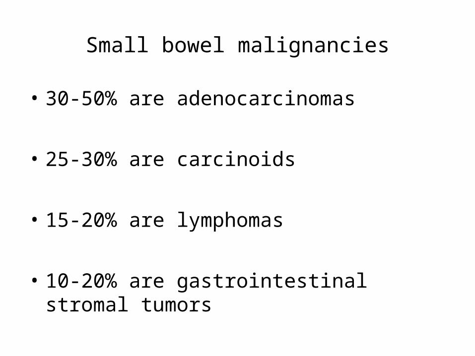
Small bowel malignancies
• 30-50% are adenocarcinomas
• 25-30% are carcinoids
• 15-20% are lymphomas
• 10-20% are gastrointestinal stromal tumors

Risk factors for small bowel adenocarcinoma
– Pre-existing adenoma, either single or multiple – 300-fold increased risk in patients with FAP
– Crohn’s – Celiac disease – IgA deficiency– Alcohol abuse– Neurofibromatosis– Urinary diversion procedures– ? Red meat

Anti-neoplastic environment of the small intestine
1. Liquid contents cause less irritation than more solid contents of large bowel
2. Rapid transit of intestinal contents provides shorter exposure of mucosa to carcinogens
3. Lower bacterial load may result in decreased conversion of bile acids into potential carcinogens
4. Benzopyrene hydroxylase, enzyme responsible for the conversion of the known carcinogen benzopyrene, is present in higher concentrations in the small bowel
5. Increased lymphoid tissue and higher levels of IgA

Metastatic disease involving small bowel
• Secondary neoplastic involvement of small intestine is more frequent than primary small bowel neoplasia
• Primary tumors of the colon, ovary, uterus, and stomach typically involve the colon by direct invasion or intraperitoneal spread
• Primary tumors from breast, lung, and melanoma metastasize to small bowel hematogenously
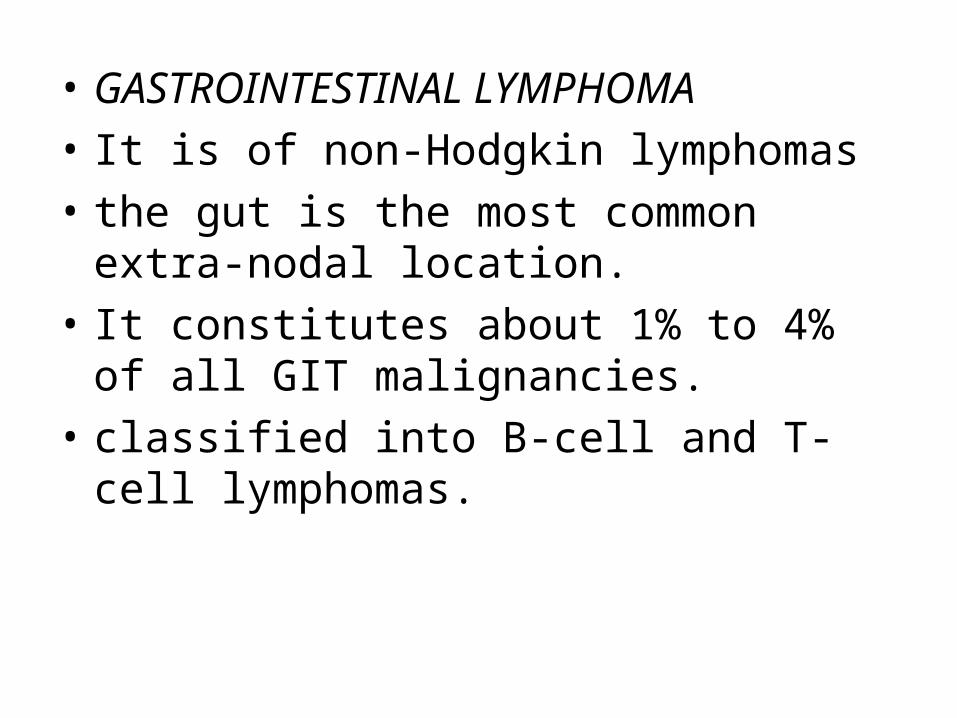
• GASTROINTESTINAL LYMPHOMA
• It is of non-Hodgkin lymphomas
• the gut is the most common extra-nodal location.
• It constitutes about 1% to 4% of all GIT malignancies.
• classified into B-cell and T-cell lymphomas.

1. MALT lymphoma is a sporadic lymphoma, which arises from the B cells of MALT (mucosa-associated lymphoid tissue
It can arise anywhere in the gut: stomach (55% to 60% of cases); small intestine (25% to 30%), proximal colon (10% to 15%), and distal colon 10%).
• History of IBD appears to increase the risk -Helicobacter-associated chronic gastritis).
.

• 2. IPSID is also referred to as Mediterranean lymphoma. immunoproliferative small-intestinal disease (IPSID),
• B-cell lymphoma arising
• children and young adults, and both sexes appear to be affected equally.
• etiology not known

• 3-The intestinal T-cell lymphoma is usually associated with a long-standing malabsorption syndrome (such as celiac disease)
• (age 30 to 40),
• Intestinal T-cell lymphoma
• In the proximal small bowel

• The large blue non-Hodgkin's lymphoma cells can be seen infiltrating through the mucosa.
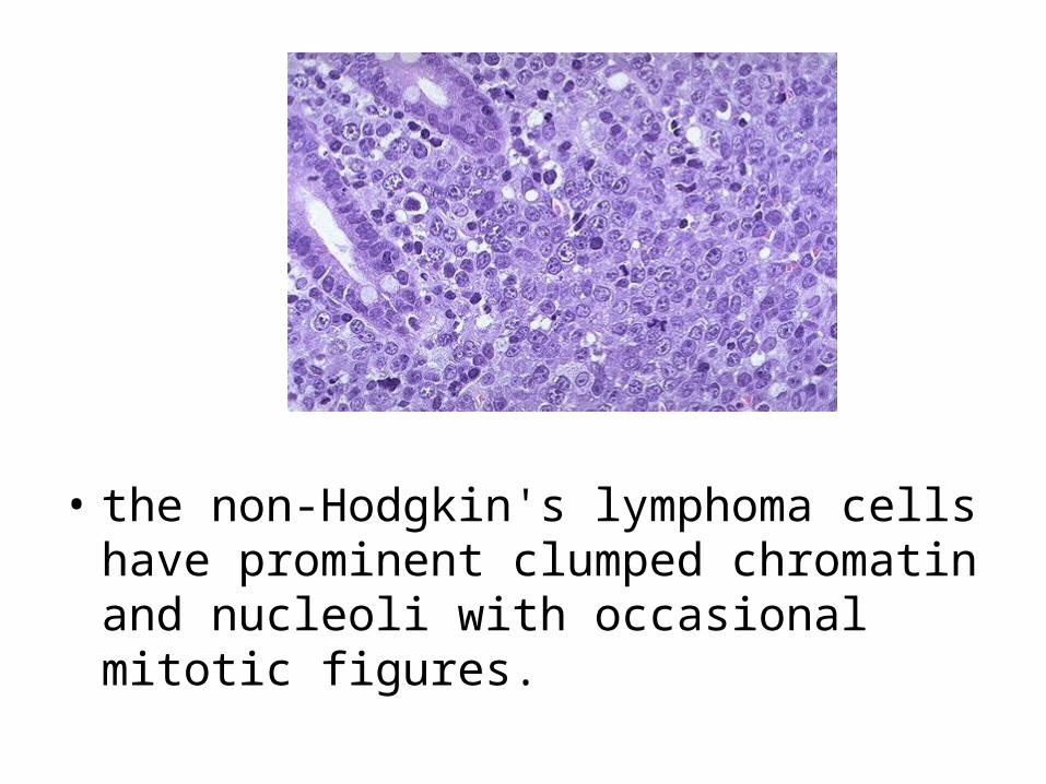
• the non-Hodgkin's lymphoma cells have prominent clumped chromatin and nucleoli with occasional mitotic figures.

Gastrointestinal stromal tumors
• Visceral sarcomas, previously classified as leiyomyomas and leiyomyosarcomas
• Now classified as GISTs with a range of biological behaviors from low grade to high grade malignancies
• Traditionally, microscopic findings were used to define malignancy including:– Increased cell size
– Increased cell irregularity
– Lack of cell differentiation
– Presence of cells with hyperchromic and multiple nuclei

GISTs – Tumor biology
• Proposed to arise from the interstitial cell of Cajal, an intestinal pacemaker cell of mesodermal origin– Similar cell markers to those of normal Cajal cells
1) myeloid stem cell antigen CD34
2) KIT receptor tyrosine kinase
3) variably positive for smooth-muscle actin
4) usually negative for desmin
• Previously thought to be smooth muscle neoplasms but now accepted to have:1) myogenic features (smooth muscle GIST)
2) neural features (GI autonomic nerve tumor)
3) myogenic and neural features (mixed GIST)
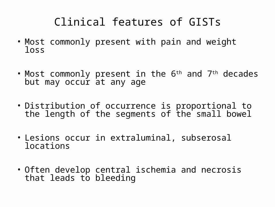
Clinical features of GISTs
• Most commonly present with pain and weight loss
• Most commonly present in the 6th and 7th decades but may occur at any age
• Distribution of occurrence is proportional to the length of the segments of the small bowel
• Lesions occur in extraluminal, subserosal locations
• Often develop central ischemia and necrosis that leads to bleeding

Carcinoid Tumors of the Intestine
• Originally described by Oberndorfer in 1907
• Arise from neuroendocrine cells
• Cells of the amine precursor uptake decarboxylase (APUD) system which have the ability to synthesize biologically active substances

Clinical features of carcinoid tumors
• Most commonly present in the 7th decade
• Often present with nonspecific complaints
• Up to 50% of patients present with obstruction
• Increasing frequency from the duodenum to the ileum with the appendix is the most common site.

Pathological features of carcinoid tumors
• Carcinoid invasion into the mesentery leads to fibrosis and often kinking of the small intestine
• Thickening of the vessel wall is also present and may lead to ischemic changes in the gut
• Serotonin is postulated to be responsible for these features

Carcinoid tumor. Multiple protruding tumors are present at the ileocecal junction

• Seen here at the ileocecal valve is a tumor that has a faint yellowish color. This is a carcinoid tumor.

Carcinoid Tumor : Clumps or cords of polygonal monotonous cells, rounded nuclei, pink granular cytoplasm, delicate stroma

• At high magnification, the nests of carcinoid tumor have a typical endocrine appearance with small round cells having small round nuclei and pink to pale blue cytoplasm. Rarely, a malignant carcinoid tumor can occur as a large bulky mass. Metastatic carcinoid to the liver can rarely result in the carcinoid syndrome.
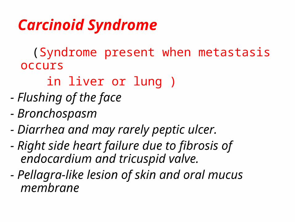
Carcinoid Syndrome
(Syndrome present when metastasis occurs in liver or lung )- Flushing of the face- Bronchospasm- Diarrhea and may rarely peptic ulcer.- Right side heart failure due to fibrosis of
endocardium and tricuspid valve.- Pellagra-like lesion of skin and oral mucus
membrane