skull, brain, cn
DESCRIPTION
Central Nervous SystemBones of SkullTRANSCRIPT
Skull, Brain and Cranial Nerves
Head and Neck Continued
Skull
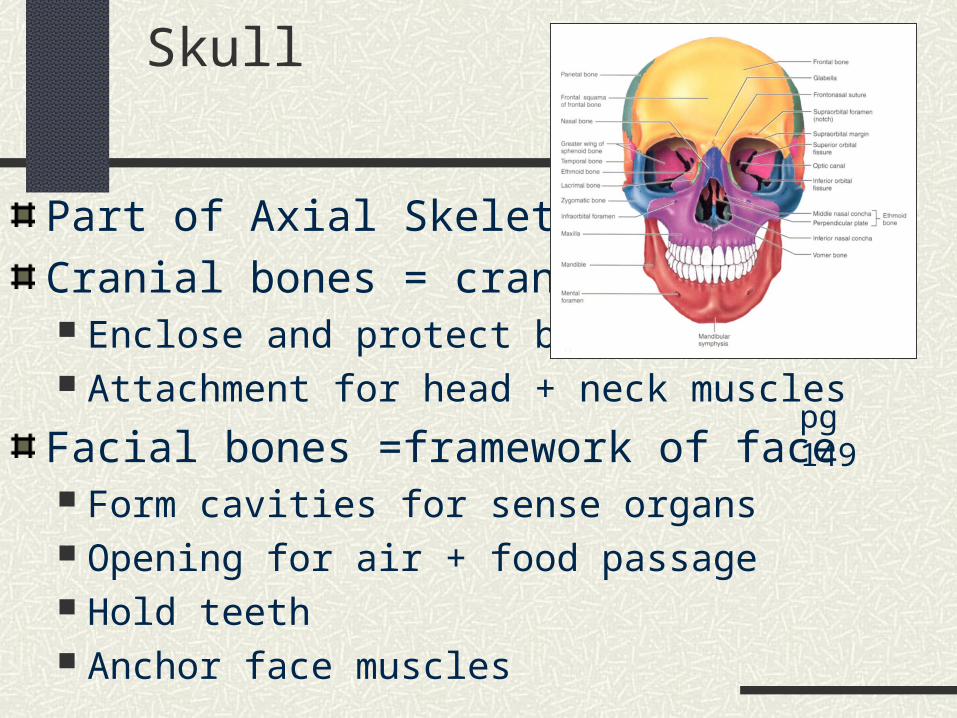
Part of Axial Skeleton
Cranial bones = cranium Enclose and protect brain Attachment for head + neck muscles
Facial bones =framework of face Form cavities for sense organs Opening for air + food passage Hold teeth Anchor face muscles
pg 149
Bones of Skull
Flat bones: thin, flattened, some curveSutures: immovable joints joining bonesCalvaria = Skullcap =Vault Superior, Lateral, Posterior part of skull
Floor = Base Inferior part of skull
85 openings in skull Spinal cord, blood vessels, nerves
Cranial Fossae
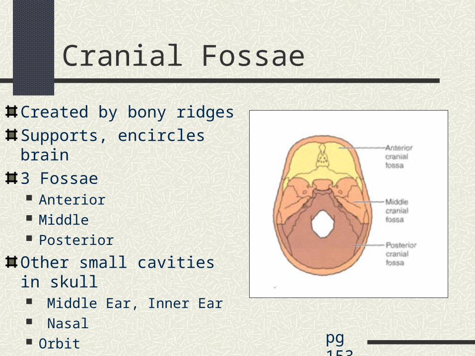
Created by bony ridges
Supports, encircles brain
3 Fossae Anterior Middle Posterior
Other small cavities in skull Middle Ear, Inner Ear Nasal Orbit pg 153
Skull through Life
Ossifies late in 2nd month of development
Frontal + Mandible start as 2 halves-then fuse
Skull bones separated by unossified membranes = Fontanels Allow compression of skull during delivery Mostly replaced w/bone after 1st year
Growth of Skull ½ adult size by age 9 months ¾ adult size by 2 years 100% adult size by 8-9 years Face enlarges between ages 6-13 years
pg 348
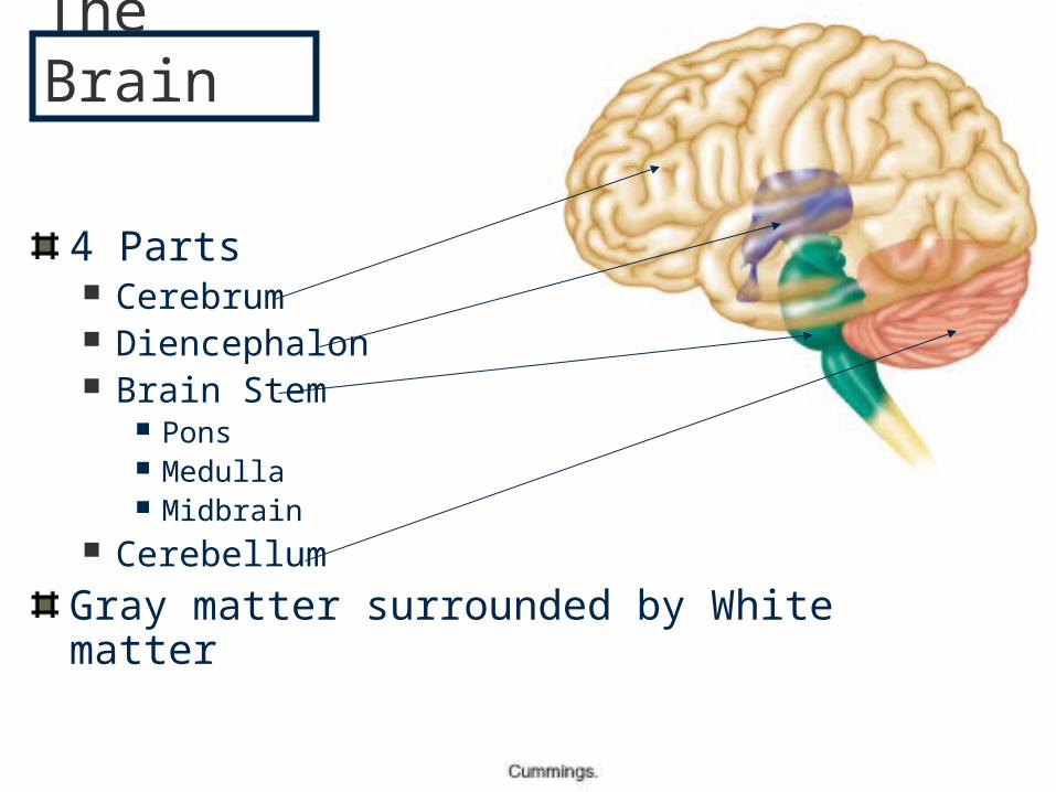
4 Parts Cerebrum Diencephalon Brain Stem
Pons Medulla Midbrain
Cerebellum
Gray matter surrounded by White matter
The Brain
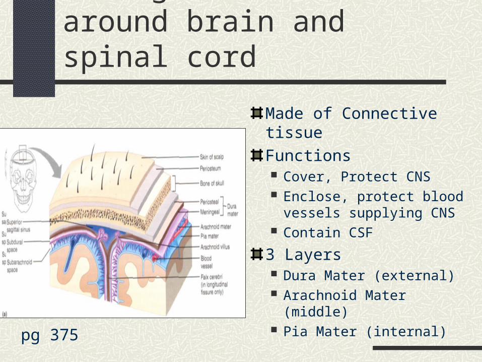
Meninges: 3 membranes around brain and spinal cord
Made of Connective tissue
Functions Cover, Protect CNS Enclose, protect blood
vessels supplying CNS Contain CSF
3 Layers Dura Mater (external) Arachnoid Mater (middle) Pia Mater (internal)pg 375
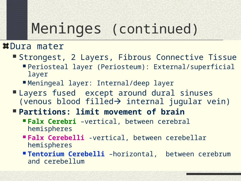
Meninges (continued)Dura mater Strongest, 2 Layers, Fibrous Connective Tissue
Periosteal layer (Periosteum): External/superficial layer Meningeal layer: Internal/deep layer
Layers fused except around dural sinuses (venous blood filled internal jugular vein)
Partitions: limit movement of brain Falx Cerebri –vertical, between cerebral hemispheres Falx Cerebelli -vertical, between cerebellar hemispheres Tentorium Cerebelli –horizontal, between cerebrum and
cerebellum
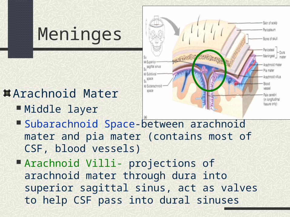
Meninges
Arachnoid Mater Middle layer Subarachnoid Space-between arachnoid mater and
pia mater (contains most of CSF, blood vessels) Arachnoid Villi- projections of arachnoid mater
through dura into superior sagittal sinus, act as valves to help CSF pass into dural sinuses
Meninges (continued)
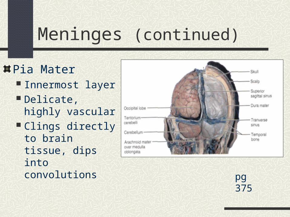
Pia Mater Innermost layer Delicate, highly
vascular Clings directly to
brain tissue, dips into convolutions
pg 375
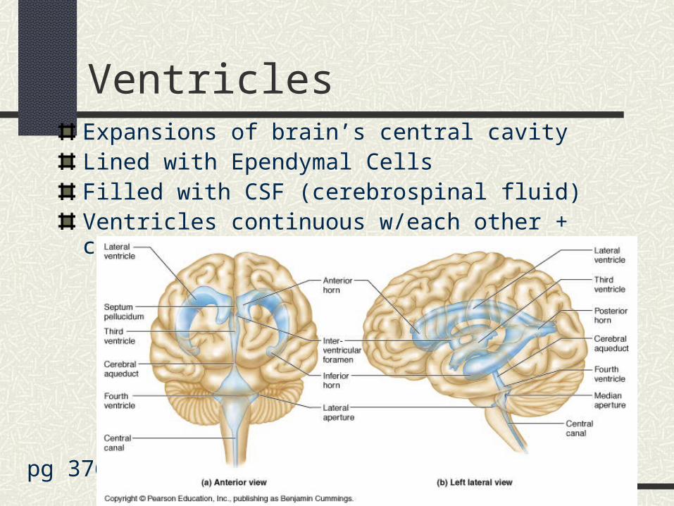
VentriclesExpansions of brain’s central cavityLined with Ependymal CellsFilled with CSF (cerebrospinal fluid)Ventricles continuous w/each other + central canal of spinal cord
pg 376
Ventricles (continued)
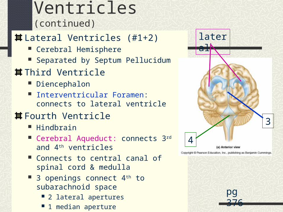
Lateral Ventricles (#1+2) Cerebral Hemisphere Separated by Septum Pellucidum
Third Ventricle Diencephalon Interventricular Foramen: connects to lateral
ventricle
Fourth Ventricle Hindbrain Cerebral Aqueduct: connects 3rd and 4th
ventricles Connects to central canal of spinal cord &
medulla 3 openings connect 4th to subarachnoid space
2 lateral apertures 1 median aperture
pg 376
3
4
lateral
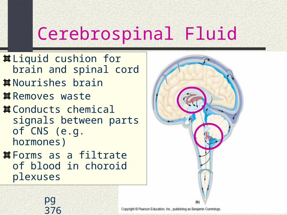
Cerebrospinal Fluid
pg 376
Liquid cushion for brain and spinal cordNourishes brainRemoves wasteConducts chemical signals between parts of CNS (e.g. hormones)Forms as a filtrate of blood in choroid plexuses
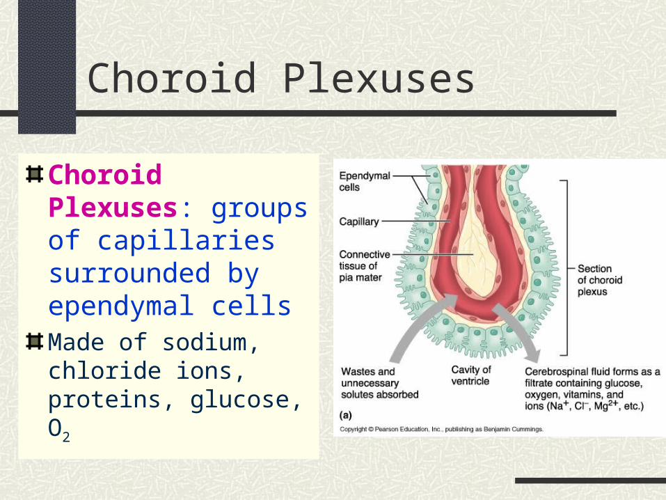
Choroid Plexuses
Choroid Plexuses: groups of capillaries surrounded by ependymal cellsMade of sodium, chloride ions, proteins, glucose, O2
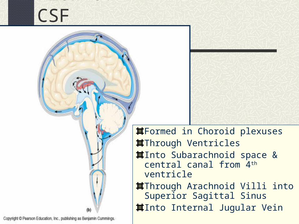
Flow of CSF
Formed in Choroid plexusesThrough VentriclesInto Subarachnoid space & central canal from 4th ventricleThrough Arachnoid Villi into Superior Sagittal Sinus Into Internal Jugular Vein
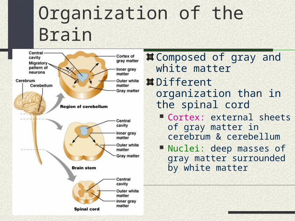
Organization of the BrainComposed of gray and white matterDifferent organization than in the spinal cord Cortex: external sheets of gray
matter in cerebrum & cerebellum
Nuclei: deep masses of gray matter surrounded by white matter
Cerebrum
“Executive Suite” of nervous system Self-awareness, initiate + control voluntary movements,
communicate, remember, understand
Made of Gray matter, White matter, Basal gangli (nuclei)Most superior region Covers diencephalon + top of brain stem like mushroom capMany small grooves + deep fissures Transverse-separates cerebral hemisphere + cerebellum Longitudinal-separates right + left cerebral hemispheres
Sulci – grooves on surfaceGyri-ridges of brain tissue between sulci
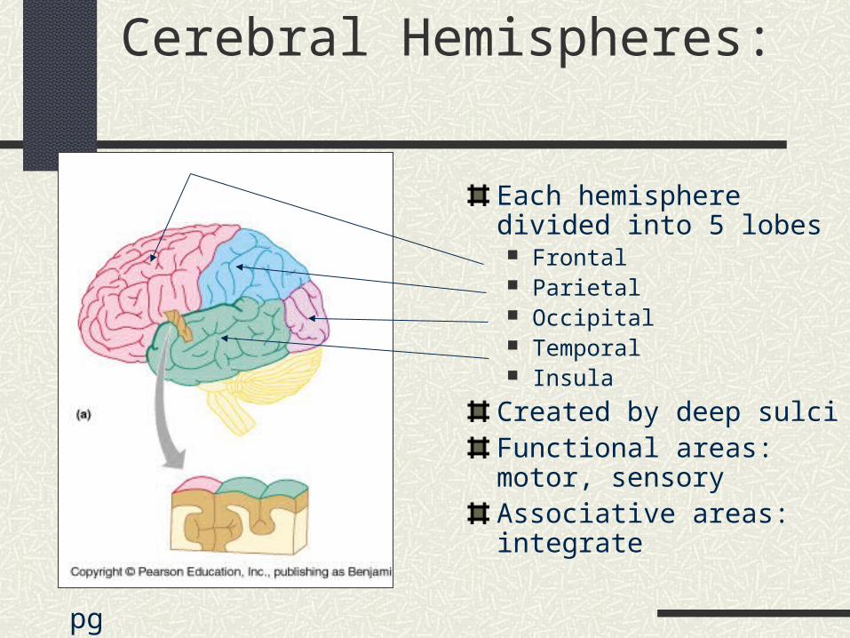
Cerebral Hemispheres:
Each hemisphere divided into 5 lobes Frontal Parietal Occipital Temporal Insula
Created by deep sulciFunctional areas: motor, sensoryAssociative areas: integrate
pg 349
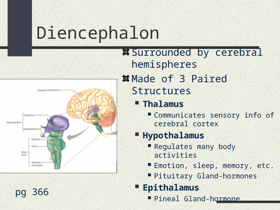
DiencephalonSurrounded by cerebral hemispheres
Made of 3 Paired Structures Thalamus
Communicates sensory info of cerebral cortex
Hypothalamus Regulates many body activities Emotion, sleep, memory, etc. Pituitary Gland-hormones
Epithalamus Pineal Gland-hormone pg 366
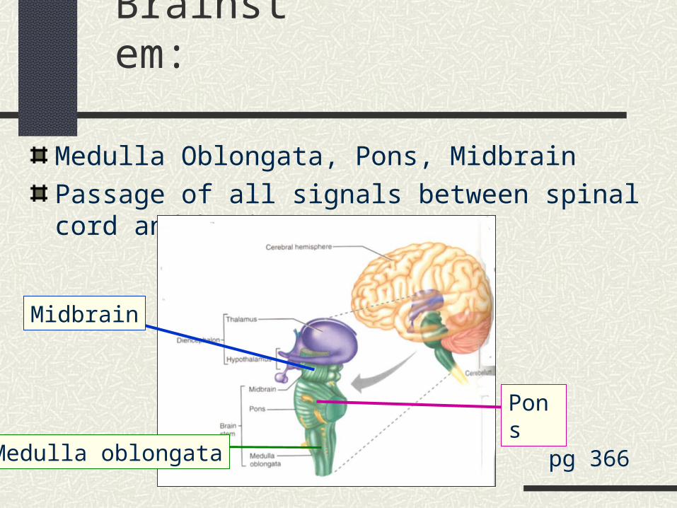
Brainstem:
Medulla Oblongata, Pons, Midbrain
Passage of all signals between spinal cord and brain
pg 366
Midbrain
Pons
Medulla oblongata
Brainstem: Medulla Oblongata
Regulates several basic physiological functions Heartbeat (rate and force) Blood pressure (vasoconstriction/dilation of
arteries) Breathing (rate and depth) Others: speech, coughing, sneezing, salivation,
swallowing, gagging, vomiting, sweating
Attachment of CN IX, X, XI, XII
Brainstem: The Pons
Contains many tracts carrying signals: from cerebrum to cerebellum & medulla up to thalamus between right and left hemispheres of cerebellum from brainstem to cerebellum
Attachment of CN V, VI, VII, VIII
Brainstem: Midbrain
Carries signals Between higher and lower brain centers From cerebellum to cerebral cortex
Visual and Auditory reflex centers
Somatic motor
Attachment for CN III, IV
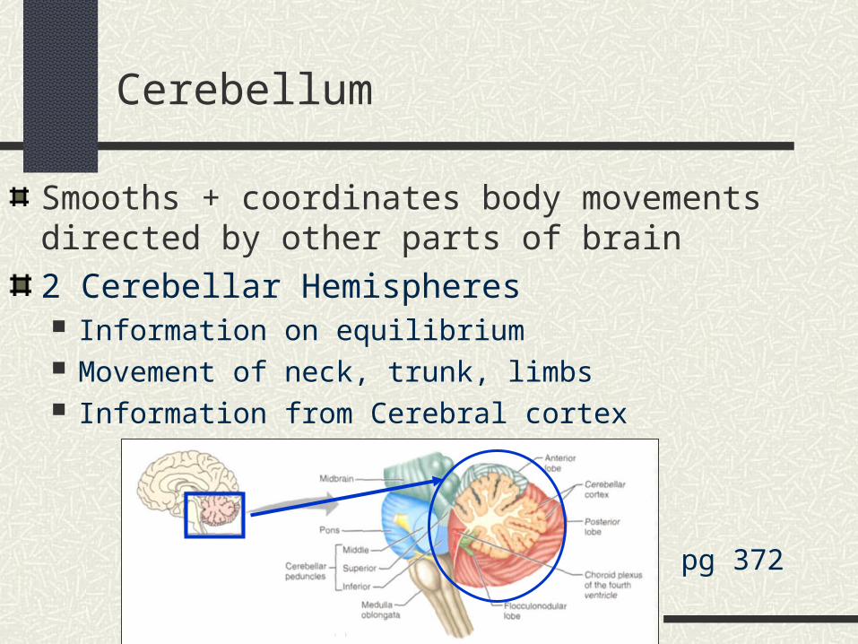
Smooths + coordinates body movements directed by other parts of brain
2 Cerebellar Hemispheres Information on equilibrium Movement of neck, trunk, limbs Information from Cerebral cortex
Cerebellum
pg 372

Blood Brain Barrier
Protects brain from blood-borne toxins (e.g. urea, food toxins, bacteria)
Endothelium of brain capillaries are loaded with tight junction to decrease permeability
Not complete protection, some things still have to get through (e.g. fat-soluble molecules can pass through)
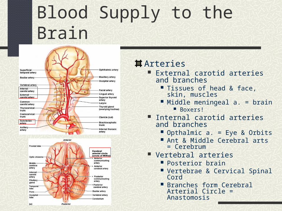
Blood Supply to the Brain
Arteries External carotid arteries and
branches Tissues of head & face, skin,
muscles Middle meningeal a. = brain
Boxers! Internal carotid arteries and branches
Opthalmic a. = Eye & Orbits Ant & Middle Cerebral arts =
Cerebrum Vertebral arteries
Posterior brain Vertebrae & Cervical Spinal Cord Branches form Cerebral Arterial
Circle = Anastomosis
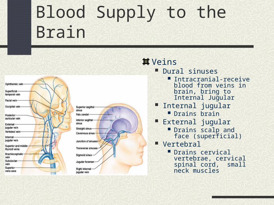
Blood Supply to the Brain
Veins Dural sinuses
Intracranial-receive blood from veins in brain, bring to Internal Jugular
Internal jugular Drains brain
External jugular Drains scalp and face
(superficial) Vertebral
Drains cervical vertebrae, cervical spinal cord, small neck muscles
Cranial Nerves: I - XII
12 Pairs
Numbered Anterior to Posterior
Attach to Ventral surface of brain
Exit brain through foramina in skull
I + II attach to Forebrain (cerebrum + diencephalon)
III-XII attach to Brainstem (midbrain, pons, medulla)
Only X goes beyond the head-neck
Foramina serving Cranial Nerves
You must know what foramina each CN leaves the skull through
(refer to handout in lab)

How to Remember CN I-XII
Oh! Oh! Oh! To Touch And Feel Very Good Velvet! Ah Heaven!
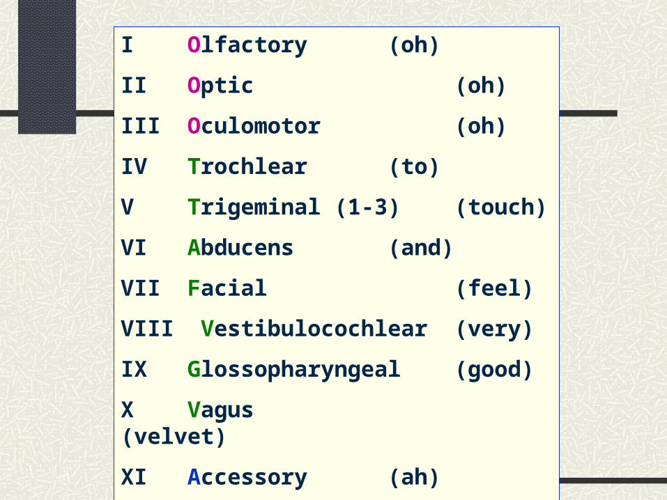
I Olfactory (oh)
II Optic (oh)
III Oculomotor (oh)
IV Trochlear (to)
V Trigeminal (1-3) (touch)
VI Abducens (and)
VII Facial (feel)
VIII Vestibulocochlear (very)
IX Glossopharyngeal (good)
X Vagus (velvet)
XI Accessory (ah)
XII Hypoglossal (heaven)

Motor vs. Sensory Nerves
Sensory = Afferent Send nervous impulse from sensory receptors to
brain to bring in information e.g. pressure, temperature, pain
Motor = Efferent Send nervous impulses from brain to body to
accomplish an action e.g. movement of a muscle, activation of a gland
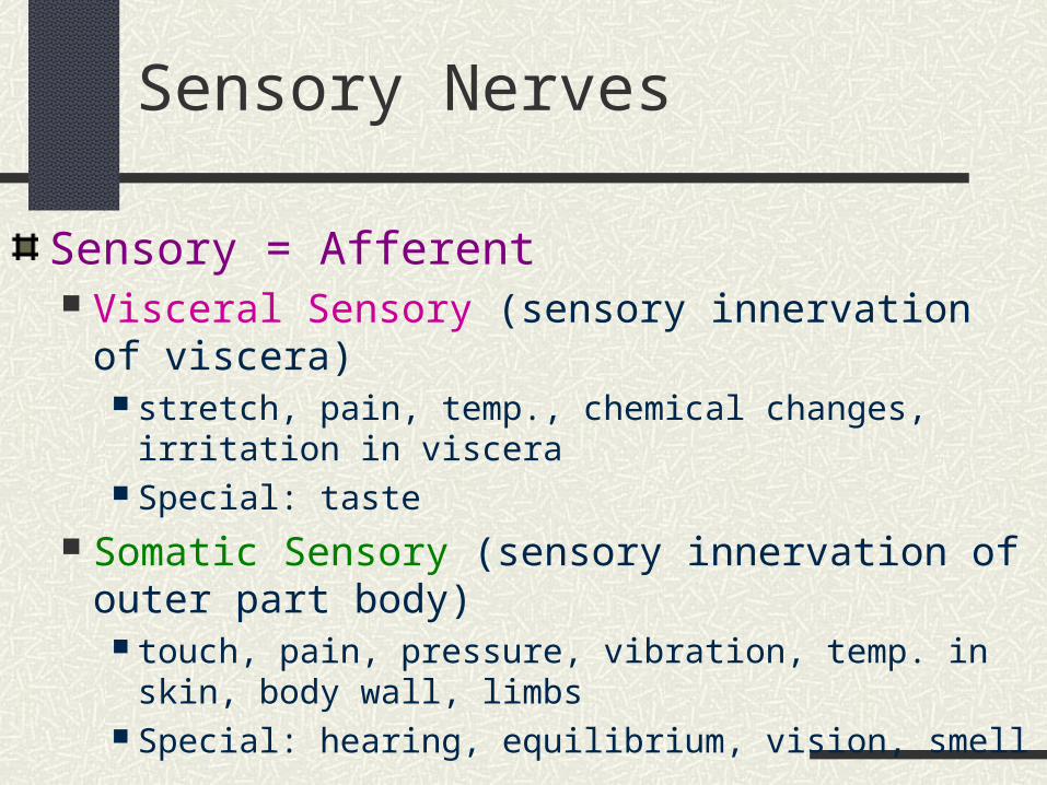
Sensory Nerves
Sensory = Afferent Visceral Sensory (sensory innervation of viscera)
stretch, pain, temp., chemical changes, irritation in viscera Special: taste
Somatic Sensory (sensory innervation of outer part body) touch, pain, pressure, vibration, temp. in skin, body wall, limbs Special: hearing, equilibrium, vision, smell
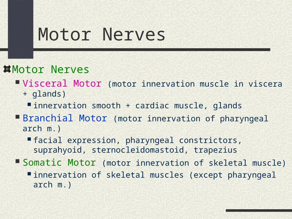
Motor Nerves
Motor Nerves Visceral Motor (motor innervation muscle in viscera + glands)
innervation smooth + cardiac muscle, glands Branchial Motor (motor innervation of pharyngeal arch m.)
facial expression, pharyngeal constrictors, suprahyoid, sternocleidomastoid, trapezius
Somatic Motor (motor innervation of skeletal muscle) innervation of skeletal muscles (except pharyngeal arch m.)

Mnemonic for CN FunctionSome (CN I)Say (CN II)Marry (CN III)Money (CN IV)But (CN V)My (CN VI)Brother (CN VII)Says (CN VIII)Big (CN IX) Brains (CN X)Matter (CN XI)Most! (CN XII)
S = Sensory function M = Motor functionB = BOTH (Sensory and Motor function)
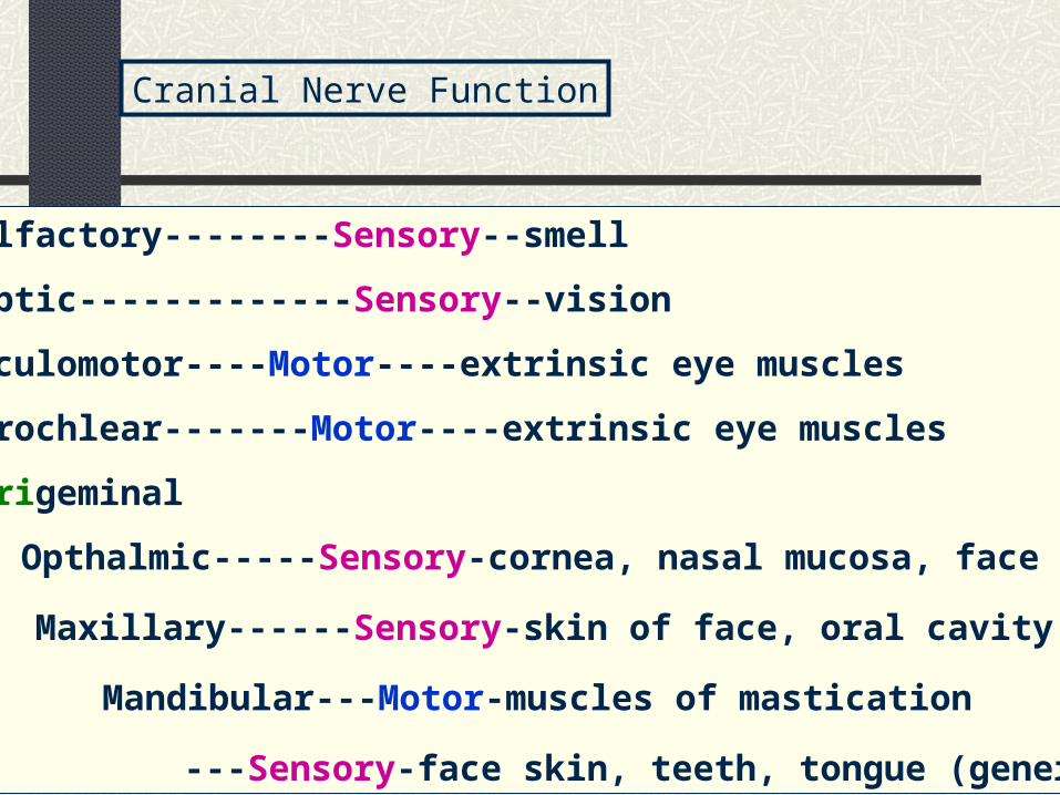
I Olfactory--------Sensory--smell
II Optic-------------Sensory--vision
III Oculomotor----Motor----extrinsic eye muscles
IV Trochlear-------Motor----extrinsic eye muscles
V Trigeminal
V1 Opthalmic-----Sensory-cornea, nasal mucosa, face skin
V2 Maxillary------Sensory-skin of face, oral cavity, teeth
V3 Mandibular---Motor-muscles of mastication
---Sensory-face skin, teeth, tongue (general)
Cranial Nerve Function
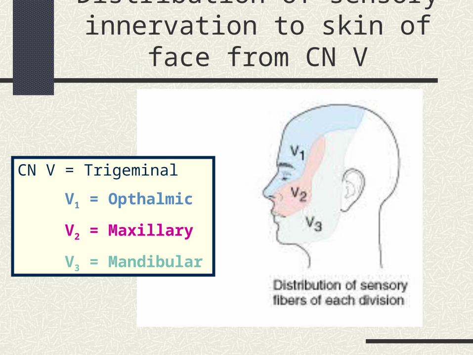
Distribution of sensory innervation to skin of face from CN V
CN V = Trigeminal
V1 = Opthalmic
V2 = Maxillary
V3 = Mandibular
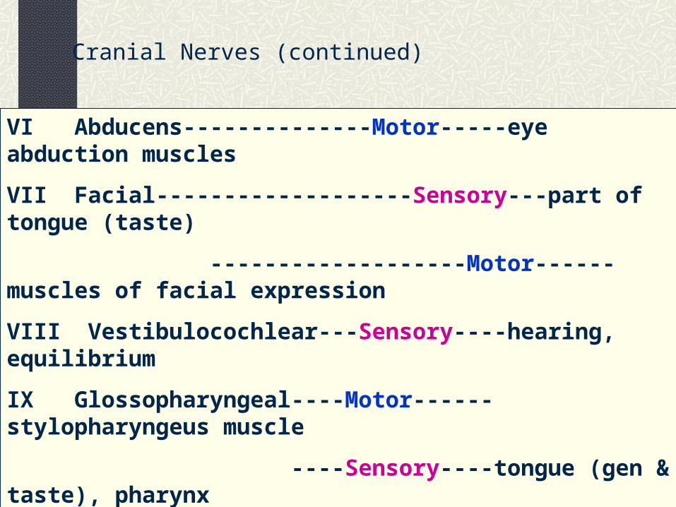
VI Abducens--------------Motor-----eye abduction muscles
VII Facial-------------------Sensory---part of tongue (taste)
-------------------Motor------muscles of facial expression
VIII Vestibulocochlear---Sensory----hearing, equilibrium
IX Glossopharyngeal----Motor------stylopharyngeus muscle
----Sensory----tongue (gen & taste), pharynx
X Vagus------------------Motor-------pharynx, larynx
-------------------Sensory----pharynx, larynx, abd. organs
XI Accessory-------------Motor------trapezius, sternocleidomastoid
XII Hypoglossal----------Motor-------tongue muscles
Cranial Nerves (continued)

Summary of Functional Groups
Purely Sensory = I, II, VIII
Primarily Motor = III, IV, VI, XI, XII
Mixed = V, VII, IX, X
Parasympathetic Fibers = III, VII, IX, X(Division of Autonomic NS = Visceral Motor)
pg 449
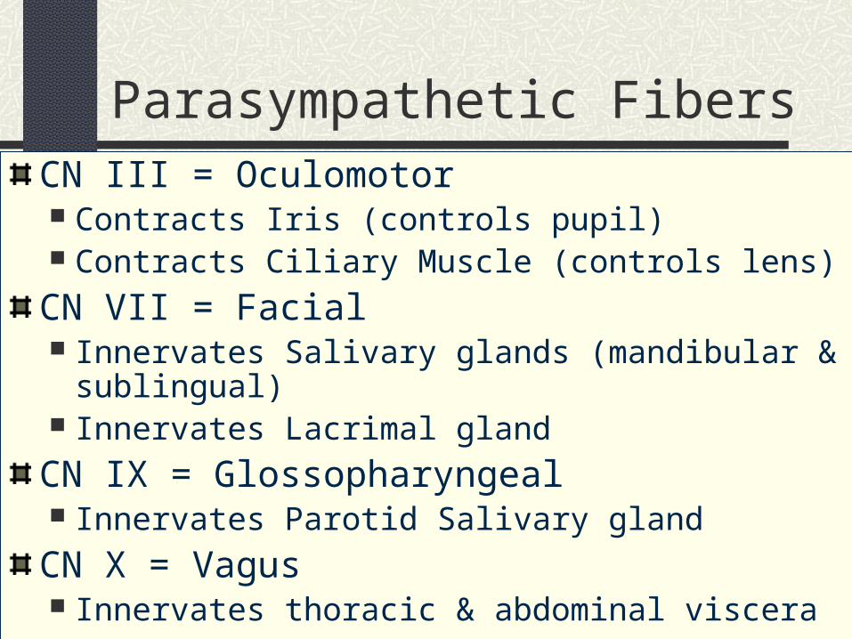
Parasympathetic FibersCN III = Oculomotor Contracts Iris (controls pupil) Contracts Ciliary Muscle (controls lens)
CN VII = Facial Innervates Salivary glands (mandibular & sublingual) Innervates Lacrimal gland
CN IX = Glossopharyngeal Innervates Parotid Salivary gland
CN X = Vagus Innervates thoracic & abdominal viscera

X
III
VII
IX
Anatomy of the Eye and Ear
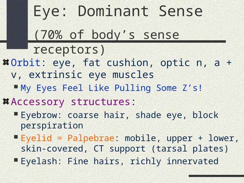
Orbit: eye, fat cushion, optic n, a + v, extrinsic eye muscles My Eyes Feel Like Pulling Some Z’s!
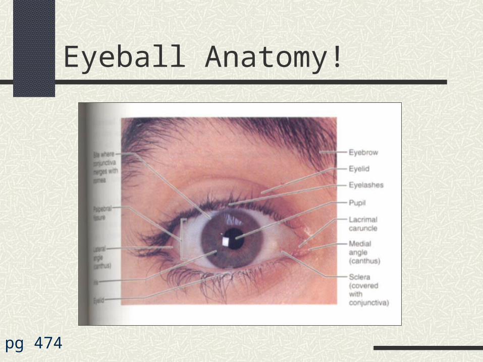
Accessory structures: Eyebrow: coarse hair, shade eye, block perspiration Eyelid = Palpebrae: mobile, upper + lower, skin-covered,
CT support (tarsal plates) Eyelash: Fine hairs, richly innervated
Eye: Dominant Sense
(70% of body’s sense receptors)
Glands Associated w/Eyelids
Types of Glands Tarsal Gland: (sebaceous glands)
Embedded in tarsal plates, open at edge of eyelids Ciliary Gland: (modified sweat glands)
Within eyelids Sebaceous glands – open into hair follicles
Function of Secretions Slow evaporation of fluid on eye surface Soften and lubricate eyelashes, skin Kill bacteria Collect dirt
Eye (continued)
More Accessory structures Conjunctiva-transparent mucous membrane on inner
eyelid + anterior surface of eye, mucus keeps eye moist Lacrimal Apparatus-gland + ducts flow into nasal cavity
Tears-keep eye moist, wash out irritant Contain mucus, antibodies, lysozome
Lacrimal Gland-Superolateral to eye, produce fluid Innervated by CN VII (parasympathetic fibers)
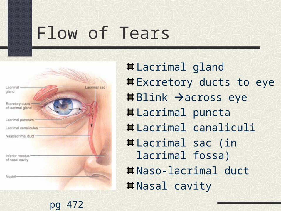
Flow of Tears
Lacrimal gland
Excretory ducts to eye
Blink across eye
Lacrimal puncta
Lacrimal canaliculi
Lacrimal sac (in lacrimal fossa)
Naso-lacrimal duct
Nasal cavity
pg 472
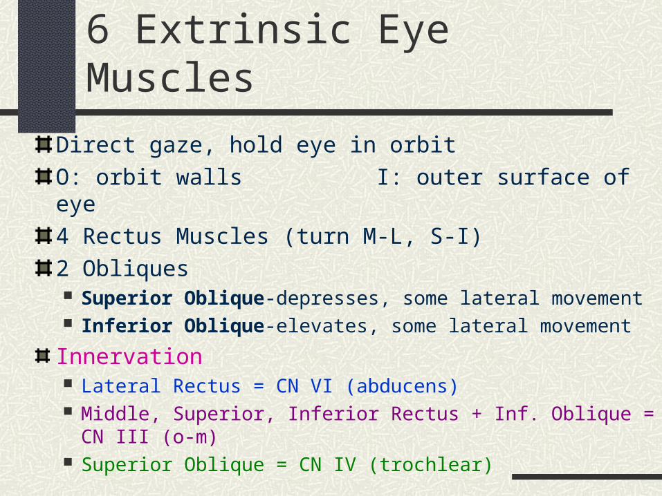
6 Extrinsic Eye Muscles
Direct gaze, hold eye in orbit
O: orbit walls I: outer surface of eye
4 Rectus Muscles (turn M-L, S-I)
2 Obliques Superior Oblique-depresses, some lateral movement Inferior Oblique-elevates, some lateral movement
Innervation Lateral Rectus = CN VI (abducens) Middle, Superior, Inferior Rectus + Inf. Oblique = CN III (o-m) Superior Oblique = CN IV (trochlear)
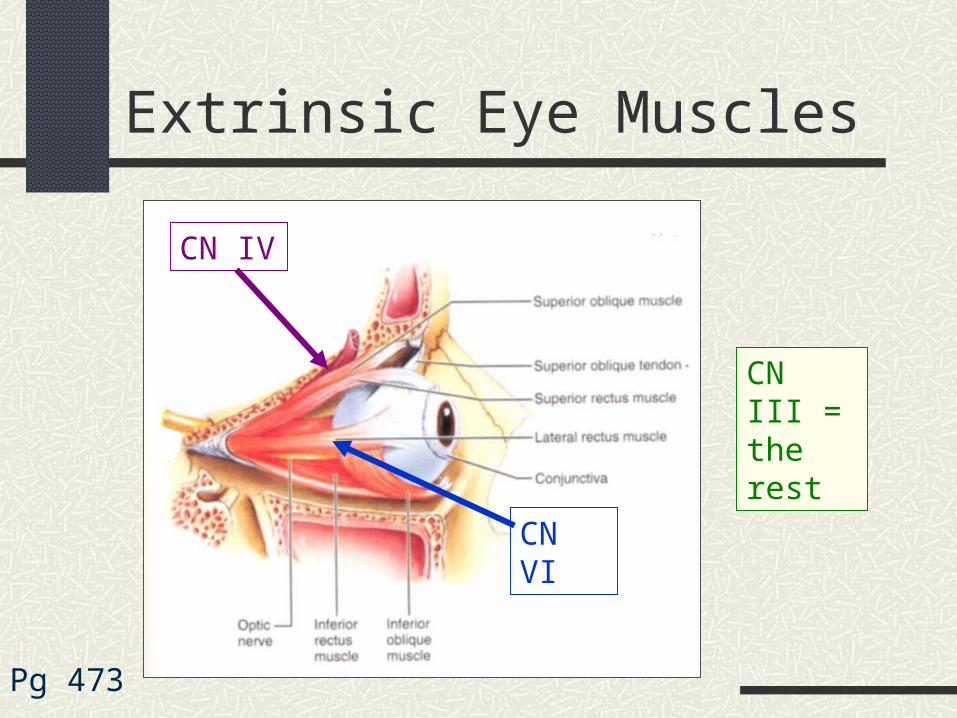
Extrinsic Eye Muscles
Pg 473
CN IV
CN VI
CN III = the rest
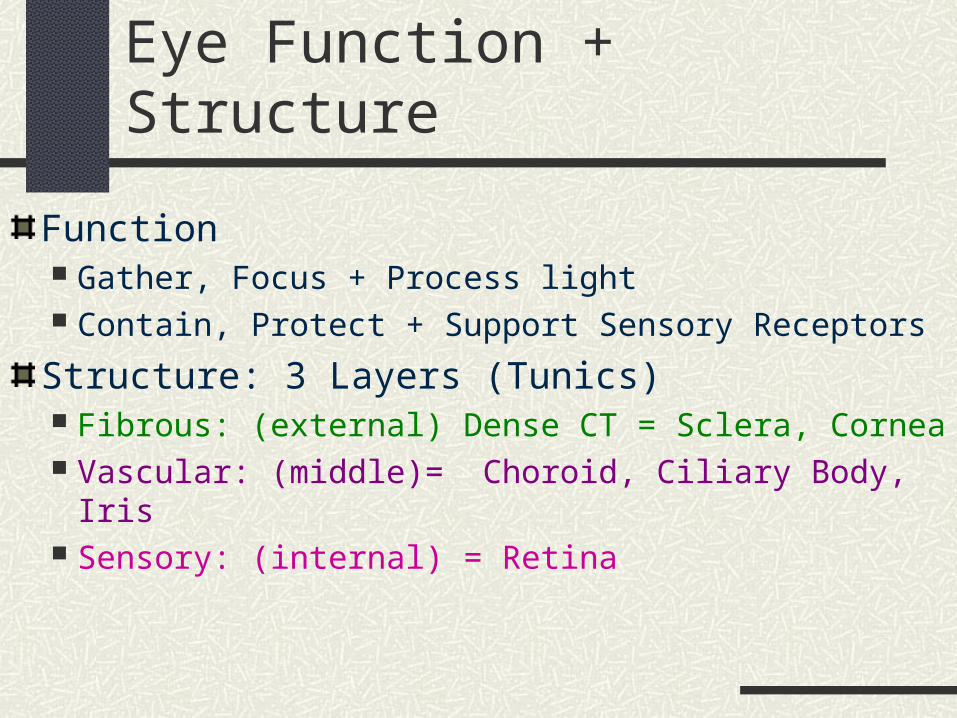
Eye Function + Structure
Function Gather, Focus + Process light Contain, Protect + Support Sensory Receptors
Structure: 3 Layers (Tunics) Fibrous: (external) Dense CT = Sclera, Cornea Vascular: (middle)= Choroid, Ciliary Body, Iris Sensory: (internal) = Retina
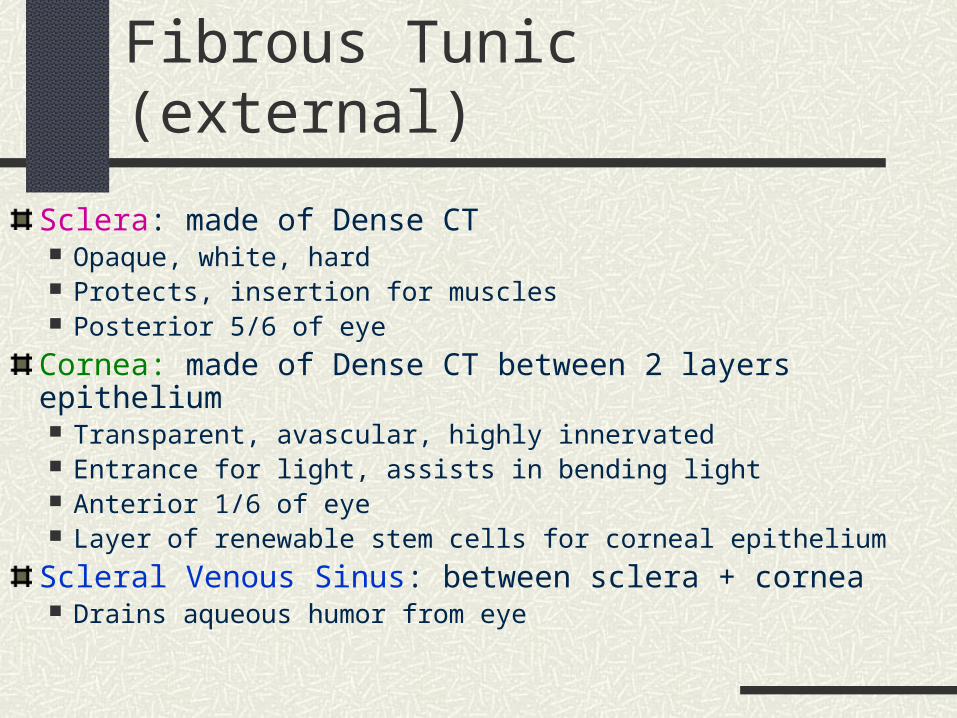
Fibrous Tunic (external)
Sclera: made of Dense CT Opaque, white, hard Protects, insertion for muscles Posterior 5/6 of eye
Cornea: made of Dense CT between 2 layers epithelium Transparent, avascular, highly innervated Entrance for light, assists in bending light Anterior 1/6 of eye Layer of renewable stem cells for corneal epithelium
Scleral Venous Sinus: between sclera + cornea Drains aqueous humor from eye
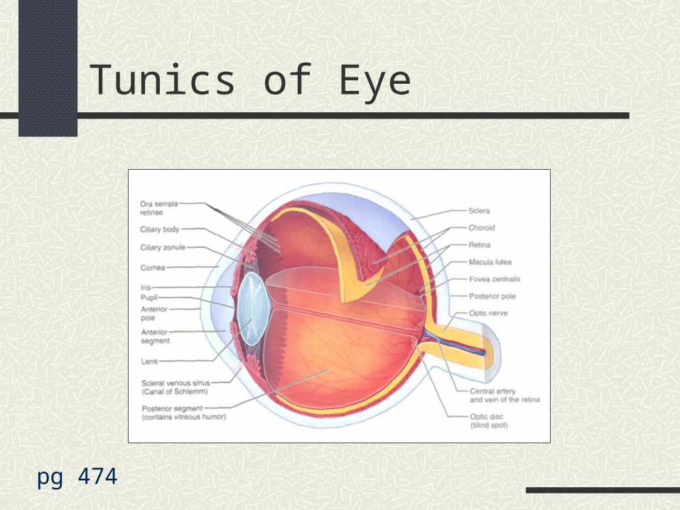
Tunics of Eye
pg 474
Vascular Tunic (middle)
Choroid: highly vascularized, darkly pigmented membrane, post. 5/6 nourishes other tunics absorbs light, prevent scattering & confusion
Ciliary Body: continuous w/choroid, thick ring of tissue around lens smooth muscle (ciliary muscle) = focuses lens
Iris: visible, colored part between cornea + lens Attached to ciliary body Pupil = opening in iris smooth muscle = dilate + constrict pupil = light enters
Retina = Sensory Tunic (internal)
Neural layer (inner) thick, sheets nervous tissue contain photoreceptors (rods + cones)
Pigmented layer (outer) contains melanocytes absorb light, prevent scattering
Eye Anatomy (continued)
Lens thick, transparent, biconvex disc changes shape to focus light on retina made of epithelium + fibers (contain proteins) divides eye into anterior/posterior segments
Aqueous Humor: clear fluid in anterior segment provides nutrients, O2 to lens/cornea
Vitreous Humor: jelly-like in posterior segment transmit light, support post. surface of lens + hold 2
layers of retina together, maintain intraocular pressure
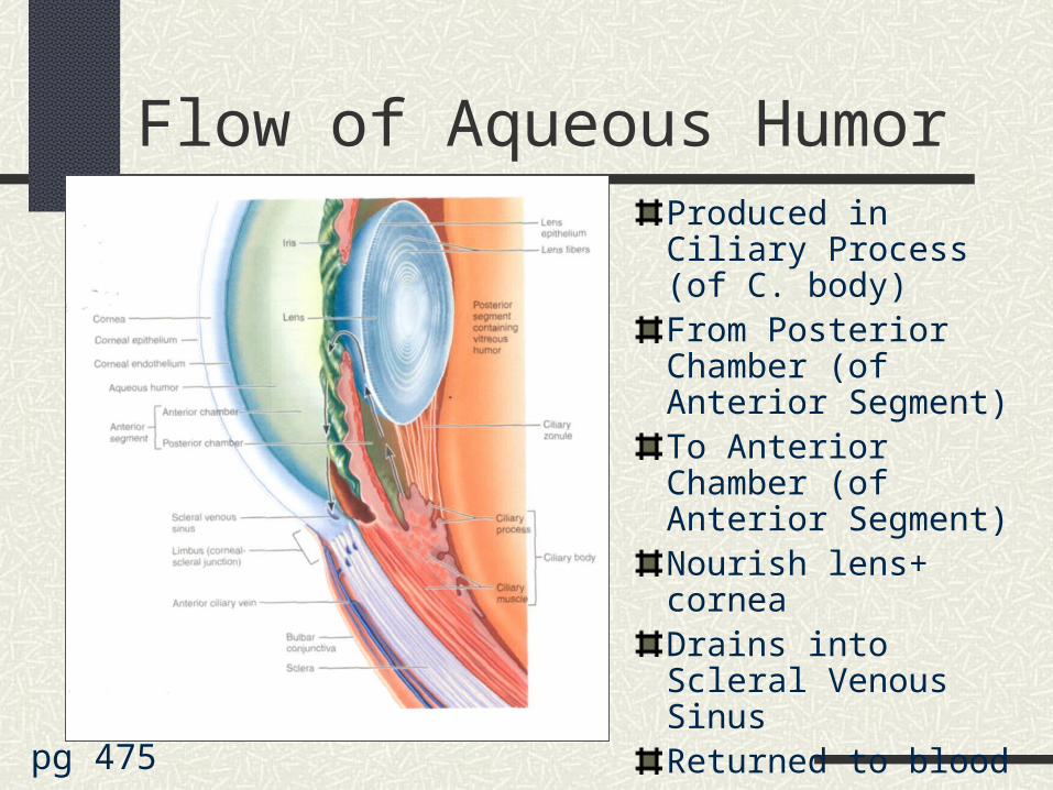
Flow of Aqueous HumorProduced in Ciliary Process (of C. body)From Posterior Chamber (of Anterior Segment)To Anterior Chamber (of Anterior Segment)Nourish lens+ corneaDrains into Scleral Venous SinusReturned to blood
pg 475
Eyeball Anatomy!
pg 474
The Ear: Outer, Middle, Inner
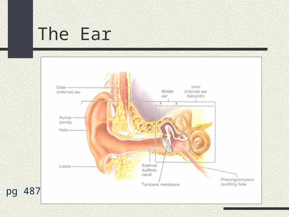
Outer: Hearing Auricle = Pinna
external elastic cartilage gathers + funnels sound into ear opening
External Auditory Meatus (canal) short tube from auricle to ear drum lateral 1/3 = elastic cartilage medial 2/3 = temporal bone Lined w/skin containing hair + glands produce ear wax
The Ear
pg 487
The Ear: Outer, Middle, Inner
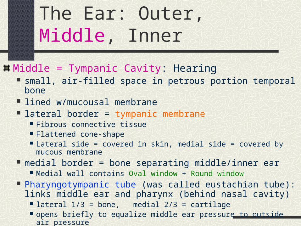
Middle = Tympanic Cavity: Hearing small, air-filled space in petrous portion temporal bone lined w/mucousal membrane lateral border = tympanic membrane
Fibrous connective tissue Flattened cone-shape Lateral side = covered in skin, medial side = covered by mucous membrane
medial border = bone separating middle/inner ear Medial wall contains Oval window + Round window
Pharyngotympanic tube (was called eustachian tube): links middle ear and pharynx (behind nasal cavity)
lateral 1/3 = bone, medial 2/3 = cartilage opens briefly to equalize middle ear pressure to outside air pressure
Middle Ear (continued)
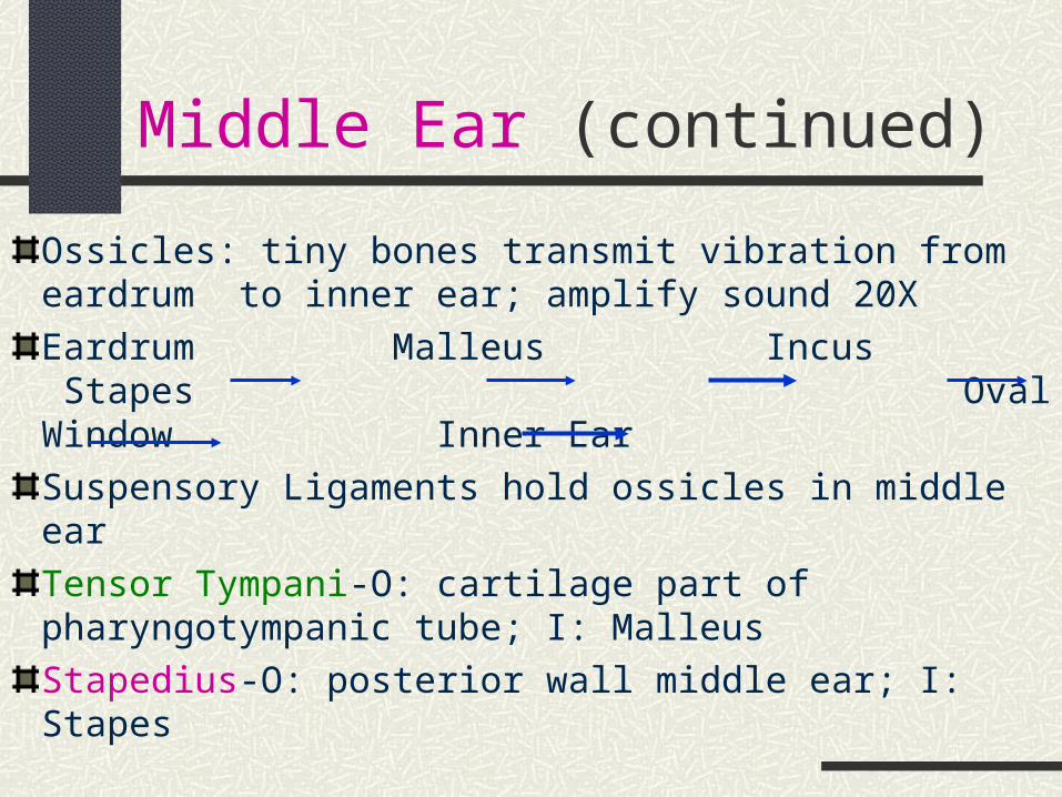
Ossicles: tiny bones transmit vibration from eardrum to inner ear; amplify sound 20X
Eardrum Malleus Incus Stapes Oval Window Inner Ear
Suspensory Ligaments hold ossicles in middle ear
Tensor Tympani-O: cartilage part of pharyngotympanic tube; I: Malleus
Stapedius-O: posterior wall middle ear; I: Stapes
The Ear: Outer, Middle, Inner
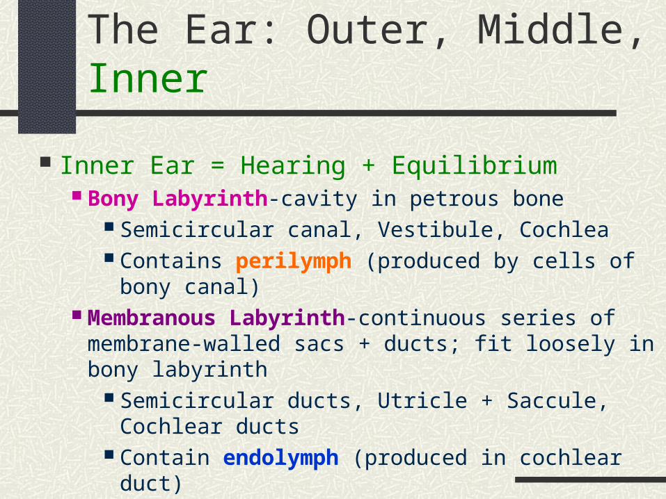
Inner Ear = Hearing + Equilibrium Bony Labyrinth-cavity in petrous bone
Semicircular canal, Vestibule, Cochlea Contains perilymph (produced by cells of bony canal)
Membranous Labyrinth-continuous series of membrane-walled sacs + ducts; fit loosely in bony labyrinth
Semicircular ducts, Utricle + Saccule, Cochlear ducts Contain endolymph (produced in cochlear duct)
Inner Ear: structures + functions
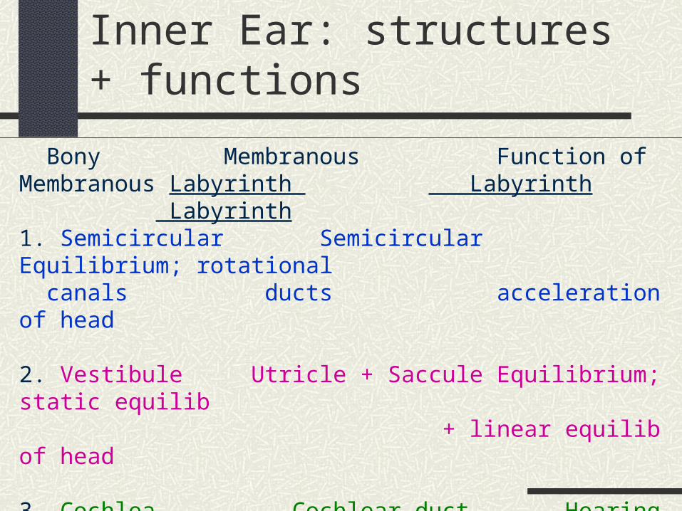
Bony Membranous Function of Membranous Labyrinth Labyrinth Labyrinth1. Semicircular Semicircular Equilibrium; rotational canals ducts acceleration of head
2. Vestibule Utricle + Saccule Equilibrium; static equilib + linear equilib of head
3. Cochlea Cochlear duct Hearing
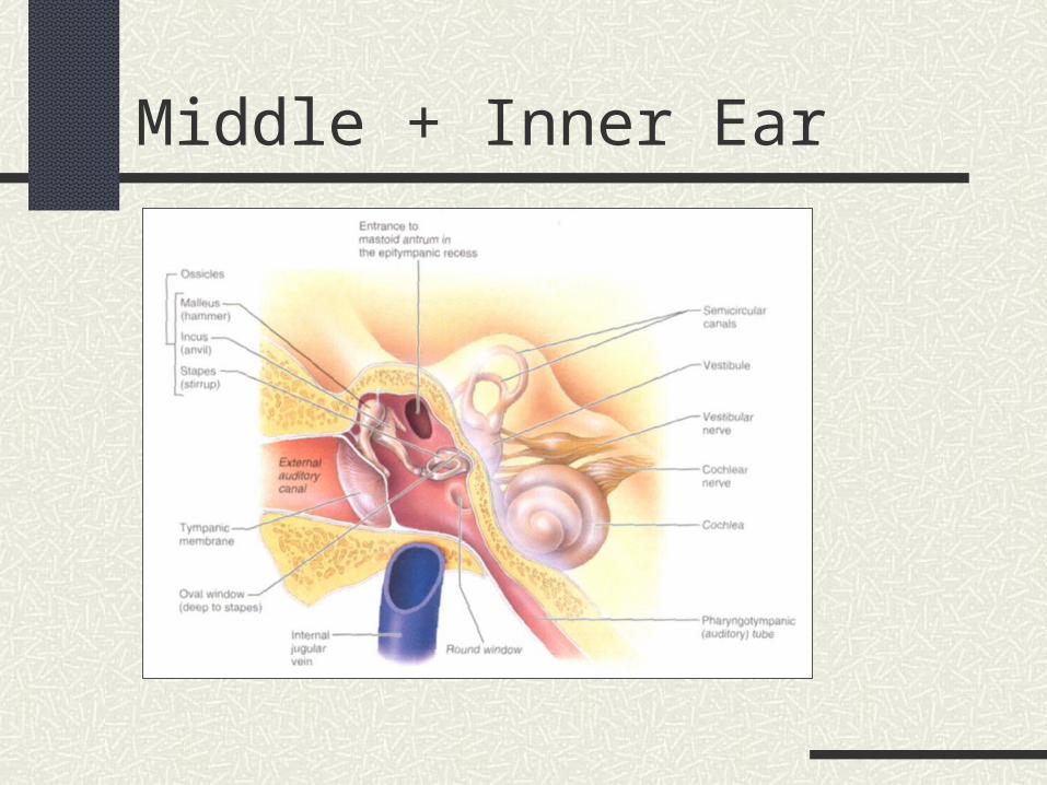
Middle + Inner Ear

STOP
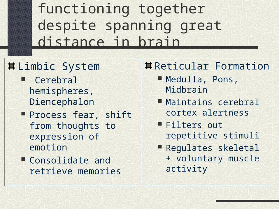
Functional Brain Systems: networks of neurons functioning together despite spanning great distance in brain
Limbic System Cerebral hemispheres,
Diencephalon Process fear, shift from
thoughts to expression of emotion
Consolidate and retrieve memories
Reticular Formation Medulla, Pons, Midbrain Maintains cerebral cortex
alertness Filters out repetitive
stimuli Regulates skeletal +
voluntary muscle activity