skindersoe, m., krogfelt, k., blom, a., zhang, j., jiang ... · ness for a particular purpose or...
TRANSCRIPT

Skindersoe, M., Krogfelt, K., Blom, A., Zhang, J., Jiang, G., Prest-wich, G. and Mansell, J. (2015) Dual Action of Lysophosphatidate-Functionalised Titanium: Interactions with Human (MG63) Osteoblastsand Methicillin Resistant Staphylococcus aureus. PLoS ONE, 10(11). e0143509. ISSN 1932-6203 Available from: http://eprints.uwe.ac.uk/28293
We recommend you cite the published version.The publisher’s URL is:http://dx.doi.org/10.1371/journal.pone.0143509
Refereed: Yes
(no note)
Disclaimer
UWE has obtained warranties from all depositors as to their title in the materialdeposited and as to their right to deposit such material.
UWE makes no representation or warranties of commercial utility, title, or fit-ness for a particular purpose or any other warranty, express or implied in respectof any material deposited.
UWE makes no representation that the use of the materials will not infringeany patent, copyright, trademark or other property or proprietary rights.
UWE accepts no liability for any infringement of intellectual property rightsin any material deposited but will remove such material from public view pend-ing investigation in the event of an allegation of any such infringement.
PLEASE SCROLL DOWN FOR TEXT.

RESEARCH ARTICLE
Dual Action of Lysophosphatidate-Functionalised Titanium: Interactions withHuman (MG63) Osteoblasts and MethicillinResistant Staphylococcus aureusMette Elena Skindersoe1,2, Karen A. Krogfelt2, Ashley Blom3, Guowei Jiang4, GlennD. Prestwich4, Jason Peter Mansell5*
1 Department of Systems Biology, Technical University of Denmark, Kgs. Lyngby, Denmark, 2 Departmentfor Infection and Microbiology Control, Statens Serum Institut, Copenhagen S, Denmark, 3 MusculoskeletalResearch Unit, University of Bristol, Southmead Hospital, Bristol, BS10 5NB, United Kingdom, 4 Departmentof Medicinal Chemistry, The University of Utah, 419WakaraWay, Suite 205, Salt Lake City, Utah 84108,United States of America, 5 Department of Biological, Biomedical & Analytical Sciences, University of theWest of England, Frenchay Campus, Bristol, BS16 1QY, United Kingdom
AbstractTitanium (Ti) is a widely used material for surgical implants; total joint replacements (TJRs),
screws and plates for fixing bones and dental implants are forged from Ti. Whilst Ti inte-
grates well into host tissue approximately 10% of TJRs will fail in the lifetime of the patient
through a process known as aseptic loosening. These failures necessitate revision arthro-
plasties which are more complicated and costly than the initial procedure. Finding ways of
enhancing early (osseo)integration of TJRs is therefore highly desirable and continues to
represent a research priority in current biomaterial design. One way of realising improve-
ments in implant quality is to coat the Ti surface with small biological agents known to sup-
port human osteoblast formation and maturation at Ti surfaces. Lysophosphatidic acid
(LPA) and certain LPA analogues offer potential solutions as Ti coatings in reducing aseptic
loosening. Herein we present evidence for the successful bio-functionalisation of Ti using
LPA. This modified Ti surface heightened the maturation of human osteoblasts, as sup-
ported by increased expression of alkaline phosphatase. These functionalised surfaces
also deterred the attachment and growth of Staphylococcus aureus, a bacterium often
associated with implant failures through sepsis. Collectively we provide evidence for the
fabrication of a dual-action Ti surface finish, a highly desirable feature towards the develop-
ment of next-generation implantable devices.
IntroductionDeveloping surface finishes to encourage osteoblastogenesis is a continuing theme of contem-porary bone biomaterials research. Enhancing human osteoblast (hOB) formation and
PLOSONE | DOI:10.1371/journal.pone.0143509 November 25, 2015 1 / 17
OPEN ACCESS
Citation: Skindersoe ME, Krogfelt KA, Blom A, JiangG, Prestwich GD, Mansell JP (2015) Dual Action ofLysophosphatidate-Functionalised Titanium:Interactions with Human (MG63) Osteoblasts andMethicillin Resistant Staphylococcus aureus. PLoSONE 10(11): e0143509. doi:10.1371/journal.pone.0143509
Editor: Christophe Egles, Université de Technologiede Compiègne, FRANCE
Received: July 14, 2015
Accepted: November 5, 2015
Published: November 25, 2015
Copyright: © 2015 Skindersoe et al. This is an openaccess article distributed under the terms of theCreative Commons Attribution License, which permitsunrestricted use, distribution, and reproduction in anymedium, provided the original author and source arecredited.
Data Availability Statement: All relevant data arewithin the paper only.
Funding: This work was supported by the DanishCouncil for Strategic Research to the Danish Centrefor Antibiotic Research and Development, grantnumber: 09-067075 (http://ufm.dk/en/research-and-innovation/councils-and-commissions/former-councils-and-commissions/the-danish-council-for-strategic-research); Orthopaedic Research UK, grant number:496 (http://www.oruk.org/funding-research/introduction/); Noth Bristol NHS Trust (Southmead Hospital) (http://

maturation at prosthetic surfaces is predicted to secure superior implant integration and lon-gevity. Novel ways of fabricating unique substrates includes coating bone biomaterials withsmall biological agents. Importantly the selected molecules should target hOBs and their bonemarrow-derived stromal cell (BMSC) precursors. Particularly attractive are agents that partici-pate in signalling co-operation of “cross-coupling” with key molecules central to bone forma-tion, health and homeostasis. Candidate factors fulfilling these criteria are the simple bioactivelysophosphatidic acids (LPAs) and/or their more stable, phosphatase-resistant analogues. Theterm LPA (1-acyl-2-hydroxy-sn-glycero-3-phosphate) actually refers to a group of lysopho-spholipids that are characterized as having a fatty acyl chain, glycerol backbone and a phos-phate head group, different LPAs vary according to chain length and degree of saturation. Thedifferent LPAs interact with a wide range of G protein-coupled receptors (GCPR), and mem-bers of the LPA family are involved in diverse physiological activities such as wound healing,cell survival, apoptosis, motility and differentiation (reviewed in [1]). Skeletal cells, includinghOBs and BMSCs, are also targets for LPAs [2]. Of particular relevance to bone formation isthe discovery that LPA co-operates with the active metabolite of vitamin D3, 1,25-dihydroxyvitamin D3 (1,25D), to secure hOB maturation [3–5]. It is the mature osteoblast phenotypethat is responsible for the provision of a mechanically robust, mineralised matrix. The signal-ling cross-coupling that occurs between LPA and active vitamin D3 metabolites culminates insynergistic increases in alkaline phosphatase (ALP), an enzyme absolutely essential for bonematrix calcification [6]. These features of LPA, its small size and ability to co-operate with1,25D make it an especially desirable molecule for the functionalisation of titanium andhydroxyapatite, two widely used bone biomaterials. The only other molecules reported to co-operate with 1,25D in stimulating hOB maturation are TGF-β [7] and epidermal growth factor[8]. Their larger size and greater cost make them less desirable contenders for biomaterial con-jugation and they are less likely to withstand conventional sterilisation protocols.
In addition to its reported effects on hOBs, 16:0 MPPA, an LPA with an unsaturated C16chain, has been reported to inhibit virulence factor production and biofilm formation of thehuman opportunistic pathogen Pseudomonas aeruginosa [9]. 16:0 MPPA has together withseveral other phospholipids such as dipalmitoyl phosphatidyl serine, dipalmitoyl phosphatidicacid and monomyristoyl phosphatidic acid been shown to sensitize otherwise resistant P. aeru-ginosa isolates to the actions of betalactams [10]. We have also shown that 16:0 MPPA andmonopalmitoyl phosphatidyl choline are antimicrobial against a range of gram positive bacte-ria such as Staphylococcus aureus and Enterococcus spp [11]. Collectively LPA-functionaliseddevices could be beneficial in two important ways; the enhancement of early osseointegrationby promoting hOB maturation at the surface and secondly, a substrate that is less appealing tobacteria at the time of implantation.
Biological coating of contemporary bone biomaterials can be particularly challenging, oftenrequiring time consuming, complex and costly procedures. However there is an attractive, fac-ile method for functionalising titanium devices; the use of alkane phosphonic acids (APAs)which have a natural, high affinity for metal oxides [12] including titania (TiO2), the naturalcoating of titanium. The bonds formed between TiO2 and APAs are robust, iono-covalent innature and are superior to those formed using silanes as no further cross-linking is required.The nature of the APA-TiO2 bond has been reported to be mono and/or bidentate and can alsoinclude electrostatic interactions [13,14].
Herein we report the facile functionalisation of Ti using octadecylphosphonic acid (ODPA)for the subsequent attachment of 16:0 MPPA and a palmitoyl, phosphatase-resistant LPA ana-logue, O-2-(hexadecyloxy)-3-methoxypropyl O-hydrogenphosphorothioate (16:0 OMPT)[15]. The rationale for using a phosphatase-resistant LPA species over 16:0 MPPA is to pro-duce a Ti surface finish with increased “residence time” of the tethered bioactive as native
Bio-Functionalised Titanium for Bone Regenerative Applications
PLOS ONE | DOI:10.1371/journal.pone.0143509 November 25, 2015 2 / 17
www.nbt.nhs.uk/research-and-innovation); TheCarlsberg Foundation to MES, grant number:2012_01_0659, (http://www.carlsberggroup.com/Company/Foundations/CARLSBERGFOUNDATION/Pages/Default.aspx); and The Bristol OrthopaedicTrust. The funders had no role in study design, datacollection and analysis, decision to publish, orpreparation of the manuscript.
Competing Interests: The authors have declaredthat no competing interests exist.
Abbreviations: 1,25D, 1,25-dihydroxy vitamin D3;ALP, alkaline phosphatase; APAs, alkane phosphonicacids; BMSC, bone marrow-derived stromal cell;hOB, human osteoblast; LPAs, lysophosphatidicacids; 16:0 MPPA, monopalmitoyl phosphatidic acid(also known as 1-palmitoyl-2-hydroxy-sn-glycero-3-phosphate); MTS, 3-(4,5-dimethylthiazol-2-yl)-5-(3-carboxymethoxy-phenyl)-2-(4-sulfophenyl)-2H-tetrazolium; OC, osteocalcin; 16:0 OMPT, O-2-(hexadecyloxy)-3-methoxypropyl O-hydrogenphosphorothioate; PMS, phenazinemethosulphate; Ti, Titanium; TJR, total jointreplacement.

LPAs are susceptible to rapid hydrolysis by lipid phosphate phosphatases [16]. These modifiedsurfaces were evaluated for their ability to support 1,25D-induced hOB maturation. In parallelthe same surfaces were exposed to S. aureus to investigate the effect of these functionalisedmaterials on bacterial attachment and growth.
Materials and Methods
GeneralUnless stated otherwise, all reagents were of analytical grade from Sigma-Aldrich (Poole, UK).Stocks of 16:0 MPPA (Cambridge Bioscience, Cambridge, UK) were prepared in 1:1 ethanol:tissue culture grade water to a final concentration of 10 mM and stored at -20°C. Likewise,1,25D (100 micromolar) was prepared in ethanol and stored at -20°C. Stable, phosphatase-resistant 16:0 OMPT (Fig 1) was synthesized at The University of Utah as detailed below. Thecompound was reconstituted in 1:1 ethanol:tissue culture grade water to a final concentrationof 10 mM and stored at -20°C. Octadecylphosphonic acid (ODPA) was dissolved in anhydroustetrahydrofuran to a final concentration of 1 mM in glass and stored at ambient temperature.
Synthesis of O-2-(hexadecyloxy)-3-methoxypropyl O-hydrogenphosphorothioate (16:0 OMPT)
2-(hexadecyloxy)-3-methoxypropan-1-ol. A solution of 1-trityl-3-methyl-glycerol (2.537g, 7.29 mmole) in 40 ml of dimethylformamide (DMF) was added sodium hydride (60% inmineral oil, 335 mg, and 1.15 eq. 8.39 mmole). Then the reaction mixture was stirred at roomtemperature for 1 hour before 1-bromohexadecane (2.67 g, 8.75 mmole, 1.2 eq.) in 10 mL
Fig 1. Structures of 16:0 MPPA, 16:0 OMPT and octadecylphosphonic acid (ODPA). The alkane phosphonic acid, ODPA, was used as a tether for thesubsequent functionalisation of titanium with 16:0 OMPT, a phosphatase-resistant LPA receptor agonist.
doi:10.1371/journal.pone.0143509.g001
Bio-Functionalised Titanium for Bone Regenerative Applications
PLOS ONE | DOI:10.1371/journal.pone.0143509 November 25, 2015 3 / 17

DMF and tributylammonia iodide (0.1 eq, 0.73 mmole, 269 mg) were added. The mixture wasstirred overnight at room temperature. Then 1N HCl (60 mL) was added to quench the reac-tion, and the mixture was extracted with 100 mL EtOAc twice. The organic extract was washedwith saturated NaHCO3 solution, followed by a saturated NaCl solution. Solvent was removedat reduced pressure and the crude mixture was purified on silica column (EtOAc:hexane 5/95)to give crude hexadecyl ether (1.537 g, 2.67 mmole, 37%), which was directly dissolved into 30mL dichloromethane and then 3 mL trifluoroacetic acid was added. The reaction mixture wasstirred for 1 hour at room temperature. Then solid sodium bicarbonate was added slowly toneutralize the reaction mixture, followed by 100 mL of water. The organic layer was separatedand the water layer was extracted by dichloromethane (100 mL) twice. All organic layers werecombined and dried over sodium sulphate. The organic solvent was concentrated and the resi-due was purified on silica column (EtOAc:hexane 5/95, 15/85) to give 2-(hexadecyloxy)-3-methoxypropan-1-ol (722 mg, 2.19 mmole, 82%) as a waxy solid.
1HNMR (CDCl3) 3.70–3.75 (m, 1H), 3.55–3.65 (m, 2H), 3.42–3.52 (m, 4H), 3.34 (s, 3H),1.51–1.58 (m, 2H), 1.20–1.38 (m, 26H).
O,O-Bis(2-cyanoethyl) O-2-(hexadecyloxy)-3-methoxypropylphosphorothioate. Di(2-cyanoethyl) diisopropylphosphorodiamidite (271 mg, 1 mmole) in 2 mL dichloromethanewas added under an argon atmosphere to a solution of the above hexadecyl ether (330 mg 1mmole) and 1H-tetrazole (70 mg 1 mmole) in 10 mL dry dichloromethane. After stirring for 1hour, sulfur (32 mg, 1 mmole) and CS2/pyridine (0.2 mL, 1:1 v/v) were added. After stirring atroom temperature for another 2 hours, the reaction mixture was filtered and filtrate waswashed with brine, dried over sodium sulphate, and concentrated. The residue was purified onsilica column (EtOAc:hexane 1:1, 1:2) to give O,O-Bis(2-cyanoethyl) O-2-(hexadecyloxy)-3-methoxypropylphosphorothioate (280 mg, 0.53 mmole, 53%) as a waxy solid.
1HNMR (CDCl3) 4.21–4.35 (m, 5H), 4.08–4.12 (m, 1H), 3.51–3.62 (m, 3H), 3.46–3.48 (m,2H), 3.37 (s, 3H), 2.78 (t, J = 6.0 Hz, 4H), 1.51–1.71 (m, 2H), 1.20–1.38 (m, 26H), 0.88 (t,J = 6.8 Hz, 3H); 31P NMR (CDCl3) 68.8.
Sodium O-2-(hexadecyloxy)-3-methoxypropyl O-hydrogenphosphorothioate (“16:0OMPT”). Tert-butylamine (0.2 mL) was added to a solution of the above O,O-bis(2-cya-noethyl) O-2-(hexadecyloxy)-3-methoxypropylphosphorothioate (150 mg, 0.27 mmole) inCH3CN (1 mL) under N2 followed by the addition of bistrimethylsilylacetamide (0.2 mL).After 48 hours, the reaction mixture was concentrated and the residue was purified on silicacolumn (EtOAc:CH3OH, 9:1) to afford 91 mg of the ammonium salt of the 16:0 analogue ofOMPT as a light yellow oil (0.21 mmole, 78%). The monosodium salt was obtained via ionexchange resin (Dowex 200 Na+ form).
1HNMR (CDCl3) 4.05–4.22 m, 2H), 3.41–3.75 (m, 5H), 3.37 (s, 3H), 1.51–1.71 (m, 2H),1.20–1.38 (m, 26H), 0.88 (t, J = 7.2 Hz, 3H); 31P NMR (CDCl3) 52.7. HRMS (MALDI) forC20H42NaO5PS [M
+ +1]: found 448.9057, calculated 448.2388.
Lysophosphatidate-functionalisation of Ti specimensTitanium (Ti) discs (12.7 mm diameter and 2.5 mm thickness) of orthopaedic grade titaniumwere a generous gift from Corin (Cirencester, UK). To encourage oxide formation Ti discswere either oven baked at 160°C overnight (~18 hours) or treated with Piranha solution asdescribed by us previously [17]. Briefly, Piranha solution was prepared by combining equal vol-umes (20 mL) of ice-cold concentrated sulphuric acid and 30% (w/v) hydrogen peroxide.[Please note that Piranha solution is highly corrosive and oxidative and should be handled withcare]. Once mixed the resultant solution was allowed to reach room temperature before use. Tidiscs were steeped in Piranha solution for 2 hours under gentle stirring at ambient temperature.
Bio-Functionalised Titanium for Bone Regenerative Applications
PLOS ONE | DOI:10.1371/journal.pone.0143509 November 25, 2015 4 / 17

Discs were subsequently washed and patted dry. Oven-baked Ti, Piranha-treated Ti and un-pre-treated Ti discs were then immersed in 1 mMODPA in tetrahydrofuran (THF) for 1 hourat ambient temperature. The Ti specimens were allowed to dry in a fume cupboard at ambienttemperature, and then given two 5-minute rinses in THF, allowed to dry followed by bakingovernight at 160°C. Each Ti disc was then placed into a well of a 24-well tissue culture plateand bathed in 1 ml of 100 micromolar 16:0 OMPT in 50% aqueous (tissue culture grade water)ethanol, and samples left overnight at ambient temperature. Then the discs were rinsed in tis-sue culture-grade water and allowed to dry, at ambient temperature in a tissue-culture cabinet.
Human MG63 osteoblastsHuman osteoblast-like cells (MG63, ECACC, item code: 86051601) were cultured in conven-tional tissue culture flasks (250 mL, Greiner, Frickenhausen, Germany) in a humidified atmo-sphere at 37°C and 5% CO2. Although osteosarcoma derived, MG63 cells exhibit features incommon with human osteoblast precursors or poorly differentiated osteoblasts. Specifically,these cells produce type I collagen with no or low basal osteocalcin (OC) and ALP [18]. How-ever, when MG63 cells are treated with 1,25D, cell proliferation is inhibited and there is anaccompanying, enhanced expression of OC. When MG63 cells are co-stimulated with 1,25Dand selected growth factors, e.g., LPAs, then there is a marked, synergistic increase in ALPreflecting transition to the mature phenotype. Thus, the application of these cells to assess thecompatibility of LPA-functionalised biomaterials for orthopaedic applications is entirelyappropriate.
MG63 cells were grown to confluence in Dulbecco’s modified Eagle medium (DMEM)/F12nutrient mix (Gibco, Paisley, Scotland) supplemented with sodium pyruvate (1 mM final con-centration), L-glutamine (4 mM), streptomycin (100 ng/mL), penicillin (0.1 units/mL) and10% v/v foetal calf serum (Gibco, Paisley, Scotland). The growth media was also supplementedwith 1% of a 100x stock of non-essential amino acids. Once confluent, MG63 cells were subse-quently dispensed into 24-well plates (Greiner, Frickenhausen, Germany) containing controland 16:0 OMPT functionalised Ti surfaces. In each case, wells were seeded with 1 mL of a4x104 cells/mL suspension (as assessed by haemocytometry) in serum free medium supple-mented with 100 nM 1,25D. Cells were then cultured for 3 days, the Ti specimens removed andtransferred to clean plates to ascertain cell number and total ALP activity for cells associatedwith the metal samples.
Enumeration of MG63 cellsAn assessment of cell number was performed using a combination of the tetrazolium com-pound 3-(4,5-dimethylthiazol-2-yl)-5-(3-carboxymethoxy-phenyl)-2-(4-sulfophenyl)-2H-tet-razolium, innersalt (MTS, Promega, UK) and the electron-coupling reagent phenazinemethosulphate (PMS). Each compound was prepared separately in pre-warmed (37°C) phenolred-free DMEM/F12, allowed to dissolve, and then combined so that 1 mL of a 1 mg/mL solu-tion of PMS was combined to 19 mL of a 2 mg/mL solution of MTS. A stock suspension ofMG63 cells (1x10⁶ cells/mL) was serially diluted in growth medium to give a series of knowncell concentrations down to 2.5x10⁴ cells/mL. Each sample (0.5 mL in a microcentrifuge tube)was spiked with 0.1 mL of the MTS/PMS reagent mixture and left for 45 minutes within a tis-sue culture incubator. Once incubated, the samples were centrifuged at 900 rpm to pellet thecells and 0.1 mL of the supernatants dispensed onto a 96-well microtitre plate and the absor-bance read at 492 nm using a multiplate reader. Plotting the absorbances against known cellnumber, as assessed initially using haemocytometry, enabled extrapolation of cell numbers forthe experiments described herein.
Bio-Functionalised Titanium for Bone Regenerative Applications
PLOS ONE | DOI:10.1371/journal.pone.0143509 November 25, 2015 5 / 17

Total alkaline phosphatase activityAn assessment of ALP activity is reliably measured by the generation of p-nitrophenol (p-NP)from p-nitrophenylphosphate (p-NPP) under alkaline conditions. The treatment of cells toquantify ALP activity was similar to that described by us recently [19]. Briefly, the MTS/PMSreagent was removed and the monolayers incubated for a further 15 minutes in fresh phenolred-free DMEM/F12 to remove the residual formazan. Following this incubation period, themedium was removed and the monolayers lysed with 0.1 mL of 25 mM sodium carbonate (pH10.3), 0.1% (v/v) Triton X-100. After 2 minutes, each well was treated with 0.2 mL of 15 mM p-NPP (di-Tris salt, Sigma, UK) in 250 mM sodium carbonate (pH 10.3), 1 mMMgCl2. Lysateswere then left under conventional cell culturing conditions for 1 h. After the incubation period,0.1 mL aliquots were transferred to 96-well microtitre plates and the absorbance read at 405nm. An ascending series of p-NP (25–500 micromolar) prepared in the incubation bufferenabled quantification of product formation. Unless stated otherwise, total ALP activity isexpressed as the mean micromolar concentration of p-NP per 100,000 cells, as extrapolatedfrom the MTS/PMS assay described above.
Growth kinetics of S. aureusUnless stated otherwise all media components used for growth of bacteria were from Sigma.Bacterial growth of the Methicillin Resistant strain S. aureus 43484 [20] was investigated in a96 well microtiter plate. A single colony of strain MRSA 43484 from an overnight LB plate wasinoculated into Luria Broth (LB) media and grown until exponential phase (around 4 hours) at37°C and shaking of 200 rpm. Bacteria were then diluted 100 times in LB or minimal media asoutlined and added calcium chloride when described. Minimal media consisted of M9 minimalmedia containing 2% glycerol, 0.5% cas-amino acids, 0.2 microgram/mL niacin, and 0.2 micro-gram/mL thiamine, and 2 mMmagnesium chloride. EDTA, 16:0 OMPT and 16:0 MPPA weredissolved and diluted in PBS in two times the final concentration in a 96 well plate. Then thediluted compounds were mixed with equal amounts of the bacterial culture in the relevantmedia (LB or minimal media). Absorbance at OD 450 nm of the 96 well microtiter plate wasread every 20 minutes the following 15 hours using a BMG Labtech FLUOstar OPTIMAMicroplate Reader running at 37°C.
Flow cytometryA single colony of MRSA 43484 was inoculated into LB. After 4 hours incubation, this culturewas used to inoculate LB media containing either 16:0 OMPT, 16:0 MPPA or EDTA at the con-centrations specified or control, and incubated over night at 37°C, shaking at 200 rpm. Bacte-rial cultures were then washed in PBS (GIBCO, Grand Island, NY) and adjusted toapproximately OD 600 nm 0,002. The bacterial suspension were then stained with 500nM pro-pidium iodide for five minutes at 30°C before analysis on an Accuri C6 standard set-up flowcytometer (Accuri, Ann Arbor, MI). Initial gating of bacteria was performed using forwardscatter (FSC-H) and side scatter (SSC-H). The gated population was further analysed usingexcitation of PI at 488 nm and emission at 670/LP nm (FL-3). The flow cytometer was operatedat the Slow Flow Rate setting (14 microliter sample/minute).
Bacterial adhesion to functionalised TiA single colony of MRSA 43484 was inoculated into LB media and grown overnight to station-ary phase at 37°C and shaking (200 rpm). The culture was then washed once in LB and dilutedto OD 600 nm 0,010 (corresponding to approximately 3x10⁷ CFU pr. mL) with fresh LB. 1 mL
Bio-Functionalised Titanium for Bone Regenerative Applications
PLOS ONE | DOI:10.1371/journal.pone.0143509 November 25, 2015 6 / 17

pr. well of the bacterial suspension was added to the wells of a 24 well plate (Nunc, Roskilde,Denmark) and Ti discs were added to the wells. The 24 well plate with Ti discs and bacteriawas incubated for 8 h at 37°C. The Ti discs were then washed briefly in PBS and transferred tonew wells with sterile PBS to loosen adhering bacteria. The plate was then stored at 4°C for 24–72 hours. The Ti discs were removed and CFU pr. mL of the detached bacteria (in PBS) wasdetermined by spot plating (in duplicate). Four discs per group (control, OMPT or MPPA)were used and the experiment performed four times. To determine whether the coating on theTi discs remained their activity after one time use, in one experiment the discs were reused:after the experiment the slides rinsed with soap and distilled water, padded dry and finallyautoclaved and used as described above. During the rinsing procedure, mixing of the Ti discswas avoided by using separate glass vials for the different types of surface modification.
Scanning electron microscopyBacterial adhesion was performed as described above, but instead of detaching bacteria byincubation in PBS, the briefly rinsed Ti discs with adhering bacteria were fixed in 2% glutaral-dehyde in PBS, and postfixed in 1% OsO4. Samples were then dehydrated in ethanol, criticalpoint-dried using CO2, and sputter-coated with gold. Samples were investigated using a PhilipsXL Feg30 scanning electron microscope operated at 2 kV. The number of adherent bacteria pr.micrometer2 was determined by manual counting of resulting images. Three images capturedfrom different, random locations at the Ti disc were counted for each condition. For each sam-ple the number of bacteria adhering to at least 10000 micrometer2 was determined. The SEMexperiment was performed once.
Statistical analysesUnless stated otherwise, all the cell culture experiments described above were performed threetimes and all data were subject to a one-way analysis of variance (ANOVA) to test for statisticalsignificance as we have reported previously [5]. When a p value of< 0.05 was found, a Tukeymultiple comparisons post-test was performed between all groups. All data are expressed as themean together with the standard deviation. Investigations of adhesion of MRSA to Ti discswere also performed at least three times unless otherwise stated and data were analysed with aone-way ANOVA using Dunnett’s multiple comparison post-test between control and treat-ment groups (functionalised Ti discs) for p values< 0.05.
Results
3.1 16:0 OMPT and 1,25D co-operate to promote human osteoblastmaturationCell number increased significantly in the presence of 16:0 OMPT (10 micromolar), while1,25D (100 nM) caused a significant reduction in cell number. However, the combined effectof 16:0 OMPT and 1,25D gave cell numbers similar to untreated cells (Fig 2A). After three daysof culture, the extent of maturation of the osteoblasts in response to the active vitamin D3
metabolite; 1,25D and 16:0 OMPT was determined by measurement of alkaline phosphataseactivity via quantification of p-NP from p-NPP. Co-stimulation of MG63 osteoblasts with1,25D and 16:0 OMPT led to clear, synergistic, increases in ALP activity, as supported by ele-vated p-NP production (Fig 2B).
Bio-Functionalised Titanium for Bone Regenerative Applications
PLOS ONE | DOI:10.1371/journal.pone.0143509 November 25, 2015 7 / 17

Fig 2. Growth andmaturation of human (MG63) osteoblasts in response to 16:0 OMPT and 1,25D. A.MG63 osteoblasts were treated with 1,25D(100nM), 16:0 OMPT (micromolarμM) or a combination of these agents and left for three days under conventional cell culturing conditions prior to anassessment of cell growth. As anticipated for the pro-differentiating effects of 1,25D, there were fewer cells (*p<0.01 1,25D versus control) at the end of theculture period. In contrast the application of 16:0 OMPT led to a significant increase in cell numbers (**p<0.01 16:0 OMPT versus control). B. Alkalinephosphatase (ALP) is expressed in greater abundance as osteoblasts progress from an immature to a more differentiated phenotype. Enzyme activity isreliably monitored via the hydrolysis of p-nitrophenyl phosphate to p-nitrophenol (p-NP). The co-stimulation of MG63 cells with 100nM 1,25D and 16:0 OMPTyielded significant increases in total ALP activity by 72 h (*p < 0.001 versus agents in isolation). In each case the data shown are the mean + SD of 4 replicatesamples and are a representative from three independent experiments.
doi:10.1371/journal.pone.0143509.g002
Bio-Functionalised Titanium for Bone Regenerative Applications
PLOS ONE | DOI:10.1371/journal.pone.0143509 November 25, 2015 8 / 17
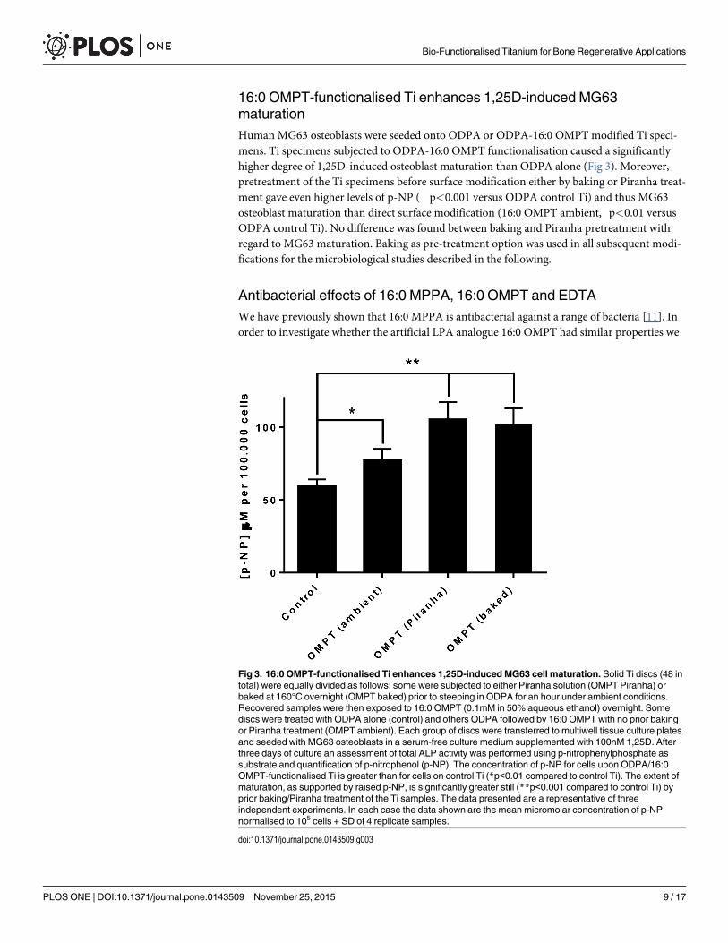
16:0 OMPT-functionalised Ti enhances 1,25D-induced MG63maturationHuman MG63 osteoblasts were seeded onto ODPA or ODPA-16:0 OMPT modified Ti speci-mens. Ti specimens subjected to ODPA-16:0 OMPT functionalisation caused a significantlyhigher degree of 1,25D-induced osteoblast maturation than ODPA alone (Fig 3). Moreover,pretreatment of the Ti specimens before surface modification either by baking or Piranha treat-ment gave even higher levels of p-NP (��p<0.001 versus ODPA control Ti) and thus MG63osteoblast maturation than direct surface modification (16:0 OMPT ambient, �p<0.01 versusODPA control Ti). No difference was found between baking and Piranha pretreatment withregard to MG63 maturation. Baking as pre-treatment option was used in all subsequent modi-fications for the microbiological studies described in the following.
Antibacterial effects of 16:0 MPPA, 16:0 OMPT and EDTAWe have previously shown that 16:0 MPPA is antibacterial against a range of bacteria [11]. Inorder to investigate whether the artificial LPA analogue 16:0 OMPT had similar properties we
Fig 3. 16:0 OMPT-functionalised Ti enhances 1,25D-induced MG63 cell maturation. Solid Ti discs (48 intotal) were equally divided as follows: some were subjected to either Piranha solution (OMPT Piranha) orbaked at 160°C overnight (OMPT baked) prior to steeping in ODPA for an hour under ambient conditions.Recovered samples were then exposed to 16:0 OMPT (0.1mM in 50% aqueous ethanol) overnight. Somediscs were treated with ODPA alone (control) and others ODPA followed by 16:0 OMPT with no prior bakingor Piranha treatment (OMPT ambient). Each group of discs were transferred to multiwell tissue culture platesand seeded with MG63 osteoblasts in a serum-free culture medium supplemented with 100nM 1,25D. Afterthree days of culture an assessment of total ALP activity was performed using p-nitrophenylphosphate assubstrate and quantification of p-nitrophenol (p-NP). The concentration of p-NP for cells upon ODPA/16:0OMPT-functionalised Ti is greater than for cells on control Ti (*p<0.01 compared to control Ti). The extent ofmaturation, as supported by raised p-NP, is significantly greater still (**p<0.001 compared to control Ti) byprior baking/Piranha treatment of the Ti samples. The data presented are a representative of threeindependent experiments. In each case the data shown are the mean micromolar concentration of p-NPnormalised to 105 cells + SD of 4 replicate samples.
doi:10.1371/journal.pone.0143509.g003
Bio-Functionalised Titanium for Bone Regenerative Applications
PLOS ONE | DOI:10.1371/journal.pone.0143509 November 25, 2015 9 / 17

exposed the MRSA clinical strain 43484 to serial dilutions of 16:0 OMPT and 16:0 MPPA andobserved growth. We also included the chelator EDTA. Fig 4 shows that 200 μg/mL of 16:0MPPA, 16:0 OMPT or EDTA (corresponding to 0.4 mMMPPA or OMPT or 0.5 mM EDTA)completely abolished growth of the MRSA strain. We found that the MRSA strain was muchmore sensitive to 16:0 MPPA and 16:0 OMPT in minimal media (Fig 5), where 25 microgram/mL of either 16:0 MPPA or 16:0 OMPT was enough to inhibit growth. We further investigatedthe effect of media composition. Fig 6 shows that addition of 2 mM Ca2+ abolished the growthinhibitory effect of 16:0 MPPA and EDTA while 16:0 OMPT still inhibited growth in the pres-ence of extra calcium.
Flow cytometryTo investigate whether 16:0 OMPT and 16:0 MPPA were bactericidal or bacteriostatic weinvestigated the uptake of the nonviable dye propidium iodide (PI) by MRSA grown in the
Fig 4. Lysophosphatidic acid species inhibit the growth of MRSA in Luria broth. The effect of different concentrations of 16:0 MPPA, 16:0 OMPT andEDTA on growth of MRSA 43484 in Luria broth. Legend: 16:0 MPPA; black (control), red (1,56 microgram/mL), green (6,3 microgram/mL), blue (25microgram/mL). 16:0 OMPT; black (control), red (1,56 microgram/mL), green (6,3 microgram/mL), blue (25 microgram/mL). EDTA; black (control), green (6,3microgram/mL), blue (25 microgram/mL), grey (0.1mg/mL).
doi:10.1371/journal.pone.0143509.g004
Fig 5. Lysophosphatidic acid species inhibit the growth of MRSA in minimal media. The effect of different concentrations of 16:0 MPPA, 16:0 OMPTand EDTA on growth of MRSA 43484 in minimal media. Legend: 16:0 MPPA; black (control), red (1,56 microgram/mL), green (6,3 microgram/mL), blue (25microgram/mL). 16:0 OMPT; black (control), red (1,56 microgram/mL), green (6,3 microgram/mL), blue (25 microgram/mL). Notice that the symbols of thecontrol almost hides the symbols of 6,3 microgram/mL EDTA.
doi:10.1371/journal.pone.0143509.g005
Bio-Functionalised Titanium for Bone Regenerative Applications
PLOS ONE | DOI:10.1371/journal.pone.0143509 November 25, 2015 10 / 17

presence of these compounds. The concentration of MPPA, OMPT and EDTA were adjustedto achieve limited growth; that is OD600 1.5 compared to untreated bacteria that achieved anOD600 of 3.0. Fig 7 shows that the three compounds in the concentrations tested caused anincreased uptake of PI, suggesting all three compounds to be bactericidal.
Adhesion of MRSA to 16:0 OMPT and 16:0 MPPA functionalised TidiscsAiming to apply the 16:0 OMPT and 16:0 MPPA to functionalise Ti for implant use we investi-gated the adhesion of MRSA strain 43484 to 16:0 MPPA or 16:0 OMPT functionalised Ti discs.Fig 8A shows that both 16:0 MPPA and 16:0 OMPT reduced adhesion of MRSA to Ti discs.Functionalisation using a linker such as ODPA should make the binding of compounds to thesurface very durable through interaction of the hydrophobic alkyl chains of both the ODPAand the LPA/LPA analogue. To test this, we reused the Ti discs in a new experiment after care-ful rinsing and autoclaving of the Ti discs. Fig 8B shows that in the reuse experiment the anti-adhesive effect reached the same level as the first round of the experiment. As mentioned abovewe found that adding Ca2+ abolished the growth inhibitory effect of 16:0 MPPA, and wewanted to investigate whether extra Ca2+ would also affect the anti-adhesive effect of 16:0
Fig 6. Growth of MRSA in response to lysophosphatidic acids and the influence of calcium.Growth curves of MRSA 43484 in minimal media showingthe effect of Ca2+. Black (control), red (control + 2 mM Ca2+), green (100 μg/mL 16:0 MPPA/16:0 OMPT/EDTA), Blue 0.1mg/mL 16:0 MPPA/16:0 OMPT/EDTA + 2 mMCa2+).
doi:10.1371/journal.pone.0143509.g006
Fig 7. Flow cytometry of MRSA stained with PI: effect of lysophosphatidic acid species and EDTA on viability.Histograms of MRSA 43484 stainedwith propidium iodide (PI), gated on forward and side scatter. Bacteria were grown in the presence of 0.1mg/mL 16:0 MPPA, 50 microgram/mL 16:0 OMPT,0.1mg/mL EDTA or in the absence of compounds (control). Markers show the percentage of PI stained, and hence nonviable, population.
doi:10.1371/journal.pone.0143509.g007
Bio-Functionalised Titanium for Bone Regenerative Applications
PLOS ONE | DOI:10.1371/journal.pone.0143509 November 25, 2015 11 / 17

MPPA and/or 16:0 OMPT. Fig 8C shows that adding 2 mM Ca2+ did not affect the anti-adhe-sive properties of 16:0 MPPA or 16:0 OMPT functionalisation. Fig 8D–8G shows that growthin the media above the functionalised Ti discs (Fig 8A–8C) were not affected, suggesting thatlow/no 16:0 MPPA or 16:0 OMPT was released from the Ti discs into the media.
Scanning Electron MicroscopyScanning Electron Microscopy (SEM) was performed on functionalised Ti discs with adherentMRSA bacteria. Examples of resulting images are shown in Fig 9. Manual counting of adherentbacteria/micrometerm2 showed that significantly fewer bacteria adhered to the 16:0 OMPTand 16:0 MPPA functionalised Ti compared to the control. Interestingly, the images showedthat while the bacteria adhering to the control Ti discs had a very rough appearance, bacteriaadhering the 16:0 OMPT and especially the 16:0 MPPA functionalised Ti discs appearedsmooth and with less extracellular matrix material.
DiscussionDeveloping novel biomaterial coatings that enhance early osseointegration yet deter the attach-ment of bacteria are especially desirable properties for future orthopaedic and dental implantdevices. The simple pleiotropic lipid, LPA and/or selected LPA analogues, could represent can-didate molecules for coating Ti. Over a decade ago Laux and colleagues [9] described how 16:0MPPA (palmitoyl LPA) inhibited the growth and virulence of P. aeruginosa. 16:0 MPPA is a
Fig 8. Lysophosphatidiate-functionalised titanium inhibits MRSA adhesion. Effect of functionalised Ti on bacterial adhesion. A-C) CFU of adherentMRSA pr. Ti disc after detachment. A) CFU pr. Ti disc; control or functionalised with either 16:0 OMPT or 16:0 MPPA. B) *Reuse of the same discs as in A)after washing and autoclaving. C) Effect of addition of 2 mM Ca2+ on MRSA adhesion to Ti discs. In each instance four discs were used for each of thedifferent Ti surface treatments. D-F) End point OD450 of planktonic bacteria in the wells containing the Ti discs in A-C. (*p<0.05, **p<0.005 and ***p<0.0005compared to control). Each of the figures is a representative of four independent experiments.
doi:10.1371/journal.pone.0143509.g008
Bio-Functionalised Titanium for Bone Regenerative Applications
PLOS ONE | DOI:10.1371/journal.pone.0143509 November 25, 2015 12 / 17

naturally occurring LPA found in biological fluids including bronchoalveolar lavage (BAL),serum and plasma [21–23]. Interestingly, exposure of bacteria to this lipid made them increas-ingly susceptible to certain antibiotics. This particular lipid species sits amongst several struc-turally similar lysophospholipids that serve as signalling molecules to most mammalian celltypes [24] In the context of human osteoblast biology LPAs co-operate with 1,25D to enhancetheir maturation [3,4] an event synonymous with bone tissue formation. Given the reported
Fig 9. Lysophosphatidate-functionalisation of titanium deters bacterial attachment and alters bacterial surface morphology. A. Scanning ElectronMicroscopy images of MRSA 43484 on control and functionalised Ti surfaces. The left column shows unmodified Ti, in the centre, Ti functionalised with 16:0OMPT, and in the right column images of bacteria upon 16:0 MPPA functionalised Ti. B.Graph depicting the data obtained for the number of bacteriaassociated for each of the different Ti surfaces.
doi:10.1371/journal.pone.0143509.g009
Bio-Functionalised Titanium for Bone Regenerative Applications
PLOS ONE | DOI:10.1371/journal.pone.0143509 November 25, 2015 13 / 17

effects of 16:0 MPPA on bacteria suggest LPA/LPA analogues as potential adjuncts in a boneregenerative setting and one way in which this could be realised is by coating biomaterials withLPA/LPA analogues. Such a “dual-action” coating would be particularly appealing to reducepotential infection risk of implantable devices whilst encouraging superior earlyosseointegration.
Human osteoblasts and bone marrow stromal cells express at least three different LPAreceptor types. When stimulated with LPA/LPA analogues effects ranging from proliferation,fibronectin binding, cytoskeletal reorganization and differentiation have been reported [2]. Inthis particular investigation we found that 16:0 OMPT stimulated MG63 cell growth. Whenthe same cells were co-stimulated with 1,25D and 16:0 OMPT cell growth was modestly attenu-ated and there was a synergistic increase in total ALP activity indicating a change towards amore mature or differentiated phenotype. All of these changes essentially mirror the effects wehave seen for other LPA species on this cell type. In this study we also show that the functiona-lisation of Ti with 16:0 OMPT, enhances 1,25D-induced MG63 maturation, as supported bythe greater total ALP activity. The effect of 16:0 OMPT functionalisation on maturation ofMG63 is particularly encouraging; mature osteoblasts are responsible for bone matrix synthesisand mineralisation and if these events can be enhanced at the Ti surface then this could facili-tate the process of early osseointegration.
We have previously shown that 16:0 MPPA sensitises otherwise betalactam resistant P. aer-uginosa to the actions of ampicillin [9,10]. We have also shown that this LPA is antibacterialagainst a number of Gram positive bacteria including S. aureus [11]. Here we further show thatthe phosphatase resistant LPA analogue, 16:0 OMPT, is antibacterial against MRSA strain43484. The mechanism by which the two compounds exert their effects is unclear. We havepreviously reported that 16:0 MPPA precipitates in the presence of the divalent cations calciumand magnesium and that precipitation is most noticeable with the former ion. These observa-tions imply that 16:0 MPPA can function as a chelator [10]. Due to the clear structural similari-ties between 16:0 MPPA and 16:0 OMPT, it is likely that 16:0 OMPT is also capable of formingchelating micelles in the presence of divalent cations. In 1968 it was reported that EDTAreversed ampicillin resistance in P. aeruginosa; the minimum inhibitory concentrations (MIC)of ampicillin was reduced from 0.5mg/mL to less than 2 microgram/mL in the presence of 3.4mM EDTA [25]. The lipopolysaccharide (LPS) layer in Gram negative bacteria such as P. aeru-ginosa are stabilised by divalent cations (mainly Ca2+). If calcium ions are removed by treat-ment with a chelator, LPS is liberated, and through this disruption the membrane becomesmore permeable to other agents, causing a potentiating action (reviewed in [26]). Instead ofLPS, Gram positive bacteria have surface sugars, phosphate groups and basic residues orga-nised as teichoic acid. S. aureus teichoic acids are composed of alternating phosphate and ribi-tol or glycerol groups, and modified with D-alanine and N-acetylglucosamine. Teichoic acidsare important for binding of Mg2+ to the S. aureus cell wall [27] and possibly other divalent cat-ions such as Ca2+. The cell surface of S. aureus has a moderately negative net charge at neutralpH, which is probably due to the fact that the teichoic acid contain less positively charged D-alanine residues than negatively charged [28]. The charge of teichoic acid has been reported tobe important for primary adhesion of bacteria to a substrate surface [29], which is the initialstep of biofilm formation. Moreover, the negative charge have been reported to play a role forresistance against glycopeptide antibiotics [30]. It is thus possible that the chelating ability of16:0 OMPT and 16:0 MPPA is important for their antibacterial activity through membranedestabilisation. On the other hand, chelators could also cause simple growth inhibition bydepriving the bacteria of essential cations. To investigate this we performed flow cytometryanalyses (Fig 7), which revealed that a high proportion of the cultures treated with 16:0 MPPAand 16:0 OMPT stained positive with PI suggesting that the two compounds directly kill the
Bio-Functionalised Titanium for Bone Regenerative Applications
PLOS ONE | DOI:10.1371/journal.pone.0143509 November 25, 2015 14 / 17

bacteria, and not just reduce proliferation. Interestingly we found that the while the effect ongrowth of 16:0 MPPA and EDTA was abolished by addition of 2 mM Ca2+, 16:0 OMPT stillinhibited growth in the presence of extra calcium (Fig 6).
The antibacterial and osteoblast maturation enhancing activity of the LPA (analogue) com-pounds lead us to investigate bacterial adhesion to 16:0 MPPA and 16:0 OMPT functionalisedTi discs. S. aureus adhesion to the functionalised Ti discs were significantly reduced, however,growth in the media above the functionalised Ti discs were not affected (Fig 8). Moreover, theantiadhesive effect was not reduced in experiments with reuse of functionalised Ti discs. Thus,it seems that only very low or no 16:0 MPPA or 16:0 OMPT were released from the Ti discsinto the media. As we found that addition of extra calcium abolished the growth inhibitoryeffect of 16:0 MPPA, we also investigated the effect of adding additional calcium to the mediain which the functionalised Ti discs were placed. However the extra added calcium did notalter bacterial adhesion to neither 16:0 MPPA nor 16:0 OMPT-functionalised surfaces.
We also performed SEM on functionalised Ti discs with adherent MRSA bacteria. In accor-dance with findings obtained using the CFU method, we found that significantly fewer bacteriaadhered to the 16:0 OMPT and 16:0 MPPA functionalised Ti compared to the control. TheSEM images revealed an interesting and clear difference in morphology between the bacteriaadhering to the control, which had a very uneven and coarse surface, and bacteria adhering the16:0 OMPT and especially the 16:0 MPPA functionalised Ti discs, which appeared smooth andwith less extracellular matrix material. This suggests that 16:0 OMPT and 16:0 MPPA affectthe teichoic acid layer, destabilising the bacterial outer membrane and thereby affecting bothsurface attachment and viability.
Whilst our findings offer a novel route to the biological modification of Ti we are cognisantthat considerably more research will be required to optimise the surface coating. In additionsteps to ascertain coating stability and robustness will be priority areas in realising the appli-cation of this technology for bone regenerative applications. Specifically an assessment ofcoating persistence to washing, sterilisation and the physical forces encountered duringimplantation will need to be part of a wider research plan prior to any in vivo evaluation inthe future.
ConclusionTo summarise we present evidence for the successful bio-functionalisation of Ti with 16:0MPPA/OMPT by adopting the facile pre-attachment of an APA via the natural oxide layerof Ti. This surface finish exhibited two noteworthy properties that may help realise the appli-cation of selected LPAs in future bone biomaterial design; in the first instance the surfacesenhanced 1,25D-induced hOB maturation. In addition this same surface deterred the attach-ment of S.aureus and there were noticeable differences in bacterial surface morphologypointing to the possible loss of teichoic acid. Collectively our findings point towards thedevelopment of a “dual action” Ti device of which similar technologies have yet to bereported.
AcknowledgmentsThe authors wish to thank the Core Facility for Integrated Microscopy, Faculty of Health andMedical Sciences, University of Copenhagen for technical assistance in connection to SEM. Wewould also like to thank Dr. Charl Faul, University of Bristol, for his assistance with Piranhasolution preparation and handling. The authors report no conflict of interest.
Bio-Functionalised Titanium for Bone Regenerative Applications
PLOS ONE | DOI:10.1371/journal.pone.0143509 November 25, 2015 15 / 17
Author ContributionsConceived and designed the experiments: MES KAK GJ JPM. Performed the experiments:MES GJ JPM. Analyzed the data: MES KAK AB GJ GDP JPM. Contributed reagents/materials/analysis tools: MES KAK AB GJ GDP JPM. Wrote the paper: MES JPM.
References1. Choi JW, Herr DR, Noguchi K, Yung YC, Lee CW, Mutoh T, et al. LPA receptors: subtypes and biologi-
cal actions. Annu. Rev. Pharmacol. Toxicol. 2010; 50: 157–186. doi: 10.1146/annurev.pharmtox.010909.105753 PMID: 20055701
2. Blackburn J, Mansell JP. The emerging role of lysophosphatidic acid (LPA) in skeletal biology. Bone.2012; 50: 756–762. doi: 10.1016/j.bone.2011.12.002 PMID: 22193551
3. Gidley J, Openshaw S, Pring ET, Sale S, Mansell JP. Lysophosphatidic acid cooperates with1alpha,25(OH)2D3 in stimulating humanMG63 osteoblast maturation. Prostaglandins Other Lipid Med-iat. 2006; 80: 46–61. PMID: 16846786
4. Mansell JP, Blackburn J. Lysophosphatidic acid, human osteoblast formation, maturation and the roleof 1α,25-Dihydroxyvitamin D3 (calcitriol). Biochim. Biophys. Acta. 2013; 1831: 105–108. doi: 10.1016/j.bbalip.2012.04.005 PMID: 22561288
5. Lancaster ST, Blackburn J, Blom A, Makishima M, Ishizawa M, Mansell JP. 24,25-Dihydroxyvitamin D3cooperates with a stable, fluoromethylene LPA receptor agonist to secure human (MG63) osteoblastmaturation. Steroids. 2014; 83: 52–61. doi: 10.1016/j.steroids.2014.01.010 PMID: 24513053
6. Whyte MP. Physiological role of alkaline phosphatase explored in hypophosphatasia. Ann. N. Y. Acad.Sci. 2010; 1192: 190–200. doi: 10.1111/j.1749-6632.2010.05387.x PMID: 20392236
7. Bonewald LF, Kester MB, Schwartz Z, Swain LD, Khare A, Johnson TL, et al. Effects of combiningtransforming growth factor beta and 1,25-dihydroxyvitamin D3 on differentiation of a human osteosar-coma (MG-63). J. Biol. Chem. 1992; 267: 8943–8949. PMID: 1577731
8. Yarram SJ, Tasman C, Gidley J, Clare M, Sandy JR, Mansell JP. Epidermal growth factor and calcitriolsynergistically induce osteoblast maturation. Mol. Cell. Endocrinol. 2004; 220: 9–20. PMID: 15196695
9. Laux DC, Corson JM, Givskov M, Hentzer A, Moller A, Wosencroft KA, et al. Lysophosphatidic acidinhibition of the accumulation of Pseudomonas aeruginosa PAO1 alginate, pyoverdin, elastase andLasA. Microbiology. 2002; 148: 1709–1723. PMID: 12055291
10. Krogfelt KA, Utley M, Krivan HC, Laux DC, Cohen PS. Specific phospholipids enhance the activity ofbeta-lactam antibiotics against Pseudomonas aeruginosa. J. Antimicrob. Chemother. 2000; 46: 377–384. PMID: 10980163
11. Cohen PS, Krogfelt KA, Laux DC, Utley M. Phospholipids having antimicrobial activity with the pres-ence of antimicrobials EP1051180. Patent (2002).
12. Queffélec C, Petit M, Janvier P, Knight DA, Bujoli B. Surface modification using phosphonic acids andesters. Chem. Rev. 2012; 112: 3777–3807. doi: 10.1021/cr2004212 PMID: 22530923
13. Paramonov PB, Paniagua SA, Hotchkiss PJ, Jones SC, Armstrong NR, Marder SR, et al. TheoreticalCharacterization of the Indium Tin Oxide Surface and of Its Binding Sites for Adsorption of PhosphonicAcid Monolayers. Chem. Mater. 2008; 20: 5131–5133.
14. Paz Y. Self-assembled monolayers and titanium dioxide: From surface patterning to potential applica-tions. Beilstein J. Nanotechnol. 2011; 2: 845–861. doi: 10.3762/bjnano.2.94 PMID: 22259769
15. Qian L, Xu Y, Hasegawa Y, Aoki J, Mills GB, Prestwich GD. Enantioselective responses to a phosphor-othioate analogue of lysophosphatidic acid with LPA3 receptor-selective agonist activity. J. Med.Chem. 2003; 46: 5575–5578. PMID: 14667211
16. Brindley DN, Pilquil C. Lipid phosphate phosphatases and signaling. J. Lipid Res. 2009; 50: SupplS225–S230. doi: 10.1194/jlr.R800055-JLR200 PMID: 19066402
17. Mansell JP, Brown J, Knapp JG, Faul CFJ, Blom AW. Lysophosphatidic acid-functionalised titanium asa superior surface for supporting human osteoblast (MG63) maturation. Eur. Cell. Mater. 2012; 23:348–361. PMID: 22573454
18. Clover J, GowenM. Are MG-63 and HOS TE85 human osteosarcoma cell lines representative modelsof the osteoblastic phenotype? Bone 1994; 15: 585–591. PMID: 7873286
19. Lancaster S, Mansell JP. The role of lysophosphatidic acid on human osteoblast formation, maturationand the implications for bone health and disease. Clin. Lipidol. 2013; 8: 123–135.
Bio-Functionalised Titanium for Bone Regenerative Applications
PLOS ONE | DOI:10.1371/journal.pone.0143509 November 25, 2015 16 / 17

20. Vingsbo Lundberg C, Frimodt-Møller N. Efficacy of topical and systemic antibiotic treatment of meticil-lin-resistant Staphylococcus aureus in a murine superficial skin wound infection model. Int. J. Antimi-crob. Agents. 2013; 42: 272–275. doi: 10.1016/j.ijantimicag.2013.05.008 PMID: 23837927
21. Saga H, Ohhata A, Hayashi A, Katoh M, Maeda T, Mizuno H,Y. et al. A novel highly potent autotaxin/ENPP2 inhibitor produces prolonged decreases in plasma lysophosphatidic acid formation in vivo andregulates urethral tension. PLoS One. 2014; 9: e93230. doi: 10.1371/journal.pone.0093230 PMID:24747415
22. Tokumura A, Carbone LD, Yoshioka Y, Morishige J, Kikuchi M, Postlethwaite A, et al. Elevated serumlevels of arachidonoyl-lysophosphatidic acid and sphingosine 1-phosphate in systemic sclerosis. Int. J.Med. Sci. 2009; 6: 168–176. PMID: 19521548
23. Park GY, Lee YG, Berdyshev E, Nyenhuis S, Du J, Fu P, et al. Autotaxin production of lysophosphatidicacid mediates allergic asthmatic inflammation. Am. J. Respir. Crit. Care Med. 2013; 188: 928–940. doi:10.1164/rccm.201306-1014OC PMID: 24050723
24. MoolenaarWH, vanMeeteren LA, Giepmans BNG. The ins and outs of lysophosphatidic acid signaling.Bioessays. 2004; 26: 870–881. PMID: 15273989
25. Weiser R, Asscher AW, Wimpenny J. In vitro reversal of antibiotic resistance by ethylenediamine tetra-acetic acid. Nature. 1968; 219: 1365–1366. PMID: 4971170
26. Lambert RJW, Hanlon GW, Denyer SP. The synergistic effect of EDTA/antimicrobial combinations onPseudomonas aeruginosa. J. Appl. Microbiol. 2004; 96: 244–253. PMID: 14723685
27. Heptinstall S, Archibald AR, Baddiley J. Teichoic acids and membrane function in bacteria. Nature.1970; 225: 519–521. PMID: 5411858
28. Peschel A, Otto M, Jack RW, Kalbacher H, Jung G, Götz F. Inactivation of the dlt operon in Staphylo-coccus aureus confers sensitivity to defensins, protegrins, and other antimicrobial peptides. J. Biol.Chem. 1999; 274: 8405–8410. PMID: 10085071
29. Gross M, Cramton SE, Götz F, Peschel A. Key role of teichoic acid net charge in Staphylococcusaureus colonization of artificial surfaces. Infect. Immun. 2001; 69: 3423–3426. PMID: 11292767
30. Peschel A, Vuong C, Otto M, Götz F. The D-alanine residues of Staphylococcus aureus teichoic acidsalter the susceptibility to vancomycin and the activity of autolytic enzymes, Antimicrob. Agents Che-mother. 2000; 44: 2845–2847.
Bio-Functionalised Titanium for Bone Regenerative Applications
PLOS ONE | DOI:10.1371/journal.pone.0143509 November 25, 2015 17 / 17