six keys to effectively using alveolar corticotomy: a
TRANSCRIPT
1 2018
Six keys to effectively using alveolar corticotomy: A different perspective on surgically assisted tooth movementDr Raffaele Spena, Italy
Introduction
Alveolar decortication (corticotomy) has long been used with orthodontic treatment in order to accelerate orthodontic tooth movement (OTM) while reducing the undesired effects of root resorption, loss of vitality, peri-odontal problems and relapse of the corrections. The acceleration of tooth movement should shorten the ther-apy. However, the scientific and clinical assumptions of the early days were totally different from the more recent ones: we moved from a pure mechanical approach to a biological and physiological one.
In 1983, Suya1 proposed a great improvement of the surgical approach described in 1959 by Kole2 modifying the horizontal osteotomy in a corticotomy, avoiding the alveolar crest in the vertical cuts and eliminating the luxation of the blocks. He proposed this “corticotomy- facilitated orthodontics” to treat adult patients, anky-losed teeth and crowded malocclusions to avoid premo-lar extractions. Like Kole, Suya believed he was creating bony blocks and suggested accomplishing most of the movements in the first three to four months of treatment before the fusion of the blocks (healing of the bone).
The concept of corticotomy-assisted OTM drastically changed in 2001 after the publication of Wilcko et al.3 In this key case report, two adult patients received a selec-tive corticotomy, along with alloplastic resorbable grafts, to increase the bone level and avoid the risk of reces-sions. An accurate evaluation with CT scans before and after treatment, and histological sections in one case, allowed the authors to formulate a new hypothesis about what really happens at the bone level after corticotomy. No movement of tooth–bone blocks, but a transient reduction of mineralisation of the alveolar bone and mod-ifications similar to those described by Frost4–7 during the healing of fractured bones and named “regional acceler-atory phenomenon” (RAP) most likely occur. The surgery -orthodontic protocol proposed by Wilcko et al.3 has been subsequently patented as Periodontally Accelerated Osteogenic Orthodontics (PAOO). The claims of PAOO are
(a) accelerated tooth movement with reduction of the total treatment time; (b) osteogenic modifications with trans-portation of the bony matrix, and final improvement of hard- and soft-tissue support of the teeth treated ortho-dontically; (c) increase of the short- and long-term stabil-ity of the orthodontic treatment. So far, scientific evidence has been given only on the acceleration of tooth move-ment that is transient, and lasts as long as there is a RAP modification in the alveolar bone surrounding the teeth.
After more than one and a half decades of clinical experience with alveolar corticotomy, in light of the cur-rent literature published on this topic, six rules have been established that should be taken into account when con-sidering using alveolar corticotomy in a complex ortho-dontic case. These keys are the best way to ensure effec-tiveness and reduce the risk of producing no positive effect or, worse, causing damage. The six keys are as follows:
1. Alveolar corticotomy is to facilitate OTM.2. Alveolar corticotomy has limited effect in time.3. Alveolar corticotomy has limited effect in space.4. A proper surgical procedure must be followed.5. Proper orthodontic management after corticotomy
must be performed.6. Proper patient selection for corticotomy is essential.
A detailed description of each rule follows.
1. Alveolar corticotomy is to facilitate orthodontic tooth movement (Periodontally Facilitated Orthodontics)
Speed is a fascinating issue in life. We like to go fast in cars, motorbikes, boats, airplanes and so forth. Speed in orthodontics is a different matter. It is one of the main objectives of modern orthodontics to reduce treatment time, but we must recognise that a great number of vari-ables may affect it.8–11
The initial difficulty of the malocclusion and tooth mal-position, the age of the patient, the variability of the indi-vidual response to the treatment, the quality of the end
20 ortho
| research
1 2018
result, and the patient’s compliance are just a few of the variables that should be considered. Numerous case reports have been published showing how treatment time can be reduced when patients are treated with cor-ticotomy. Case reports, however, have limited scientific validity.
The predictability and quantification of treatment time reduction are still not scientifically possible. The addi-tional expenses and morbidity associated with the use of alveolar corticotomy should always be carefully evaluated to determine whether they are worth the saving of few months. A shorter orthodontic treatment is desirable, but certainly not at the expense of a high- quality end result.
Regarding OTM, numerous studies have shown that its speed is influenced by bone turnover and the individual response to mechanical forces and it is not related to the level of the forces.12–15 Clinical experience confirms this: there are slow movers and fast movers, but we are still far from recognising them. In addition to this variability, there is the temporary effect of alveolar corticotomy, which we will discuss under the third key. A faster treatment may be a secondary advantage and may be obtained in a sub-stantial way only in those “simple” orthodontic cases that require a naturally short treatment.
In conclusion, alveolar decortication should not be combined with orthodontic treatment with the only objec-tive of accelerating OTM and reducing treatment time: the risk of not obtaining either as desired may be high.
Despite this scientific evidence against its major claims, alveolar corticotomy has its place in orthodontic therapy. Let us consider the surgical insult and the associated RAP reaction produced at a biomechanical level: the increased metabolism, the transient reduced regional density (osteopenia) created by the increased osteoclas-tic activity, the reduced undermining resorption and hya-linisation (we still do not know exactly what happens in humans) facilitate OTM. The decorticated tooth is less resistant to orthodontic forces and will be easier to move and will require less anchorage.
Spena et al. in two studies conducted on a total of 12 adult patients with Class II malocclusions treated with distalisation of the maxillary molars showed how maxil-lary molars could be bodily distalised with simple buccal mechanics and no anterior anchorage.16, 17 Corticotomy was performed only on the teeth to be moved, thus reducing the anchorage needs and their resistance to distal forces.
The term “Periodontally Facilitated Orthodontics”, instead of “Periodontally Accelerated Osteogenic Ortho-dontics”, is used to describe a procedure that has the pri-
mary goal of simplifying, enhancing and improving OTMs that are difficult or risky, from a biomechanical and biolog-ical point of view. The surgical procedure and the asso-ciated orthodontic treatment and biomechanics depend on the initial problems and the goals of every single spe-cific treatment. This is in agreement with Oliveira et al.: corticotomies should be used to “…facilitate the imple-mentation of mechanically challenging orthodontic movements and enhance the correction of moderate to severe skeletal malocclusions”.18
2. Alveolar corticotomy has limited effect in time
Since the early studies of Frost on the biology of frac-ture healing, it is known that the altered metabolism of bone after a traumatic (or surgical) event has lim-ited duration: it is the natural search for equilibrium or homeostasis.
The burst of hard- and soft-tissue remodelling starts a few days after the insult, peaks at the first or second month, and returns to a normal pace after a maximum of four to six months. This RAP reaction, when applied to the alveolar bone, causes an accelerated/facilitated movement of the teeth subjected to applied orthodontic forces. The effect lasts for as long as there is this reac-tion, so for a limited part of an orthodontic therapy. This has been confirmed by experimental studies on animals and by clinical studies on patients.19 Clinically, this tem-porary phenomenon leads to the need to perform the alveolar corticotomy when the RAP is necessary. Timing is fundamental.
Alveolar corticotomy may be repeated during the treatment with the objective of prolonging the effect.20 The effective benefit, cost and risks must be taken into account. Sanjideh et al. in a split-mouth study on fox-hounds found that a second corticotomy performed after 28 days in the mandible produced a higher rate of tooth movement and a greater total tooth movement.21 How-ever, they concluded that proper timing for a second cor-ticotomy needed to be better determined.
Wilcko,22–24 Dibart25 and Murphy26, 27 claimed that con- tinuously activated orthodontic forces applied after decortication may maintain a constant mechanical stim-ulation, and allow a prolonged osteopenic state during which teeth can be moved rapidly.
In order to achieve this effect, they recommended seeing patients frequently (every two weeks) and con-tinuing the activation of the applied orthodontic forces. If not, remineralisation would complete the healing process and bring the bone metabolism to a normal level. It must be said that these claims have never been demonstrated either clinically or histologically.
21ortho
research |
1 2018
3. Alveolar corticotomy has limited effect in space
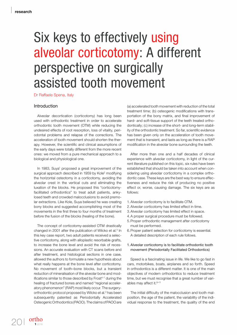
The effects of alveolar corticotomy are localised to the area immediately adjacent to the site of injury.28 This finding is of outmost importance. Different surgeries may affect differently the resulting OTM. Glenn et al.29 and Tuncay and Killiany,30 in two experimental studies on animals published before the new trend on corti-cotomy, found that fiberotomy (a corticotomy limited to the crestal side of the alveolar bone) affected the rate of OTM and shifted the centre of rotation toward the apex of the roots, thus modifying the biomechanical behaviour of the teeth under the orthodontic forces. If the surgical insult is applied to a limited area of the alveolar bone (i.e. middle third and only buccal surface; Fig. 1), the RAP reaction will not be extended to the entire root area. The modifications at the bone level will be limited at the area of the decortication, and control of the apical and lingual sides will not be influenced as desired.
As a general rule, if a mesiodistal bodily movement or better control of the apical area are the biomechanical needs of the OTM to be achieved and enhanced (i.e. intru-sion/extrusion), the decortication needs to be extended to the entire alveolar bone surrounding the roots of the teeth, buccally and lingually (Fig. 2); if the movement is less complex or anatomical limitations of the surgical site
impede an extended decortication, the cuts may be lim-ited in the direction of the OTM. These biomechanical needs determine the type of procedure in both the open-flap and the flapless surgeries.
4. A proper surgical procedure must be followed
Several surgical protocols for performing alveolar cor-ticotomy have been proposed. Most of them have been tried in the last 15 years on several patients. These sur-geries may be divided into two groups: the open-flap and the flapless corticotomies (Tab. 1).
The original corticotomies were performed after rais-ing a flap. This type of surgery is still preferred when an extended or critical area of decortication has to be man-aged and when an extended grafting is planned.
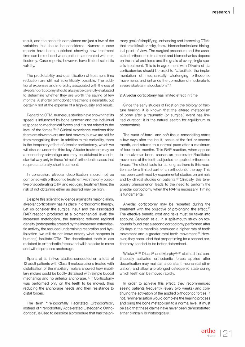
The flap can be designed according to the periodontal characteristics of the site and has to be full thickness in the area of decortication and split thickness below this area to ensure a good blood supply. Interproximal and subapical cuts of 1–2 mm in the cortical bone (Figs. 3 & 4) are performed together with a light scraping of the exter-nal cortex in between the cuts. This extended surgical insult will produce a wide RAP reaction and prepare a bleeding bed for any grafting material eventually placed
Fig. 2Fig. 1
Fig. 3 Fig. 4
22 ortho
| research
1 2018
in association with the decortication. Piezo- surgical calibrated micro-saws are preferred to rotating surgical burs because of their selective, safer, micrometric and more precise cuts; better irrigation/cooling effect from cavitation; better comfort for the surgeon; and better healing for the patient. The open-flap corticotomy pro-cedure is routinely used during orthognathic surgery, when exposing impacted teeth, to treat transverse max-illary deficiencies and periodontally involved cases.
Flapless surgery has been proposed as an alternative way of performing a corticotomy. Corticision31 and Piezo-cision32 have been an attempt to reduce the invasive-ness of the decortication and the possible periodontal damage and postoperative discomfort with raising a flap. Even if attractive, they seem to have surgical and biome-chanical limitations.
The surgical limitations include risks when per-formed in crowded arches, limited visibility when pro-ducing the cuts, limitation of the cuts to the inter-proximal areas and to the middle third of the roots, difficult control of the grafting in the apico - coronal direction and need for optimal extension of the attached gingiva in the area of decortication. The biome-chanical limitations are strictly related to the fact that cor-
ticotomy is performed only on the buccal side and middle third of the roots.
They are definitely not minimally invasive surgeries as claimed and are quite expensive for the patient, since only a well-trained periodontist/oral surgeon can perform them and they often require complex planning with digi-tally designed 3-D surgical guides.33
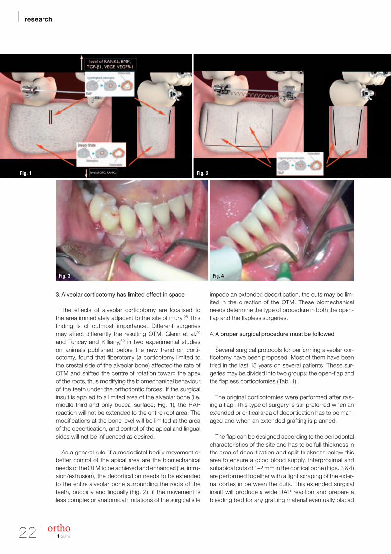
The Micro-Osteo-Perforations (MOPs) described by Alikhani et al.34 and Teixeira et al.35 are an effective and min-imally invasive way of producing insult to the cortical alveo-lar bone. These MOPs may be created with manual instru-ments (Excellerator, Propel Orthodontics) or with dedicated burs on a reduced-speed electric handpiece (Fig. 5).
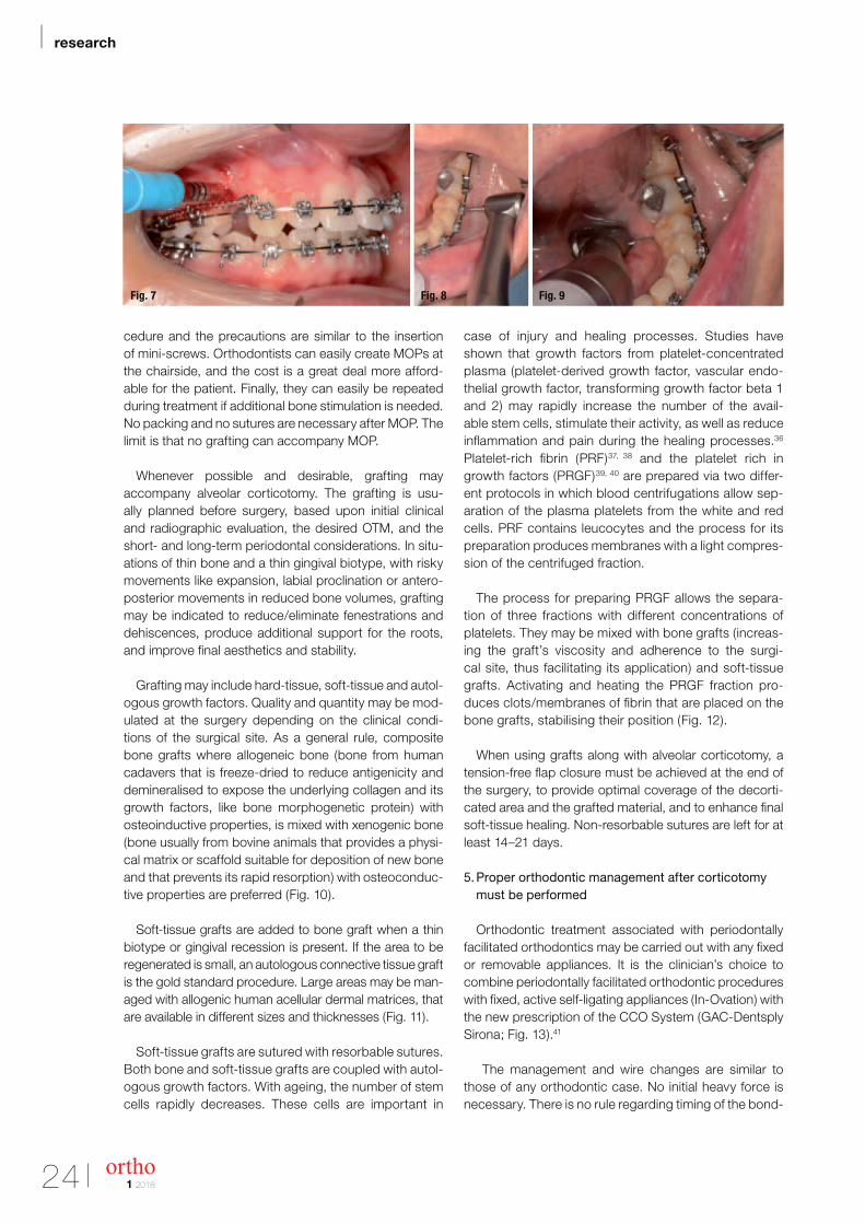
MOPs are produced with a penetration in the cortex of a maximum of 1–2 mm. Instead of conventional local anaesthesia, a strong anaesthetic gel placed on the mucosa for three minutes is sufficient to control the patient’s pain and discomfort. It is advisable to produce two to three MOPs in each interproximal area of the teeth and both buccally and lingually (Fig. 6), to ensure that the metabolic changes are extended around the entire radic-ular alveolar bone. Manual MOP is usually created in the frontal areas, whereas drilled MOP is usually performed in the posterior and lingual areas (Figs. 7–9). The pro-
Open-flap corticotomies Flapless corticotomies
· Periodontally Accelerated Osteogenic Orthodontics · Fiberotomy
· Segmental corticotomy · Corticision
· Any corticotomy performed during an open-flap surgery · Piezocision
· Micro-osteoperforations
Tab. 1: Surgical protocols for performing alveolar corticotomy.
Fig. 5 Fig. 6
23ortho
research |
1 2018
cedure and the precautions are similar to the insertion of mini-screws. Orthodontists can easily create MOPs at the chairside, and the cost is a great deal more afford-able for the patient. Finally, they can easily be repeated during treatment if additional bone stimulation is needed. No packing and no sutures are necessary after MOP. The limit is that no grafting can accompany MOP.
Whenever possible and desirable, grafting may accompany alveolar corticotomy. The grafting is usu-ally planned before surgery, based upon initial clinical and radiographic evaluation, the desired OTM, and the short- and long-term periodontal considerations. In situ-ations of thin bone and a thin gingival biotype, with risky movements like expansion, labial proclination or antero -posterior movements in reduced bone volumes, grafting may be indicated to reduce/eliminate fenestrations and dehiscences, produce additional support for the roots, and improve final aesthetics and stability.
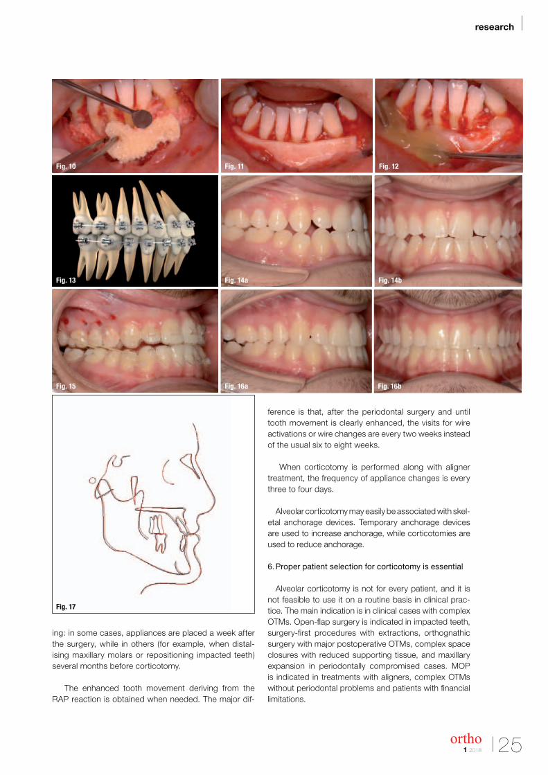
Grafting may include hard-tissue, soft-tissue and autol-ogous growth factors. Quality and quantity may be mod-ulated at the surgery depending on the clinical condi-tions of the surgical site. As a general rule, composite bone grafts where allogeneic bone (bone from human cadavers that is freeze-dried to reduce antigenicity and demineralised to expose the underlying collagen and its growth factors, like bone morphogenetic protein) with osteoinductive properties, is mixed with xenogenic bone (bone usually from bovine animals that provides a physi-cal matrix or scaffold suitable for deposition of new bone and that prevents its rapid resorption) with osteoconduc-tive properties are preferred (Fig. 10).
Soft-tissue grafts are added to bone graft when a thin biotype or gingival recession is present. If the area to be regenerated is small, an autologous connective tissue graft is the gold standard procedure. Large areas may be man-aged with allogenic human acellular dermal matrices, that are available in different sizes and thicknesses (Fig. 11).
Soft-tissue grafts are sutured with resorbable sutures. Both bone and soft-tissue grafts are coupled with autol-ogous growth factors. With ageing, the number of stem cells rapidly decreases. These cells are important in
case of injury and healing processes. Studies have shown that growth factors from platelet- concentrated plasma (platelet- derived growth factor, vascular endo-thelial growth factor, transforming growth factor beta 1 and 2) may rapidly increase the number of the avail-able stem cells, stimulate their activity, as well as reduce inflammation and pain during the healing processes.36 Platelet- rich fibrin (PRF)37, 38 and the platelet rich in growth factors (PRGF)39, 40 are prepared via two differ-ent protocols in which blood centrifugations allow sep-aration of the plasma platelets from the white and red cells. PRF contains leucocytes and the process for its preparation produces membranes with a light compres-sion of the centrifuged fraction.
The process for preparing PRGF allows the separa-tion of three fractions with different concentrations of platelets. They may be mixed with bone grafts (increas-ing the graft’s viscosity and adherence to the surgi-cal site, thus facilitating its application) and soft -tissue grafts. Activating and heating the PRGF fraction pro-duces clots/membranes of fibrin that are placed on the bone grafts, stabilising their position (Fig. 12).
When using grafts along with alveolar corticotomy, a tension-free flap closure must be achieved at the end of the surgery, to provide optimal coverage of the decorti-cated area and the grafted material, and to enhance final soft-tissue healing. Non-resorbable sutures are left for at least 14–21 days.
5. Proper orthodontic management after corticotomy must be performed
Orthodontic treatment associated with periodontally facilitated orthodontics may be carried out with any fixed or removable appliances. It is the clinician’s choice to combine periodontally facilitated orthodontic procedures with fixed, active self-ligating appliances (In-Ovation) with the new prescription of the CCO System (GAC-Dentsply Sirona; Fig. 13).41
The management and wire changes are similar to those of any orthodontic case. No initial heavy force is necessary. There is no rule regarding timing of the bond-
Fig. 7 Fig. 9Fig. 8
24 ortho
| research
1 2018
ing: in some cases, appliances are placed a week after the surgery, while in others (for example, when distal-ising maxillary molars or repositioning impacted teeth) several months before corticotomy.
The enhanced tooth movement deriving from the RAP reaction is obtained when needed. The major dif-
ference is that, after the periodontal surgery and until tooth movement is clearly enhanced, the visits for wire activations or wire changes are every two weeks instead of the usual six to eight weeks.
When corticotomy is performed along with aligner treatment, the frequency of appliance changes is every three to four days.
Alveolar corticotomy may easily be associated with skel-etal anchorage devices. Temporary anchorage devices are used to increase anchorage, while corticotomies are used to reduce anchorage.
6. Proper patient selection for corticotomy is essential
Alveolar corticotomy is not for every patient, and it is not feasible to use it on a routine basis in clinical prac-tice. The main indication is in clinical cases with complex OTMs. Open-flap surgery is indicated in impacted teeth, surgery-first procedures with extractions, orthognathic surgery with major postoperative OTMs, complex space closures with reduced supporting tissue, and maxillary expansion in periodontally compromised cases. MOP is indicated in treatments with aligners, complex OTMs without periodontal problems and patients with financial limitations.
Fig. 10 Fig. 11 Fig. 12
Fig. 13 Fig. 14a Fig. 14b
Fig. 15 Fig. 16a Fig. 16b
Fig. 17
25ortho
research |
1 2018
One case treated with open-flap corticotomy and two cases treated with MOP will be shown to elucidate the concepts described in this article.
Case 1A 19-year-old male patient with a Class III dental mal-
occlusion with anterior midline discrepancy wanted to be treated only with aligners (Figs. 14a & b). Treatment was carried out with 71 aligners and two MOPs performed at the second month and at the fifth month of treat-ment, only on the premolar and molar maxillary dentition (Fig. 15). Class III elastics were prescribed throughout the therapy. Treatment was completed in seven months with acceptable intercuspation in the buccal segments and correction of the midlines (Figs. 16a & b) and with good anchorage control in the lower arch (Fig. 17).
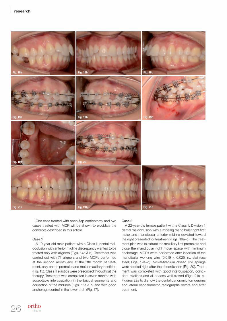
Case 2A 22-year-old female patient with a Class II, Division 1
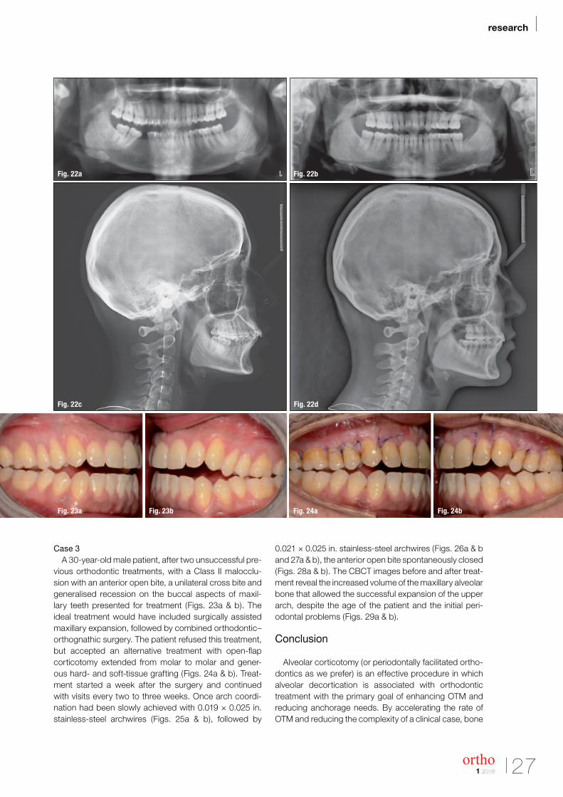
dental malocclusion with a missing mandibular right first molar and mandibular anterior midline deviated toward the right presented for treatment (Figs. 18a–c). The treat-ment plan was to extract the maxillary first premolars and close the mandibular right molar space with minimum anchorage. MOPs were performed after insertion of the mandibular working wire (0.019 × 0.025 in., stainless steel; Figs. 19a–d). Nickel-titanium closed coil springs were applied right after the decortication (Fig. 20). Treat-ment was completed with good intercuspation, coinci-dent midlines and all spaces well closed (Figs. 21a–c). Figures 22a to d show the dental panoramic tomograms and lateral cephalometric radiographs before and after treatment.
Fig. 18a Fig. 18b Fig. 18c
Fig. 19a Fig. 19b Fig. 19c
Fig. 19d Fig. 20
Fig. 21a Fig. 21b Fig. 21c
26 ortho
| research
1 2018
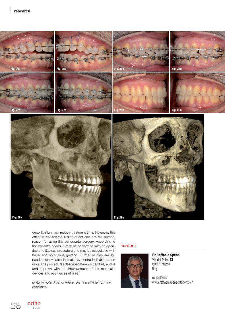
Case 3A 30-year-old male patient, after two unsuccessful pre-
vious orthodontic treatments, with a Class II malocclu-sion with an anterior open bite, a unilateral cross bite and generalised recession on the buccal aspects of maxil-lary teeth presented for treatment (Figs. 23a & b). The ideal treatment would have included surgically assisted maxillary expansion, followed by combined orthodontic–orthognathic surgery. The patient refused this treatment, but accepted an alternative treatment with open-flap corticotomy extended from molar to molar and gener-ous hard- and soft-tissue grafting (Figs. 24a & b). Treat-ment started a week after the surgery and continued with visits every two to three weeks. Once arch coordi-nation had been slowly achieved with 0.019 × 0.025 in. stainless-steel archwires (Figs. 25a & b), followed by
0.021 × 0.025 in. stainless-steel archwires (Figs. 26a & b and 27a & b), the anterior open bite spontaneously closed (Figs. 28a & b). The CBCT images before and after treat-ment reveal the increased volume of the maxillary alveolar bone that allowed the successful expansion of the upper arch, despite the age of the patient and the initial peri-odontal problems (Figs. 29a & b).
Conclusion
Alveolar corticotomy (or periodontally facilitated ortho-dontics as we prefer) is an effective procedure in which alveolar decortication is associated with orthodontic treatment with the primary goal of enhancing OTM and reducing anchorage needs. By accelerating the rate of OTM and reducing the complexity of a clinical case, bone
Fig. 22a Fig. 22b
Fig. 22c Fig. 22d
Fig. 23a Fig. 23b Fig. 24a Fig. 24b
27ortho
research |
1 2018
Dr Raffaele SpenaVia dei Mille, 1380121 NapoliItaly
contact
decortication may reduce treatment time. However, this effect is considered a side-effect and not the primary reason for using this periodontal surgery. According to the patient’s needs, it may be performed with an open-flap or a flapless procedure and may be associated with hard- and soft-tissue grafting. Further studies are still needed to evaluate indications, contra-indications and risks. The procedures described here will certainly evolve and improve with the improvement of the materials, devices and appliances utilised.
Editorial note: A list of references is available from the publisher.
Fig. 25a
Fig. 27a
Fig. 29a
Fig. 25b
Fig. 27b
Fig. 26a
Fig. 28a
Fig. 29b
Fig. 26b
Fig. 28b
28 ortho
| research