sip structural materials – platform of innovative
TRANSCRIPT
SIP Structural Materials – Platform ofInnovative Measurement and Analysis for Structural Materials (SIP-IMASM)
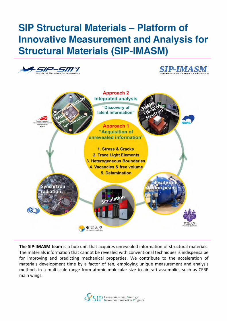
The SIP-IMASM team is a hub unit that acquires unrevealed information of structural materials.The materials information that cannot be revealed with conventional techniques is indispensalbefor improving and predicting mechanical properties. We contribute to the acceleration ofmaterials development time by a factor of ten, employing unique measurement and analysismethods in a multiscale range from atomic-molecular size to aircraft assemblies such as CFRPmain wings.
Concept and approach
The Subjects and Roles of the five institutes
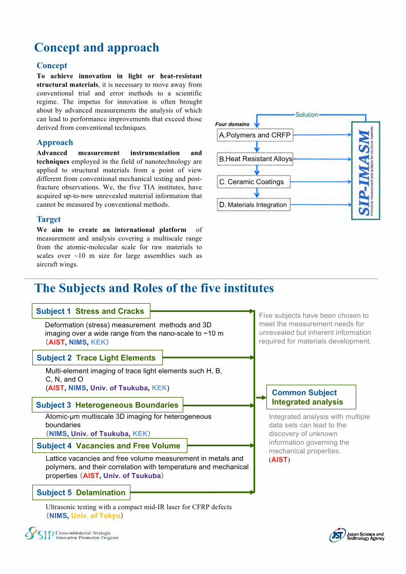
ConceptTo achieve innovation in light or heat-resistantstructural materials, it is necessary to move away fromconventional trial and error methods to a scientificregime. The impetus for innovation is often broughtabout by advanced measurements the analysis of whichcan lead to performance improvements that exceed thosederived from conventional techniques.
ApproachAdvanced measurement instrumentation andtechniques employed in the field of nanotechnology areapplied to structural materials from a point of viewdifferent from conventional mechanical testing and post-fracture observations. We, the five TIA institutes, haveacquired up-to-now unrevealed material information thatcannot be measured by conventional methods.
TargetWe aim to create an international platform ofmeasurement and analysis covering a multiscale rangefrom the atomic-molecular scale for raw materials toscales over ~10 m size for large assemblies such asaircraft wings.
Common SubjectIntegrated analysis
Subject 4 Vacancies and Free Volume
Subject 1 Stress and Cracks
Subject 2 Trace Light Elements
Subject 5 Delamination
Deformation (stress) measurement methods and 3D imaging over a wide range from the nano-scale to ~10 m (AIST, NIMS, KEK)
Multi-element imaging of trace light elements such H, B, C, N, and O(AIST, NIMS, Univ. of Tsukuba, KEK)
Atomic-µm multiscale 3D imaging for heterogeneous boundaries (NIMS, Univ. of Tsukuba, KEK)
Lattice vacancies and free volume measurement in metals and polymers, and their correlation with temperature and mechanical properties (AIST, Univ. of Tsukuba)
Ultrasonic testing with a compact mid-IR laser for CFRP defects(NIMS, Univ. of Tokyo)
Subject 3 Heterogeneous Boundaries
Five subjects have been chosen to meet the measurement needs for unrevealed but inherent information required for materials development.
Integrated analysis with multiple data sets can lead to the discovery of unknown information governing the mechanical properties.(AIST)
Polymers and CRFP
Heat Resistant Alloys
Ceramic Coatings
Materials Integration
Prospect for innovative measurement and analysis
Measurement and analysis are essential for designing structural materials. I hope that the SIP-IMASMteam plays an important role as the hub platform in order to change the conventional material designbased on experience-and-intuition into a scientific approach.
Teruo KishiProgram Director of the SIP-Structural Materials for Innovation (SM4I)
Innovative Measurement and Analysis PlatformD66 Innovative Measurement and Analysis for
Structural MaterialsMasataka OhkuboDirector, the D66 unit
Conventional strategy for structural materials development, which is based on experience, know-how,and intuition, is insufficient in development time in order to meet the COP21 target in a low carbonsociety. We are pursuing an objective of being the international measurement and analysis hub forinnovative light or heat-resistant structural materials. There are many unrevealed material informationthat governs mechanical properties and lifetime. The acquisition of the unrevealed materialinformation contributes to realize an acceleration of material development time with a factor of ten.
From a measurement and analysis point of view different from conventional structural materials testingand evaluation, we cover a multi-scale range from atomic or molecular scale for raw materials toassembly size such as man wings. Instruments for nanotechnologies are applied to detection ofdeterioration sign at a precursory stage before crack initiation, elucidation of the correlation betweenphysical and chemical state of additive elements and lifetime, as well as dynamic observation offracture progress.
MasatakaOhkubo
YoshihisaHarada
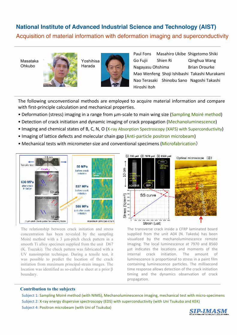
The transverse crack inside a CFRP laminated boardsupplied from the unit A04 (N. Takeda) has beenvisualized by the mechanoluminescence remoteimaging. The local luminescence at 7970 and 8560µst indicates the locations and moments of theinternal crack initiation. The amount ofluminescence is proportional to stress in a paint filmcontaining luminescence particles. The millisecondtime response allows detection of the crack initiationtiming and the dynamics observation of crackpropagation.
Contribution to the subjectsSubject1:Sampling Moirémethod (withNIMS),Mechanoluminescenceimaging,mechanicaltestwithmicro-specimensSubject2:X-rayenergydispersivespectroscopy(EDS)withsuperconductivity(withUniTsukubaandKEK)Subject4:Positronmicrobeam(withUniofTsukuba)
PaulFonsMasahiroUkibeShigetomoShikiGoFujiiShienRi QinghuaWangNagayasuOhshima BrianOrourkeMaoWenfeng ShojiIshibashiTakashiMurakamiNaoTerasaki ShinobuSanoNagoshiTakashiHiroshiItoh
National Institute of Advanced Industrial Science and Technology (AIST)Acquisition of material information with deformation imaging and superconductivity
The following unconventional methods are employed to acquire material information and comparewith first-principle calculation and mechanical properties.• Deformation (stress) imaging in a range from µm-scale to main wing size (Sampling Moiré method)• Detection of crack initiation and dynamic imaging of crack propagation (Mechanoluminescence)• Imaging and chemical states of B, C, N, O (X-ray Absorption Spectroscopy (XAFS) with Superconductivity)• Imaging of lattice defects and molecular chain gap (Anti-particle positron microbeam)• Mechanical tests with micrometer-size and conventional specimens (Microfabrication)
The relationship between crack initiation and stressconcentration has been revealed by the samplingMoiré method with a 3 µm-pitch check pattern in asmooth Ti alloy specimen supplied from the unit D67(K. Tsuzaki). The check pattern was fabricated with aUV nanoimprint technique. During a tensile test, itwas possible to predict the location of the crackinitiation from maximum principal-strain images. Thelocation was identified as so-called a sheet at a prior bboundary.
Makoto Watanabe
HiroakiMamiya
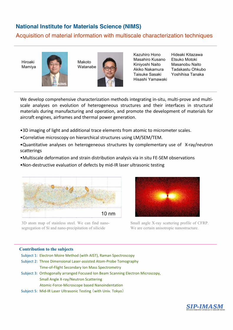
3D atom map of stainless steel. We can find nano-segregation of Si and nano-precipitation of silicide
Small angle X-ray scattering profile of CFRP.We are certain anisotropic nanostructure.
National Institute for Materials Science (NIMS) Acquisition of material information with multiscale characterization techniques
Contribution to the subjectsSubject1: ElectronMoireMethod(withAIST),RamanSpectroscopySubject2: ThreeDimensionalLaser-assistedAtom-ProbeTomography
Time-of-FlightSecondaryIonMassSpectrometrySubject3: OrthogonallyarrangedFocusedIonBeamScanningElectronMicroscopy,
SmallAngleX-ray/NeutronScatteringAtomic-Force-MicroscopebasedNanoindentation
Subject5: Mid-IRLaserUltrasonicTesting(withUniv.Tokyo)
We develop comprehensive characterization methods integrating in-situ, multi-prove and multi-scale analyses on evolution of heterogeneous structures and their interfaces in structuralmaterials during manufacturing and operation, and promote the development of materials foraircraft engines, airframes and thermal power generation.
•3D imaging of light and additional trace elements from atomic to micrometer scales.•Correlative microscopy on hierarchical structures using LM/SEM/TEM.•Quantitative analyses on heterogeneous structures by complementary use of X-ray/neutronscatterings•Multiscale deformation and strain distribution analysis via in situ FE-SEM observations•Non-destructive evaluation of defects by mid-IR laser ultrasonic testing
Kazuhiro Hono Hideaki KitazawaMasahiro Kusano Etsuko MotokiKiniyoshi Naito Masanobu NaitoAkiko Nakamura Tadakastu OhkuboTaisuke Sasaki Yoshihisa TanakaHisashi Yamawaki
University of TsukubaAcquisition of material information with ion beams and positron
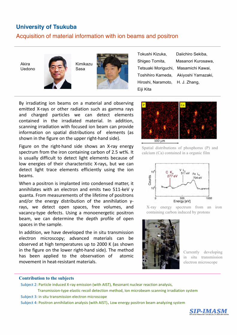
By irradiating ion beams on a material and observingemitted X-rays or other radiation such as gamma raysand charged particles we can detect elementscontained in the irradiated material. In addition,scanning irradiation with focused ion beam can provideinformation on spatial distributions of elements (asshown in the figure on the upper right-hand side).Figure on the right-hand side shows an X-ray energyspectrum from the iron containing carbon of 2.5 wt%. Itis usually difficult to detect light elements because oflow energies of their characteristic X-rays, but we candetect light trace elements efficiently using the ionbeams.When a positron is implanted into condensed matter, itannihilates with an electron and emits two 511-keV γquanta. From measurements of the lifetime of positronsand/or the energy distribution of the annihilation γ-rays, we detect open spaces, free volumes, andvacancy-type defects. Using a monoenergetic positronbeam, we can determine the depth profile of openspaces in the sample.In addition, we have developed the in situ transmissionelectron microscopy; advanced materials can beobserved at high temperatures up to 2000 K (as shownin the figure on the lower right-hand side). The methodhas been applied to the observation of atomicmovement in heat-resistant materials.
Spatial distributions of phosphorus (P) andcalcium (Ca) contained in a organic film
KimikazuSasa
AkiraUedono
Tokushi Kizuka, Daiichiro Sekiba,
Shigeo Tomita, Masanori Kurosawa,
Tetsuaki Moriguchi, Masamichi Kawai,
Toshihiro Kameda, Akiyoshi Yamazaki,
Hiroshi, Naramoto, H. J. Zhang,
Eiji Kita
Contribution to the subjectsSubject2: ParticleinducedX-rayemission(withAIST),Resonantnuclearreactionanalysis,
Transmission-typeelasticrecoildetectionmethod,IonmicrobeamscanningirradiationsystemSubject3:insitutransmissionelectronmicroscopeSubject4: Positronannihilationanalysis(withAIST)、Lowenergypositronbeamanalyzingsystem
X-ray energy spectrum from an ironcontaining carbon induced by protons
Currently developingin situ transmissionelectron microscope
Contribution to the subjectsSubject1: Observationofpropagationofcracks,andchemicalstatemappingofcarbonSubject2: Chemicalstatemappingoflightelements(withAIST)Subject3: Chemicalstatemappingofmetallicelements(withNIMS)Subject4: Developmentofpositronbeams(withAISTandUT)
MasaoKimura
YasuoTakeichi
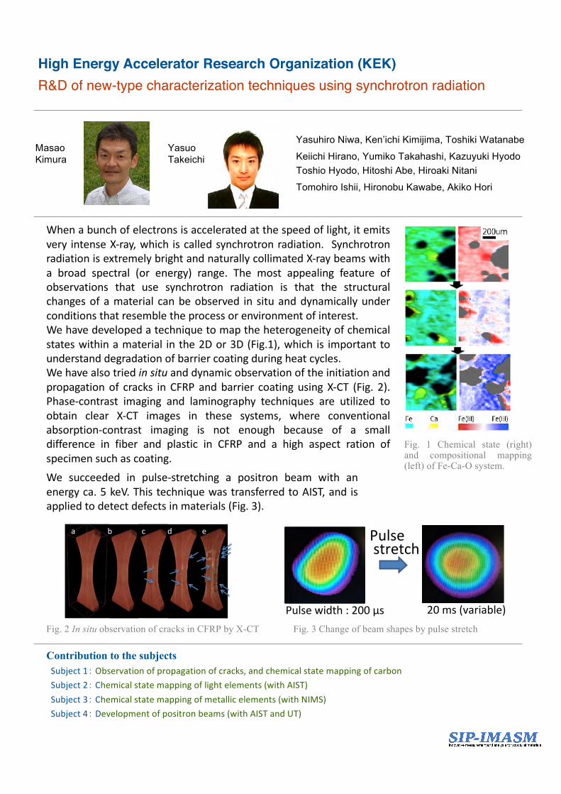
When a bunch of electrons is accelerated at the speed of light, it emitsvery intense X-ray, which is called synchrotron radiation. Synchrotronradiation is extremely bright and naturally collimated X-ray beams witha broad spectral (or energy) range. The most appealing feature ofobservations that use synchrotron radiation is that the structuralchanges of a material can be observed in situ and dynamically underconditions that resemble the process or environment of interest.We have developed a technique to map the heterogeneity of chemicalstates within a material in the 2D or 3D (Fig.1), which is important tounderstand degradation of barrier coating during heat cycles.We have also tried in situ and dynamic observation of the initiation andpropagation of cracks in CFRP and barrier coating using X-CT (Fig. 2).Phase-contrast imaging and laminography techniques are utilized toobtain clear X-CT images in these systems, where conventionalabsorption-contrast imaging is not enough because of a smalldifference in fiber and plastic in CFRP and a high aspect ration ofspecimen such as coating.
Fig. 1 Chemical state (right)and compositional mapping(left) of Fe-Ca-O system.
We succeeded in pulse-stretching a positron beam with anenergy ca. 5 keV. This technique was transferred to AIST, and isapplied to detect defects in materials (Fig. 3).
a� b� c� d� e�
Fig. 2 In situ observation of cracks in CFRP by X-CT Fig. 3 Change of beam shapes by pulse stretch
High Energy Accelerator Research Organization (KEK)R&D of new-type characterization techniques using synchrotron radiation
Yasuhiro Niwa, Ken’ichi Kimijima, Toshiki Watanabe
Keiichi Hirano, Yumiko Takahashi, Kazuyuki Hyodo Toshio Hyodo, Hitoshi Abe, Hiroaki Nitani
Tomohiro Ishii, Hironobu Kawabe, Akiko Hori
Pulse&&stretch�
Pulse&width&:&200&μs� 20&ms&(variable)�
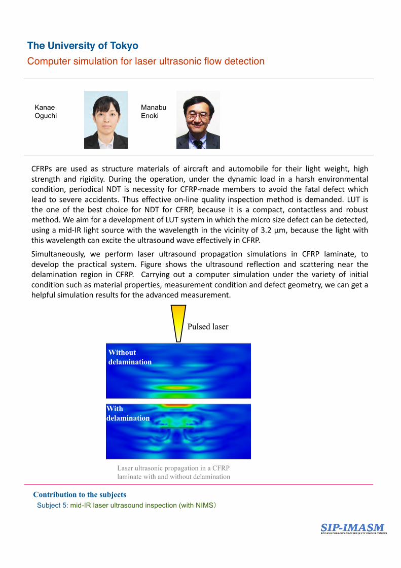
CFRPs are used as structure materials of aircraft and automobile for their light weight, highstrength and rigidity. During the operation, under the dynamic load in a harsh environmentalcondition, periodical NDT is necessity for CFRP-made members to avoid the fatal defect whichlead to severe accidents. Thus effective on-line quality inspection method is demanded. LUT isthe one of the best choice for NDT for CFRP, because it is a compact, contactless and robustmethod. We aim for a development of LUT system in which the micro size defect can be detected,using a mid-IR light source with the wavelength in the vicinity of 3.2 µm, because the light withthis wavelength can excite the ultrasound wave effectively in CFRP.Simultaneously, we perform laser ultrasound propagation simulations in CFRP laminate, todevelop the practical system. Figure shows the ultrasound reflection and scattering near thedelamination region in CFRP. Carrying out a computer simulation under the variety of initialcondition such as material properties, measurement condition and defect geometry, we can get ahelpful simulation results for the advanced measurement.
The University of TokyoComputer simulation for laser ultrasonic flow detection
Laser ultrasonic propagation in a CFRPlaminate with and without delamination
はく離あり
はく離なし
Pulsed laser
With delamination
Without delamination
KanaeOguchi
ManabuEnoki
Contribution to the subjectsSubject 5: mid-IR laser ultrasound inspection (with NIMS)
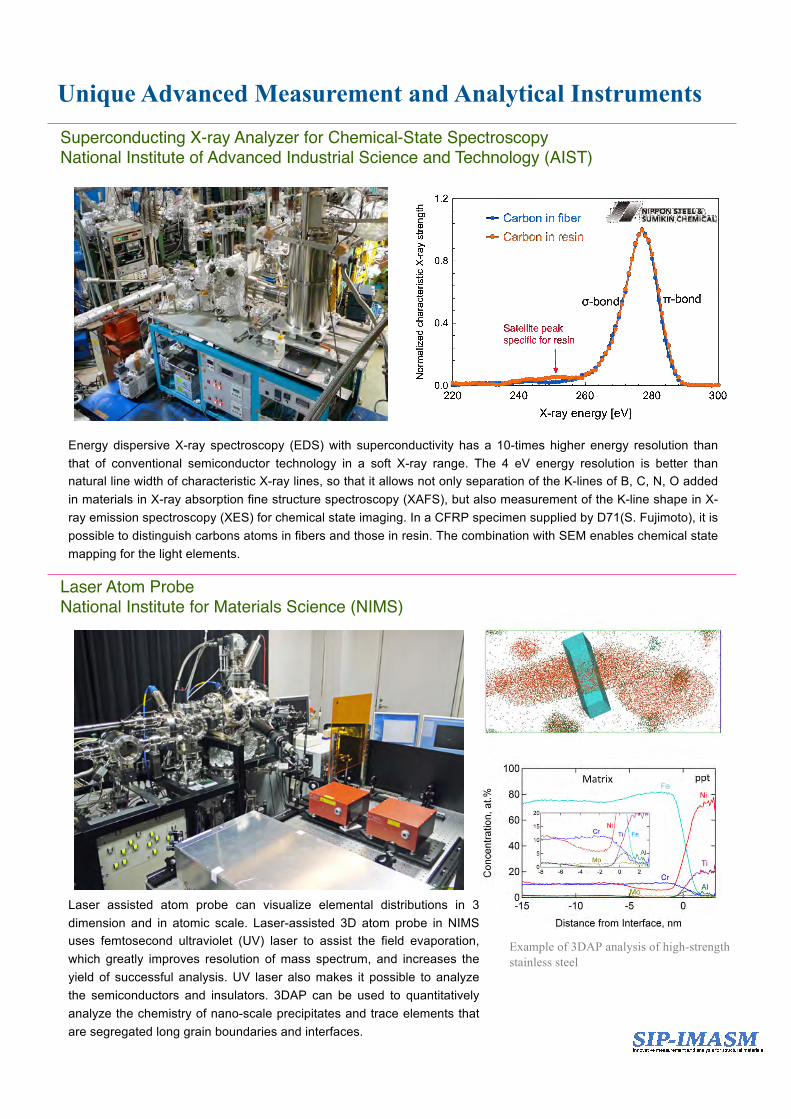
Unique Advanced Measurement and Analytical Instruments
Energy dispersive X-ray spectroscopy (EDS) with superconductivity has a 10-times higher energy resolution thanthat of conventional semiconductor technology in a soft X-ray range. The 4 eV energy resolution is better thannatural line width of characteristic X-ray lines, so that it allows not only separation of the K-lines of B, C, N, O addedin materials in X-ray absorption fine structure spectroscopy (XAFS), but also measurement of the K-line shape in X-ray emission spectroscopy (XES) for chemical state imaging. In a CFRP specimen supplied by D71(S. Fujimoto), it ispossible to distinguish carbons atoms in fibers and those in resin. The combination with SEM enables chemical statemapping for the light elements.
Superconducting X-ray Analyzer for Chemical-State Spectroscopy National Institute of Advanced Industrial Science and Technology (AIST)
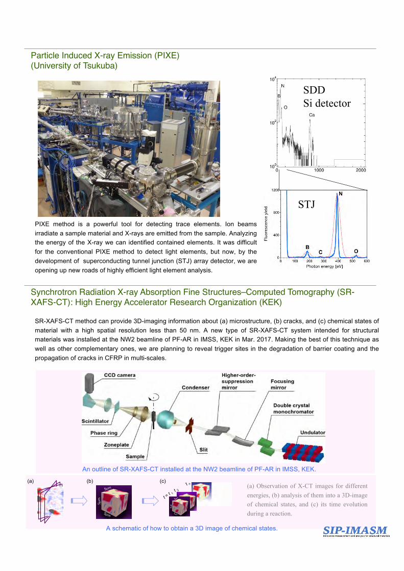
Laser assisted atom probe can visualize elemental distributions in 3dimension and in atomic scale. Laser-assisted 3D atom probe in NIMSuses femtosecond ultraviolet (UV) laser to assist the field evaporation,which greatly improves resolution of mass spectrum, and increases theyield of successful analysis. UV laser also makes it possible to analyzethe semiconductors and insulators. 3DAP can be used to quantitativelyanalyze the chemistry of nano-scale precipitates and trace elements thatare segregated long grain boundaries and interfaces.
Example of 3DAP analysis of high-strength stainless steel
Laser Atom ProbeNational Institute for Materials Science (NIMS)
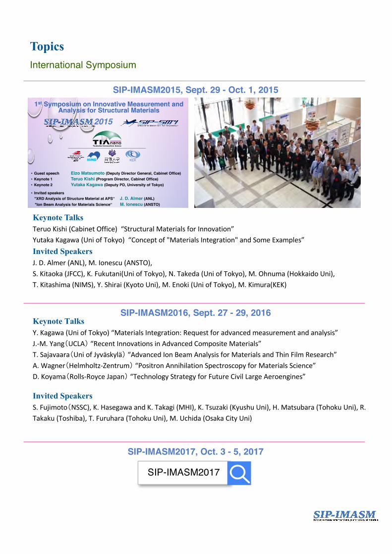
Particle Induced X-ray Emission (PIXE)(University of Tsukuba)
PIXE method is a powerful tool for detecting trace elements. Ion beamsirradiate a sample material and X-rays are emitted from the sample. Analyzingthe energy of the X-ray we can identified contained elements. It was difficultfor the conventional PIXE method to detect light elements, but now, by thedevelopment of superconducting tunnel junction (STJ) array detector, we areopening up new roads of highly efficient light element analysis.
SDDSi detector
STJ
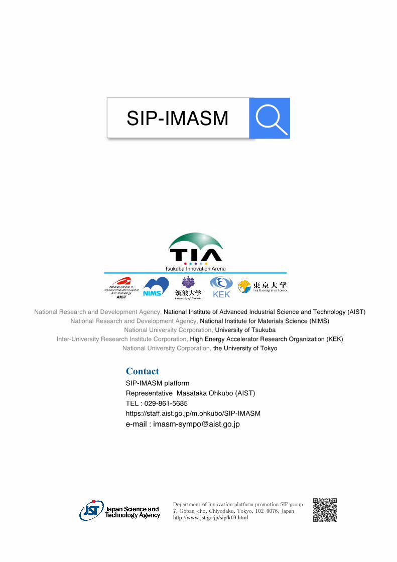
Synchrotron Radiation X-ray Absorption Fine Structures–Computed Tomography (SR-XAFS-CT): High Energy Accelerator Research Organization (KEK)
SR-XAFS-CT method can provide 3D-imaging information about (a) microstructure, (b) cracks, and (c) chemical states ofmaterial with a high spatial resolution less than 50 nm. A new type of SR-XAFS-CT system intended for structuralmaterials was installed at the NW2 beamline of PF-AR in IMSS, KEK in Mar. 2017. Making the best of this technique aswell as other complementary ones, we are planning to reveal trigger sites in the degradation of barrier coating and thepropagation of cracks in CFRP in multi-scales.
Z
An outline of SR-XAFS-CT installed at the NW2 beamline of PF-AR in IMSS, KEK.
(a) Observation of X-CT images for differentenergies, (b) analysis of them into a 3D-imageof chemical states, and (c) its time evolutionduring a reaction.
t = t 1�
t 2�t n�
(b)�(a)� (c)�
A schematic of how to obtain a 3D image of chemical states.
TopicsInternational Symposium
SIP-IMASM2015, Sept. 29 - Oct. 1, 2015
• Guest speech Eizo Matsumoto (Deputy Director General, Cabinet Office) • Keynote 1 Teruo Kishi (Program Director, Cabinet Office)• Keynote 2 Yutaka Kagawa (Deputy PD, University of Tokyo) • Invited speakers "XRD Analysis of Structure Material at APS“ J. D. Almer (ANL) "Ion Beam Analysis for Materials Science“ M. Ionescu (ANSTO)
参加登録 Registrationhttps://staff.aist.go.jp/m.ohkubo/SIP-IMASM/
2015�
1st Symposium on Innovative Measurement and Analysis for Structural Materials
The SIP-IMASM is supported by Cross-ministerial Strategic Innovation Promotion Program (SIP).
KEK
Keynote TalksTeruoKishi(CabinetOffice)“StructuralMaterialsforInnovation”YutakaKagawa(UniofTokyo)“Conceptof"MaterialsIntegration"andSomeExamples”Invited SpeakersJ.D.Almer(ANL),M.Ionescu(ANSTO),S.Kitaoka(JFCC),K.Fukutani(UniofTokyo),N.Takeda(UniofTokyo),M.Ohnuma(HokkaidoUni),T.Kitashima(NIMS),Y.Shirai(KyotoUni),M.Enoki(UniofTokyo),M.Kimura(KEK)
SIP-IMASM2016, Sept. 27 - 29, 2016Keynote TalksY.Kagawa(UniofTokyo)“MaterialsIntegration:Requestforadvancedmeasurementandanalysis”J.-M.Yang(UCLA)“RecentInnovationsinAdvancedCompositeMaterials”T.Sajavaara(UniofJyvaskyla)“AdvancedIonBeamAnalysisforMaterialsandThinFilmResearch”A.Wagner(Helmholtz-Zentrum)“PositronAnnihilationSpectroscopyforMaterialsScience”D.Koyama(Rolls-RoyceJapan)“TechnologyStrategyforFutureCivilLargeAeroengines”
Invited SpeakersS.Fujimoto(NSSC),K.HasegawaandK.Takagi(MHI),K.Tsuzaki(KyushuUni),H.Matsubara(TohokuUni),R.Takaku(Toshiba),T.Furuhara(TohokuUni),M.Uchida(OsakaCityUni)
SIP-IMASM2017, Oct. 3 - 5, 2017
SIP-IMASM2017
National Research and Development Agency, National Institute of Advanced Industrial Science and Technology (AIST)National Research and Development Agency, National Institute for Materials Science (NIMS)
National University Corporation, University of TsukubaInter-University Research Institute Corporation, High Energy Accelerator Research Organization (KEK)
National University Corporation, the University of Tokyo
ContactSIP-IMASM platformRepresentative Masataka Ohkubo (AIST)TEL : 029-861-5685https://staff.aist.go.jp/m.ohkubo/SIP-IMASMe-mail : [email protected]
Tsukuba Innovation Arena
KEK
SIP-IMASM
Department of Innovation platform promotion SIP group 7, Goban-cho, Chiyodaku, Tokyo, 102-0076, Japanhttp://www.jst.go.jp/sip/k03.html