simulation study and comparative evaluation of viral
TRANSCRIPT

Simulation study and comparative evaluation of viral contiguous sequence identification toolsCody Glickman1,2*, Jo Hendrix1,2 and Michael Strong1,2
Abstract
Background: Viruses, including bacteriophages, are important components of envi-ronmental and human associated microbial communities. Viruses can act as extracellu-lar reservoirs of bacterial genes, can mediate microbiome dynamics, and can influence the virulence of clinical pathogens. Various targeted metagenomic analysis techniques detect viral sequences, but these methods often exclude large and genome integrated viruses. In this study, we evaluate and compare the ability of nine state-of-the-art bioinformatic tools, including Vibrant, VirSorter, VirSorter2, VirFinder, DeepVirFinder, MetaPhinder, Kraken 2, Phybrid, and a BLAST search using identified proteins from the Earth Virome Pipeline to identify viral contiguous sequences (contigs) across simu-lated metagenomes with different read distributions, taxonomic compositions, and complexities.
Results: Of the tools tested in this study, VirSorter achieved the best F1 score while Vibrant had the highest average F1 score at predicting integrated prophages. Though less balanced in its precision and recall, Kraken2 had the highest average precision by a substantial margin. We introduced the machine learning tool, Phybrid, which demon-strated an improvement in average F1 score over tools such as MetaPhinder. The tool utilizes machine learning with both gene content and nucleotide features. The addi-tion of nucleotide features improves the precision and recall compared to the gene content features alone.Viral identification by all tools was not impacted by underlying read distribution but did improve with contig length. Tool performance was inversely related to taxonomic complexity and varied by the phage host. For instance, Rhizobium and Enterococcus phages were identified consistently by the tools; whereas, Neisseria prophage sequences were commonly missed in this study.
Conclusion: This study benchmarked the performance of nine state-of-the-art bioin-formatic tools to identify viral contigs across different simulation conditions. This study explored the ability of the tools to identify integrated prophage elements traditionally excluded from targeted sequencing approaches. Our comprehensive analysis of viral identification tools to assess their performance in a variety of situations provides valu-able insights to viral researchers looking to mine viral elements from publicly available metagenomic data.
Keywords: Virus, Bacteriophage, Prophage, Metagenomics, Tool comparison
Open Access
© The Author(s), 2021. Open Access This article is licensed under a Creative Commons Attribution 4.0 International License, which permits use, sharing, adaptation, distribution and reproduction in any medium or format, as long as you give appropriate credit to the original author(s) and the source, provide a link to the Creative Commons licence, and indicate if changes were made. The images or other third party material in this article are included in the article’s Creative Commons licence, unless indicated otherwise in a credit line to the mate-rial. If material is not included in the article’s Creative Commons licence and your intended use is not permitted by statutory regulation or exceeds the permitted use, you will need to obtain permission directly from the copyright holder. To view a copy of this licence, visit http:// creat iveco mmons. org/ licen ses/ by/4. 0/. The Creative Commons Public Domain Dedication waiver (http:// creat iveco mmons. org/ publi cdoma in/ zero/1. 0/) applies to the data made available in this article, unless otherwise stated in a credit line to the data.
RESEARCH
Glickman et al. BMC Bioinformatics (2021) 22:329 https://doi.org/10.1186/s12859-021-04242-0
*Correspondence: [email protected] 2 Computational Bioscience, University of Colorado Anschutz, 12801 E 17th Avenue, Aurora, CO 80045, USAFull list of author information is available at the end of the article

Page 2 of 19Glickman et al. BMC Bioinformatics (2021) 22:329
BackgroundViruses are the most abundant biological entities on Earth [1]. However, the collective knowledge of environmental viral sequences, including bacteriophages, remains under-represented relative to the amount of genetic information for eukaryotic viruses and bacteria. Bacteriophages are viruses that infect bacteria and are commonly referred to as phages. Phages are obligate parasites that play an important role in the genomic composition and evolution of their bacterial hosts. Phages directly contribute to bacte-rial infections in humans by acting as a genetic reservoir for virulent genes in bacteria such as Escherichia coli, Salmonella enterica, Pseudomonas aeruginosa, Vibrio cholerae, Corynebacterium diphtheriae, and Streptococcus pyogenes [2, 3].
In addition, some phages utilize Ig-like domains to attach to mucosal layers in humans to lie in wait for bacterial prey. This bacteriophage adherence to mucus (BAM) model suggests that phages may act as a non-host derived innate immunity system to modu-late the bacterial microbiome [4]. A longitudinal study of the human virome revealed composition conservation that mimicked the stability of healthy bacterial microbiomes [5, 6]. Dysbiosis in the virome has been observed in disease states such as inflammatory bowel disease (IBD), Crohn’s disease, and asthma [7–9].
The study of viruses has traditionally relied on the ability to cultivate viral particles from a cultured host; however, many bacteria cannot be cultured in a laboratory setting [10]. The limited number of culturable hosts, in combination with the additional com-plexities of viral isolation limit the study of viruses. The advancements in next generation sequencing technologies created an opportunity to study viruses with culture independ-ent methods. However, because viruses do not share a common universal marker gene, like the bacterial small subunit RNA, sequencing techniques such as metagenomics are a necessity [11]. Metagenomics is a non-targeted sequencing approach to elucidate the totality of genetic material within a sample, either DNA or RNA. However, in part due to small genomes, viruses are traditionally underrepresented in metagenomic stud-ies from a read abundance perspective. It is common for viral reads to comprise less than 5% of metagenomic sequences [12]. A way to enrich viral reads in metagenomic studies is to filter or directly select viral like particles (VLPs). However, these tech-niques tend to remove large viruses and viruses integrated into bacterial genomes called prophages, before sequencing. Therefore, the ability to identify viral elements directly from metagenomic sequencing studies is also important for understanding the composi-tion of the virome. The advent of computational tools dedicated to the identification of viral sequences in metagenomics has improved our ability to identify known, novel, and integrated viruses.
MetaPhinder is an approach that uses BLASTn and average nucleotide identity thresh-olds to classify viral contigs in metagenomics [13]. Methods that use sequence similar-ity suffers worsening performance with smaller contig lengths. Domain recognition is utilized by more tools to counter the limitations of contig length on traditional sequence homology approaches, but these tools are often reliant on specialized viral domains like those from pVOGs (prokaryotic virus orthologous groups) [14]. Unlike prophage iden-tification methods that use viral domain enrichment or presence/absence to calculate a score, a new method, called Vibrant, uses domain abundances in a neural network framework to classify contigs having more than 4 proteins [15]. VirSorter2 follows a

Page 3 of 19Glickman et al. BMC Bioinformatics (2021) 22:329
similar methodology, using domain percentages, gene content features, and key homol-ogy genes in a tree-based machine learning framework to classify viral reads [16].
Homology of viral protein domains is limited to known viruses, which are thought to represent only a small slice of the vast viral dark matter [17]. Another homology approach sought to expand known viral hidden Markov models (HMMs) through a semi-supervised expansion of existing viral protein families. Paez-Espino et al. (Earth Virome Pipeline) collected viral coding regions from NCBI servers and known viral metagenomic contigs; then clustered those peptides into protein families to create new viral HMMs [18]. This initial set was used as bait to identify potential viral contigs in thousands of metagenomic data sets. Predicted proteins from these captured viral con-tigs were added to the original set of peptides and re-clustered to create thousands of new viral protein families and HMMs. Even with the expansion of viral families, both VirSorter and the BLASTp search using the Earth Virome protein set are at least par-tially reliant on domain homology. A reference-free viral identification tool was devel-oped using machine learning to address limitations of homology searching. VirFinder is a logistic regression classification using nucleotide sequence 8-mers as features [19]. The authors of VirFinder expanded the concept of using k-mers as features to identify viral contiguous sequences with DeepVirFinder, a convolutional neural network that takes raw sequences as inputs and learns features that are useful for viral contig pre-diction [20]. VirFinder relies solely on sequence-based features, which is analogous to another k-mer approach, Kraken2. Kraken2 uses discriminatory 35-mers to uniquely identify sequences to the species and even subspecies level [21]. In order to use Kraken2 in a viral identification context, we created the tool, VirKraken, that parses the Kraken2 classification output to assign viral contigs in metagenomic reads. VirKraken is avail-able on PyPI and at https:// github. com/ Strong- Lab/ VirKr aken. VirKraken references the Kraken2 assigned taxonomy identification number against an edited NCBI Taxonomy database to assign kingdom and to filter sequences when requested [22].
Another approach to identify viral elements in metagenomics involves negation of known bacteria contigs. VirMine uses a homology search against a bacterial protein database; if hits of bacterial genes outnumber the number of unknown hits the contig is removed, thus leaving viral contigs [23]. All previously described tools identify viral ele-ments from assembled sequences. MARVEL is a machine learning method that classifies binned contigs as viral clusters using a random forest approach with three features (gene density, strand shifts, and fraction of homology hits to a viral protein database) [24].
The authors of VirFinder put forth a call to create a hybrid tool that utilizes both k-mer features and gene content features to offset the weaknesses of both methods [19]. To answer that call, we developed a machine learning model called Phybrid that uses both gene content features such as gene density and strand shift frequency, in addition to sequence-based features to classify viral contigs using an additive boosting model. The addition of gene content features is hypothesized to offset the dip in performance of sequence-based machine learning models compared to homology methods on longer contigs [19].
Many approaches exist to identify viral elements in metagenomics. However, a sys-temic evaluation among many of these tools has not been performed. This study is meant to provide information and guidance to researchers regarding when to use a
Page 4 of 19Glickman et al. BMC Bioinformatics (2021) 22:329
specific viral identification tool to further study viral elements or to remove them for downstream analyses. The characterization of more viral elements in the public domain could lead to the discovery of novel viruses [25] and provide insight into the functional potential residing in an extracellular genetic reservoir [2].
MethodsPhybrid, a hybrid gene content and nucleotide feature set for viral classification
To build Phybrid, 1849 complete phage and 2327 complete archaeal/bacterial genomes were compiled from RefSeq (Accessed on January 8th, 2020). Prophage elements in the archaeal and bacterial genomes were identified using VirSorter [22]. Category 4 prophages were selected, and the predicted nucleotide sequences were added to the complete viral genomes. Custom scripts were used to identify and remove the predicted prophage sequences from the host genome. The total number of prophages predicted was 730 in 339 bacterial genomes (14.57% of genomes contained at least 1 prophage) culminating in an average prophage per genome ratio of 0.314.
After removing integrated prophages, the complete genomes were fragmented into k-mers of 4 sizes using an n-step kmerization method. The n-step method removes contig end-overlap and ensures that the maximum number of k-mers is the length of the base sequence over the length k. The complete genomes were fragmented into sizes of 1 KB, 3 KB, 5 KB, and 10 KB sequences. Due to the size of bacterial and archaeal genomes relative to phage genomes, the fragments from non-phage sampling were down sampled to evenly distribute the classes. The four different fragment lengths were used to train four separate models.
The n-step fragments were subjected to a sliding window kmerization of size 8 using a k-mer counting program written in C [26]. A sliding window kmerization calculates k-mer abundance with significant overlap and the maximum number of k-mers is the length of the base sequences minus 1. The program stores all 8-mer values (65,536 pos-sible 8-mers) in a hash table. In real world metagenomic sampling, the directionality of a sequence fragment may be ambiguous. Therefore, similar to VirFinder [19], we devel-oped custom scripts to sum complement, reverse, and reverse complement sequences thus reducing our feature space from 65,536 possible k-mers to 16,384 possible k-mers. The nucleotide feature space is further reduced to 888 k-mers using Gini importance or total decrease in node impurity above 0.001, which is a weighted probability of reaching a feature averaged over all trees in a random forest [27].
Gene content feature set creation
The use of gene content features are built into tools such as MARVEL and VirSorter [24, 28]. MARVEL and VirSorter both utilize gene density as a marker of viral elements. In this study, four gene content features associated with viral genomes were included as part of the feature set in Phybrid; gene density, operon length, average peptide length, and percentage of overlapping peptides. Due to the physical restraints of some viral capsids, viral genomes are commonly tightly packed and translate shorter proteins than bacterial genomes [29]. Operon length in the context of this study is the length of a continuous set of genes on the same strand. Viruses tend have long stretches of genes located on the same strand [30]. In addition, viral genomes often have overlapped genes

Page 5 of 19Glickman et al. BMC Bioinformatics (2021) 22:329
for different life cycles [30]. Custom scripts were used to calculate the four protein char-acteristics from the output of the Prodigal gene prediction software [31]. Figure 1 shows the observed distribution of protein features in the training data of the 10 KB model.
Model and hyperparameter selection
After combining the complementary nucleotide features and the gene content features, the total feature space of Phybrid was 892 features. During training, the performance of a random forest, multi-layer perceptron, and an additive boosting model were com-pared using 5-fold cross validation [32]. At every fragment size, the additive boosting model performed the best. We selected XGBoost (version 0.81) and performed a Ran-domSearchCV (version 0.20.1) analysis to determine hyperparameters [32, 33]. The pre-trained models were added to the tool repository for use classifying metagenomic fasta sequences. Phybrid generates outputs as a header file containing the header sequences of viral elements and a fasta file containing the nucleotide sequences of the predicted viral elements.
Building simulated Illumina metagenomes
To build a simulated test set, all complete genomes were downloaded from NCBI RefSeq (accessed on 12/15/2020). The genomes deposited since May 1st, 2020, were selected to test the viral contig identification tools because many of the tools were trained or relied on databases last updated prior to this date. Bacterial hosts of phages were col-lected using a dataset from Virus-Host DB [34] (Accessed on December 17, 2020). Phage were assigned bacteria genera values by their host organism. Using information from the Earth Microbiome Project (EMP) and from Qiita, the recently submitted genomes were further filtered by 53 genera commonly found in soils (37 genera) and in clinical samples (26 genera) with 8 genera in both niches [35, 36]. This resulted in 297 unique bacterial genomes being used for the simulations with 82 genomes found in both clinical sampling (160 genomes) and soil sampling (219 genomes). The reliance on recently sub-mitted genomes to produce the testing set did not produce traditional bacterial distribu-tions seen in clinical and soil microbiomes. For example, while the genera Bacteroides are commonly present in the clinical microbe samples, the amount in this study does not represent a substantial portion of the community as seen in other clinical microbiome studies [37]. The distribution of bacterial genera was used as a confounder for viral clas-sification in this study. The goal of this study was to observe the performance of phage identification in the presence of genetically similar bacteria.
Phage genomes were also filtered by their host bacterial genera and randomly down sampled to match the number of bacterial genomes in the simulations. While phages are thought to outnumber bacteria ten to one in the environment [38], we matched the complexity of phage and bacteria in our simulations across taxonomic levels due to limi-tations in the number of available phage genomes for the full datasets. In order to test the impact of taxonomic complexity on viral identification tool performance, we sub-sampled phage and bacterial genomes into medium (50 bacterial genomes and 50 phage genomes) and low (10 bacterial genomes and 10 phage genomes) complexity subsets. Additional file 2: Table S1 (clinical) and Additional file 2: Table S2 (soil) detail the taxo-nomic abundance of the top 6 genera and phage host genera in the testing set across

Page 6 of 19Glickman et al. BMC Bioinformatics (2021) 22:329
Fig.
1 G
ene
cont
ent f
eatu
re p
erfo
rman
ce. T
he p
erfo
rman
ce o
f the
4 g
ene
cont
ent f
eatu
res
in th
e 10
KB
trai
ning
dat
aset
. a G
ene
dens
ity re
pres
ente
d by
num
ber o
f gen
es p
er 1
KB.
b M
edia
n op
eron
leng
th is
a re
pres
enta
tive
mea
sure
of s
tran
d sw
itchi
ng fr
eque
ncy.
An
oper
on is
defi
ned
as a
s a
set o
f clo
sely
link
ed g
enes
on
the
sam
e st
rand
. c P
erce
ntag
e of
ove
rlapp
ing
pept
ides
m
easu
red
as a
per
cent
age
of a
ll pr
edic
ted
gene
s. Vi
ruse
s th
at h
ave
a ly
soge
ny p
hase
are
kno
wn
over
lap
gene
s fo
r diff
eren
t life
cyc
les.
d M
edia
n am
ino
acid
leng
th a
s vi
ral p
eptid
es a
re c
omm
only
sh
orte
r tha
n ba
cter
ial p
eptid
es
Page 7 of 19Glickman et al. BMC Bioinformatics (2021) 22:329
taxonomic complexity levels. While both lower complexities draw from the full distribu-tion of genomes, there is no overlap in the selected genomes between the medium and low taxonomic levels. This was accomplished through setting a random seed in the sub-sampling procedure and using set operations to confirm no overlap of genomes.
Simulated metagenomes were created using InSilicoSeq (version 1.2.0). InSilicoSeq and another popular metagenomic simulator, CAMISIM use a lognormal read distribu-tion by default, however, four additional read distributions are provided as a part of the InSilicoSeq software suite: uniform, exponential, zero inflated lognormal, and halfnor-mal [39, 40]. Due to the enormous diversity of naturally occurring communities, read distribution profiles are likely to fluctuate. To understand the impact of read distribution and taxonomic complexities on the performance of viral identification, we created 30 MiSeq simulations with 12 million 2x300 reads. The 30 simulations were composed of two environmental conditions (clinical and soil microbes) with five read distributions across three taxonomic levels (full, medium, low). Bacterial reads represented 93.75% of the total composition in each simulation and phages represented 6.25%. Prior studies suggest phages commonly represent less than 5% of metagenomic sequencing reads [12] due to genomes that are orders of magnitude smaller than prokaryotic genomes. Our decision to exceed the 5% of viral reads in metagenomics was driven by the need to iden-tify an expanded set of phages from taxonomically diverse testing sets. After assembly and filtration of contigs less than 1KB in length, phages comprise an average of 1.54% of total contig abundance.
After simulating, the reads were perfectly binned by sequence origin to limit the crea-tion of chimeric contigs. Chimeric contigs are assembly errors when reads from different organisms are assembled together resulting in a shorter fragmented assembly or taxo-nomic misclassification downstream. The decision to bin prior to assembly was to allow for genera labeled contigs in order to explore false positive and recall rates of bacteria and phage, respectively. The perfect bins were assembled using metaSpades (version 3.11.1) and only contigs of length 1KB or greater were retained [41]. The relative abun-dance of bacteria genera in the simulations are shown in Fig. 2.
Integrated prophage identification
Integrated prophage elements were identified in complete bacterial genomes using VirSorter prior to read simulations [28]. Integrated prophages were selected for down-stream processing if assigned as category 4, the highest confidence category for prophages within VirSorter [28]. A nucleotide BLAST database was created with the identified prophage elements. After read simulation and assembly, bacterial contigs were identified as prophages using a BLASTn search against the prophage database with a bitscore greater than 1000 and a percent identity greater than 95%. Additional file 2: Fig-ure S1 shows the genera distribution of the identified prophage elements separated by read distribution and sampling site.
Tools used in simulation study
The tools used in the study shown in Table 1 were tested on their performance to identify viral elements from assembled contigs in the simulations. The tools used in this study to identify viral contigs were Vibrant (Version 1.2.0), VirSorter, VirSorter2, VirFinder,

Page 8 of 19Glickman et al. BMC Bioinformatics (2021) 22:329
Fig.
2 R
elat
ive
abun
danc
e of
gen
era
in s
imul
atio
ns. T
hese
figu
res
high
light
the
rela
tive
abun
danc
e of
the
cont
igs
grea
ter t
han
1 KB
. Bac
teria
l con
tigs
repr
esen
t 98.
46%
of c
ontig
s, w
hile
pha
ges
and
prop
hage
s co
mbi
ne fo
r the
rem
aini
ng 1
.54%
. a T
he c
ontig
dis
trib
utio
n w
ithin
15
soil
sim
ulat
ions
. b T
he c
ontig
dis
trib
utio
n w
ithin
15
clin
ical
sim
ulat
ions

Page 9 of 19Glickman et al. BMC Bioinformatics (2021) 22:329
DeepVirFinder, MetaPhinder, Kraken 2, Phybrid, and a BLAST search using identified proteins from the Earth Virome Pipeline [15, 16, 19, 28].
Any VirSorter predictions that were classified to the lowest confidence category were removed via evidence by the tool developers [28]. VirFinder and DeepVirFinder assign a probability value and any contigs that had a value less than 0.01 were classified as viral. A diamond blast database was created with the viral proteins from the Earth Virome Pipeline [18, 42]. Proteins from the simulation contigs were predicted using Prodi-gal and searched for viral homology using diamond BLASTp against proteins from the Earth Virome Pipeline with matches retained that had a bit score greater than 100 and an e-value less than 1e−05 [31]. Contigs with more than one hit were classified as viral. MetaPhinder, Phybrid, Phybrid Proteins, and Vibrant were run with default parameters [13, 15]. Double-stranded DNA phages and single stranded DNA viruses were selected with the groups parameter of VirSorter2 as described by the authors [16]. Kraken 2 was run with default parameters using the minikraken database from March 2020 [21]. The resulting Kraken 2 report was parsed for viral reads using VirKraken (Version 0.0.5).
Tool performance scoring
The structure of the simulation allowed for each contig to possess a true origin label. These labels were used to identify the performance of the tools to identify viral elements in the simulations. The performance was measured by precision, recall, and F1 score. Prophages were considered viral in this study and an additional analysis of tool per-formance on prophage identification was performed. The performance measures were used in a simulation performance ranking system to determine the best performing tool across different scenarios. The performance of each tool was ranked within each con-dition with 1 representing the best performing tool. The highest-ranking value (worst performing tool) changes as some tools were unable to properly calculate a score. This occurs when a tool did not predict any viral element in a simulation.
In addition to overall performance, tool performance is evaluated at four discretized contig lengths: 1 KB–2.5 KB, 2.5 KB–5 KB, 5 KB–10 KB, 10 KB+. The recall of the tools to identify viral elements by genera was used to determine any systematic biases for or against specific viral groups. Visualizations of scoring metrics were performed in Python using a combination of Matplotlib (version 2.2.3) and Seaborn (version 0.9.0) plotting
Table 1 Tools used in viral identification benchmarking study
Tool Last updated Target Viral homology matching
Compositional protein features
Machine learning classification
Programming skills required
VirSorter 2015 Virus Yes Yes No No
VirSorter2 2020 Virus Yes Yes Yes No
VirFinder 2017 Virus No No Yes Yes
DeepVirFinder 2020 Virus No No Yes Yes
Vibrant 2020 Virus Yes No Yes Yes
MetaPhinder 2016 Phage Yes No No Yes
Earth Virome 2020 Virus Yes No No Yes
Phybrid 2020 Phage No Yes Yes Yes
Kraken2+VirKraken 2020 Virus Yes No No Yes

Page 10 of 19Glickman et al. BMC Bioinformatics (2021) 22:329
software [43, 44]. Kruskal-Wallace nonparametric testing was performed to determine if the scoring values arose from the same distributions.
ResultsOverall tool performance
The F1 performance across different read simulation conditions was not significantly different (H = 4.02, p = 0.404, Kruskal–Wallis). The F1 performance was significantly different by taxonomic complexity with better tool performance in lower complex-ity simulations relative to both medium and full complexity simulations (H = 47.65, p = 4.50e−11, Kruskal–Wallis). The F1 performance, as well as precision and recall, of longer contigs specially the 10 KB+ bin was higher relative to other contig length bins (H = 275.7, p = 1.82e−59, Kruskal–Wallis). Table 2 contains the mean performance of the tools and the average ranking across the 30 simulations. The F1 performance of the tools in the simulation discretized by taxonomic complexity is shown in Fig. 3.
Kraken2 led both average precision and precision rank. In this study, the BLASTp search of proteins from the Earth Virome Pipeline performed best in both recall and recall rank. The tool with the highest average F1 score and best F1 rank was VirSorter. VirSorter was also the tool used to perform prophage identification. This may provide VirSorter with an advantage over other tools in prophage identification.
Prophage identification performance
The prophage performance of the low complexity simulations are removed due to the presence of only a single prophage contig in all 10 simulations. The F1 performance of the tools to identify prophage in 20 medium and high complexity simulations is shown in Table 3.
Tool performance by contig length
As the length of the contigs increased, the performance of the tools improved. Mean contig length of the simulations was affected by the taxonomic complexity in this study as shown in Additional file 2: Figure S2. Figure 4 demonstrates the F1 performance of each tool within defined contig length bins. If the F1 score of a tool was 0, the record was removed as some lower complexity simulations lacked shorter contiguous sequences.
Table 2 Average performance and simulation rankings of tools at identifying phage
Score of best performing tool is bolded in each column
Tool F1 rank Precision rank Recall rank Average F1 Average precision
Average recall
VirSorter 2.10 3.10 6.40 0.636 0.640 0.658
Kraken2 2.93 1.07 7.80 0.609 0.962 0.467
Vibrant 3.52 4.10 7.26 0.560 0.573 0.598
VirFinder 3.93 2.30 9.32 0.548 0.717 0.450
DeepVirFinder 5.04 5.38 7.90 0.432 0.392 0.496
VirSorter2 5.27 5.93 3.10 0.463 0.341 0.797
Phybrid 5.40 6.00 3.60 0.413 0.317 0.755
Phybrid Proteins 7.47 7.70 4.63 0.142 0.213 0.717
MetaPhinder 8.73 8.67 2.83 0.082 0.138 0.842
Earth Virome 9.73 9.73 1.78 0.023 0.044 0.872

Page 11 of 19Glickman et al. BMC Bioinformatics (2021) 22:329
Fig.
3 F
1 sc
ores
of t
ools
by
taxo
nom
ic c
ondi
tions
. Dod
ge b
oxpl
ots
by ta
xono
mic
com
plex
ity a
rran
ged
by a
vera
ge F
1 pe
rfor
man
ce w
ith th
e be
st p
erfo
rmin
g to
ols
on th
e rig
ht s
ide
of th
e x
axis

Page 12 of 19Glickman et al. BMC Bioinformatics (2021) 22:329
Viral recall by host genera
Recall scores of viral elements from the medium and full distributions were calcu-lated across 30 host genera. Recall was only retained if greater than 0 to prevent the absence of a phage host genera by niche. Figure 5 shows the recall of viral contigs by host genera across all tools. The viral host genera with the best recall was Xan-thomonas, however, phage with Xanthomonas as a host were not well represented in the data set. Phage known to infect Enterococcus achieved an average recall over 0.83 across all tools. DeepVirFinder performed the best at identifying phage known to infect Enterococcus with an average recall rate of 0.97. Neisseria prophage sequences had the lowest average recall performance across all tools (0.23), with only 7 tools correctly predicting at least one Neisseria prophage contig. While multiple contigs were derived only a single Neisseria prophage was included in this study and that may be affecting tool performance. The BLASTp search using the proteins from the Earth Virome Pipeline performed the best at identifying this elusive prophage (0.68) and the next best tool was MetaPhinder with a recall rate of 0.24.
False positive genera
In addition to the recall rate of viral elements by host genera, the percent of genera associated with bacterial false positives was calculated for each tool in medium and full complexity simulations. Bacterial genera that represent more than one third of false positives of a tool in a simulation were retained. Eleven genera were represented with Streptomyces present in 9 of 10 tools. Additionally, Citrobacter and Pseudomonas were major false positive genera in more than 5 tools. Additional file 2: Figure S3 shows the genera of false positives that represent more than 33% by tool.
DiscussionThis study benchmarked and evaluated the ability of nine viral classification tools to identify viral and prophage elements within shotgun metagenomics. The study con-sisted of 30 Illumina MiSeq simulations across two communities, five read abun-dance distributions, and three taxonomic levels. The performance of the tools was consistent across read distributions (H = 4.02, p = 0.404, Kruskal–Wallis), whereas,
Table 3 Average performance and simulation rankings of tools at identifying prophage
Score of best performing tool is bolded in each column
Tool F1 rank Precision rank Recall rank Prophage F1 Prophage precision
Prophage recall
Vibrant 1.15 1.95 7.45 0.169 0.146 0.231
VirSorter 2.11 1.66 8.76 0.147 0.144 0.164
VirSorter2 2.70 3.40 4.70 0.117 0.0685 0.453
Phybrid 3.95 4.45 7.13 0.0446 0.0269 0.252
Phybrid Proteins 5.20 5.50 6.05 0.0188 0.00978 0.342
Kraken2 6.70 1.85 9.90 0.0169 0.172 0.00896
MetaPhinder 6.65 6.85 2.90 0.0152 0.00776 0.588
Earth Virome 6.85 7.00 1.60 0.0117 0.00588 0.728VirFinder 8.88 8.88 2.73 0.00725 0.00365 0.705
DeepVirFinder 8.79 9.03 2.79 0.00647 0.00325 0.637

Page 13 of 19Glickman et al. BMC Bioinformatics (2021) 22:329
Fig.
4 F
1 sc
ores
of t
ools
acr
oss
cont
ig le
ngth
bin
s in
all
sim
ulat
ions
. The
ave
rage
F1
perf
orm
ance
of a
ll to
ols
incr
ease
s as
the
bin
repr
esen
ting
cont
ig le
ngth
s in
crea
ses.
All
thirt
y si
mul
atio
ns a
re
incl
uded
as
part
of t
his
figur
e, h
owev
er in
som
e si
mul
atio
ns, p
redi
cted
vira
l con
tigs
of a
spe
cific
leng
th a
re a
bsen
t. Th
is m
ay c
ause
som
e to
ols
to h
ave
mor
e da
ta p
oint
s th
an o
ther
s

Page 14 of 19Glickman et al. BMC Bioinformatics (2021) 22:329
Fig.
5 V
iral r
ecal
l by
host
gen
era
in m
ediu
m a
nd fu
ll co
mpl
exity
sim
ulat
ions
. The
30
host
gen
era
of p
hage
are
list
ed in
ord
er o
f mea
n re
call
alon
g th
e x-
axis
. The
dot
ted
line
in th
e fig
ure
dem
arca
tes
0.5
reca
ll
Page 15 of 19Glickman et al. BMC Bioinformatics (2021) 22:329
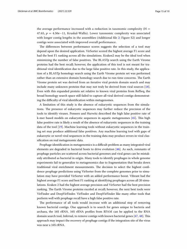
the average performance increased with a reduction in taxonomic complexity (H = 47.65, p = 4.50e−11, Kruskal-Wallis). Lower taxonomic complexity was associated with longer contig lengths in the assemblies (Additional file 2: Figure S2) and longer contigs were associated with improved overall performance.
The differences between performance scores suggests the selection of a tool may depend upon the desired application. VirSorter scored the highest average F1 score and had the best F1 ranking across all the simulations. Kraken2 may be the ideal tool when minimizing the number of false positives. The BLASTp search using the Earth Virome proteins had the best recall; however, the application of this tool is not meant for tra-ditional viral identification due to the large false positive rate. In this study, the applica-tion of a BLASTp homology search using the Earth Virome protein set was performed rather than an extensive domain homology search due to run-time concerns. The Earth Virome protein set was derived from an iterative viral protein domain search and may include many unknown proteins that may not truly be derived from viral sources [18]. Even with this expanded protein set relative to known viral proteins from RefSeq, the broad homology search space still failed to capture all viral derived contigs demonstrat-ing the difficulty of viral identification within metagenomes.
A limitation of this study is the absence of eukaryotic sequences from the simula-tions. The presence of eukaryotic sequences may further reduce the precision of the tools to identify viruses. Ponsero and Hurwitz described the high false positive rate of k-mer based models on eukaryotic sequences in aquatic metagenomes [45]. This high false positive rate is likely a result of the absence of eukaryotic sequences in the training data of the tool [19]. Machine learning tools without eukaryotic sequences in the train-ing set may produce additional false positives. Any machine learning tool with gaps of eukaroytic or novel viral sequences in the training data may produce errors in viral clas-sification on real metagenomic data.
Prophage identification in metagenomics is a difficult problem as many integrated viral elements are degraded in bacterial hosts to drive evolution [46]. As such, remnants of prophage particles are scattered across bacterial genomes and viral genes can be mistak-enly attributed as bacterial in origin. Many tools to identify prophages in whole genome experiments fail to generalize to metagenomics due to fragmentation that breaks down traditional viral enrichment measurements. The decision to select the highest confi-dence prophage predictions using VirSorter from the complete genomes prior to simu-lation may have provided VirSorter with an added performance boost. Vibrant had the highest average F1 score and best F1 ranking at identifying prophages across all 20 simu-lations. Kraken 2 had the highest average precision and VirSorter had the best precision ranking. The Earth Virome proteins exceled at recall; however, the next best tools were VirFinder and DeepVirFinder. VirFinder and DeepVirFinder like many other tools that perform well with prophage recall have a high false positive rate.
The performance of all tools would increase with an additional step of removing known bacterial contigs. One approach is to search for genes unique to bacteria and archaea, the 16S rRNA. 16S rRNA profiles from RFAM can be applied to the RNA domain search tool, Infernal, to remove contigs with known bacterial genes [47, 48]. This approach may impact the recovery of prophage contigs if the integration site of the virus was near a 16S rRNA.

Page 16 of 19Glickman et al. BMC Bioinformatics (2021) 22:329
Viral identification tools performed well at identifying phages known to infect genera such as Enterococcus (0.83), Mycobacterium (0.77), and Salmonella (0.81). The performance of the tools to identify phages that infect genera such as Neisseria (0.23), Brevibacterium (0.30), and Mesorhizobium (0.33) dropped substantially. Detecting the presence of Neisse-ria phage and prophage may be important for a diagnostic of invasive meningococcal dis-ease as prophage-like elements are commonly found throughout the Neisseria genera [49]. The results of the tools on individual genera are meant to demonstrate the variability of tool performance on different genera of phage. Phage genera in this study are defined by host range. Phage host range is poorly understood and there exists sampling bias towards phages affecting more well studied bacterial pathogens. The results of this study seemed to imply that sampling bias of phage genera in public databases may be affecting many overall tool performances. Mycobacterium phages and Enterococcus phage are the most abundant phages in public databases. The results of phage genera performance should not be over-interpreted in this study as the number of unique phages known to affect a bacterial genera are not uniformly distributed as seen in Additional file 2: Tables S1 and S2.
The performance of Phybrid including the nucleotide features showed improvement over the gene content features alone. The precision of Phybrid dramatically improved with con-tigs over 10KB, however, smaller bins were plagued with many false positives. Integrated prophages added to the viral class in the training data represented 28.3% of the total viral genomes. Prophages are commonly degraded in bacterial hosts to drive evolution [46], therefore degraded viral elements in bacterial contigs with similar nucleotide structures as the complete prophages may be misclassified. In addition, the use of k-mer profiles for smaller contig classification created sparse data sets, which may have led to overfitting.
The performance of the tools presented needs to be weighted with the computational cost to run each tool. This study was performed on a shared high performance computing cluster and individual tool performance and memory requirements were not captured on an isolated node. However, the mechanism of viral identification can be used infer the rela-tive time and memory consumption of the tools. The fastest tool in this study was Kraken2, which uses discriminatory k-mers to compare against a pre-computed hash table. The amount of memory needed to build the full hash table may be a drawback against using Kraken2 on a personal machine. The tool, Vibrant, uses protein features derived from mul-tiple HMM searches. As a result of a large domain space, this tool ran for a significantly longer amount of time (1 week for full complexity simulations) relative to the other tools on the shared compute cluster.
This study benchmarked and compared the performance of viral identification tools in metagenomics. The viral identification performance measures, in conjunction with the genera and prophage recall, highlights the advantages and challenges of using specific viral identification tools, and can be used as a guide to assist the selection of tools for subsequent research.
ConclusionIn summary, we tested the performance of nine viral identification tools on 30 simu-lated metagenomes. The underlying read distribution has little impact on average tool performance. Increasing contig length and decreasing taxonomic complexity improved the average performance of the tools. Vibrant performed the best at the identification of

Page 17 of 19Glickman et al. BMC Bioinformatics (2021) 22:329
prophages in metagenomics. Overall, the tool that averaged the best F1 score was Vir-Sorter, while Kraken2 lead all other tools in precision. The results of these simulations should provide researchers with a guide to selecting the appropriate tool for their own viral identification research.
Abbreviationscontig: Contiguous sequence; BAM: Bacteriophage adherence to mucus; IBD: Inflammatory bowel disease; VLP: Viral like particles; pVOG: Prokaryotic virus orthologous group; HMM: Hidden Markov model; EMP: Earth Microbiome Project; KB: Kilo-basepairs.
Supplementary InformationThe online version contains supplementary material available at https:// doi. org/ 10. 1186/ s12859- 021- 04242-0.
Additional file 1. Supplemental figures and tables referenced in the article.
Additional file 2. Captions of supplemental figures and tables referenced in the article.
AcknowledgementsThe authors would like to thank Chris Miller, James Costello, Catherine Lozupone, and Kirk Harris for their helpful comments.
Authors’ contributionsAll authors read and approved the final manuscript. CG and MS conceived the study. CG and JH performed data analysis. CG and MS wrote the manuscript.
FundingCG is supported by NLM 5 T15 LM009451-12. MS and CG thank Stephen and Betty Thorp for support.
Availability of data and materialsAll scripts used to derive the figures and additional preprocessing workflows are available on the Strong Lab GitHub at https:// github. com/ Strong- Lab/ Viral_ Class ifica tion_ in_ Metag enomi cs. Phybrid is available on the Strong Lab GitHub at https:// github. com/ Strong- Lab/ Phybr id. VirKraken, the Kraken2 extension used in this study is available on the Strong Lab GitHub at https:// github. com/ Strong- Lab/ VirKr aken and on PyPI. The fasta files of the simulations are found at https:// tinyu rl. com/ fasta vm.
Declarations
Ethics approval and consent to participateNot applicable.
Consent for publicationNot applicable.
Competing interestsThe authors have declared no competing interests in this work.
Author details1 Center for Genes, Environment, and Health, National Jewish Health, 1400 Jackson Street, Denver, CO 80206, USA. 2 Com-putational Bioscience, University of Colorado Anschutz, 12801 E 17th Avenue, Aurora, CO 80045, USA.
Received: 1 March 2021 Accepted: 4 June 2021
References 1. Ackermann H-W. 5500 phages examined in the electron microscope. Adv Virol. 2007;152(2):227–43. 2. Modi SR, Lee HH, Spina CS, Collins JJ. Antibiotic treatment expands the resistance reservoir and ecological network
of the phage metagenome. Nature. 2013;499(7457):219–22. 3. Brüssow H, Canchaya C, Hardt W-D. Phages and the evolution of bacterial pathogens: from genomic rearrange-
ments to lysogenic conversion. Microbiol Mol Biol Rev. 2004;68(3):560–602. 4. Barr JJ, Auro R, Furlan M, Whiteson KL, Erb ML, Pogliano J, Stotland A, Wolkowicz R, Cutting AS, Doran KS, et al. Bacte-
riophage adhering to mucus provide a non-host-derived immunity. Proc Natl Acad Sci. 2013;110(26):10771–6. 5. Martínez I, Muller CE, Walter J. Long-term temporal analysis of the human fecal microbiota revealed a stable core of
dominant bacterial species. PLoS ONE. 2013;8(7):69621.

Page 18 of 19Glickman et al. BMC Bioinformatics (2021) 22:329
6. Minot S, Bryson A, Chehoud C, Wu GD, Lewis JD, Bushman FD. Rapid evolution of the human gut virome. Proc Natl Acad Sci. 2013;110(30):12450–5.
7. Gogokhia L, Buhrke K, Bell R, Hoffman B, Brown DG, Hanke-Gogokhia C, Ajami NJ, Wong MC, Ghazaryan A, Valentine JF, et al. Expansion of bacteriophages is linked to aggravated intestinal inflammation and colitis. Cell Host Microbe. 2019;25(2):285–99.
8. Wagner J, Maksimovic J, Farries G, Sim WH, Bishop RF, Cameron DJ, Catto-Smith AG, Kirkwood CD. Bacteriophages in gut samples from pediatric crohn’s disease patients: metagenomic analysis using 454 pyrosequencing. Inflamm Bowel Dis. 2013;19(8):1598–608.
9. Megremis S, Constantinides B, Xepapadaki P, Bachert C, Neurath-Finotto S, Jartti T, Kowalski ML, Sotiropoulos AG, Tapinos A, Vuorinen T, et al. Bacteriophage deficiency characterizes respiratory virome dysbiosis in childhood asthma. bioRxiv. 2020.
10. Vartoukian SR, Palmer RM, Wade WG. Strategies for culture of ‘unculturable’bacteria. FEMS Microbiol Lett. 2010;309(1):1–7.
11. Rohwer F, Edwards R. The phage proteomic tree: a genome-based taxonomy for phage. J Bacteriol. 2002;184(16):4529–35.
12. Edwards RA, Rohwer F. Viral metagenomics. Nat Rev Microbiol. 2005;3(6):504–10. 13. Jurtz VI, Villarroel J, Lund O, Voldby Larsen M, Nielsen M. Metaphinder-identifying bacteriophage sequences in
metagenomic data sets. PLoS ONE. 2016;11(9):0163111. 14. Grazziotin AL, Koonin EV, Kristensen DM. Prokaryotic virus orthologous groups (PVOGs): a resource for comparative
genomics and protein family annotation. Nucleic acids Res. 2016;975:51. 15. Kieft K, Zhou Z, Anantharaman K. Vibrant: automated recovery, annotation and curation of microbial viruses, and
evaluation of viral community function from genomic sequences. Microbiome. 2020;8(1):1–23. 16. Guo J, Bolduc B, Zayed AA, Varsani A, Dominguez-Huerta G, Delmont TO, Pratama AA, Gazitúa MC, Vik D, Sullivan
MB, et al. Virsorter2: a multi-classifier, expert-guided approach to detect diverse dna and rna viruses. Microbiome. 2021;9(1):1–13.
17. Martínez-García M, Santos F, Moreno-Paz M, Parro V, Antón J. Unveiling viral-host interactions within the ‘microbial dark matter’. Nat Commun. 2014;5(1):1–8.
18. Paez-Espino D, Eloe-Fadrosh EA, Pavlopoulos GA, Thomas AD, Huntemann M, Mikhailova N, Rubin E, Ivanova NN, Kyrpides NC. Uncovering earth’s virome. Nature. 2016;536(7617):425–30.
19. Ren J, Ahlgren NA, Lu YY, Fuhrman JA, Sun F. Virfinder: a novel k-mer based tool for identifying viral sequences from assembled metagenomic data. Microbiome. 2017;5(1):69.
20. Ren J, Song K, Deng C, Ahlgren NA, Fuhrman JA, Li Y, Xie X, Poplin R, Sun F. Identifying viruses from metagenomic data using deep learning. Quant Biol. 2020;8:1–14.
21. Wood DE, Lu J, Langmead B. Improved metagenomic analysis with Kraken 2. Genome Biol. 2019;20(1):1–13. 22. Federhen S. The ncbi taxonomy database. Nucleic Acids Res. 2012;40(D1):136–43. 23. Garretto A, Hatzopoulos T, Putonti C. virMine: automated detection of viral sequences from complex metagenomic
samples. PeerJ. 2019;7:6695. 24. Amgarten D, Braga LP, da Silva AM, Setubal JC. Marvel, a tool for prediction of bacteriophage sequences in
metagenomic bins. Front Genet. 2018;9:304. 25. Dutilh BE, Cassman N, McNair K, Sanchez SE, Silva GG, Boling L, Barr JJ, Speth DR, Seguritan V, Aziz RK, et al. A highly
abundant bacteriophage discovered in the unknown sequences of human faecal metagenomes. Nat Commun. 2014;5(1):1–11.
26. Alex Reynolds: Kmer-counter. https:// github. com/ alexp reyno lds/ kmer- count er. 27. Breiman L, Friedman J, Stone CJ, Olshen RA. Classification and regression trees. Boca Raton: CRC Press; 1984. 28. Roux S, Enault F, Hurwitz BL, Sullivan MB. Virsorter: mining viral signal from microbial genomic data. PeerJ.
2015;3:985. 29. Hatfull GF, Jacobs-Sera D, Lawrence JG, Pope WH, Russell DA, Ko C-C, Weber RJ, Patel MC, Germane KL, Edgar RH,
et al. Comparative genomic analysis of 60 mycobacteriophage genomes: genome clustering, gene acquisition, and gene size. J Mol Biol. 2010;397(1):119–43.
30. Hatfull GF, Cresawn SG, Hendrix RW. Comparative genomics of the mycobacteriophages: insights into bacterio-phage evolution. Res Microbiol. 2008;159(5):332–9.
31. Hyatt D, Chen G-L, LoCascio PF, Land ML, Larimer FW, Hauser LJ. Prodigal: prokaryotic gene recognition and transla-tion initiation site identification. BMC Bioinform. 2010;11(1):1–11.
32. Pedregosa F, Varoquaux G, Gramfort A, Michel V, Thirion B, Grisel O, Blondel M, Prettenhofer P, Weiss R, Dubourg V, et al. Scikit-learn: machine learning in python. J Mach Learn Res. 2011;12:2825–30.
33. Chen T, Guestrin C. Xgboost: a scalable tree boosting system. In: Proceedings of the 22nd ACM SIGKDD international conference on knowledge discovery and data mining, pp. 785–794; 2016.
34. Mihara T, Nishimura Y, Shimizu Y, Nishiyama H, Yoshikawa G, Uehara H, Hingamp P, Goto S, Ogata H. Linking virus genomes with host taxonomy. Viruses. 2016;8(3):66.
35. Thompson LR, Sanders JG, McDonald D, Amir A, Ladau J, Locey KJ, Prill RJ, Tripathi A, Gibbons SM, Ackermann G, et al. A communal catalogue reveals earth’s multiscale microbial diversity. Nature. 2017;551(7681):457–63.
36. Gonzalez A, Navas-Molina JA, Kosciolek T, McDonald D, Vázquez-Baeza Y, Ackermann G, DeReus J, Janssen S, Swaf-ford AD, Orchanian SB, et al. Qiita: rapid, web-enabled microbiome meta-analysis. Nat Methods. 2018;15(10):796–8.
37. Wexler HM. Bacteroides: the good, the bad, and the nitty-gritty. Clin Microbiol Rev. 2007;20(4):593–621. 38. Labrie SJ, Samson JE, Moineau S. Bacteriophage resistance mechanisms. Nat Rev Microbiol. 2010;8(5):317–27. 39. Gourlé H, Karlsson-Lindsjö O, Hayer J, Bongcam-Rudloff E. Simulating illumina metagenomic data with insilicoseq.
Bioinformatics. 2019;35(3):521–2. 40. Fritz A, Hofmann P, Majda S, Dahms E, Dröge J, Fiedler J, Lesker TR, Belmann P, DeMaere MZ, Darling AE, et al. Cami-
sim: simulating metagenomes and microbial communities. Microbiome. 2019;7(1):1–12. 41. Nurk S, Meleshko D, Korobeynikov A, Pevzner PA. metaSPAdes: a new versatile metagenomic assembler. Genome
Res. 2017;27(5):824–34.

Page 19 of 19Glickman et al. BMC Bioinformatics (2021) 22:329
• fast, convenient online submission
•
thorough peer review by experienced researchers in your field
• rapid publication on acceptance
• support for research data, including large and complex data types
•
gold Open Access which fosters wider collaboration and increased citations
maximum visibility for your research: over 100M website views per year •
At BMC, research is always in progress.
Learn more biomedcentral.com/submissions
Ready to submit your researchReady to submit your research ? Choose BMC and benefit from: ? Choose BMC and benefit from:
42. Buchfink B, Xie C, Huson DH. Fast and sensitive protein alignment using diamond. Nat Methods. 2015;12(1):59–60. 43. Hunter JD. Matplotlib: a 2D graphics environment. IEEE Ann Hist Comput. 2007;9(03):90–5. 44. Waskom M. The seaborn development team: Mwaskom/seaborn. https:// doi. org/ 10. 5281/ zenodo. 592845. 45. Ponsero AJ, Hurwitz BL. The promises and pitfalls of machine learning for detecting viruses in aquatic metagen-
omes. Front Microbiol. 2019;10:806. 46. Bobay L-M, Touchon M, Rocha EP. Pervasive domestication of defective prophages by bacteria. Proc Natl Acad Sci.
2014;111(33):12127–32. 47. Griffiths-Jones S, Bateman A, Marshall M, Khanna A, Eddy SR. Rfam: an rna family database. Nucleic Acids Res.
2003;31(1):439–41. 48. Nawrocki EP, Eddy SR. Infernal 1.1: 100-fold faster rna homology searches. Bioinformatics. 2013;29(22):2933–5. 49. Al Suwayyid BA, Rankine-Wilson L, Speers DJ, Wise MJ, Coombs GW, Kahler CM. Meningococcal disease-associated
prophage-like elements are present in neisseria gonorrhoeae and some commensal neisseria species. Genome Biol Evol. 2020;12(2):3938–50.
Publisher’s NoteSpringer Nature remains neutral with regard to jurisdictional claims in published maps and institutional affiliations.