silencing disease genes in the laboratory and the...
TRANSCRIPT

INVITED REVIEWJournal of PathologyJ Pathol 2012; 226: 365–379Published online 9 November 2011 in Wiley Online Library(wileyonlinelibrary.com) DOI: 10.1002/path.2993
Silencing disease genes in the laboratory and the clinicJonathan K Watts and David R Corey*
Departments of Pharmacology and Biochemistry, University of Texas Southwestern Medical Center, Dallas, TX 75390-9041, USA
*Correspondence to: David R Corey, Departments of Pharmacology and Biochemistry, University of Texas Southwestern Medical Center, Dallas,TX 75390-9041, USA. e-mail: [email protected]
AbstractSynthetic nucleic acids are commonly used laboratory tools for modulating gene expression and have the potentialto be widely used in the clinic. Progress towards nucleic acid drugs, however, has been slow and many challengesremain to be overcome before their full impact on patient care can be understood. Antisense oligonucleotides(ASOs) and small interfering RNAs (siRNAs) are the two most widely used strategies for silencing gene expression.We first describe these two approaches and contrast their relative strengths and weaknesses for laboratoryapplications. We then review the choices faced during development of clinical candidates and the current state ofclinical trials. Attitudes towards clinical development of nucleic acid silencing strategies have repeatedly swungfrom optimism to depression during the past 20 years. Our goal is to provide the information needed to designrobust studies with oligonucleotides, making use of the strengths of each oligonucleotide technology.Copyright 2011 Pathological Society of Great Britain and Ireland. Published by John Wiley & Sons, Ltd.
Keywords: antisense oligonucleotides; siRNA; mRNA; gene silencing; therapeutics
Received 14 July 2011; Revised 23 August 2011; Accepted 30 August 2011
No conflicts of interest were declared.
Overview
The ability to use synthetic agents to control geneexpression facilitates many aspects of biological re-search and would have a transformative impact on thetreatment of many diseases. Oligonucleotides are onepromising class of synthetic agents. Such compoundscan be designed to recognize any species of cellularDNA or RNA and, in theory, have the potential tomodulate gene expression and affect the course ofalmost any disease.
This concept is not new. In 1978, Zamecnik firstreported that a synthetic oligonucleotide (at that time,a rare and difficult to obtain type of compound)complementary to Rous sarcoma virus 35S RNA actedas an efficient inhibitor of protein expression [1–3]. Inspite of the obvious promise of this approach, progresshas been slow because of the need to overcomemany technical hurdles. Now, fuelled by advancesin antisense technology and the emergence of RNAinterference, the modulation of gene expression bynucleic acids has become a routine tool for laboratoryresearch. For patient care, the field has seen repeateddisappointments that often mask the underlying steadyprogress being made.
The concept is simple: a target RNA is chosen basedon a hypothesis about its physiological significance;a complementary oligonucleotide is synthesized; geneexpression is assayed; and phenotypes are examined.Reality is more complex. Here we describe strategies
for using nucleic acids to control gene expression,with a focus on translating the technology into theclinic.
Basic principles of oligonucleotide-mediated genesilencing
Single-stranded antisense oligonucleotides (ASOs) andRNA interference (RNAi) share their fundamental prin-ciple: an oligonucleotide binds a target RNA throughWatson–Crick base pairing. An ASO must survive andfunction as a single strand (Figure 1A). In contrast,during RNAi, a small RNA duplex associates with theRNA-induced silencing complex (RISC), one strand(the passenger strand) is lost, and the remaining strand(the guide strand) cooperates with RISC to bind com-plementary RNA. In contrast to ASOs, the guide strandis always associated with a complementary strand or aprotein (Figure 1B). This difference between the twoapproaches leads to different strengths and weaknessesthat affect drug development.
Optimization strategies for antisenseoligonucleotides: chemical modification
Unmodified single-stranded DNA or RNA oligonu-cleotides are too unstable to use in cells. The firsttype of optimization to be developed was therefore the
Copyright 2011 Pathological Society of Great Britain and Ireland. J Pathol 2012; 226: 365–379Published by John Wiley & Sons, Ltd. www.pathsoc.org.uk www.thejournalofpathology.com

366 JK Watts and DR Corey
A B
Figure 1. Comparison of the ASO and siRNA mechanisms. (A) ASOs must be stable as single-stranded oligonucleotides and find theirtarget alone. (B) siRNAs are delivered as duplexes and then taken up by Argonaute (AGO), part of the RNA-induced silencing complex.Thus, the ‘antisense oligonucleotide’ that guides AGO to complementary mRNA is always present in the cell as a duplex or as part of aprotein complex. Note that RISC also engages in complex biology with endogenous small RNAs [71].
use of chemical modifications to increase the nucleaseresistance. The earliest major breakthrough was theintroduction of phosphorothioate (PS) linkages in placeof the phosphodiester bond [4] (Figure 2A). This mod-ification greatly improved stability towards digestionby nucleases. PS linkages also improved binding toserum proteins in vivo, increasing half-life and per-mitting greater delivery of active compound to tissues[5,6]. ASOs that only contain PS modifications werecapable of producing antisense effects inside cells,but potencies were not always high nor were reliableresults routine [7].
Another obstacle was inadequate affinity for in-tended target sequences leading to low potencies.Poor potencies force the use of high concentrationsof oligonucleotides, which can lead to ‘off-target’effects—unintended phenotypes that are unrelated toinhibition of the intended target gene. Off-target effectscan be due to recognition of other genes by bindingto sequences that are similar to the intended target.Oligonucleotides can also bind directly to proteins andaffect their function. While direct binding to proteinsis generally considered an unwanted off-target effect inthe gene silencing field, it has spawned a field of itsown (see, for example, ref 8).
Chemical modifications can improve potency andselectivity by increasing the binding affinity of oligonu-cleotides for their complementary sequences. Widelyused modifications include 2′-O-methyl (2′-O-Me) [9],2′-fluoro (2′-F) [10–13], and 2′-O-methoxyethyl (2′MOE) [14,15] RNA (Figure 2B). Even more affinitycan be gained using oligonucleotides modified withlocked nucleic acid (LNA), which contains a methy-lene bridge between the 2′ and 4′ position of the ribose[16,17]. This bridge ‘locks’ the ribose ring in a confor-mation that is ideal for binding, leading to high affinityfor complementary sequences [18]. Related bridged
nucleic acid (BNA) compounds have been developedand share these favourable properties [19–27]. Theirhigh affinity has permitted the development of farshorter oligonucleotides than previously thought pos-sible which nonetheless retain high potency [28].
The chemistry for introducing 2′-O-Me, 2′-MOE, 2′F, or LNA into oligonucleotides is compatible withDNA or RNA synthesis, allowing chimeras with DNAor RNA bases to be easily obtained. This compat-ibility allows the properties of chemically modifiedoligonucleotides to be fine-tuned for specific applica-tions—a major advantage for development that makesLNAs and other BNAs convenient tools for manyapplications.
Amplifying the effectiveness of ASOs: RNase H
The RNA strand of DNA/RNA hybrids is cleaved bythe enzyme RNase H, an enzyme that exists in boththe nucleus and the cytoplasm of eukaryotic cells [29].This catalytic cleavage can be exploited by syntheticoligonucleotides to increase potency [30,31]. ‘Gap-mer’ oligonucleotides contain two to five chemicallymodified nucleotides (eg LNA or 2′-MOE) on eachterminus flanking a central eight to ten base ‘gap’of DNA [13,32]. The chemically modified oligonu-cleotides increase nuclease resistance and affinity fortarget sequences, while the DNA gap permits the for-mation of a DNA/RNA hybrid that can be a goodsubstrate for RNase H. Most of the ASOs currentlyin clinical development are gapmers of this type. Itis also possible to use chemically modified oligonu-cleotides that mimic the DNA structure and can recruitRNase H, yet bind strongly to complementary RNA. 2′-Fluoroarabinonucleic acid (2′F-ANA, Figure 2B) is thebest example of this type of oligonucleotide [33,34].
Copyright 2011 Pathological Society of Great Britain and Ireland. J Pathol 2012; 226: 365–379Published by John Wiley & Sons, Ltd. www.pathsoc.org.uk www.thejournalofpathology.com
Gene silencing by synthetic nucleic acids 367
A B
C
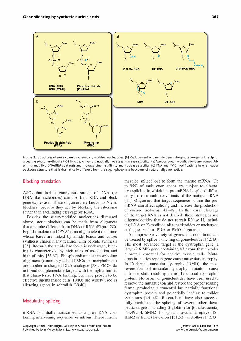
Figure 2. Structures of some common chemically modified nucleotides. (A) Replacement of a non-bridging phosphate oxygen with sulphurgives the phosphorothioate (PS) linkage, which dramatically increases nuclease stability. (B) Various sugar modifications are compatiblewith unmodified DNA/RNA synthesis and increase binding affinity and nuclease stability. (C) PNA and PMO modifications have a neutralbackbone structure that is dramatically different from the sugar-phosphate backbone of natural oligonucleotides.
Blocking translation
ASOs that lack a contiguous stretch of DNA (orDNA-like nucleotides) can also bind RNA and blockgene expression. These oligomers are known as ‘stericblockers’ because they act by blocking the ribosomerather than facilitating cleavage of RNA.
Besides the sugar-modified nucleotides discussedabove, steric blockers can be made from oligomersthat are quite different from DNA or RNA (Figure 2C).Peptide nucleic acid (PNA) is an oligonucleotide mimicwhose bases are linked by amide bonds and whosesynthesis shares many features with peptide synthesis[35]. Because the amide backbone is uncharged, bind-ing is characterized by high rates of association andhigh affinity [36,37]. Phosphorodiamidate morpholinooligomers (commonly called PMOs or ‘morpholinos’)are another uncharged DNA analogue [38]. PMOs donot bind complementary targets with the high affinitiesthat characterize PNA binding, but have proven to beeffective agents inside cells. PMOs are widely used assilencing agents in zebrafish [39,40].
Modulating splicing
mRNA is initially transcribed as a pre-mRNA con-taining intervening sequences or introns. These introns
must be spliced out to form the mature mRNA. Upto 95% of multi-exon genes are subject to alterna-tive splicing in which the pre-mRNA is spliced differ-ently to form multiple variants of the mature mRNA[41]. Oligomers that target sequences within the pre-mRNA can affect splicing and increase the productionof desired isoforms [42–48]. In this case, cleavageof the target RNA is not desired; these strategies useoligonucleotides that do not recruit RNase H, includ-ing LNA or 2′-modified oligonucleotides or unchargedanalogues such as PNA or PMO oligomers.
An impressive variety of genes and conditions canbe treated by splice-switching oligonucleotides [42,43].The most advanced target is the dystrophin gene, alarge (2.6 Mb) gene containing 97 exons that encodesa protein essential for healthy muscle cells. Muta-tions in the dystrophin gene cause muscular dystrophy.In Duchenne muscular dystrophy (DMD), the mostsevere form of muscular dystrophy, mutations causea frame shift resulting in no functional dystrophinprotein. However, oligonucleotides have been used toremove the mutant exon and restore the proper readingframe, producing a truncated but partially functionaldystrophin protein and potentially leading to mildersymptoms [46–48]. Researchers have also success-fully modulated the splicing of several other thera-peutic targets, including β-globin (for β-thalassaemia)[44,49,50], SMN2 (for spinal muscular atrophy) [45],HER2 or Bcl-x (for cancer) [51,52], and others [42,43].
Copyright 2011 Pathological Society of Great Britain and Ireland. J Pathol 2012; 226: 365–379Published by John Wiley & Sons, Ltd. www.pathsoc.org.uk www.thejournalofpathology.com
368 JK Watts and DR Corey
Targeting miRNAs
MicroRNAs (miRNAs) are endogenous small RNAsthat can regulate normal physiological processes andaffect disease [53–58]. miRNAs act through the RNAipathway and recognize target mRNAs through complexpatterns of recognition that are incompletely under-stood. An increasing number of miRNAs have beenimplicated in physiological processes that affect dis-ease, and interfering with these miRNAs might increasethe expression of their target genes and provide thera-peutic lead compounds.
ASOs that are complementary to miRNAs canblock their function [59–61]. For example, miR-122is an abundant liver-specific miRNA implicated ina variety of diseases including cancer and hepatitisC [62]. Oligonucleotides complementary to miR-122have been shown to alter liver metabolism [63,64]and block hepatitis C virus replication [65,66]. Var-ious chemistries have been shown to be active asinhibitors of miRNA function, including PNA [67],LNA [65–68], 2′-O-Me [69], 2′-MOE [64], and mor-pholino [70].
siRNAs
Over the past decade, double-stranded short interferingRNAs (siRNAs) have become widely used tools forsilencing gene expression. When a duplex RNA enterscells, it binds the protein machinery of the RNA-induced silencing complex (RISC) [71]. SyntheticRNAs used for gene silencing are usually 19–22 bpduplexes. This length is sufficient to form a stableduplex and be recognized by RISC, but short enough toavoid most of the strong interferon response provokedby duplexes greater than 30 bp in length.
Since publication of the first report of gene silencingin mammalian cells in 2001 [72], siRNAs have been thesubject of thousands of experimental studies aimed atexamining function. While antisense oligonucleotidescontinue to be used for gene silencing, the robust natureof siRNAs and the relative ease of identifying activesiRNAs have made them a favoured silencing tool formany laboratories.
Chemical modifications and duplex RNAs
Unmodified duplex RNA is surprisingly stable andchemical modification of siRNAs is usually not essen-tial for silencing gene expression in cultured cells(see Table 1). In vivo, however, unmodified siRNAsare not highly active and chemical modification cansignificantly improve their properties [73]. Chemicallymodified siRNAs can feature improved nuclease stabil-ity and an associated increase in the duration of action[74–76]. Unmodified RNA is also rapidly cleared [77]and chemical modification, complexation with carrier
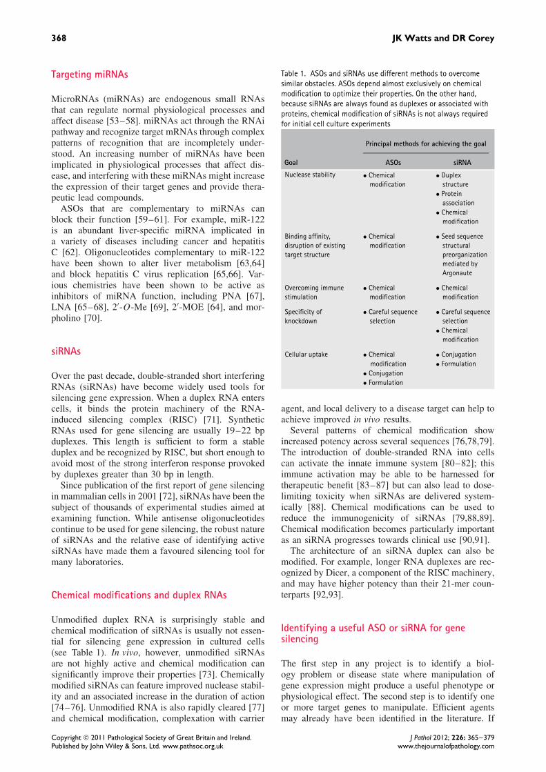
Table 1. ASOs and siRNAs use different methods to overcomesimilar obstacles. ASOs depend almost exclusively on chemicalmodification to optimize their properties. On the other hand,because siRNAs are always found as duplexes or associated withproteins, chemical modification of siRNAs is not always requiredfor initial cell culture experiments
Principal methods for achieving the goal
Goal ASOs siRNA
Nuclease stability • Chemicalmodification
• Duplexstructure
• Proteinassociation
• Chemicalmodification
Binding affinity,disruption of existingtarget structure
• Chemicalmodification
• Seed sequencestructuralpreorganizationmediated byArgonaute
Overcoming immunestimulation
• Chemicalmodification
• Chemicalmodification
Specificity ofknockdown
• Careful sequenceselection
• Careful sequenceselection
• Chemicalmodification
Cellular uptake • Chemical • Conjugationmodification • Formulation
• Conjugation• Formulation
agent, and local delivery to a disease target can help toachieve improved in vivo results.
Several patterns of chemical modification showincreased potency across several sequences [76,78,79].The introduction of double-stranded RNA into cellscan activate the innate immune system [80–82]; thisimmune activation may be able to be harnessed fortherapeutic benefit [83–87] but can also lead to dose-limiting toxicity when siRNAs are delivered system-ically [88]. Chemical modifications can be used toreduce the immunogenicity of siRNAs [79,88,89].Chemical modification becomes particularly importantas an siRNA progresses towards clinical use [90,91].
The architecture of an siRNA duplex can also bemodified. For example, longer RNA duplexes are rec-ognized by Dicer, a component of the RISC machinery,and may have higher potency than their 21-mer coun-terparts [92,93].
Identifying a useful ASO or siRNA for genesilencing
The first step in any project is to identify a biol-ogy problem or disease state where manipulation ofgene expression might produce a useful phenotype orphysiological effect. The second step is to identify oneor more target genes to manipulate. Efficient agentsmay already have been identified in the literature. If
Copyright 2011 Pathological Society of Great Britain and Ireland. J Pathol 2012; 226: 365–379Published by John Wiley & Sons, Ltd. www.pathsoc.org.uk www.thejournalofpathology.com

Gene silencing by synthetic nucleic acids 369
no such data exist, commercial suppliers may offerduplex RNAs that have been validated for inhibition ofthe target gene. If commercial RNAs are unavailable,are too expensive, or are inadequate, investigators canexperimentally identify ASOs or siRNAs themselves.
There are algorithms for siRNA design [94–96],but none is perfect and it will probably be necessaryto test several duplex RNAs to find sequences thatare sufficiently active and selective. Various groupshave proposed computational [97–99] or experimen-tal [99–104] methods for identifying potent ASOs.Aspects of mRNA secondary structure can be roughlypredicted, but algorithms such as the popular mFOLDdo not take into account factors such as tertiary struc-ture or the involvement of RNA binding proteins, andso they are of limited utility in practice [105]. Ulti-mately, if a group is serious about finding a potentASO, they should test as many oligonucleotides as timeand budget permit [106,107].
Control experiments: you can never have toomany
Regardless of whether ASOs or siRNAs are used forgene silencing, it is essential to use proper controls[108]. Understanding off-target effects can help inves-tigators choose controls appropriately. ASOs and siR-NAs are large synthetic molecules and can act inways independent of Watson–Crick base pairing. ForASOs, this includes non-specific binding to proteinsboth in serum and inside cells. Phosphorothioate back-bone oligonucleotides are particularly liable to bindproteins—this is the source of their slower clearancefrom serum, but also the source of much of their tox-icity [109]. Oligonucleotides can fold into complexsecondary structures and bind proteins in a sequence-specific manner related to shape rather than pairing[110].
For siRNAs, cells can recognize double-strandedRNA and activate the innate immune system [80–82]and this immunostimulatory activity has been misinter-preted as RNA interference effects [111,112]. Finally,high concentrations of siRNA can saturate the RNAimachinery, leading to a global perturbation of miRNA-mediated regulation [113–115].
An ASO or siRNA will always have partial comple-mentarity to non-target transcripts, and this can causeunintended gene repression and misleading phenotypes[116,117]. For siRNAs, one of the most common off-target effects occurs through 7–8 nucleotide comple-mentarity at the 5′-end of the guide strand (the so-called‘seed region’) to sites in the 3′-untranslated region ofother genes. This type of base pairing is a prerequi-site for miRNA-mediated gene repression, and partiallycomplementary siRNA duplexes can enter the miRNApathway and repress non-target transcripts [117,118].Careful use of chemistry and duplex design can helpto alleviate this type of off-target effect. For example,
A B
Figure 3. Typical mismatched and scrambled controls. (A) A duplexwith several pairs of bases exchanged (from one strand to the other)should maintain very similar properties to the parent duplex butlose affinity for its target mRNA. (B) A scrambled control canbe made by moving blocks of bases with respect to the parentoligomer.
including two 2′-O-Me-RNA nucleotides at the 5′-endof the siRNA reduces its ability to enter the miRNApathway [117,119]. siRNA designs that ensure loadingof the correct guide strand into RISC can also help toreduce off-target effects [120–126].
It is impossible to avoid some level of off-target effects in oligonucleotide-mediated gene silenc-ing—the goal is to minimize them by careful use ofchemistry and to use multiple approaches with non-overlapping off-target effects so that an observed phe-notype can be confidently ascribed to recognition ofthe desired target. For example, an investigator maybe testing the hypothesis that inhibition of a proteinwill lead to decreased cell proliferation. How can onebuild a case that the observed protein knockdown anddecreased cell proliferation are not indirect effects?Several types of controls are recommended for anyoligonucleotide experiment:
• When multiple ASOs or siRNAs target the samegene, they should have the same on-target effectbut different off-target effects. Thus, possessionof multiple ASOs or siRNAs that are complemen-tary to the target mRNA and produce the samephenotype provides one piece of evidence sup-porting a relationship between recognition of thetarget mRNA, gene silencing, and phenotype.
• Experiments should include negative controlcompounds containing mismatched bases relativeto the target mRNA or having blocks of basesmoved relative to the sequence of the parentoligomer (Figure 3). Sequences that are closelyrelated to the oligonucleotide of interest are morelikely to have similar immunogenic or other off-target effects, making them better controls thantotally unrelated sequences. An ideal mismatchedor scrambled control does not significantly per-turb the levels of a gene of interest.
• Protein and RNA levels of the target gene shouldboth be tested where possible. If using siRNAor an RNase H competent ASO, both proteinand RNA should decrease. A different result
Copyright 2011 Pathological Society of Great Britain and Ireland. J Pathol 2012; 226: 365–379Published by John Wiley & Sons, Ltd. www.pathsoc.org.uk www.thejournalofpathology.com

370 JK Watts and DR Corey
should raise suspicion about the mechanism ofthe silencing observed.
• The dose–response of any knockdown should betested and experiments should be carried out atthe lowest possible concentration.
• For gapmers or siRNAs, a technique called rapidamplification of 5′-cDNA ends (5′-RACE) can beused to verify that the targeted transcript is beingcleaved at the predicted site [127–130]. Whilevaluable, it is important to note that 5′-RACEmerely detects cleavage; it is not quantitative andis not an indication of efficiency.
• Finally, when possible, the ultimate control forgene silencing experiments is a functional rescueby an exogenous copy of the gene of interest con-taining silent mutations at the oligonucleotide’starget site. Nevertheless, these experiments canbe quite challenging, depending on the biologicalsystem being studied.
Off-target effects are a significant concern for lab-oratory use and clinical development of nucleic acidacids. Unintended phenotypes are, however, a concernfor the development of any other type of drug includingsmall molecules and proteins. The solution for mini-mizing off-target effects for nucleic acids is the same asfor other classes of drug—iterative testing and rationaloptimization.
Introducing ASOs or siRNAs into cultured cells
The most common method for promoting uptake ofASOs and siRNAs in cell culture involves the useof cationic lipids to transfect nucleic acids. Mixingcationic lipid with negatively charged nucleic acidsyields a complex that can cross cell membranes andrelease active oligonucleotide into the cytoplasm ofcells.
Many different cationic lipids are available andactivity depends on the cell line used. Even for agiven cell line, the preferred lipid may vary dependingon whether an ASO or siRNA is being transfected.Not all cell lines can be transfected using cationiclipid, and if literature precedent is unavailable, it maybe necessary to experimentally test different cationiclipids to find one that can successfully transfect acell line of interest. It is also possible to electroporateoligonucleotides into cells [131–133]. This method canbe highly effective and useful for cell lines that cannotbe readily transfected by lipid, but requires specializedequipment and expertise.
Recently, it has become apparent that active ASOscan freely enter some cell lines without the need toadd lipid [134,135]. The transfection protocol is thussimplified and any off-target effects due to exposureof cells to lipid are avoided. The method can also beused with cell lines that are not compatible with lipid-mediated transfection. However, higher concentrations
of ASO are needed relative to the amounts used inlipid-mediated transfections.
Delivery of ASOs and siRNAs in vivo
As gene silencing technologies move from culturedcells into animal models and ultimately clinical appli-cation, the challenge of delivery increases. Deliveringoligonucleotides in whole organisms requires cross-ing many barriers [136,137]. Degradation by serumnucleases, clearance by the kidney, or inappropriatebiodistribution can prevent the oligonucleotide fromever reaching its target organ. The oligonucleotide mustpass through the blood vessel wall and navigate theinterstitial space and extracellular matrix. Finally, ifthe oligonucleotide succeeds in reaching the appropri-ate cell membrane, it will usually be taken up into anendosome, from which it must escape to be active.
ASOs are usually delivered in saline and relyon chemical modifications to enable uptake. Theirphosphorothioate backbone binds to serum proteins,slowing excretion by the kidney [109]. The aro-matic nucleobases also interact with other hydrophobicmolecules in serum and on cell surfaces. Many typesof cells in vivo express surface receptors that activelytake up oligonucleotides; these are often lost when cellsare cultured, which explains why lipid seems moreimportant for delivering ASOs in culture than in vivo[138].
Delivery is even more challenging for duplex RNAsthan single-stranded oligonucleotides. In an siRNA, allof the aromatic nucleobases are on the inside, leav-ing only heavily hydrated phosphates on the outsideof the duplex. This hydrated surface interacts poorlywith cell surfaces and is rapidly excreted in the urine.Thus, researchers have invested heavily in the develop-ment of delivery vehicles for siRNAs [137,139–142].The predominant technologies for delivering siRNAsinvolve complexing the RNA with cationic and neu-tral lipids [139,143,144], although encouraging resultshave also been obtained using peptide transductiondomains [145] and cationic polymers [130]. IncludingPEGylated lipids in the formulation prolongs the cir-culating half-life of the particles [146]. Conjugation ofcholesterol to one strand of the siRNA gave effectiveknockdown in the liver of mice [147], but the quantitiesof material required (50 mg/kg) were several orders ofmagnitude higher than current lipid-based formulations(as low as 0.01 mg/kg [148]).
Lipid-based formulations are ideal for targeting theliver, since lipid nanoparticles are readily taken upby liver cells. For targeting other organs follow-ing systemic administration, researchers have conju-gated various ligands to the siRNA itself or includedthem as part of a formulation. Promising strategiesinclude the use of aptamers [149], antibodies/fragments[150], vitamins [151], and other targeting ligands[130,152].
Copyright 2011 Pathological Society of Great Britain and Ireland. J Pathol 2012; 226: 365–379Published by John Wiley & Sons, Ltd. www.pathsoc.org.uk www.thejournalofpathology.com

Gene silencing by synthetic nucleic acids 371
siRNA or ASO?
For cell culture experiments, siRNAs will often be thebetter choice. It is relatively simpler to discover potentsiRNAs and it may be easier to obtain siRNAs sinceunmodified RNA works with high potency. ASOs, onthe other hand, must contain chemical modificationsto be active inside cells. For experiments where thegoal is to develop compounds for testing in animalsor investigate therapeutic development, the choice ismore complex.
Several studies have directly compared the activityof various ASOs and siRNAs [105,153–157]. Thesestudies can be misleading if one of the compoundscontains suboptimal chemistry or sequence selection(eg first-generation PS-DNA ASOs); both potent andeffective antisense oligonucleotides or siRNAs cangenerally be found if researchers are willing to investin optimizing each technology [158]. An ideal targetsequence for an ASO is not necessarily ideal for ansiRNA, and vice versa.
In vivo, the choice of ASO versus siRNA is unset-tled and will continue to evolve over the next decade.The challenges involved in delivering oligonucleotidesto a given target tissue should be considered beforechoosing between them. For example, in animal mod-els of Huntington’s disease, antisense oligonucleotidesor siRNAs have been infused directly into the cen-tral nervous system. In the case of single-strandedoligonucleotides, researchers observed wide distribu-tion throughout the mouse CNS including deep-brainpenetration [159]. In contrast, others found that siRNAinfused into the monkey brain penetrated into brain tis-sue only up to about 12 mm from the site of infusion[159].
ASOs and siRNAs share important similarities asdrug candidates. Both platforms are intended to mod-ulate gene expression. Both are nucleic acids and con-tain an antisense strand intended to recognize a targetmRNA. They also have important differences. ASOshave one strand, while siRNAs have two, a basic factthat may lower cost and simplify delivery. On the otherhand, siRNAs have proven to be a more robust tech-nology in cell culture in the hands of most users. It isnot clear whether this will be true in vivo, but the pos-sibility that siRNAs might have superior potency forat least some applications is a major driving force fortheir continued development.
Clinical progress
The clinical progress of oligonucleotide drugs has beenslow because realizing the potential for ASOs andduplex RNAs requires inventing a new model forpharmaceutical development that allows large, highlycharged molecules to be synthesized economically,distribute to target tissues, enter cells, and functionwithin acceptable limits for toxicity. Oligonucleotides
are unlike traditional small molecule drugs (<500–700molecular weight) and much effort has been requiredto understand their properties and optimize them.Antibody therapeutics provide a useful comparison.This class of molecules is now a major source of newdrugs, but they also required many years to develop.Oligonucleotides may eventually have similar success.One ASO has been approved by the FDA and at least22 oligonucleotide drugs are in phase II or III clinicaltrials (Table 2). Many more are earlier in the processof clinical development [91,158,160,161].
ASO-mediated gene inhibition in the clinic
ASOs began clinical development in the 1990s withfirst-generation compounds consisting of phosphoroth-ioate DNA. One programme from ISIS Pharmaceuti-cals succeeded, leading to FDA approval of fomivirsenfor treatment of CMV retinitis [162,163]. Developmentwas facilitated by the location of the disease target andmode of administration. Fomivirsen is administereddirectly into the eye, reducing the amount of materialneeded and decreasing concerns about systemic sideeffects. One lesson for future work from these studieswas that local delivery of oligonucleotides can sim-plify clinical studies and contribute to efficient trialdesign.
Other trials, however, were not successful. Drugsfrom Genta, Hybridon, and ISIS Pharmaceuticals failedin phase III clinical trials. Reasons that contributeto the lack of success include (i) incomplete under-standing of the biological target and the consequencesof its repression; (ii) the use of relatively inefficientfirst-generation chemically-modified PS-DNA oligonu-cleotides; and (iii) targeting disease tissues that arenot prime locations for oligonucleotide biodistribution.
More recent trials have begun to revive optimism.Gapmer designs with optimized chemistry have ledto improved potencies [28,164]. Biodistribution ofoligonucleotides is better understood and some of themore promising trials involve inhibiting the expressionof genes in the liver, an organ known to accumulateASO [165]. The importance of convenient markers ofactivity has been recognized. Such markers allow anASO-mediated down-regulation of target gene expres-sion to be demonstrated early in clinical trials, permit-ting resources to be devoted to the most promising drugcandidates.
An example of the new wave of more promisingASOs is mipomersen, an ASO from ISIS Pharma-ceuticals designed to inhibit the expression of ApoB(Figure 4) [166]. Mipomersen is a gapmer contain-ing phosphorothioate-modified DNA and 2′-O-MOE-RNA. Data from animal models show a robust andprolonged repression of ApoB expression [167]. Inpatients, the desired physiological response upon sys-temic administration was demonstrated by monitor-ing serum LDL-cholesterol: all primary, secondary,
Copyright 2011 Pathological Society of Great Britain and Ireland. J Pathol 2012; 226: 365–379Published by John Wiley & Sons, Ltd. www.pathsoc.org.uk www.thejournalofpathology.com

372 JK Watts and DR Corey
Tabl
e2.
Sele
cted
olig
onuc
leot
ide
drug
cand
idat
esin
adva
nced
clin
ical
tria
ls(p
hase
IIor
high
er).
Besi
des
refe
renc
eslis
ted
inth
eta
ble,
info
rmat
ion
was
take
nfr
omco
mpa
nyw
ebsi
tes
and
refs
158
and
160
Drug
Com
pany
Phas
eTa
rget
gene
and
dise
ase
Not
esan
dre
fere
nces
mRN
A-ta
rget
edan
tisen
seol
igon
ucle
otid
edr
ugca
ndid
ates
Mip
omer
sen
ISIS
/Gen
zym
eIII
ApoB
forh
yper
chol
este
rola
emia
PS-M
OE
gapm
er,i
ntra
veno
usde
liver
y;m
etal
lend
poin
tsin
four
phas
eIII
tria
ls[1
66]
OG
X-01
1(C
ustir
sen)
ISIS
/Tev
a/O
ncoG
enex
IIICl
uste
rinfo
rpro
stat
e,N
SCLC
and
brea
stca
ncer
PS-M
OE
gapm
er[1
84]
AP12
009
(Tra
bede
rsen
)An
tisen
sePh
arm
aIII
TGF-
β2
forb
rain
canc
erPS
-DN
AG
S-10
1(A
gani
rsen
)G
ene
Sign
alIII
Insu
linre
cept
orsu
bstr
ate-
1fo
rcor
neal
neov
ascu
lariz
atio
nPS
-DN
A,to
pica
ldel
iver
y(e
yedr
ops)
LOR-
2040
Loru
sII
Ribo
nucl
eotid
ere
duct
ase
forc
ance
rPS
-DN
AAS
M8
Phar
max
isII
Mul
tiple
targ
ets
fora
llerg
icas
thm
aTw
oPS
-DN
AAS
Os,
deliv
ered
byin
hala
tion
Arch
exin
Rexa
hnII
AKT-
1fo
rcan
cer
PS-D
NA
LY21
8130
8IS
IS/L
illy
IISu
rviv
info
rcan
cer
PS-M
OE
gapm
erIS
IS-E
IF4E
RxIS
IS/L
illy
IIeI
F4E
forc
ance
rPS
-MO
Ega
pmer
OG
X-42
7IS
IS/O
ncoG
enex
IIH
sp27
forc
ance
rPS
-MO
Ega
pmer
Alic
afor
sen
ISIS
/Atla
ntic
IIIC
AM-1
foru
lcer
ativ
eco
litis
Avai
labl
ew
hen
requ
este
dby
phys
icia
nsfo
rpat
ient
sw
ithin
flam
mat
ory
bow
eldi
seas
eAE
G35
156
Aege
raII
XIAP
forc
ance
rPS
2′-O
-Me
gapm
er
Splic
e-sw
itchi
ngol
igon
ucle
otid
edr
ugca
ndid
ates
PRO
051
(GSK
2402
968)
Pros
ensa
/GSK
IIIDy
stro
phin
(exo
n51
skip
ping
)for
DMD
20-m
erPS
,ful
ly-2
′ -O
-Me
ASO
[46]
AVI-
4658
AVIB
ioPh
arm
aII
Dyst
roph
in(e
xon
51sk
ippi
ng)f
orDM
D30
-mer
mor
phol
ino
[48]
PRO
044
Pros
ensa
I/II
Dyst
roph
in(e
xon
44sk
ippi
ng)f
orDM
DPS
,ful
ly-2
′ -O
-Me
ASO
Anti-
miR
olig
onuc
leot
ide
drug
cand
idat
eM
iravi
rsen
(SPC
3649
)Sa
ntar
isII
miR
-122
forh
epat
itis
Cvi
rus
LNA-
mod
ified
15-m
eran
ti-m
iRol
igon
ucle
otid
e,de
liver
edin
trav
enou
sly
[66]
siRN
Adr
ugca
ndid
ates
ALN
-RSV
01Al
nyla
m/C
ubis
t/Ky
owa
Kirin
IIRS
V(v
iraln
ucle
ocap
sid)
Unm
odifi
edsi
RNA,
intr
anas
alor
inha
led
PF-6
55Q
uark
/Pfiz
er/S
ilenc
eII
RTP8
01fo
rwet
AMD
and
diab
etic
mac
ular
oede
ma
Chem
ical
lym
odifi
ed,i
ntra
ocul
arQ
PI-1
002
Qua
rk/N
ovar
tis/S
ilenc
eII
p53
fora
cute
rena
lfai
lure
Chem
ical
lym
odifi
ed,i
ntra
veno
usEx
cella
irZa
BeCo
rII
SYK
kina
sefo
rath
sma
Inha
led
Oth
erad
vanc
edol
igon
ucle
otid
edr
ugca
ndid
ates
IMO
-205
5Id
era/
Mer
ckII
TLR-
9ac
tivat
ion
forc
ance
rtre
atm
ent
CpG
-ric
hol
igon
ucle
otid
eca
uses
activ
atio
nof
TLR-
9[1
85]
GRN
163L
(Imet
elst
at)
Ger
onII
Telo
mer
ase
inhi
bito
rfor
canc
ertr
eatm
ent
13-m
erN
3′th
ioph
osph
oram
idat
eol
igon
ucle
otid
e(li
pid
conj
ugat
e);i
nhib
itste
lom
eras
eby
dire
ctbi
ndin
g,no
tan
antis
ense
effe
ct[1
86,1
87]
Copyright 2011 Pathological Society of Great Britain and Ireland. J Pathol 2012; 226: 365–379Published by John Wiley & Sons, Ltd. www.pathsoc.org.uk www.thejournalofpathology.com

Gene silencing by synthetic nucleic acids 373
Figure 4. Mechanism of action of mipomersen.
and tertiary endpoints have been met in four separatephase III clinical trials [166]. Some toxic effects havebeen noted, and while these have been relativelymild, they may (at least initially) limit the patientpopulation to patients who are at the most severe riskfor atherosclerosis [168].
Eleven other traditional antisense oligonucleotidesare in advanced clinical trials (Table 2). Targets rele-vant to cancer are the most highly represented, but thereare also ASOs in trials against asthma, corneal neovas-cularisation, and ulcerative colitis. Many of these ASOscontain optimized chemistry and are taking advantageof the lessons learned over the past two decades interms of delivery.
Modulating splicing in the clinic
Three splice-switching ASOs are in phase II or IIIclinical trials, all of them for treatment of Duchennemuscular dystrophy (DMD) (Figure 5). Prosensa hasdeveloped 2′-O-Me phosphorothioate oligonucleotides[46], while AVI BioPharma has favoured the devel-opment of morpholino oligomers [48]. Both drugsshow promise in clinical development, but two partic-ular challenges face splice-switching oligomers againstDMD—the first is that of delivery. For significant clin-ical benefit, the ASOs would need to enter muscletissue [144], including heart tissue. Currently, all ofthe splice-switching ASOs are delivered naked and arenot efficiently taken up into heart muscle cells (car-diomyocytes) in particular. Delivery is aided to somedegree by the weakened, ‘leaky’ nature of dystrophicmuscle cells, but as the drug begins to take effect andthe muscle cells recover, they are not such easy targetsfor further drug uptake. One possible solution is to usea targeted delivery system such as a cell-penetratingpeptide [169,170]. Peptide-conjugated PMOs (PPMOs)take advantage of an active cell-internalization processand are taken up far more efficiently than unconju-gated PMOs [171–177]. Even at lower doses, theydistribute to more muscle cells body-wide, includinghealthy muscle cells and cardiomyocytes.
The second challenge is that DMD is caused by alarge family of mutations. Exon 51 skipping would
in principle be helpful for ∼13% of DMD patients,including those with deletions of exons 50, 52, 45–50,48–50 or 49–50. Exon 44 skipping could help another∼6% of DMD patients. Further splice-switching ASOscould be developed that would help other patients,but the populations become increasingly small. Itis unclear whether the regulatory process could oneday be adjusted to approve, for example, personal-ized dystrophin-targeted splice-switching ASOs as aclass [170]. Extensive clinical trials for a sequencewith a small target population might be prohibitivelyexpensive.
Clinical trials for ASOs targeting microRNA
The first anti-miR oligonucleotide to be tested inhumans is being developed by Santaris Pharmaceuti-cals. Miravirsen is a 15-mer phosphorothioate oligonu-cleotide containing eight LNA modifications. It iscomplementary to miR-122 and designed to inhibitreplication of hepatitis C virus (HCV) [65,66](Figure 6). Since miravirsen was the first anti-miR tobe tested in humans, no one could predict the effect ofinhibiting a miRNA, thus simultaneously de-repressinga family of mRNA targets in humans. Accordingly,phase I testing started conservatively at 0.2 mg/kgdelivered intravenously or subcutaneously. However,the drug was so well tolerated at the planned end-point dose of 6 mg/kg that the trial was extended toa 12 mg/kg upper dose.
While miravirsen is designed to treat HCV, inhi-bition of miR-122 also lowers plasma cholesterol(Figure 6). Researchers at Santaris made use of thisfact to demonstrate dose-dependent pharmacology intheir phase I trial in spite of the fact that the trialenrolled healthy volunteers. Phase II clinical trials onHCV patients began in September 2010.
siRNAs in the clinic
A decade after the first siRNA experiments [72,178],a dozen siRNA drugs are in clinical development [91].The four most advanced are in phase II trials (Table 2).As with ASOs, some of the earliest drugs to enter trialswere very simple ‘first-generation’ siRNAs containingno chemical modifications. Two of these drugs, bothtargeting the VEGF pathway in the eye, had reachedadvanced clinical trials (phase II and phase III) butwere withdrawn [91]. The therapeutic siRNA field isstill young; however, many valuable lessons have beenlearned from 20 years of clinical work with ASOs thatcan now be applied to siRNA clinical development[179].
While cationic lipid-based formulations are clearlydominant in terms of potency, they deliver siRNAs intothe endosome, the part of the cell where innate immunereceptors are most intensely displayed. This means that
Copyright 2011 Pathological Society of Great Britain and Ireland. J Pathol 2012; 226: 365–379Published by John Wiley & Sons, Ltd. www.pathsoc.org.uk www.thejournalofpathology.com

374 JK Watts and DR Corey
A
B
Figure 5. Mode of action of drug candidates PRO051 and AVI-4568. (A) This DMD patient is missing dystrophin exon 50. Splicing ofthe pre-mRNA gives mature mRNA that is out-of-frame and so no functional dystrophin can be produced. (B) In the presence of asplice-switching ASO that favours exclusion of exon 51, the cell splices exon 49 to exon 52, which restores the reading frame and causestranslation of a shorter but partially functional dystrophin protein.
Figure 6. miR-122 is a liver-specific miRNA that regulates multiplepathways. Therapeutic inhibition of miR-122 by miravirsen blocksHCV replication and lowers plasma cholesterol.
siRNAs delivered by cationic lipid-based formulationsare particularly vulnerable to immune stimulation. Assuch, a phase I drug candidate targeting ApoB fromTekmira Pharmaceuticals (http://www.tekmirapharm.com) was withdrawn from clinical trials after onepatient at the highest dose level showed severe flu-like symptoms typical of an immune response. Tek-mira halted the trial in autumn 2009 and opti-mized both the lipid delivery agent and the siRNAcargo, decreasing the immunogenicity of both, beforereturning to clinical testing of the improved candi-date.
Conclusions and future prospects
The field of oligonucleotide therapeutics has oftenswung from irrational optimism to irrational despair.In the laboratory as well, gene knockdown experimentshave fallen in and out of favour with researchers. Inreality, oligonucleotides are useful tools with strengthsand weaknesses. Different oligonucleotide technolo-gies have different strengths, and many of the pen-dulum swings in the field have caused a switch fromone oligonucleotide technology to another [180–183].If researchers approach the oligonucleotide toolboxwith careful experimentation and the broadest possi-ble understanding, we believe that they will continueto find useful tools for both laboratory experiments andtherapeutic development.
AcknowledgmentThis work was supported by the National Institutes ofHealth (NIGMS 77253 and 73042 to DRC), the RobertA Welch Foundation (I-1244 to DRC), and the NaturalSciences and Engineering Research Council of Canada(postdoctoral fellowship to JKW).
Author contribution statement
JKW and DRC wrote the manuscript.
References
1. Stephenson ML, Zamecnik PC. Inhibition of Rous sarcoma viralRNA translation by a specific oligodeoxyribonucleotide. Proc NatlAcad Sci U S A 1978; 75: 285–288.
Copyright 2011 Pathological Society of Great Britain and Ireland. J Pathol 2012; 226: 365–379Published by John Wiley & Sons, Ltd. www.pathsoc.org.uk www.thejournalofpathology.com

Gene silencing by synthetic nucleic acids 375
2. Zamecnik PC, Stephenson ML. Inhibition of Rous sarcoma virus
replication and cell transformation by a specific oligodeoxynu-
cleotide. Proc Natl Acad Sci USA 1978; 75: 280–284.
3. Agrawal S Remembering Paul C. Zamecnik, M.D., ‘Father of
antisense’ (1912–2009). Oligonucleotides 2010; 20: 47–50.
4. Eckstein F. Developments in RNA chemistry, a personal view.
Biochimie 2002; 84: 841–848.
5. Geary RS, Yu RZ, Levin AA. Pharmacokinetics of phosphoroth-
ioate antisense oligodeoxynucleotides. Curr Opin Invest Drugs
2001; 2: 562–573.
6. Watanabe TA, Geary RS, Levin AA. Plasma protein binding of an
antisense oligonucleotide targeting human ICAM-1 (ISIS 2302).
Oligonucleotides 2006; 16: 169–180.
7. Stein CA, Krieg AM. Problems in interpretation of data derived
from in vitro and in vivo use of antisense oligodeoxynucleotides.
Antisense Res Dev 1994; 4: 67–69.
8. Thiel KW, Giangrande PH. Therapeutic applications of DNA and
RNA aptamers. Oligonucleotides 2009; 19: 209–222.
9. Chiang MY, Chan H, Zounes MA, et al . Antisense oligonu-
cleotides inhibit intercellular adhesion molecule 1 expression by
two distinct mechanisms. J Biol Chem 1991; 266: 18162–18171.
10. Williams DM, Benseler F, Eckstein F. Properties of 2′-fluoro-
thymidine-containing oligonucleotides: interaction with restriction
endonuclease EcoRV. Biochemistry 1991; 30: 4001–4009.
11. Pieken WA, Olsen DB, Aurup H, et al . Structure–function rela-
tionship of hammerhead ribozymes as probed by 2′-modifications.
Nucleic Acids Symp Ser 1991; 24: 51–53.
12. Kawasaki AM, Casper MD, Freier SM, et al . Uniformly modified
2′-deoxy-2′-fluoro-phosphorothioate oligonucleotides as nuclease-
resistant antisense compounds with high affinity and specificity
for RNA targets. J Med Chem 1993; 36: 831–841.
13. Monia BP, Lesnik EA, Gonzalez C, et al . Evaluation of 2′-modified oligonucleotides containing 2′-deoxy gaps as anti-
sense inhibitors of gene expression. J Biol Chem 1993; 268:
14514–14522.
14. Freier SM, Altmann K-H. The ups and downs of nucleic
acid duplex stability: structure–stability studies on chemically-
modified DNA : RNA duplexes. Nucleic Acids Res 1997; 25:
4429–4443.
15. Martin P. A new access to 2′-O-alkylated ribonucleosides and
properties of 2′-O-alkylated oligoribonucleotides. Helv Chim Acta
1995; 78: 486–504.
16. Koshkin AA, Singh SK, Nielsen P, et al . LNA (locked
nucleic acids): synthesis of the adenine, cytosine, guanine, 5-
methylcytosine, thymine and uracil bicyclonucleoside monomers,
oligomerisation, and unprecedented nucleic acid recognition.
Tetrahedron 1998; 54: 3607–3630.
17. Obika S, Nanbu D, Hari Y, et al . Stability and structural
features of the duplexes containing nucleoside analogues with a
fixed N-type conformation, 2′-O,4′-C-methyleneribonucleosides.
Tetrahedron Lett 1998; 39: 5401–5404.
18. Braasch DA, Corey DR. Locked nucleic acid (LNA): fine-tuning
the recognition of DNA and RNA. Chem Biol 2001; 8: 1–7.
19. Leumann CJ. DNA analogues: from supramolecular principles to
biological properties. Bioorg Med Chem 2002; 10: 841–854.
20. Obika S. Development of bridged nucleic acid analogues for anti-
gene technology. Chem Pharm Bull (Tokyo) 2004; 52: 1399–1404.
21. Christensen NK, Petersen M, Nielsen P, et al . A novel
class of oligonucleotide analogues containing 2′-O,3′-C-linked
[3.2.0]bicycloarabinonucleoside monomers: synthesis, thermal
affinity studies, and molecular modeling. J Am Chem Soc 1998;
120: 5458–5463.
22. Rajwanshi VK, Hakansson AE, Sorensen MD, et al . The eight
stereoisomers of LNA (locked nucleic acid): a remarkable family
of strong RNA binding molecules. Angew Chem Int Ed Engl 2000;39: 1656–1659.
23. Morita K, Hasegawa C, Kaneko M, et al . 2′-O,4′-C-ethylene-bridged nucleic acids (ENA): highly nuclease-resistant and ther-modynamically stable oligonucleotides for antisense drug. Bioorg
Med Chem Lett 2002; 12: 73–76.24. Renneberg D, Bouliong E, Reber U, et al . Antisense properties
of tricyclo-DNA. Nucleic Acids Res 2002; 30: 2751–2757.25. Hari Y, Obika, S, Ohnishi R, et al . Synthesis and properties of
2′-O,4′-C-methyleneoxymethylene bridged nucleic acid. Bioorg
Med Chem 2006; 14: 1029–1038.26. Srivastava P, Barman J, Pathmasiri W, et al . Five- and
six-membered conformationally locked 2′,4′-carbocyclic ribo-thymidines: synthesis, structure, and biochemical studies. J Am
Chem Soc 2007; 129: 8362–8379.27. Bramsen JB, Laursen MB, Nielsen AF, et al . A large-scale
chemical modification screen identifies design rules to generatesiRNAs with high activity, high stability and low toxicity. Nucleic
Acids Res 2009; 37: 2867–2881.28. Straarup EM, Fisker N, Hedtjarn M, et al . Short locked nucleic
acid antisense oligonucleotides potently reduce apolipoprotein BmRNA and serum cholesterol in mice and non-human primates.Nucleic Acids Res 2010; 38: 7100–7111.
29. Wu H, Lima WF, Zhang H, et al . Determination of the role ofthe human RNase H1 in the pharmacology of DNA-like antisensedrugs. J Biol Chem 2004; 279: 17181–17189.
30. Minshull J, Hunt T. The use of single-stranded DNA andRNase H to promote quantitative ‘hybrid arrest of translation’ ofmRNA/DNA hybrids in reticulocyte lysate cell-free translations.Nucleic Acids Res 1986; 14: 6433–6451.
31. Nakamura H, Oda Y, Iwai S, et al . How does RNase H recognizea DNA.RNA hybrid? Proc Natl Acad Sci U S A 1991; 88:11535–11539.
32. Wahlestedt C, Salmi P, Good L, et al . Potent and nontoxicantisense oligonucleotides containing locked nucleic acids. Proc
Natl Acad Sci U S A 2000; 97: 5633–5638.33. Damha MJ, Wilds CJ, Noronha A, et al . Hybrids of RNA
and arabinonucleic acids (ANA and 2′F-ANA) are substrates ofribonuclease H. J Am Chem Soc 1998; 120: 12976–12977.
34. Watts JK, Damha MJ. 2′F-Arabinonucleic acids (2′F-ANA)—history, properties, and new frontiers. Can J Chem
2008; 86: 641–656.35. Nielsen PE, Egholm M, Berg RH, et al . Sequence-selective
recognition of DNA by strand displacement with a thymine-substituted polyamide. Science 1991; 254: 1497–1500.
36. Bentin T, Nielsen PE. Enhanced peptide nucleic acid bindingto supercoiled DNA: possible implications for DNA ‘breathing’dynamics. Biochemistry 1996; 35: 8863–8869.
37. Smulevitch SV, Simmons CG, Norton JC, et al . Enhancementof strand invasion by oligonucleotides through manipulation ofbackbone charge. Nature Biotechnol 1996; 14: 1700–1704.
38. Summerton J, Weller D. Morpholino antisense oligomers: design,preparation, and properties. Antisense Nucleic Acid Drug Dev
1997; 7: 187–195.39. Corey DR, Abrams JM. Morpholino antisense oligonucleotides:
tools for investigating vertebrate development. Genome Biol
2001; 2: reviews1015.1–reviews1015.3.40. Bill BR, Petzold AM, Clark KJ, et al . A primer for morpholino
use in zebrafish. Zebrafish 2009; 6: 69–77.41. Pan Q, Shai O, Lee LJ, et al . Deep surveying of alternative splic-
ing complexity in the human transcriptome by high-throughputsequencing. Nature Genet 2008; 40: 1413–1415.
42. Aartsma-Rus A, van Ommen GJ. Antisense-mediated exon skip-ping: a versatile tool with therapeutic and research applications.RNA 2007; 13: 1609–1624.
Copyright 2011 Pathological Society of Great Britain and Ireland. J Pathol 2012; 226: 365–379Published by John Wiley & Sons, Ltd. www.pathsoc.org.uk www.thejournalofpathology.com

376 JK Watts and DR Corey
43. Bauman J, Jearawiriyapaisarn N, Kole R. Therapeutic potentialof splice-switching oligonucleotides. Oligonucleotides 2009; 19:1–13.
44. Dominski Z, Kole R. Restoration of correct splicing in thalassemicpre-mRNA by antisense oligonucleotides. Proc Natl Acad Sci
U S A 1993; 90: 8673–8677.45. Hua Y, Sahashi K, Hung G, et al . Antisense correction of SMN2
splicing in the CNS rescues necrosis in a type III SMA mousemodel. Genes Dev 2010; 24: 1634–1644.
46. Goemans NM, Tulinius M, van den Akker JT, et al . Systemicadministration of PRO051 in Duchenne’s muscular dystrophy.N Engl J Med 2011; 364: 1513–1522.
47. Aartsma-Rus A, van Ommen GJ. Less is more: therapeutic exonskipping for Duchenne muscular dystrophy. Lancet Neurol 2009;8: 873–875.
48. Alter J, Lou F, Rabinowitz A, et al . Systemic delivery ofmorpholino oligonucleotide restores dystrophin expression body-wide and improves dystrophic pathology. Nature Med 2006; 12:175–177.
49. Suwanmanee T, Sierakowska H, Fucharoen S, et al . Repair ofa splicing defect in erythroid cells from patients with beta-thalassemia/HbE disorder. Mol Ther 2002; 6: 718–726.
50. Suwanmanee T, Sierakowska H, Lacerra G, et al . Restorationof human beta-globin gene expression in murine and humanIVS2-654 thalassemic erythroid cells by free uptake of antisenseoligonucleotides. Mol Pharmacol 2002; 62: 545–553.
51. Wan J, Sazani P, Kole R. Modification of HER2 pre-mRNAalternative splicing and its effects on breast cancer cells. Int
J Cancer 2009; 124: 772–777.52. Bauman JA, Li SD, Yang A, et al . Anti-tumor activity of
splice-switching oligonucleotides. Nucleic Acids Res 2010; 38:8348–8356.
53. Bartel DP. MicroRNAs: genomics, biogenesis, mechanism, andfunction. Cell 2004; 116: 281–297.
54. Farazi TA, Spitzer JI, Morozov P, et al . miRNAs in humancancer. J Pathol 2011; 223: 102–115.
55. Garzon R, Marcucci G, Croce CM. Targeting microRNAs incancer: rationale, strategies and challenges. Nature Rev Drug
Discov 2010; 9: 775–789.56. Soifer HS, Rossi JJ, Saetrom P. MicroRNAs in disease and poten-
tial therapeutic applications. Mol Ther 2007; 15: 2070–2079.57. Lu M, Zhang Q, Deng M, et al . An analysis of human microRNA
and disease associations. PLoS One 2008; 3: e3420.58. Jiang Q, Wang Y, Hao Y, et al . miR2Disease: a manually curated
database for microRNA deregulation in human disease. Nucleic
Acids Res 2009; 37: D98–D104.59. Esau CC. Inhibition of microRNA with antisense oligonu-
cleotides. Methods 2008; 44: 55–60.60. Davis S, Propp S, Freier SM, et al . Potent inhibition of microRNA
in vivo without degradation. Nucleic Acids Res 2009; 37: 70–77.61. Montgomery RL, van Rooij E. Therapeutic advances in
microRNA targeting. J Cardiovasc Pharmacol 2011; 57: 1–7.62. Girard M, Jacquemin E, Munnich A, et al . miR-122, a paradigm
for the role of microRNAs in the liver. J Hepatol 2008; 48:648–656.
63. Krutzfeldt J, Rajewsky N, Braich R, et al . Silencing of microR-NAs in vivo with ‘antagomirs’. Nature 2005; 438: 685–689.
64. Esau C, Davis S, Murray SF, et al . miR-122 regulation of lipidmetabolism revealed by in vivo antisense targeting. Cell Metab
2006; 3: 87–98.65. Elmen J, Lindow M, Schutz S, et al . LNA-mediated microRNA
silencing in non-human primates. Nature 2008; 452: 896–899.66. Lanford RE, Hildebrandt-Eriksen ES, Petri A, et al . Therapeutic
silencing of microRNA-122 in primates with chronic hepatitisC virus infection. Science 2010; 327: 198–201.
67. Fabani MM, Gait MJ. miR-122 targeting with LNA/2′-O-methyloligonucleotide mixmers, peptide nucleic acids (PNA), and PNA-peptide conjugates. RNA 2008; 14: 336–346.
68. Orom UA, Kauppinen S, Lund AH. LNA-modified oligonu-cleotides mediate specific inhibition of microRNA function. Gene
2006; 372: 137–141.69. Meister G, Landthaler M, Dorsett Y, et al . Sequence-specific
inhibition of microRNA- and siRNA-induced RNA silencing.RNA 2004; 10: 544–550.
70. Kloosterman WP, Lagendijk AK, Ketting RF, et al . Targetedinhibition of miRNA maturation with morpholinos reveals a rolefor miR-375 in pancreatic islet development. PLoS Biol 2007; 5:e203.
71. Siomi H, Siomi MC. On the road to reading the RNA-interferencecode. Nature 2009; 457: 396–404.
72. Elbashir SM, Harborth J, Lendeckel W, et al . Duplexes of 21-nucleotide RNAs mediate RNA interference in cultured mam-malian cells. Nature 2001; 411: 494–498.
73. Watts JK, Deleavey GF, Damha MJ. Chemically modified siRNA:tools and applications. Drug Discov Today 2008; 13: 842–855.
74. Morrissey DV, Lockridge JA, Shaw L, et al . Potent and persistentin vivo anti-HBV activity of chemically modified siRNAs. Nature
Biotechnol 2005; 23: 1002–1007.75. Morrissey DV, Blanchard K, Shaw L, et al . Activity of stabilized
short interfering RNA in a mouse model of hepatitis B virusreplication. Hepatology 2005; 41: 1349–1356.
76. Allerson CR, Sioufi N, Jarres R, et al . Fully 2′-modified oligonu-cleotide duplexes with improved in vitro potency and stabilitycompared to unmodified small interfering RNA. J Med Chem
2005; 48: 901–904.77. Braasch DA, Paroo Z, Constantinescu A, et al . Biodistribution of
phosphodiester and phosphorothioate siRNA. Bioorg Med Chem
Lett 2004; 14: 1139–1143.78. Koller E, Propp S, Murray H, et al . Competition for RISC binding
predicts in vitro potency of siRNA. Nucleic Acids Res 2006; 34:4467–4476.
79. Deleavey GF, Watts JK, Alain T, et al . Synergistic effectsbetween analogs of DNA and RNA improve the potency ofsiRNA-mediated gene silencing. Nucleic Acids Res 2010; 38:4547–4557.
80. Marques JT, Williams BRG. Activation of the mammalianimmune system by siRNAs. Nature Biotechnol 2005; 23:1399–1405.
81. Robbins M, Judge A, MacLachlan I. siRNA and innate immunity.Oligonucleotides 2009; 19: 89–102.
82. Olejniczak M, Galka P, Krzyzosiak WJ. Sequence-non-specificeffects of RNA interference triggers and microRNA regulators.Nucleic Acids Res 2010; 38: 1–16.
83. Schlee M, Hornung V, Hartmann G. siRNA and isRNA: twoedges of one sword. Mol Ther 2006; 14: 463–470.
84. Poeck H, Besch R, Maihoefer C, et al . 5′-Triphosphate-siRNA:turning gene silencing and Rig-I activation against melanoma.Nature Med 2008; 14: 1256–1263.
85. Kortylewski M, Swiderski P, Herrmann A, et al . In vivo deliveryof siRNA to immune cells by conjugation to a TLR9 agonistenhances antitumor immune responses. Nature Biotechnol 2009;27: 925–932.
86. Gantier MP, Tong S, Behlke MA, et al . Rational design ofimmunostimulatory siRNAs. Mol Ther 2010; 18: 785–795.
87. Khairuddin N, Gantier MP, Blake SJ, et al . siRNA-inducedimmunostimulation through TLR7 promotes antitumoral activityagainst HPV-driven tumors in vivo. Immunol Cell Biol 2011;DOI: 10.1038/icb.2011.19.
88. Judge A, MacLachlan I. Overcoming the innate immune responseto small interfering RNA. Hum Gene Ther 2008; 19: 111–124.
Copyright 2011 Pathological Society of Great Britain and Ireland. J Pathol 2012; 226: 365–379Published by John Wiley & Sons, Ltd. www.pathsoc.org.uk www.thejournalofpathology.com

Gene silencing by synthetic nucleic acids 377
89. Judge AD, Bola G, Lee ACH, et al . Design of noninflammatorysynthetic siRNA mediating potent gene silencing in vivo. MolTher 2006; 13: 494–505.
90. Corey DR. Chemical modification: the key to clinical applicationof RNA interference? J Clin Invest 2007; 117: 3615–3622.
91. Watts JK, Corey DR. Clinical status of duplex RNA. Bioorg MedChem Lett 2010; 20: 3203–3207.
92. Kim D-H, Behlke MA, Rose SD, et al . Synthetic dsRNA Dicersubstrates enhance RNAi potency and efficacy. Nature Biotechnol2005; 23: 222–226.
93. Collingwood MA, Rose SD, Huang L, et al . Chemical modifica-tion patterns compatible with high potency dicer-substrate smallinterfering RNAs. Oligonucleotides 2008; 18: 187–200.
94. Yuan B, Latek R, Hossbach M, et al . siRNA Selection Server:an automated siRNA oligonucleotide prediction server. NucleicAcids Res 2004; 32: W130–W134.
95. Boese Q, Leake D, Reynolds A, et al . Mechanistic insights aidcomputational short interfering RNA design. Methods Enzymol2005; 392: 73–96.
96. Huesken D, Lange J, Mickanin C, et al . Design of a genome-wide siRNA library using an artificial neural network. NatureBiotechnol 2005; 23: 995–1001.
97. Sczakiel G, Homann M, Rittner K. Computer-aided search foreffective antisense RNA target sequences of the human immuno-deficiency virus type 1. Antisense Res Dev 1993; 3: 45–52.
98. Stull RA, Taylor LA, Szoka FC Jr. Predicting antisense oligonu-cleotide inhibitory efficacy: a computational approach using his-tograms and thermodynamic indices. Nucleic Acids Res 1992; 20:3501–3508.
99. Sczakiel G. Theoretical and experimental approaches to designeffective antisense oligonucleotides. Front Biosci 2000; 5:D194–D201.
100. Gifford LK, Opalinska JB, Jordan D, et al . Identification ofantisense nucleic acid hybridization sites in mRNA moleculeswith self-quenching fluorescent reporter molecules. Nucleic AcidsRes 2005; 33: e28.
101. Rittner K, Burmester C, Sczakiel G. In vitro selection of fast-hybridizing and effective antisense RNAs directed against thehuman immunodeficiency virus type 1. Nucleic Acids Res 1993;21: 1381–1387.
102. Ho SP, Bao Y, Lesher T, et al . Mapping of RNA accessible sitesfor antisense experiments with oligonucleotide libraries. NatureBiotechnol 1998; 16: 59–63.
103. Kronenwett R, Haas R, Sczakiel G. Kinetic selectivity of com-plementary nucleic acids: bcr-abl-directed antisense RNA andribozymes. J Mol Biol 1996; 259: 632–644.
104. Milner N, Mir KU, Southern EM. Selecting effective antisensereagents on combinatorial oligonucleotide arrays. Nature Biotech-nol 1997; 15: 537–541.
105. Rudnick SI, Swaminathan J, Sumaroka M, et al . Effects of localmRNA structure on posttranscriptional gene silencing. Proc NatlAcad Sci U S A 2008; 105: 13787–13792.
106. Bacon TA, Wickstrom E. Walking along human c-myc mRNAwith antisense oligodeoxynucleotides: maximum efficacy at the5′ cap region. Oncogene Res 1991; 6: 13–19.
107. Stewart AJ, Canitrot Y, Baracchini E, et al . Reduction of expres-sion of the multidrug resistance protein (MRP) in human tumorcells by antisense phosphorothioate oligonucleotides. BiochemPharmacol 1996; 51: 461–469.
108. [No authors listed]. Whither RNAi? Nature Cell Biol 2003; 5:489–490.
109. Levin AA. A review of issues in the pharmacokinetics and tox-icology of phosphorothioate antisense oligonucleotides. BiochimBiophys Acta 1999; 1489: 69–84.
110. Bunka DHJ, Stockley PG. Aptamers come of age - at last. NatRev Microbiol 2006; 4: 588–596.
111. Robbins M, Judge A, Ambegia E, et al . Misinterpreting the
therapeutic effects of small interfering RNA caused by immune
stimulation. Hum Gene Ther 2008; 19: 991–999.112. Kleinman ME, Yamada K, Takeda A, et al . Sequence- and
target-independent angiogenesis suppression by siRNA via TLR3.
Nature 2008; 452: 591–597.113. Yi R, Doehle BP, Qin Y, et al . Overexpression of exportin 5
enhances RNA interference mediated by short hairpin RNAs and
microRNAs. RNA 2005; 11: 220–226.114. Grimm D, Streetz KL, Jopling CL, et al . Fatality in mice
due to oversaturation of cellular microRNA/short hairpin RNA
pathways. Nature 2006; 441: 537–541.115. Khan AA, Betel D, Miller ML, et al . Transfection of small RNAs
globally perturbs gene regulation by endogenous microRNAs.
Nature Biotechnol 2009; 27: 549–555.116. Jackson AL, Bartz SR, Schelter J, et al . Expression profiling
reveals off-target gene regulation by RNAi. Nature Biotechnol
2003; 21: 635–637.117. Fedorov Y, Anderson EM, Birmingham A, et al . Off-target
effects by siRNA can induce toxic phenotype. RNA 2006; 12:
1188–1196.118. Jackson AL, Burchard J, Schelter J, et al . Widespread siRNA
‘off-target’ transcript silencing mediated by seed region sequence
complementarity. RNA 2006; 12: 1179–1187.119. Jackson AL, Burchard J, Leake D, et al . Position-specific chemi-
cal modification of siRNAs reduces ‘off-target’ transcript silenc-
ing. RNA 2006; 12: 1197–1205.120. Bramsen JB, Laursen MB, Damgaard CK, et al . Improved
silencing properties using small internally segmented interfering
RNAs. Nucleic Acids Res 2007; 35: 5886–5897.121. Chen PY, Weinmann L, Gaidatzis D, et al . Strand-specific 5′-O-
methylation of siRNA duplexes controls guide strand selection
and targeting specificity. RNA 2008; 14: 263–274.122. Sano M, Sierant M, Miyagishi M, et al . Effect of asymmetric
terminal structures of short RNA duplexes on the RNA interfer-
ence activity and strand selection. Nucleic Acids Res 2008; 36:
5812–5821.123. Sun X, Rogoff HA, Li CJ. Asymmetric RNA duplexes mediate
RNA interference in mammalian cells. Nature Biotechnol 2008;
26: 1379–1382.124. Ui-Tei K, Naito Y, Zenno S, et al . Functional dissection of siRNA
sequence by systematic DNA substitution: modified siRNA with
a DNA seed arm is a powerful tool for mammalian gene silencing
with significantly reduced off-target effect. Nucleic Acids Res
2008; 36: 2136–2151.125. Chang CI, Yoo JW, Hong SW, et al . Asymmetric shorter-duplex
siRNA structures trigger efficient gene silencing with reduced
nonspecific effects. Mol Ther 2009; 17: 725–732.126. Lapierre J, Salomon W, Cardia J, et al . Potent and systematic
RNAi mediated silencing with single oligonucleotide compounds.
RNA 2011; 17: 1032–1037.127. Lasham A, Herbert M, Coppieters ‘t Wallant N, et al . A rapid
and sensitive method to detect siRNA-mediated mRNA cleavage
in vivo using 5′ RACE and a molecular beacon probe. Nucleic
Acids Res 2010; 38: e19.128. Alvarez R, Elbashir S, Borland T, et al . RNA interference-
mediated silencing of the respiratory syncytial virus nucleocapsid
defines a potent antiviral strategy. Antimicrob Agents Chemother
2009; 53: 3952–3962.129. Judge AD, Robbins M, Tavakoli I, et al . Confirming the RNAi-
mediated mechanism of action of siRNA-based cancer therapeu-
tics in mice. J Clin Invest 2009; 119: 661–673.130. Davis ME, Zuckerman JE, Choi CHJ, et al . Evidence of RNAi
in humans from systemically administered siRNA via targeted
nanoparticles. Nature 2010; 464: 1067–1070.
Copyright 2011 Pathological Society of Great Britain and Ireland. J Pathol 2012; 226: 365–379Published by John Wiley & Sons, Ltd. www.pathsoc.org.uk www.thejournalofpathology.com

378 JK Watts and DR Corey
131. Bergan R, Hakim F, Schwartz GN, et al . Electroporation ofsynthetic oligodeoxynucleotides: a novel technique for ex vivobone marrow purging. Blood 1996; 88: 731–741.
132. Spiller DG, Giles RV, Grzybowski J, et al . Improving the intracel-lular delivery and molecular efficacy of antisense oligonucleotidesin chronic myeloid leukemia cells: a comparison of streptolysin-O permeabilization, electroporation, and lipophilic conjugation.Blood 1998; 91: 4738–4746.
133. Li S (ed). Electroporation Protocols: Preclinical and ClinicalGene Medicine(Methods in Molecular Biology, vol 423). HumanaPress: New York, 2008.
134. Stein CA, Hansen JB, Lai J, et al . Efficient gene silencingby delivery of locked nucleic acid antisense oligonucleotides,unassisted by transfection reagents. Nucleic Acids Res 2010; 38:e3.
135. Zhang Y, Qu Z, Kim S, et al . Down-modulation of cancer targetsusing locked nucleic acid (LNA)-based antisense oligonucleotideswithout transfection. Gene Ther 18: 326–333.
136. Wang J, Lu Z, Wientjes MG, et al . Delivery of siRNA therapeu-tics: barriers and carriers. AAPS J 2010; 12: 492–503.
137. Juliano R, Alam MR, Dixit V, et al . Mechanisms and strategiesfor effective delivery of antisense and siRNA oligonucleotides.Nucleic Acids Res 2008; 36: 4158–4171.
138. Koller E, Vincent TM, Chappell A, et al . Mechanisms ofsingle-stranded phosphorothioate modified antisense oligonu-cleotide accumulation in hepatocytes. Nucleic Acids Res 2011;39: 4795–4807.
139. Whitehead KA, Langer R, Anderson DG. Knocking down barri-ers: advances in siRNA delivery. Nature Rev Drug Discov 2009;8: 129–138.
140. Grimm D, Kay MA. Therapeutic application of RNAi: is mRNAtargeting finally ready for prime time? J Clin Invest 2007; 117:3633–3641.
141. De Paula D, Bentley MVLB, Mahato RI. Hydrophobization andbioconjugation for enhanced siRNA delivery and targeting. RNA2007; 13: 431–456.
142. Akhtar S, Benter IF. Nonviral delivery of synthetic siRNAsin vivo. J Clin Invest 2007; 117: 3623–3632.
143. Li W, Szoka FC Jr. Lipid-based nanoparticles for nucleic aciddelivery. Pharm Res 2007; 24: 438–449.
144. Tseng YC, Mozumdar S, Huang L. Lipid-based systemic deliveryof siRNA. Adv Drug Deliv Rev 2009; 61: 721–731.
145. Eguchi A, Meade BR, Chang YC, et al . Efficient siRNA deliveryinto primary cells by a peptide transduction domain-dsRNAbinding domain fusion protein. Nature Biotechnol 2009; 27:567–571.
146. Papahadjopoulos D, Allen TM, Gabizon A, et al . Stericallystabilized liposomes: improvements in pharmacokinetics andantitumor therapeutic efficacy. Proc Natl Acad Sci U S A 1991;88: 11460–11464.
147. Soutschek J, Akinc A, Bramlage B, et al . Therapeutic silencingof an endogenous gene by systemic administration of modifiedsiRNAs. Nature 2004; 432: 173–178.
148. Semple SC, Akinc A, Chen J, et al . Rational design of cationiclipids for siRNA delivery. Nature Biotechnol 2010; 28: 172–176.
149. Dassie JP, Liu XY, Thomas GS, et al . Systemic administrationof optimized aptamer-siRNA chimeras promotes regression ofPSMA-expressing tumors. Nature Biotechnol 2009; 27: 839–849.
150. Song E, Zhu P, Lee S-K, et al . Antibody mediated in vivodelivery of small interfering RNAs via cell-surface receptors.Nature Biotechnol 2005; 23: 709–717.
151. Sato Y, Murase K, Kato J, et al . Resolution of liver cirrhosis usingvitamin A-coupled liposomes to deliver siRNA against a collagen-specific chaperone. Nature Biotechnol 2008; 26: 431–442.
152. Kim SS, Ye C, Kumar P, et al . Targeted delivery of siRNA tomacrophages for anti-inflammatory treatment. Mol Ther 2010; 18:993–1001.
153. Ferrari N, Bergeron D, Tedeschi A-L, et al . Characterization ofantisense oligonucleotides comprising 2′-deoxy-2′-fluoro-beta-D-arabinonucleic acid (FANA): specificity, potency, and duration ofactivity. Ann N Y Acad Sci 2006; 1082: 91–102.
154. Stein D, Foster E, Huang SB, et al . A specificity comparison offour antisense types: morpholino, 2′-O-methyl RNA, DNA, andphosphorothioate DNA. Antisense Nucleic Acid Drug Dev 1997;7: 151–157.
155. Summerton JE. Morpholino, siRNA, and S-DNA compared:impact of structure and mechanism of action on off-target effectsand sequence specificity. Curr Top Med Chem 2007; 7: 651–660.
156. Gruenweller A, Wyszko E, Bieber B, et al . Comparison ofdifferent antisense strategies in mammalian cells using lockednucleic acids, 2′-O-methyl RNA, phosphorothioates and smallinterfering RNA. Nucleic Acids Res 2003; 31: 3185–3193.
157. Kretschmer-Kazemi Far R, Sczakiel G. The activity of siRNAin mammalian cells is related to structural target accessibility:a comparison with antisense oligonucleotides. Nucleic Acids Res
2003; 31: 4417–4424.158. Bennett CF, Swayze EE. RNA targeting therapeutics: molecular
mechanisms of antisense oligonucleotides as a therapeutic plat-form. Annu Rev Pharmacol Toxicol 2010; 50: 259–293.
159. Gagnon KT. HD Therapeutics—CHDI Fifth Annual Conference(Meeting Report). IDrugs 2010; 13: 219–223.
160. Kunze D, Kraemer K, Fuessel, S. Antisense oligonucleotides:insights from preclinical studies and clinical trials. In RNA
Technologies and Their Applications, Erdmann V A, BarciszewskiJ (eds). Springer-Verlag: Berlin, 2010; 285–303.
161. Haussecker D. The business of RNAi therapeutics. Hum Gene
Ther 2008; 19: 451–462.162. Azad RF, Driver VB, Tanaka K, et al . Antiviral activity of a
phosphorothioate oligonucleotide complementary to RNA of thehuman cytomegalovirus major immediate-early region. Antimi-
crob Agents Chemother 1993; 37: 1945–1954.163. Grillone LR, Lanz R. Fomivirsen. Drugs Today (Barc) 2001; 37:
245–255.164. Bennett CF. Pharmacological properties of 2′-O-methoxyethyl-
modified oligonucleotides. In Antisense Drug Technology: Prin-
ciples, Strategies, and Applications (2nd edn), Crooke ST (ed).CRC Press: Boca Raton, 2008; 273–303.
165. Levin AA, Yu RZ, Geary RS. Basic principles of the pharma-cokinetics of antisense oligonucleotide drugs. In Antisense Drug
Technology: Principles, Strategies, and Applications (2nd edn),Crooke ST (ed). CRC Press: Boca Raton, 2008; 183–215.
166. Raal FJ, Santos RD, Blom DJ, et al . Mipomersen, an apolipopro-tein B synthesis inhibitor, for lowering of LDL cholesterolconcentrations in patients with homozygous familial hypercholes-terolaemia: a randomised, double-blind, placebo-controlled trial.Lancet 2010; 375: 998–1006.
167. Crooke RM, Graham MJ, Lemonidis KM, et al . An apolipopro-tein B antisense oligonucleotide lowers LDL cholesterol in hyper-lipidemic mice without causing hepatic steatosis. J Lipid Res
2005; 46: 872–884.168. Kling J. Safety signal dampens reception for mipomersen anti-
sense. Nature Biotechnol 2010; 28: 295–297.169. Moulton HM, Moulton JD. Morpholinos and their peptide conju-
gates: therapeutic promise and challenge for Duchenne musculardystrophy. Biochim Biophys Acta 2010; 1798: 2296–2303.
170. Goyenvalle A, Davies KE. Challenges to oligonucleotides-basedtherapeutics for Duchenne muscular dystrophy. Skeletal Muscle
2011; 1: 8.171. Wu B, Moulton HM, Iversen PL, et al . Effective rescue of
dystrophin improves cardiac function in dystrophin-deficient miceby a modified morpholino oligomer. Proc Natl Acad Sci U S A
2008; 105: 14814–14819.
Copyright 2011 Pathological Society of Great Britain and Ireland. J Pathol 2012; 226: 365–379Published by John Wiley & Sons, Ltd. www.pathsoc.org.uk www.thejournalofpathology.com

Gene silencing by synthetic nucleic acids 379
172. Yin H, Moulton HM, Betts C, et al . Functional rescue ofdystrophin-deficient mdx mice by a chimeric peptide-PMO. MolTher 2010; 18: 1822–1829.
173. Yin H, Moulton HM, Betts C, et al . A fusion peptide directsenhanced systemic dystrophin exon skipping and functionalrestoration in dystrophin-deficient mdx mice. Hum Mol Genet2009; 18: 4405–4414.
174. Yin H, Moulton HM, Seow Y, et al . Cell-penetrating peptide-conjugated antisense oligonucleotides restore systemic muscle andcardiac dystrophin expression and function. Hum Mol Genet 2008;17: 3909–3918.
175. Jearawiriyapaisarn N, Moulton HM, Sazani P, et al . Long-termimprovement in mdx cardiomyopathy after therapy with peptide-conjugated morpholino oligomers. Cardiovasc Res 2010; 85:444–453.
176. Jearawiriyapaisarn N, Moulton HM, Buckley B, et al . Sustaineddystrophin expression induced by peptide-conjugated morpholinooligomers in the muscles of mdx mice. Mol Ther 2008; 16:1624–1629.
177. Amantana A, Moulton HM, Cate ML, et al . Pharmacokinetics,biodistribution, stability and toxicity of a cell-penetrating pep-tide–morpholino oligomer conjugate. Bioconjug Chem 2007; 18:1325–1331.
178. Elbashir SM, Lendeckel W, Tuschl T. RNA interference ismediated by 21- and 22-nucleotide RNAs. Genes Dev 2001; 15:188–200.
179. Corey DR. RNAi learns from antisense. Nature Chem Biol 2007;3: 8–11.
180. [No authors listed]. Silence is golden. Nature Biotechnol 2006;24: 306–308.
181. Frederickson RM. RNAi gets vote of confidence from big pharma.Mol Ther 2007; 15: 221–222.
182. Potera C. Antisense—down, but not out. Nature Biotechnol 2007;25: 497–499.
183. Schmidt C. RNAi momentum fizzles as pharma shifts priorities.Nature Biotechnol 2011; 29: 93–94.
184. Zoubeidi A, Chi K, Gleave M. Targeting the cytoprotectivechaperone, clusterin, for treatment of advanced cancer. ClinCancer Res 2010; 16: 1088–1093.
185. Agrawal S, Kandimalla ER. Synthetic agonists of Toll-likereceptors 7, 8 and 9. Biochem Soc Trans 2007; 35: 1461–1467.
186. Harley CB. Telomerase and cancer therapeutics. Nature RevCancer 2008; 8: 167–179.
187. Joseph I, Tressler R, Bassett E, et al . The telomerase inhibitorimetelstat depletes cancer stem cells in breast and pancreaticcancer cell lines. Cancer Res 2010; 70: 9494–9504.
Copyright 2011 Pathological Society of Great Britain and Ireland. J Pathol 2012; 226: 365–379Published by John Wiley & Sons, Ltd. www.pathsoc.org.uk www.thejournalofpathology.com