significance of the dissociation of dna2 by flap...
TRANSCRIPT

Significance of the Dissociation of Dna2 by Flap Endonuclease 1 toOkazaki Fragment Processing in Saccharomyces cerevisiae
Jason A. Stewart†, Judith L. Campbell‡, and Robert A. Bambara†§†Department of Biochemistry and Biophysics, University of Rochester School of Medicine
and Dentistry, Rochester, New York, 14642‡Braun Laboratories, California Institute of Technology, Pasadena, California, 91125
Running Title: Significance of FEN1 Removal of Flap-Bound Dna2§Address Correspondence to: Robert A. Bambara, Department of Biochemistry and Biophysics,University of Rochester School of Medicine and Dentistry, 601 Elmwood Ave., Box 712, Rochester, NewYork 14642, Tel. 585 275 3269; Fax. 585 275 6007; E-mail: [email protected]
Okazaki fragments are initiated byshort RNA/DNA primers, which are displacedinto flap intermediates for processing. Flapendonuclease 1 (FEN1) and Dna2 areresponsible for flap cleavage. Replicationprotein A (RPA)-bound flaps inhibit cleavageby FEN1 but stimulate Dna2, requiring thatDna2 cleave prior to FEN1. Upon cleavage,Dna2 leaves a short flap, which is then cut byFEN1 forming a nick for ligation. Bothenzymes require a flap with a free 5'-end fortracking to the cleavage sites. Previously, wedemonstrated that FEN1 disengages thetracking mechanism of Dna2 to remove it fromthe flap. To determine why the disengagementmechanism evolved, we measured FEN1dissociation of Dna2 on short RNA and DNAflaps, which occur during flap processing.Dna2 tracked onto these flaps but could notcleave, presenting a block to FEN1 entry.However, FEN1 disengaged these non-productively bound Dna2 molecules,proceeding on to conduct proper cleavage.These results clarify the importance ofdisengagement. Additional results showed thatflap substrate recognition and tracking byFEN1, as occur during fragment processing,are required for effective displacement of theflap-bound Dna2. Dna2 was recently shown todissociate flap-bound RPA, independent ofcleavage. Using a nuclease-defective Dna2mutant, we reconstituted the sequentialdissociation reactions in the proposedRPA/Dna2/FEN1 pathway showing that, evenwithout cutting, Dna2 enables FEN1 to cleaveRPA-coated flaps. In summary, RPA, Dna2,and FEN1 have evolved highly coordinatedbinding properties enabling one protein to
succeed the next for proper and efficientOkazaki flap processing.
During eukaryotic DNA replication,synthesis of the leading strand occurs incontinuous fashion in the direction of DNAunwinding. In contrast, the lagging strand isreplicated in a discontinuous fashion via shortOkazaki fragments. Each Okazaki fragment isbetween 100 to 150-nucleotides (nt)1 in length. InSaccharomyces cerevisiae, approximately 100,000fragments are created per replication cycle. Thesefragments are initiated by the DNA polymerase(pol) α-primase complex, which synthesizes 10 to12-nt of RNA followed by 20 to 30-nt of DNA(1,2). The pol α -primase complex is thendisplaced by the clamp loader, replication factorC. The toroidal sliding DNA clamp, proliferatingcell nuclear antigen, and DNA pol δ are thenloaded onto the DNA.
Pol δ then synthesizes DNA until itencounters the downstream Okazaki fragment.The downstream RNA/DNA primer is thendisplaced into a flap intermediate by pol δ. Theflap must then be removed and the fragmentsjoined to avoid genome instability (3,4). Removalof the primer is proposed to occur by at least twoparallel-acting pathways in S. cerevisiae (2).
In one pathway, flap endonuclease 1(FEN1) cleaves the flap intermediate to create anicked product for ligation (5,6). FEN1 is astructure-specific, single-stranded nuclease thatrecognizes and cleaves at the base of a flapstructure on both DNA and RNA (7). The FEN1-only model suggests that strand displacementsynthesis by pol δ produces short flaps, which aresuccessively cleaved by FEN1 until the primer has
1
http://www.jbc.org/cgi/doi/10.1074/jbc.M809189200The latest version is at JBC Papers in Press. Published on January 29, 2009 as Manuscript M809189200
Copyright 2009 by The American Society for Biochemistry and Molecular Biology, Inc.
at CA
LIFO
RN
IA IN
ST
ITU
TE
OF
TE
CH
NO
LOG
Y on F
ebruary 3, 2009 w
ww
.jbc.orgD
ownloaded from

been removed. DNA ligase I then joins theresultant nicked product.
Another model of primer removalinvolves both FEN1 and the nuclease/helicaseDna2 (8). Dna2 possesses both single-strandedDNA (ssDNA) nuclease and 5' to 3' ATP-dependent helicase activities (9,10). It isfunctionally conserved from yeast to humans (11-14). Originally identified in a screen for DNAreplication mutants, S. cerevisiae Dna2 has alsobeen shown to play a role in telomere processingand DNA repair (15-21). Recently, it wasidentified as a major nuclease for resection ofdouble-strand breaks, in both S. cerevisiae andXenopus laevis (22,23).
In S. cerevisiae, Dna2 was shown tophysically interact with FEN1 (24). Also, theover-expression of FEN1 rescued the temperaturesensitive phenotype of the dna2-1 nuclease-impaired mutant and over-expression of Dna2rescued the temperature-sensitive rad27Δ (FEN1-null) strain. Moreover, Dna2 interacts with thesingle-stranded binding protein, replication proteinA (RPA), which is involved in both DNAreplication and repair (25). RPA stimulates flapcleavage by Dna2, while repressing cleavage byFEN1. Based on these findings, Seo andcolleagues proposed that pol δ displaces flaps thatbecome long enough to be coated by RPA (8).Once RPA is bound, Dna2 cleavage is required,since FEN1 is inhibited. After cleavage by Dna2,the shortened flap is then free of RPA but must befurther processed because Dna2, unlike FEN1,cannot cut at the base of the flap. Instead it leavesa short flap of approximately 5-nt (8,26), which isremoved by FEN1 to create a nick for ligation.
The FEN1-only model is consistent withresults obtained from the reconstitution of Okazakifragment processing with S. cerevisiae proteins.These results showed that the coordinationbetween pol δ and FEN1 is highly efficient,resulting in mostly mononucleotide cleavageproducts and the production of nicked replicationintermediates for ligation (6). Later, however, weshowed that although mostly short flaps werecreated during strand displacement by pol δ aminor subset of longer flaps arose (27). Thissubset reached a length at which RPA could stablybind, suggesting a role for Dna2 in processing atleast some flaps.
Relevant to this issue, Budd, et. al. (15)showed that the elimination of Pif1, a 5' to 3'helicase, rescued the lethality of the dna2Δ strainin S. cerevisiae. Cell growth was even morerobust when both Pif1 and Pol32, the non-essentialsubunit of pol δ, were simultaneously deleted inthe dna2Δ strain. Significantly, the pol δ mutantlacking Pol32 exhibits decreased stranddisplacement activity (28). Furthermore, we haverecently shown that the addition of Pif1 inreconstituted Okazaki fragment processingaugmented the subset of longer flaps that escapedFEN1 cleavage and were bound by RPA (29).These results suggest that Pif1 aids pol δ stranddisplacement in creating long flap substrates thatrequire Dna2 nuclease function. While the FEN1-only pathway is likely to be the dominantmechanism of flap removal, employment of bothpathways appears to be critical to process and joinall Okazaki fragments.
A characteristic feature of both FEN1 andDna2 is that they must enter a free flap 5'-end forsubstrate cleavage. If a double-stranded region ora streptavidin-biotin conjugate is used to block the5'-end of the flap then cleavage is inhibited(30,31). Since tracking is required for cleavage,we previously tested whether a bound nuclease-defective mutant of Dna2, E675A, inhibited FEN1cleavage (32). We were surprised to find thatcleavage was not inhibited and discovered thatFEN1 disengages the tracking mechanism of Dna2to allow dissociation. FEN1 also dissociated Dna2from RNA flaps, which Dna2 cannot cleave.Furthermore, we recently demonstrated the abilityof Dna2 to dissociate flap-bound RPA (33). Thesefindings suggest a sequential dissociation of RPAby Dna2 followed by the dissociation of Dna2 byFEN1.
Here, we are investigating the significanceof the FEN1 disengagement of Dna2 on relevantsubstrates of the proposed RPA/Dna2/FEN1pathway. Dna2 binds but cannot cleave RNA(32,34). Additionally, cleavage by Dna2 producesshort approximately 5-nt flaps, which Dna2 cannotcleave. In this study, we analyzed these substratesfor Dna2 binding and FEN1 dissociation of flap-bound Dna2. We also probed the role of trackingand flap structure for disengagement of Dna2 byFEN1. Finally, we tested the proposed sequentialdissociation reactions by reconstituting the
2
at CA
LIFO
RN
IA IN
ST
ITU
TE
OF
TE
CH
NO
LOG
Y on F
ebruary 3, 2009 w
ww
.jbc.orgD
ownloaded from

RPA/Dna2/FEN1 pathway with the nuclease-defective Dna2 E675A.
EXPERIMENTAL PROCEDURESMaterials—Synthetic oligonucleotides,
including ones with biotin modifications, weresynthesized by Integrated DNA Technologies.Radioactive [α-32P]dCTP and [γ-32P]ATP wereacquired from PerkinElmer Life Sciences. Boththe polynucleotide kinase and the Klenowfragment of Escherichia coli DNA polymerase I,used for labeling, were purchased from RocheApplied Sciences. All other reagents were the bestavailable commercial grade.
Oligonucleotides—Primers used in thisstudy are listed in Table 1. 32P was incorporated ateither the 5' or 3'-end of the downstream primersfor visualization as described (32). For 5'-endlabeling, [γ-32P]ATP was incorporated usingpolynucleotide kinase and, for 3'-end labeling, [α-32P]dCTP was added by the Klenow fragment.Substrates were then PAGE-purified andresuspended in 1 x TE. Radiolabeled primerswere then annealed in a 1:2:4 ratio of downstreamprimer to template to upstream primer to create aflap substrate. Substrates containing RNAincluded Protector RNase (Roche) during substratepurification and annealing to prevent degradation.
Protein purification—Wild type andE675A Dna2 proteins from S. cerevisiae wereover-expressed in baculovirus High Five cells andpurified as described (35). Dna2 E675A wascreated using site-directed mutagenesis asdescribed (35). S. cerevisiae FEN1 (36) and RPA(37) were over-expressed in E. coli and thenpurified as described.
Surface Plasmon Resonance—Associationand dissociation of wild-type Dna2 with a single-stranded segment of DNA was analyzed using aReichert SR7000 dual channel instrument (Depew,NY). A mixture of EDC/NHS was used toactivate the dithiol carboxyl surface of the sensorchip as described (38). Approximately 900 µRIUof Dna2 was then immobilized over one channelwhile the other served as a control to detect non-specific binding, refractive index changes, andinstrument drift. Following Dna2 immobilization,the chip was inactivated by flowing 1 Methanolamine, pH 8.5 over both chambers. Therunning buffer then consisted of 30 mM HEPES,
pH 7.5, 0.1 mg/ml bovine serum albumin, 40 mMKCl, 2 mM CaCl2, 50 µM ATP, and 0.05%Tween-20. For association, ssDNA (D4) was runover the immobilized Dna2 at a flow rate of 50µl/min for 3 min. For dissociation, the reactionbuffer only was run over the chip for 3 min at thesame flow rate of 50 µl/min. After each run thechip was regenerated for 2 min with 1 M NaCl inthe running buffer to remove the bound DNA.Regeneration was verified by a return to thebaseline established prior to each run. Theresulting data were then analyzed using Scrubber 2software (Biologic Software Pty. Ltd.).
Gel shift assay—Reactions contained 5fmol of radiolabeled substrate and variousamounts of Dna2 and/or FEN1, as indicated. Thereaction buffer contained 50 mM Tris-HCl, pH8.0, 2 mM dithiothreitol, 30 mM NaCl, 0.1 mg/mlbovine serum albumin, 5% glycerol, and 50 µMATP. Dna2 was pre-bound to the substrate for 5min at room temperature prior to the addition ofFEN1, which was then incubated with the reactionfor 5 min at room temperature. When streptavidinwas added, it was incubated with the substrate for10 min prior to the addition of protein, unlessotherwise indicated. Reactions were then loadedonto a 5% polyacrylamide gel and subjected toelectrophoresis at 150V for 30-40 min.
Nuclease assays—Samples contained 5fmol of radiolabeled substrate and variousamounts of protein, as stated in the figure legends.The reaction buffer contained 50 mM Tris-HCl,pH 8.0, 2 mM dithiothreitol, 30 mM NaCl, 0.1mg/ml bovine serum albumin, 5% glycerol, 50 µMATP, and 2 mM MgCl2. In Fig. 1A, Dna2 wasbound to the radiolabeled substrate for 5 min atroom temperature. Unlabeled substrate (1 pmol)was then added at time zero. At each time point,MgCl2 was added, to a final concentration of 2mM, to initiate the reaction. Reactions were thenincubated at 37ºC for 10 min. In Fig. 5, RPA,Dna2 E675A, and FEN1 were mixed followed bythe addition of the flap substrate. The reactionswere then incubated at 37ºC for 10 min. Afterincubation, all reactions were then stopped by theaddition of 2x termination dye, consisting of 90%formamide (v/v), 10 mM EDTA, 0.01%bromophenol blue, and 0.01% xylene cyanole.Reactions were then incubated at 95ºC for 5 min,and loaded onto a 15% polyacrylamide gel,
3
at CA
LIFO
RN
IA IN
ST
ITU
TE
OF
TE
CH
NO
LOG
Y on F
ebruary 3, 2009 w
ww
.jbc.orgD
ownloaded from

containing 7 M Urea and subjected toelectrophoresis at 80W for 1-1.5 hrs.
Gel analysis—At least two independentexperiments were performed for each figure andrepresentative gels are shown. Afterelectrophoresis, the gels were placed on filterpaper and dried on a gel dryer (Bio-Rad) withvacuum (Savant). Dried gels were then exposed toa phosphor screen, visualized by phosphor-imaging (GE Healthcare), and analyzed usingImageQuantMac, version 1.2.
Calculation of dissociation rates—Datapoints in Fig. 1B are an average of fiveindependent experiments and were fit usingnonlinear least squares regression of either thesingle exponential decay equation:
y = a*exp(-b*x)
or the double exponential decay equation:
y = a*exp(-b*x) + c*exp(-d*x)
where y is the relative cleavage, a and c are theamplitudes of each dissociation curve, and b and dare the rates of dissociation for each curve. InFig. 1C , data were fit using the Scrubber 2software (Biologics Software Pty. Ltd.).
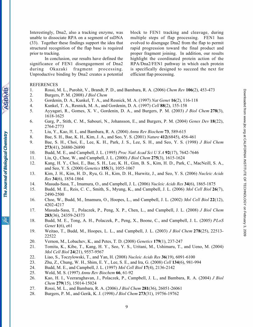
RESULTSDna2 dissociates slowly from a flap
substrate—Previously, we observed that FEN1disengages Dna2 from a flap substrate to gainaccess for cleavage (32). To understand thedetails of FEN1-promoted disengagement ofDna2, we used a DNA competition assay andsurface plasmon resonance (SPR) to assess Dna2dissociation (Fig. 1). We reasoned that if Dna2dissociates slowly from flap substrates thedisengagement reaction is likely to have evolvedto facilitate rapid joining of Okazaki fragments.However, if spontaneous dissociation of Dna2were rapid, the disengagement process has anotherpurpose.
To assess the rate of Dna2 dissociationfrom the flap, we incubated Dna2 with aradiolabeled 53-nt double flap substrate. Afterbinding, an excess of unlabeled flap substrate wasadded to the reaction followed by the addition ofMgCl2 at the indicated time points (Fig. 1A ) .Reactions were then incubated for 10 min to allow
Dna2 cleavage. Since MgCl2 is required for Dna2cleavage, the cleavage rate was proportional to theamount of Dna2 still bound to the labeled substrateat each time point. These results were comparedwith a control in which the labeled and excessunlabeled substrates were incubated prior to theaddition of Dna2 (lane 12). A graph was thengenerated and points were fit to an exponentialdecay curve to determine the dissociation rate (seeExperimental Procedures) (Fig. 1B). Initially, wefit the curve to a single exponential decayequation, which showed a half-time of about 25min (gray line). Based on the shape of the curve,we then utilized the double exponential decayequation and found a better fit (black line). Thissuggests two dissociation phases, an initial rapiddissociation followed by a much slower one.Based on the small amplitude (~20% of therelative cleavage) and the short time frame (~1min), we believe that nonspecific binding or aweak binding mode accounts for the initialdissociation phase of Dna2. The second phasewould account for the majority of Dna2 binding.Dna2 bound in this manner dissociates slowlyfrom the DNA, with a half-time of about 40 min.These data show that binding of Dna2 to thesubstrate is quite stable.
To further assess the binding anddissociation rates of Dna2 to DNA, we performedSPR. Dna2 was immobilized onto a chip andvarious amounts of ssDNA were allowed to flowover the chip while association was measured (Fig.1C). This was followed by a dissociation phasewith only buffer flowing over the chip. A secondsurface in which Dna2 was not immobilizedserved as a reference. When we attempted to fitthe curves, they did not fit a simple 1:1 bindingmodel, suggesting a complex interaction betweenDna2 and the DNA. While we were unable tosimultaneously fit both the association anddissociation rates, we could independently fit thedissociation rate using the Scrubber 2 software.Since the curves appeared strikingly similar tothose in Fig. 1B , we fit the data 30 sec into thedissociation phase. By doing so, we were able tobypass the initial dissociation phase and fit a 1:1binding model for the second dissociation phase.Again, these curves suggest a slow rate ofdissociation, with a half-time of approximately 50min. Both the excess substrate and SPRdissociation measurements clearly indicate that the
4
at CA
LIFO
RN
IA IN
ST
ITU
TE
OF
TE
CH
NO
LOG
Y on F
ebruary 3, 2009 w
ww
.jbc.orgD
ownloaded from

half-time for dissociation of Dna2 is in the rangeof one-half to one hour. These findings areconsistent with the conclusion that, because Dna2binding to the flap is stable, FEN1 has evolved theability to disengage Dna2 in order to efficientlygain access to the flap base for cleavage.
Dna2 binds, but does not cleave, shortRNA and DNA flaps—Previously, we hypothesizedthat FEN1 evolved to remove Dna2 molecules thatare unproductively bound to the flap. The needfor disengagement is envisioned to arise at twostages of Okazaki fragment processing. EachOkazaki fragment is initiated by a short segmentof RNA, 10 to 12-nt in length (1). The first stagerequiring disengagement would occur duringinitial strand displacement by pol δ, when RNAflaps begin to emerge. While FEN1 can readilycleave short RNA flap intermediates, RNA is not asubstrate for the nuclease activity of Dna2 (34). Abound, inactive Dna2 molecule could blockprogressive FEN1 cleavage.
We previously showed that Dna2 bound,but did not cleave a 30-nt RNA flap, and thatDna2 was dissociated by FEN1 (32). Here weemployed a substrate with 5-nt of RNA on the flapand an additional 8-nts of RNA in the annealedportion of the labeled primer. This substratesimulates the initial partial displacement of theRNA primer by pol δ. The substrate was used totest Dna2 cleavage and binding. Consistent withprevious findings, Dna2 was unable to cleave theRNA flap (Fig. 2A). By way of a control, wemeasured robust Dna2 cleavage activity on a 30-ntDNA flap substrate. We then tested the ability ofDna2 to bind the 5-nt RNA flap. Dna2 wasincubated with the substrate and the reactions werethen analyzed by gel shift (Fig. 2B). The labeledsubstrate band shifted upon the addition of Dna2to indicate formation of a higher molecular weightcomplex.
Next, we assessed FEN1 dissociation ofDna2 on the 5-nt RNA flap substrate (Fig. 2C).Dna2 was pre-bound to the flap. FEN1 was thenadded with the Dna2-bound substrate. Thereactions were then analyzed by gel shift toseparate the products and determine which proteinremained bound to the substrate. Since Dna2 isthree times the size of FEN1, the bound complexesof these proteins with the labeled substrate areeasily distinguished (Fig. 2C, lanes 2 and 7). Withincreasing amounts of FEN1, the bands were
shifted from a Dna2-bound substrate to a FEN1-bound substrate (lanes 3-6). This shift isindicative of the removal of Dna2 from the 5-ntRNA flap by FEN1. These results suggest thatFEN1 disengagement of flap-bound Dna2 wouldpromote FEN1 cleavage on initially displacedflaps consisting only of RNA.
The second stage requiring Dna2disengagement would occur after Dna2 cleavageon RPA-coated flaps. When flaps become longenough to bind RPA, Dna2 is needed to shortenthem so that RPA will no longer bind and blockFEN1. The properties of the Dna2 nucleasefunction appear ideal for this task, in that it cleavesflaps to a terminal length of about five nucleotides.However, Dna2 may remain bound unlessdissociated by FEN1.
We designed a substrate with a 5-nt DNAflap to simulate the terminal product of long flapcleavage by Dna2. We then tested this flap forDna2 cleavage and binding. As expected, Dna2cleavage did not occur (Fig. 2A). Dna2 bindingwas then measured (Fig. 2D). Gel shift analysisshowed that the addition of Dna2 shifted thelabeled substrate band. Finally, Dna2 was pre-bound to the 5-nt DNA flap followed by theaddition of FEN1 to test for Dna2 removal (Fig.2E). Again, the addition of FEN1 shifted the banddistribution from a Dna2-bound substrate to aFEN1-bound substrate, indicative of Dna2removal (lanes 3-6).
Notably, similar results were obtained forboth the 5-nt RNA and DNA flaps. Dna2unproductively bound both substrates, potentiallyblocking FEN1, but in both cases FEN1 removedDna2 to enable progressive FEN1 action. Inaddition, FEN1 showed nearly the same amount ofdisplacement and binding on both the DNA andRNA flaps, suggesting that the interactionproperties of these proteins are similar on bothDNA and RNA. These results support theconclusion that FEN1 has evolved thedisengagement mechanism to ensure that it is thedominant nuclease at all times that it shares a flapwith Dna2.
The FEN1 tracking mechanism is requiredto dissociate Dna2—Both FEN1 and Dna2 musttrack from the 5'-end of the flap to displaynuclease activity (30,31). Our previous resultsindicate that FEN1 disengages the trackingmechanism of Dna2 to dissociate it from the flap
5
at CA
LIFO
RN
IA IN
ST
ITU
TE
OF
TE
CH
NO
LOG
Y on F
ebruary 3, 2009 w
ww
.jbc.orgD
ownloaded from

(32). Here we tested whether FEN1 must be in itstracking mode, as expected during natural flapprocessing, in order to disengage Dna2.
To block FEN1 tracking, we employed a53-nt flap with a biotin attached at the 5'-end (Fig.3, lanes 1-7). Dna2 was pre-incubated with thissubstrate to allow tracking. The Dna2-boundsubstrate was then incubated with streptavidin,which blocked the FEN1 tracking mechanism.FEN1 was then added into the reaction.Interestingly, FEN1 was unable to remove theflap-bound Dna2 (lanes 3-6), indicating that itmust be tracking to dissociate the Dna2.
We then questioned whether Dna2 wouldbe disengaged if the FEN1 began tracking but wasnot allowed to track all the way to the position ofthe flap-bound Dna2. In this scenario, the FEN1would be in the tracking mode and could stillpotentially contact the Dna2 by a looping process.However, it would not likely form the exactcontacts that it could make if it tracked in thenatural manner. To achieve this situation, weemployed a substrate with a biotin attached to anucleotide in the middle of the flap (Fig. 3, lanes8-14). This substrate was previously used to testthe Dna2 tracking mechanism (30). The Dna2cleavage pattern was unaltered indicating thebiotin modification did not interfere with Dna2tracking. By placing the biotin in the middle ofthe flap, the addition of streptavidin would permitFEN1 tracking, but prevent full travel along theflap. Dna2 was bound to the flap followed by theaddition of streptavidin. FEN1 was then addedand gel shift analysis was performed (Fig. 3, lanes10-13). As with the 5'-end blocked flap, a block tothe middle of the flap prevented FEN1 removal offlap-bound Dna2. Based on these findings, wepropose that natural tracking by FEN1 is arequirement for the removal of Dna2 from theflap.
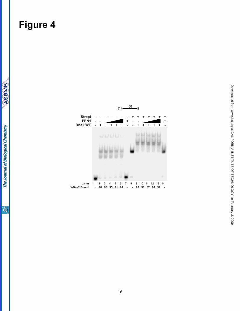
FEN1 is unable to remove Dna2 fromssDNA—FEN1 is a structure specific enzyme, thatrecognizes the 5' flap junction between ssDNAand dsDNA for cleavage, and needs a 1-nt 3' tailfor maximal cleavage efficiency (36). Wereasoned that Dna2 removal by FEN1 might alsorequire a genuine flap structure. Accordingly, alinear ssDNA segment was used to measure Dna2dissociation by FEN1 (Fig. 4). A biotin wasattached to the 3'-end of the DNA, which, whenconjugated with streptavidin, would prevent Dna2
or FEN1 from tracking off the 3'-end of the singlestrand. Experiments were done with and withoutstreptavidin to compare its effect.
Dna2 was pre-incubated with the ssDNAsubstrate and increasing concentrations of FEN1were then added into the reaction. Significantly,the addition of FEN1 did not affect the amount ofDna2 bound to the ssDNA (compare Fig. 4, lanes3-6 and lanes 10-13). In fact, FEN1 alone wasunable to bind the ssDNA substrate (lanes 7 and14). The inability of FEN1 to bind the ssDNAsuggests that FEN1 must structurally identify theflap base for binding. This result implies that acollection of specific structural features of theDNA are important for Dna2 removal by FEN1.
Nuclease-defective Dna2 E675Aovercomes RPA inhibition of FEN1—Is the abilityof FEN1 to displace Dna2 significant in thecontext of the RPA/Dna2/FEN1 Okazaki fragmentprocessing pathway? As previously described,flap-bound RPA inhibits FEN1 cleavage (8). Werecently discovered that Dna2 displaces flap-bound RPA, independent of cleavage (33). Inaddition, FEN1 dissociation of Dna2 enablesFEN1 cleavage (32). This offered us anopportunity to address the role of FEN1dissociation of Dna2 in the actualRPA/Dna2/FEN1 pathway, in which RPA isblocking FEN1 cleavage. We envision that after aflap grows long and binds RPA, Dna2 woulddisplace the RPA, cleave the flap, but then remainbound. FEN1 would then dissociate the Dna2,cleaving the flap to create a nick for ligation.Since Dna2 does not require nuclease activity toremove RPA, we asked whether Dna2 couldpermit FEN1 activity on an RPA-coated flap evenwithout cutting it. To address this question, wereconstituted the RPA/Dna2/FEN1 pathway withthe nuclease-defective Dna2 E675A.
RPA was incubated with a 21-nt DNAflap substrate (Fig. 5). Dna2 E675A and FEN1were then added to the reaction followed bydenaturing PAGE analysis of the labeled primer.Comparing the amount of substrate cleaved byFEN1 with or without RPA-bound, we measuredan approximately three-fold inhibition of FEN1cleavage activity in the presence of RPA (compareFig. 5A and B, lanes 3 and 6). Moreover, weobserved that Dna2 E675A, as expected, does notinhibit FEN1 cleavage (Fig. 5A and B, lane 5). Infact, Dna2 E675A slightly stimulated FEN1
6
at CA
LIFO
RN
IA IN
ST
ITU
TE
OF
TE
CH
NO
LOG
Y on F
ebruary 3, 2009 w
ww
.jbc.orgD
ownloaded from

nuclease activity. RPA, FEN1, and increasingamounts of Dna2 E675A were then incubated withthe 21-nt flap substrate (Fig. 5A and B , lanes 8-11). At the highest Dna2 E675A concentration,FEN1 cleavage was restored to near the levelachieved without RPA (compare lanes 3 and 11).Based on these results, we conclude that Dna2E675A actively displaced the RPA from the flapfollowed by FEN1 removal of the now flap-boundDna2 E675A. FEN1 was then free to cleave theflap devoid of either RPA or Dna2.
DISCUSSIONWe previously demonstrated that FEN1
disengages Dna2 from the flap (32). Our currentstudies questioned why FEN1 evolved to removeflap-bound Dna2 in the context of Okazakifragment processing. We conclude that Dna2binding to short RNA and DNA flaps wouldpotentially block FEN1 entry and cleavage.However, FEN1 displaces these flap-bound Dna2molecules.
Dna2 does not cleave RNA, but it bindsRNA flap substrates. In fact, it binds with asimilar affinity to RNA as DNA and can trackfrom the 5'-end of an RNA segment to cleave theDNA portion of an RNA-DNA flap substrate (34).Such properties would enable Dna2 cleavage ofthe long flap intermediates anticipated to occurnaturally as pol δ displaces an RNA-primedOkazaki fragment. However, previous resultsindicate that FEN1 alone processes most flaps(6,27). The ability of FEN1 to cleave an RNA flapindicates that it is designed to act constantly on theRNA, and then DNA, as the flap is generated. Insupport of this conclusion, biochemicalreconstitution studies suggest that coordinationbetween pol δ and FEN1 produces short cleavageproducts of 1 to 8-nt, with the majority of productsbeing mononucleotides. This coordination ishighly efficient and allows for the rapid processingof flap intermediates. We envision that a bindingcompetition between Dna2 and FEN1 can arise asthe RNA flap is beginning to be displaced. Dna2binding ahead of FEN1 on RNA flaps would proveunproductive until the RNA and more than aboutfive nucleotides of DNA have been displaced.The ability of FEN1 to remove Dna2 from shortRNA flaps allows FEN1 to act as the solenuclease, unless long flaps arise.
Once flaps escape FEN1 cleavage andbecome long, they are bound by RPA eliciting theneed for the RPA/Dna2/FEN1 pathway. Weshowed that Dna2 dissociates RPA to access theflap for cleavage (33). In addition, RPA strandmelting capacity stimulates Dna2 cleavage byremoving DNA secondary structure. While RPAbinding inhibits FEN1 cleavage, RPA would alsoprevent structured flap formation, which wouldinhibit efficient cleavage by either Dna2 or FEN1.By this reasoning, RPA binding likely prepareslong structured flaps for Dna2 cleavage. Dna2tracks down the flap removing RPA andsuccessively cleaves until the flap reachesapproximately 5-nt in length. At this length, Dna2cannot cleave, necessitating FEN1 cleavage tomake a product for ligation. Again, we found thatFEN1 can disengage Dna2 that has reached thisstatic state. In fact, the unproductively boundDna2 may even act to recruit FEN1 to theshortened RPA-free flap for final processing.
Dna2 binding kinetics were consistentwith a slow rate of dissociation from the DNAafter initial binding (Fig. 1). Slow dissociationmay be enhanced by the tracking mechanism ofDna2. When tracking, the protein behaves as abead on a string, or as if it is encircling the flap.The Dna2 may also not readily slide back off ofthe 5'-end of the flap. This resistance to 5' motionmay be accentuated when the Dna2 helicase isacting to continuously drive Dna2 toward the flapbase. Our demonstration that Dna2 can bindOkazaki fragment intermediates non-productively,together with evidence of slow naturaldissociation, highlights the reasons why FEN1 hasevolved the ability to disengage Dna2.
Using the nuclease-defective Dna2mutant, we showed that the RPA/Dna2/FEN1pathway could be reconstituted in the absence ofDna2 cleavage activity (Fig. 5). Results of thisexperiment reveal the elegant coordination offunctions that can be displayed by Okazakifragment maturation proteins. We were impressedto see that the successive binding functions ofDna2 and FEN1 were sufficient to clear a longflap of RPA and allow FEN1 cleavage. While thisexperiment allowed us to visualize more closelythe sequential steps to proper flap removal, itseemingly questions the role of the nucleaseactivity of Dna2. Genetic evidence emphasizesthe importance of Dna2 nuclease activity in DNA
7
at CA
LIFO
RN
IA IN
ST
ITU
TE
OF
TE
CH
NO
LOG
Y on F
ebruary 3, 2009 w
ww
.jbc.orgD
ownloaded from

replication (39). The nuclease activity is essentialin S. cerevisiae and the temperature sensitivemutant, dna2-1 , which has reduced nucleasefunction, showed defects associated with DNAreplication at the restrictive temperature. Theseinclude highly fragmented DNA, deficiency inDNA, but not RNA, synthesis, and undividednuclei.
An answer to this puzzle is suggested bythe results of an attempted repeat of thereconstitution of the RPA/Dna2/FEN1 pathwayshown in Fig. 5, but with a 53-nt RPA-coated flapsubstrate. With this substrate, Dna2 E675A wasunable to stimulate FEN1 cleavage (data notshown). We interpret this result to mean thatDna2 dissociated the flap-bound RPA and thenstalled at the base, unable to cleave. RPA thenrebound behind the Dna2 to inhibit FEN1cleavage. The 21-nt flap likely represents a lengthat which Dna2 removes RPA without allowingstable RPA rebinding. Reconstitutions of Okazakifragment processing suggest that a fraction offlaps grow into the 40 to 50-nt size range (27,29).If so, the need to process such flaps is anticipatedto require the nuclease activity of Dna2. Even onshorter flaps, it is likely that the nuclease functionof Dna2 accelerates the rate of flap removal in away that makes its function essential, as suggestedby the nearly but not totally complete rescue ofFEN1 cleavage in Fig. 5.
FEN1 tracking, a prerequisite forcleavage, was also necessary for the removal ofDna2 (Fig. 3). Apparently, when the flap is long,FEN1 must recognize the 5'-end, bind and trackdown the flap until it encounters Dna2. Theinteraction between FEN1 and Dna2 then resultsin disengagement of Dna2 from the flap. Incontrast, when the flap is short the situation coulddiffer. On a 5-nt flap, the natural terminal productof Dna2 cleavage, Dna2 may occlude the entireflap. If so, FEN1 could not track. Yet, it stilldissociates Dna2 from the flap. How might FEN1tracking be required on long but not short flaps?Dna2, like FEN1, is a tracking enzyme (30). Forcleavage, it must load onto the 5'-end and trackdown the flap. Interestingly, while tracking isrequired for cleavage activity, both FEN1 andDna2 can bind the flap independent of tracking(32,40), but the ability to bind the flap was notsufficient for FEN1 to promote the dissociation ofDna2 (Fig. 3). Like FEN1, it is envisioned that the
flap is threaded through Dna2. The site where the5'-end of the flap exits Dna2 is likely near therequired area for proper protein-protein contactswith FEN1. Likewise, the region on FEN1 criticalfor protein contacts with Dna2 would be locatednear the site of flap entry on the smaller nuclease.Consistent with this interpretation, FEN1 couldnot loop around a streptavidin block in the middleof a long flap to remove the flap-bound Dna2 (Fig.3). Instead the streptavidin block prevented FEN1from removing Dna2. Natural tracking by FEN1would allow direct contact of the appropriateprotein surfaces to induce the disengagement ofDna2 and release it from the flap. In the case of ashort flap, the surfaces of interaction would beunobstructed by the flap, permitting FEN1 toproperly interact with Dna2. In addition, the flaplikely facilitates proper protein contacts bybringing the proteins into close proximity. Theactual orientations of FEN1, Dna2, and DNAduring these processes await high-resolutionstructural analysis.
Finally, we showed that FEN1 did notdisengage Dna2 on a single-stranded segment ofDNA (Fig. 4). Recognition of a genuine flapsubstrate is required for efficient cleavage byFEN1 (36). FEN1 does not cleave linear ssDNAsegments at all, and here we show that FEN1 isunable even to bind such DNA. Binding was notachieved even with a streptavidin block at the 3'-end of the ssDNA, suggesting at least twopossibilities. Structural features of the flap mustbe recognized prior to binding and tracking.Alternatively, the flap base stabilizes FEN1binding after tracking.
Previously, we envisioned that FEN1begins tracking by first recognizing the 5'-end ofthe flap, followed by threading of the flap throughthe protein until it reached the base. Uponencountering the base, FEN1 would then identifythe structure features required to activate cleavageof the substrate. Based on this model, FEN1should still track on the ssDNA segment, with thestreptavidin block preventing FEN1 from trackingoff the 3'-end. Since the substrate does not possessthe flap base structure that activates FEN1 forcleavage, FEN1 would be stopped by thestreptavidin block until it tracks back off the 5'-endfor dissociation (31). In addition, FEN1 shouldstill remove flap-bound Dna2. Instead, FEN1could not bind the ssDNA or remove Dna2.
8
at CA
LIFO
RN
IA IN
ST
ITU
TE
OF
TE
CH
NO
LOG
Y on F
ebruary 3, 2009 w
ww
.jbc.orgD
ownloaded from
Interestingly, Dna2, also a tracking enzyme, wasunable to dissociate RPA on a segment of ssDNA(33). Together these findings support the idea thatstructural recognition of the flap base is requiredprior to tracking.
In conclusion, our results have defined thesignificance of FEN1 disengagement of Dna2during Okazaki fragment processing.Unproductive binding by Dna2 creates a potential
block to FEN1 tracking and cleavage, duringmultiple steps of flap processing. FEN1 hasevolved to disengage Dna2 from the flap to permitrapid progression toward the final product andproper fragment joining. In addition, our resultshighlight the coordinated protein action of theRPA/Dna2/FEN1 pathway in which each proteinis specifically designed to succeed the next forefficient flap processing.
REFERENCES1. Rossi, M. L., Purohit, V., Brandt, P. D., and Bambara, R. A. (2006) Chem Rev 106(2), 453-4732. Burgers, P. M. (2008) J Biol Chem3. Gordenin, D. A., Kunkel, T. A., and Resnick, M. A. (1997) Nat Genet 16(2), 116-1184. Kunkel, T. A., Resnick, M. A., and Gordenin, D. A. (1997) Cell 88(2), 155-1585. Ayyagari, R., Gomes, X. V., Gordenin, D. A., and Burgers, P. M. (2003) J Biol Chem 278(3),
1618-16256. Garg, P., Stith, C. M., Sabouri, N., Johansson, E., and Burgers, P. M. (2004) Genes Dev 18(22),
2764-27737. Liu, Y., Kao, H. I., and Bambara, R. A. (2004) Annu Rev Biochem 73, 589-6158. Bae, S. H., Bae, K. H., Kim, J. A., and Seo, Y. S. (2001) Nature 412(6845), 456-4619. Bae, S. H., Choi, E., Lee, K. H., Park, J. S., Lee, S. H., and Seo, Y. S. (1998) J Biol Chem
273(41), 26880-2689010. Budd, M. E., and Campbell, J. L. (1995) Proc Natl Acad Sci U S A 92(17), 7642-764611. Liu, Q., Choe, W., and Campbell, J. L. (2000) J Biol Chem 275(3), 1615-162412. Kang, H. Y., Choi, E., Bae, S. H., Lee, K. H., Gim, B. S., Kim, H. D., Park, C., MacNeill, S. A.,
and Seo, Y. S. (2000) Genetics 155(3), 1055-106713. Kim, J. H., Kim, H. D., Ryu, G. H., Kim, D. H., Hurwitz, J., and Seo, Y. S. (2006) Nucleic Acids
Res 34(6), 1854-186414. Masuda-Sasa, T., Imamura, O., and Campbell, J. L. (2006) Nucleic Acids Res 34(6), 1865-187515. Budd, M. E., Reis, C. C., Smith, S., Myung, K., and Campbell, J. L. (2006) Mol Cell Biol 26(7),
2490-250016. Choe, W., Budd, M., Imamura, O., Hoopes, L., and Campbell, J. L. (2002) Mol Cell Biol 22(12),
4202-421717. Masuda-Sasa, T., Polaczek, P., Peng, X. P., Chen, L., and Campbell, J. L. (2008) J Biol Chem
283(36), 24359-2437318. Budd, M. E., Tong, A. H., Polaczek, P., Peng, X., Boone, C., and Campbell, J. L. (2005) PLoS
Genet 1(6), e6119. Weitao, T., Budd, M., Hoopes, L. L., and Campbell, J. L. (2003) J Biol Chem 278(25), 22513-
2252220. Vernon, M., Lobachev, K., and Petes, T. D. (2008) Genetics 179(1), 237-24721. Tomita, K., Kibe, T., Kang, H. Y., Seo, Y. S., Uritani, M., Ushimaru, T., and Ueno, M. (2004)
Mol Cell Biol 24(21), 9557-956722. Liao, S., Toczylowski, T., and Yan, H. (2008) Nucleic Acids Res 36(19), 6091-610023. Zhu, Z., Chung, W. H., Shim, E. Y., Lee, S. E., and Ira, G. (2008) Cell 134(6), 981-99424. Budd, M. E., and Campbell, J. L. (1997) Mol Cell Biol 17(4), 2136-214225. Wold, M. S. (1997) Annu Rev Biochem 66, 61-9226. Kao, H. I., Veeraraghavan, J., Polaczek, P., Campbell, J. L., and Bambara, R. A. (2004) J Biol
Chem 279(15), 15014-1502427. Rossi, M. L., and Bambara, R. A. (2006) J Biol Chem 281(36), 26051-2606128. Burgers, P. M., and Gerik, K. J. (1998) J Biol Chem 273(31), 19756-19762
9
at CA
LIFO
RN
IA IN
ST
ITU
TE
OF
TE
CH
NO
LOG
Y on F
ebruary 3, 2009 w
ww
.jbc.orgD
ownloaded from

29. Rossi, M. L., Pike, J. E., Wang, W., Burgers, P. M., Campbell, J. L., and Bambara, R. A. (2008) JBiol Chem 283(41), 27483-27493
30. Kao, H. I., Campbell, J. L., and Bambara, R. A. (2004) J Biol Chem 279(49), 50840-5084931. Murante, R. S., Rust, L., and Bambara, R. A. (1995) J Biol Chem 270(51), 30377-3038332. Stewart, J. A., Campbell, J. L., and Bambara, R. A. (2006) J Biol Chem 281(50), 38565-3857233. Stewart, J. A., Miller, A. S., Campbell, J. L., and Bambara, R. A. (2008) J Biol Chem 283(46),
31356-3136534. Bae, S. H., and Seo, Y. S. (2000) J Biol Chem 275(48), 38022-3803135. Budd, M. E., Choe, W., and Campbell, J. L. (2000) J Biol Chem 275(22), 16518-1652936. Kao, H. I., Henricksen, L. A., Liu, Y., and Bambara, R. A. (2002) J Biol Chem 277(17), 14379-
1438937. Henricksen, L. A., and Wold, M. S. (1994) J Biol Chem 269(39), 24203-2420838. Subramanian, A., Irudayaraj, J., and Ryan, T. (2006) Biosens Bioelectron 21(7), 998-100639. Budd, M. E., Choe, W. C., and Campbell, J. L. (1995) J Biol Chem 270(45), 26766-2676940. Hohl, M., Dunand-Sauthier, I., Staresincic, L., Jaquier-Gubler, P., Thorel, F., Modesti, M.,
Clarkson, S. G., and Scharer, O. D. (2007) Nucleic Acids Res 35(9), 3053-3063
FOOTNOTES*We would like to thank Drs. Sara Binz and Marc Wold for the purified RPA protein. We thank Dr. TomRyan and his team at Reichert as well as the Sullivan laboratory at the University of Rochester fortraining and assistance with the SPR equipment. In addition, we thank the Bambara and Campbelllaboratories for beneficial discussion and review of the manuscript. This work was supported by NationalInstitutes of Health (NIH) Grant GM024441 to R.A.B., with additional support from NIH GM087666 toJ.L.C. J.A.S. was supported by NIH Grant T32 GM068411 and an Elon Huntington Hooker GraduateFellowship.
1The abbreviations used are: pol, polymerase; FEN1, flap endonuclease 1; RPA, replication protein A; nt,nucleotide; ss, single-stranded; PAGE, polyacrylamide gel electrophoresis; SPR, surface plasmonresonance
FIGURE LEGENDSFigure 1. Slow dissociation of Dna2 from DNA substrates. A, Dna2 (200 fmol) and 5 fmol of aradiolabeled 53-nt flap substrate (D4:U2:T2) were incubated followed by the addition of 200-fold excessunlabeled flap substrate (D4:U2:T2). MgCl2 was then added at the indicated time points. Dna2 cleavagewas then measured by denaturing PAGE. Lane 1 is the substrate alone. Lane 2 is Dna2 with labeled andunlabeled substrate without MgCl2. In lane 12, the labeled and excess unlabeled substrates were mixedprior to the addition of Dna2. B, graphical analysis of A. Points were fit to a single (gray line) or double(black line) exponential decay curve using nonlinear least squares regression. Cleavage is defined as(cleaved/(cleaved+uncleaved)) x 100. For the double exponential decay curve, the dissociation amplitudedescribed by the first curve was 21% and the second 78%. C, surface plasmon resonance was used tomeasure the affinity between Dna2 and ssDNA (D4). Dna2 was immobilized and increasing amounts ofsubstrate (62.5, 125, 250, and 500 nM) was flowed over the chip. Measurements at each concentrationwere repeated twice. After three minutes, the flow of ssDNA was discontinued and buffer alone wasflowed over the chip to measure dissociation.
Figure 2. FEN1 disengagement of Dna2 from short RNA and DNA flaps to which Dna2 binds butcannot cleave. A, Dna2 (50, 100, 200, 500, 1000 fmol) was incubated with 5 fmol of a 5-nt RNA flapsubstrate (D1:U1:T1) (squares), a 5-nt DNA flap substrate (D2:U2:T2) (circles), or a 30-nt DNA flapsubstrate (D3:U2:T2) (diamonds). Cleavage activity was then measured by denaturing PAGE. B, gelshift analysis was used to measure Dna2 (0.2, 0.5, 1 pmol) binding activity on the 5-nt RNA flap (lanes 2-4). Lane 1 is the substrate alone control. C, Dna2 (1 pmol) was pre-bound to the 5-nt RNA flap substrate
10
at CA
LIFO
RN
IA IN
ST
ITU
TE
OF
TE
CH
NO
LOG
Y on F
ebruary 3, 2009 w
ww
.jbc.orgD
ownloaded from

followed by the addition of FEN1 (5, 10, 25, 50 fmol) (lanes 3-6). The samples were then analyzed bygel shift. Lanes 1 and 7 are the substrate alone and substrate plus FEN1 (50 fmol), respectively. D, asdescribed in B, except a 5-nt DNA flap was used. E, as described in C, except a 5-nt DNA flap substratewas used. For A, points are an average of three experiments and the bars indicate the standard deviation.Percent cleavage is defined as (cleaved/(cleaved + uncleaved)) x 100. Percent Dna2 bound is defined as(Dna2 bound/(Dna2 bound + FEN1 bound + unbound substrate)) x 100. Percent FEN1 bound is definedas (FEN1 bound/(FEN1 bound + Dna2 bound + unbound substrate)) x 100. Substrates are depicted abovegels with the RNA labeled in gray and DNA in black. The asterisk indicates the site of the 3' 32-P-radiolabel.
Figure 3. FEN1 requires its tracking mechanism to disengage flap-bound Dna2. Gel shift analysiswas used to test Dna2 dissociation by FEN1 when tracking was blocked. Dna2 (500 fmol) was bound toa 53-nt flap substrate with a biotin attached at either the 5' flap end (D4:U2:T2) or in the middle of theflap (D5:U2:T2). Streptavidin was then added to the reaction for conjugation with the biotin. Followingconjugation, FEN1 (5, 10, 20, 50 fmol) was added (lanes 3-6 and 10-13). Lanes 1 and 8 are streptavidin-bound substrate alone. Lanes 2 and 9 are streptavidin-bound substrate plus Dna2 (500 fmol). Lanes 7and 14 are streptavidin-bound substrate plus FEN1 (50 fmol). Percent Dna2 bound is defined as (Dna2bound/(Dna2 bound + FEN1 bound + unbound substrate)) x 100. Percent FEN1 bound is defined as(FEN1 bound/(FEN1 bound + Dna2 bound + unbound substrate)) x 100. Substrates are depicted abovegels and the asterisk indicates the site of the 3' 32-P-radiolabel. B indicates the site of biotin modification.
Figure 4. FEN1 cannot dissociate Dna2 on a ssDNA segment. A 50-nt ssDNA segment with a biotinattached at the 3' end (D6) was used to test FEN1 disengagement of Dna2. In lanes 8-14, streptavidin waspre-incubated with the substrate. Dna2 (500 fmol) was bound to the ssDNA segment followed by theaddition of FEN1 (5, 10, 25, 50 fmol) (lanes 3-6 and 10-13). Gel shift was then used to separate theproducts. Lanes 1, 2, and 6 are substrate alone, substrate with Dna2 (500 fmol), and substrate with FEN1(50 fmol), respectively. Lanes 8, 9, and 14 are the same as lanes 1, 2, and 3, respectively, except withstreptavidin. Percent Dna2 bound is defined as (Dna2 bound/(Dna2 bound + unbound substrate)) x 100.Substrates are depicted above gels and the asterisk indicates the site of the 5' 32-P-radiolabel. B indicatesthe site of biotin modification.
Figure 5. Dna2 E675A overcomes RPA inhibition of FEN1. RPA (200 fmol), FEN1 (0.25 fmol) andDna2 E675A (10, 20, 100, and 200 fmol) were mixed followed by the addition of a 21-nt flap substrate(D7:U3:T3) (lanes 8-11). Denaturing PAGE was then used to separate the products. Lane 1 is thesubstrate alone control. Lanes 2, 3, and 4 are substrate with Dna2 E675A (200 fmol), substrate withFEN1 (0.25 fmol), and substrate with RPA (200 fmol), respectively. Lanes 5, 6, and 7 are substrate withDna2 E675A (200 fmol) and FEN1 (0.25 fmol), substrate with RPA (200 fmol) and FEN1 (0.25 fmol),and substrate with Dna2 E675A (200 fmol) and RPA (200 fmol), respectively. B, graphical analysis of A.Each bar of the graph represents the conditions shown in the corresponding lane in A. The bars are anaverage of four independent experiments and error bars represent the standard deviation. The substrate isdepicted above the gel in A and the asterisk indicates the site of the 3' 32-P-radiolabel.
11
at CA
LIFO
RN
IA IN
ST
ITU
TE
OF
TE
CH
NO
LOG
Y on F
ebruary 3, 2009 w
ww
.jbc.orgD
ownloaded from
Primer Length (nt) SequenceDownstream*^# (5' - 3')
D1 23 GCC GU C CAC CCG U CC ACC CGA CGD2 28 GCC GTC GTT TTA CAA CGA CGT GAC TGG GD3 53 TTC ACG CCT GTT AGT TAA TTC ACT GGC CGT CGT TTT ACA ACG ACG TGA CTG GGD4 76 GTA CCG AGC TCG AAT TCG CCC GTT TCA CGC CTG TTA GTT AAT TCA CTG GCC GTC
GTT TTA CAA CGA CGT GAC TGG GD5 76 GTA CCG AGC TCG AAT TCG CCC GTT TCA CGC CTG TTA GTT AAT TCA CTG GCC GTC
GTT TTA CAA CGA CGT GAC TGG GD6 50 GTA CCG AGC TCG AAT TCG CCC GTT TCA CGC CTG TTA GTT AAT TCA CTG GCD7 46 CAC TGG CCG TCG TTT TAC GGA CCC GTC CAC CCG ACG CCA CCT CCT G
Upstream (5' - 3')U1 26 CGA CCG TGC CAG CCT AAA TTT CAA GAU2 26 CGC CAG GGT TTT CCC AGT CAC GAC CAU3 26 CGA CCG TGC CAG CCT AAA TTT CAA TA
Template (3' - 5')T1 44 GCT GGC ACG GTC GGA TTT AAA GTT CGG TGG GCA GGT GGG CTG CGT2 49 GCG GTC CCA AAA GGG TCA GTG CTG GGC AAA ATG TTG CTG CAC TGA CCC GT3 51 GCT GGC ACG GTC GGA TTT AAA GTT AGG GCA GGT GGG CTG CGG TGG AGG ACG
*Bolded nucleotides are biotinylated^RNA segment is in italics#Underlined nucleotide indicates the last annealed nucleotide
Table 1: Oligonucleotides
12
at CA
LIFO
RN
IA IN
ST
ITU
TE
OF
TE
CH
NO
LOG
Y on F
ebruary 3, 2009 w
ww
.jbc.orgD
ownloaded from
13
at CA
LIFO
RN
IA IN
ST
ITU
TE
OF
TE
CH
NO
LOG
Y on F
ebruary 3, 2009 w
ww
.jbc.orgD
ownloaded from

14
at CA
LIFO
RN
IA IN
ST
ITU
TE
OF
TE
CH
NO
LOG
Y on F
ebruary 3, 2009 w
ww
.jbc.orgD
ownloaded from

15
at CA
LIFO
RN
IA IN
ST
ITU
TE
OF
TE
CH
NO
LOG
Y on F
ebruary 3, 2009 w
ww
.jbc.orgD
ownloaded from
16
at CA
LIFO
RN
IA IN
ST
ITU
TE
OF
TE
CH
NO
LOG
Y on F
ebruary 3, 2009 w
ww
.jbc.orgD
ownloaded from

Dna2 E675A - + - - + - +FEN1 - - + - + + - + + + +RPA - - - + - + + + + + +
Lanes 1 2 3 4 5 6 7 8 9 10 11
51 nt
21 nt
21
*5'3'
at CA
LIFO
RN
IA IN
ST
ITU
TE
OF
TE
CH
NO
LOG
Y on F
ebruary 3, 2009 w
ww
.jbc.orgD
ownloaded from