signaling pathways and model checking in the pancreatic...
TRANSCRIPT

Signaling Pathways and Model Checking
in the Pancreatic Cancer Studies
H. Gong1 P. Zuliani1 J. Faeder2 E. Clarke1
1Carnegie Mellon University
2University of Pittsburgh
03/05/2010

Outline
• Introduction
• Signaling Pathways
• HMGB1 and Pancreatic Cancer
• Model Checking
• Future Work
• Acknowledgement
Oncoprotein & Tumor-suppressor Protein
• Oncoproteins stimulate cell growth under normal conditions.
• Cells with mutant oncoproteins continue to grow (refuse to die) even when they are receiving no-growth signals.
• Some examples of oncoproteins are RAS, AKT, MDM2.
• Tumor-suppressor proteins can inhibit the cell cycle progress or promote apoptosis (programmed cell death).
• In normal cell, oncoproteins are regulated by the tumor-suppressor proteins.
• Some examples of tumor-suppressor proteins are P53, RB, PTEN, INK4A, and ARF.
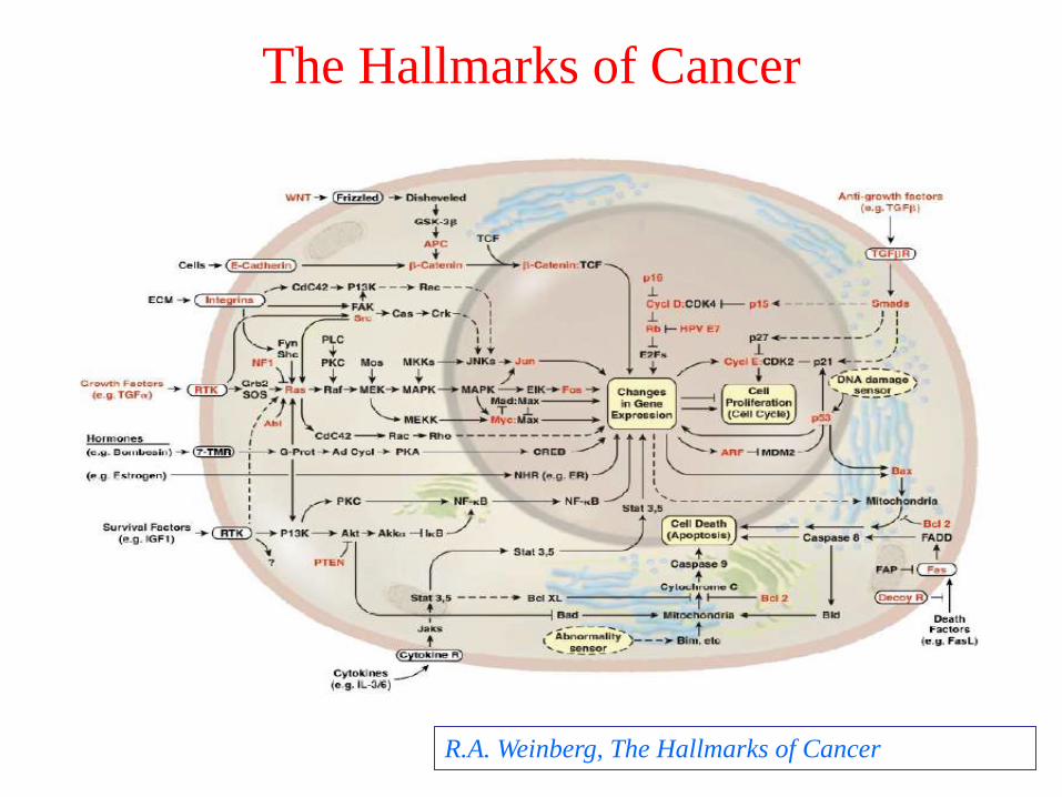
The Hallmarks of Cancer
R.A. Weinberg, The Hallmarks of Cancer
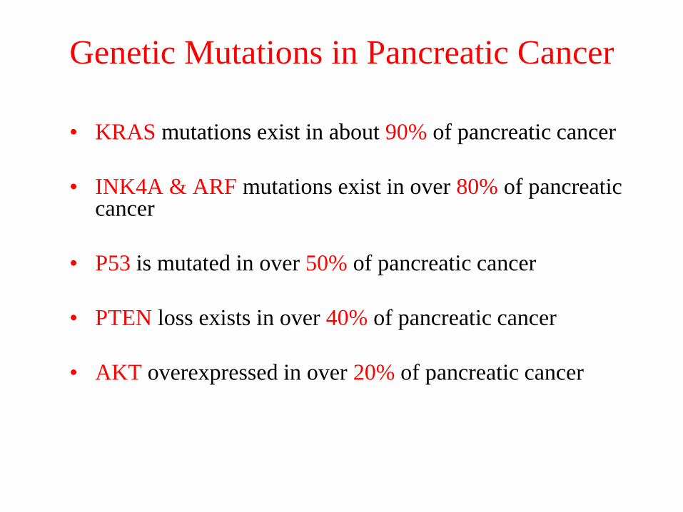
Genetic Mutations in Pancreatic Cancer
• KRAS mutations exist in about 90% of pancreatic cancer
• INK4A & ARF mutations exist in over 80% of pancreatic cancer
• P53 is mutated in over 50% of pancreatic cancer
• PTEN loss exists in over 40% of pancreatic cancer
• AKT overexpressed in over 20% of pancreatic cancer
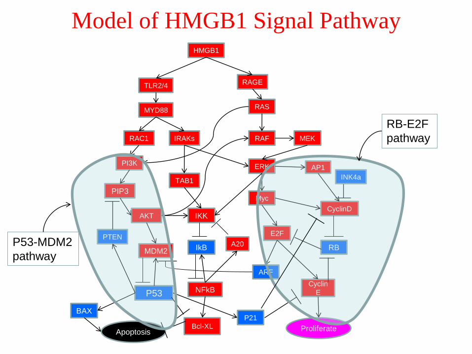
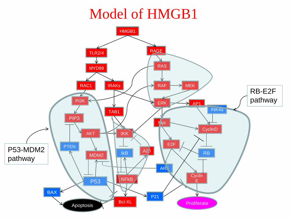
Model of HMGB1 Signal Pathway
INK4a
HMGB1
PIP3
IRAKsRAC1
MYD88
RAGETLR2/4
RAF
ERK
MEK
RAS
A20
E2F
AP1
NFkB
IKK
Myc
AKT
PI3K
TAB1
Cyclin
E
CyclinD
IkB RB
ARF
PTEN
P53
Bcl-XL
MDM2
BAX
Apoptosis Proliferate
P21
P53-MDM2
pathway
RB-E2F
pathway
I. The P53-MDM2 Pathway
• Important Proteins:1. P53
2. MDM2
3. PI3K
4. PTEN
5. AKT
• Positive and negative feedback loops in the P53-MDM2 pathway

The P53 Protein
• P53 is a tumor suppressor and regulates the cell cycle by integratingnumerous signals that control cell life and death.
• P53 is mutated in more than 50% of pancreatic cancers.
• P53 is a transcription factor for many genes including the pro-apoptosisand anti-apoptosis genes, e.g., Bax, mdm2.
• P53 is short-lived and expressed at very low levels in NORMAL cells.
BUT, P53 becomes stable and accumulates if the cell has DNA damage.
• Functions of P53:
Induces cell cycle arrest: P21, etc.
DNA repair: P53R2
Initiates apoptosis – Programmed Cell Death: Bax, etc.
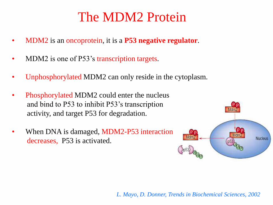
The MDM2 Protein
• MDM2 is an oncoprotein, it is a P53 negative regulator.
• MDM2 is one of P53’s transcription targets.
• Unphosphorylated MDM2 can only reside in the cytoplasm.
• Phosphorylated MDM2 could enter the nucleus
and bind to P53 to inhibit P53’s transcription
activity, and target P53 for degradation.
• When DNA is damaged, MDM2-P53 interaction
decreases, P53 is activated.
L. Mayo, D. Donner, Trends in Biochemical Sciences, 2002

The Proteins PI3K and PTEN
• PI3K is an oncoprotein, activated by some growth
factors (GF).
• PI3K can phosphorylate the lipid PIP2 to PIP3, then
activate the AKT signaling pathway.
• PTEN is a tumor suppressor protein. It is also one of
P53’s transcription targets.
• PTEN can dephosphorylate PIP3 back to PIP2, then,
inhibit the AKT signaling pathway.
• PTEN loss occurs in more than 40% of pancreatic
cancers.
PTEN
PI3K
AKT
P53
PIP3
GF
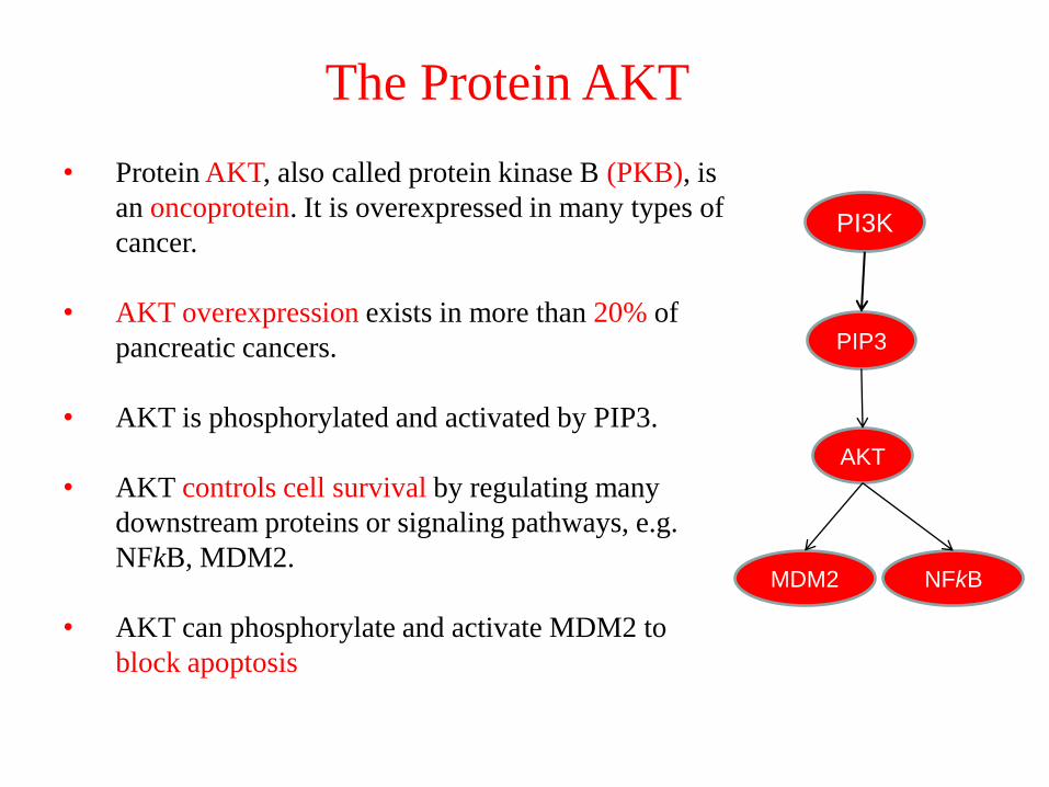
The Protein AKT
• Protein AKT, also called protein kinase B (PKB), is
an oncoprotein. It is overexpressed in many types of
cancer.
• AKT overexpression exists in more than 20% of
pancreatic cancers.
• AKT is phosphorylated and activated by PIP3.
• AKT controls cell survival by regulating many
downstream proteins or signaling pathways, e.g.
NFkB, MDM2.
• AKT can phosphorylate and activate MDM2 to
block apoptosis
PI3K
AKT
PIP3
MDM2 NFkB

P53-MDM2 Pathway
Negative feedback loop
P53 MDM2 ─┤P53
Positive feedback loop
P53 PTEN ─┤PIP3
AKT MDM2 ─┤ P53
PTEN
Damaged
DNA
PI3K
MDM2
AKT
P53
PIP3
Apoptosis
GF
II. RB-E2F Pathway
• Cell Cycle Introduction
• Important Proteins:
1. CYCLIN
2. CDK
3. RB
4. E2F
5. INK4A
6. ARF
• RB-E2F Pathway
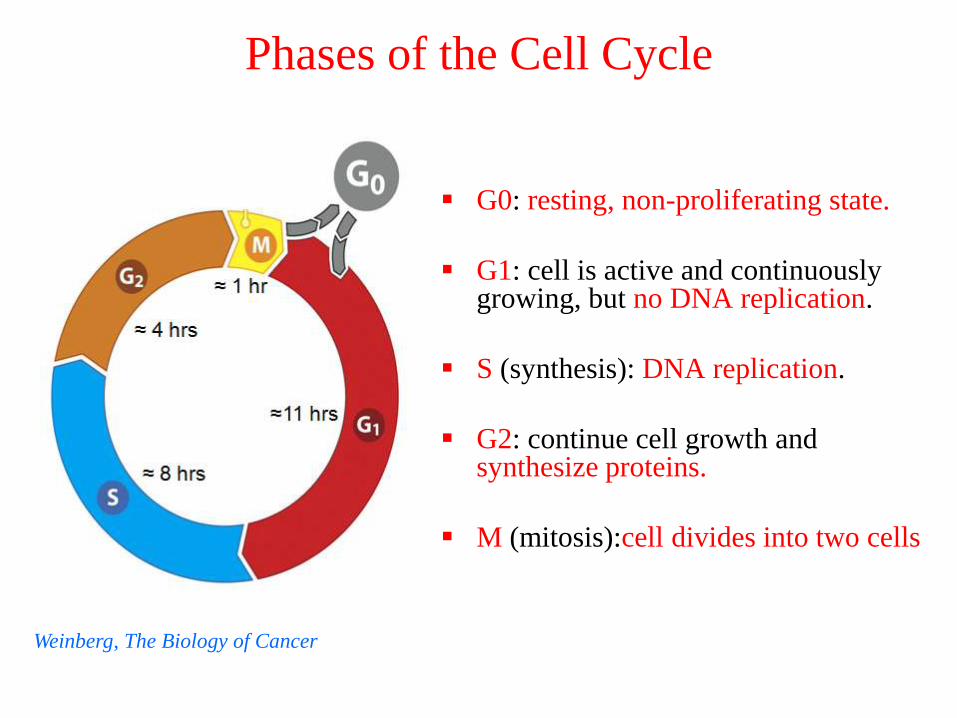
Phases of the Cell Cycle
G0: resting, non-proliferating state.
G1: cell is active and continuously growing, but no DNA replication.
S (synthesis): DNA replication.
G2: continue cell growth and synthesize proteins.
M (mitosis):cell divides into two cells
Weinberg, The Biology of Cancer
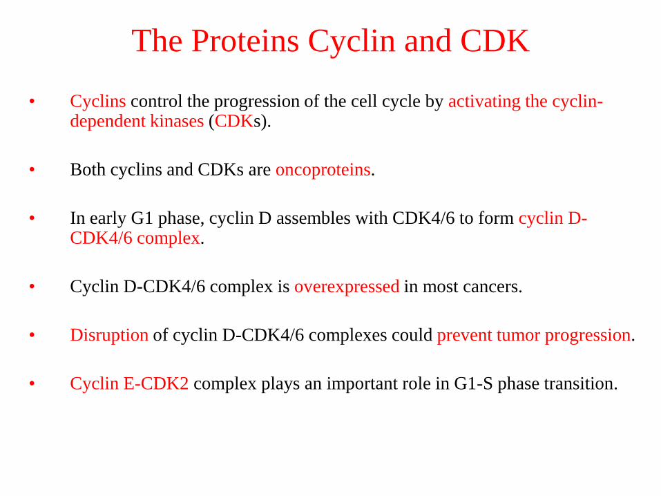
The Proteins Cyclin and CDK
• Cyclins control the progression of the cell cycle by activating the cyclin-dependent kinases (CDKs).
• Both cyclins and CDKs are oncoproteins.
• In early G1 phase, cyclin D assembles with CDK4/6 to form cyclin D-CDK4/6 complex.
• Cyclin D-CDK4/6 complex is overexpressed in most cancers.
• Disruption of cyclin D-CDK4/6 complexes could prevent tumor progression.
• Cyclin E-CDK2 complex plays an important role in G1-S phase transition.
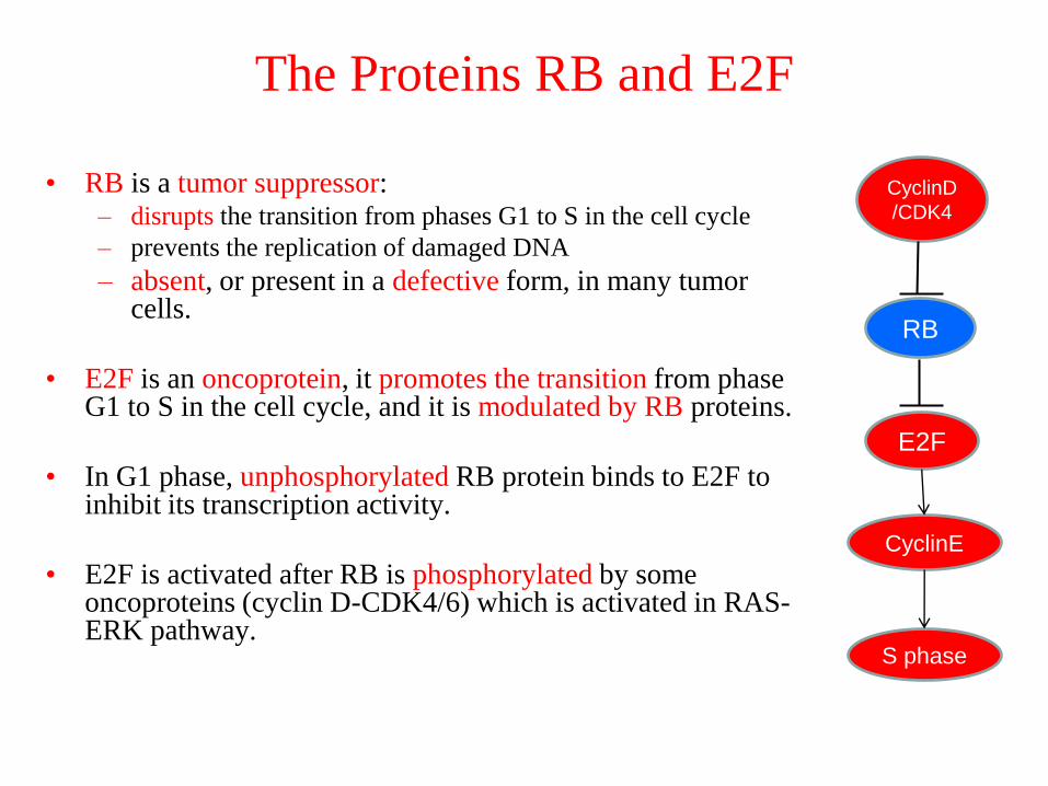
The Proteins RB and E2F
• RB is a tumor suppressor:– disrupts the transition from phases G1 to S in the cell cycle
– prevents the replication of damaged DNA
– absent, or present in a defective form, in many tumor cells.
• E2F is an oncoprotein, it promotes the transition from phase G1 to S in the cell cycle, and it is modulated by RB proteins.
• In G1 phase, unphosphorylated RB protein binds to E2F to inhibit its transcription activity.
• E2F is activated after RB is phosphorylated by some oncoproteins (cyclin D-CDK4/6) which is activated in RAS-ERK pathway.
CyclinD
/CDK4
RB
E2F
S phase
CyclinE
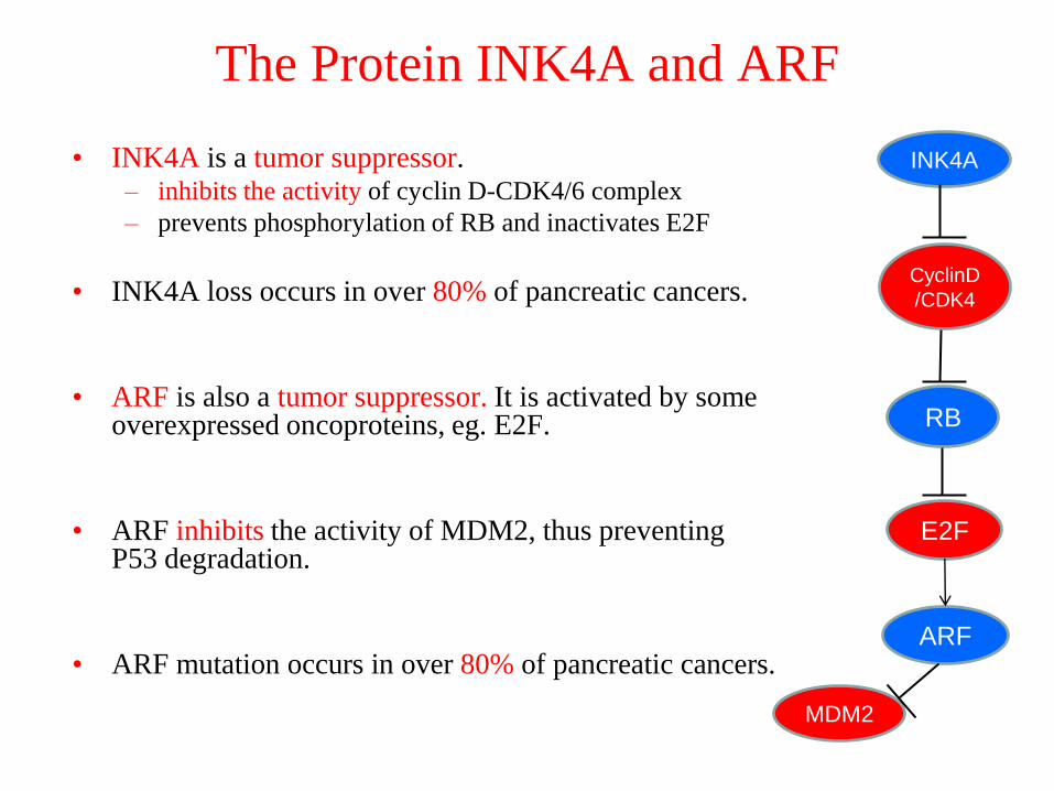
The Protein INK4A and ARF
• INK4A is a tumor suppressor. – inhibits the activity of cyclin D-CDK4/6 complex
– prevents phosphorylation of RB and inactivates E2F
• INK4A loss occurs in over 80% of pancreatic cancers.
• ARF is also a tumor suppressor. It is activated by some overexpressed oncoproteins, eg. E2F.
• ARF inhibits the activity of MDM2, thus preventing P53 degradation.
• ARF mutation occurs in over 80% of pancreatic cancers.
CyclinD
/CDK4
RB
E2F
INK4A
ARF
MDM2
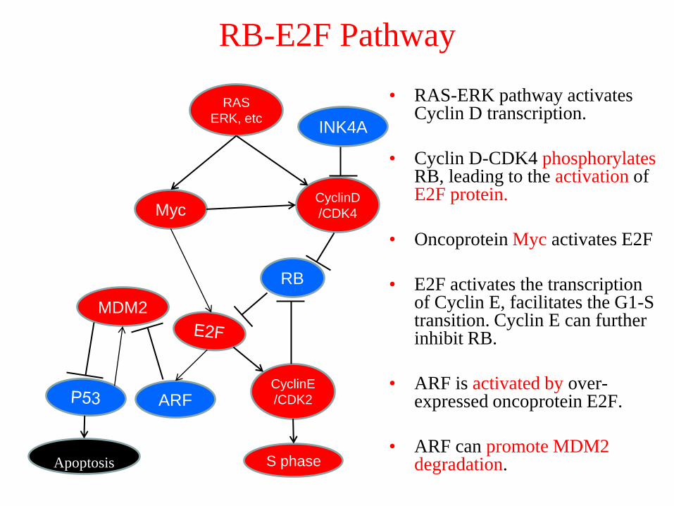
RB-E2F Pathway
• RAS-ERK pathway activates Cyclin D transcription.
• Cyclin D-CDK4 phosphorylatesRB, leading to the activation ofE2F protein.
• Oncoprotein Myc activates E2F
• E2F activates the transcription of Cyclin E, facilitates the G1-S transition. Cyclin E can further inhibit RB.
• ARF is activated by over-expressed oncoprotein E2F.
• ARF can promote MDM2 degradation.
ARF
MDM2
Apoptosis
RAS
ERK, etc
CyclinD
/CDK4
RB
Myc
CyclinE
/CDK2
S phase
INK4A
HMGB1 and Pancreatic Cancer
• HMGB1 Protein
• HMGB1 and Pancreatic Cancer
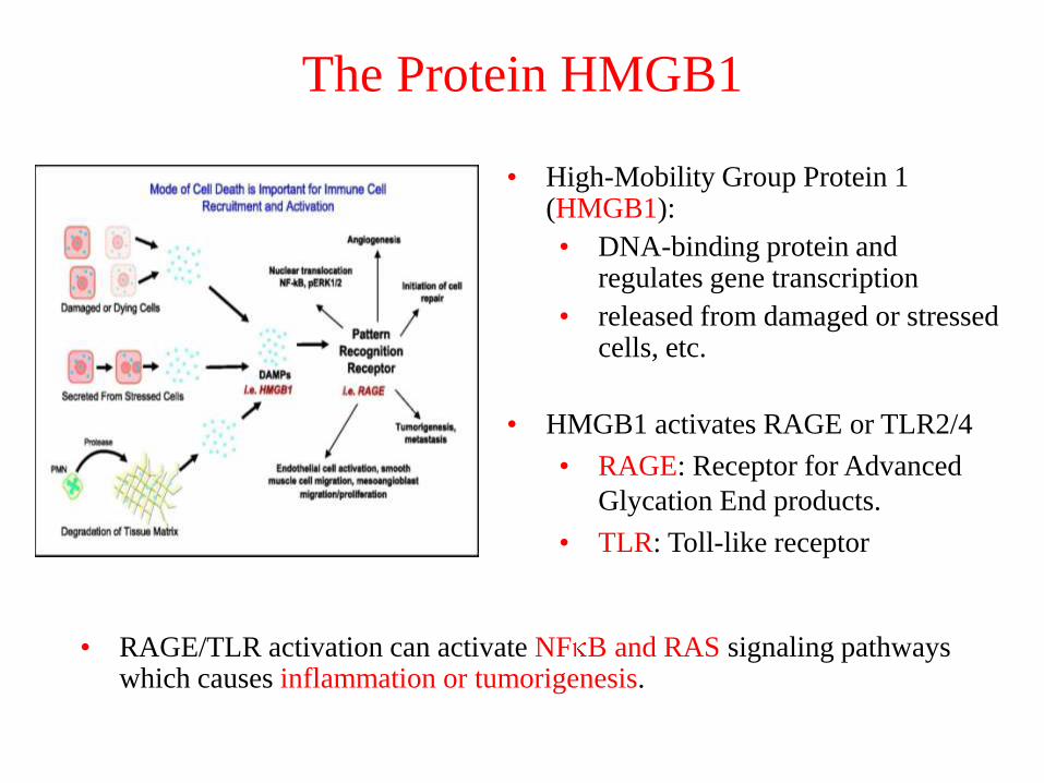
The Protein HMGB1
• High-Mobility Group Protein 1 (HMGB1):
• DNA-binding protein and regulates gene transcription
• released from damaged or stressed cells, etc.
• HMGB1 activates RAGE or TLR2/4
• RAGE: Receptor for Advanced
Glycation End products.
• TLR: Toll-like receptor
• RAGE/TLR activation can activate NF B and RAS signaling pathways which causes inflammation or tumorigenesis.

HMGB1 and Pancreatic Cancer
(Lotze et al., UPMC)
Experiments with pancreatic cancer cells:
Overexpression of HMGB1/RAGE is associated with diminished
apoptosis, and greater cancer cell survival.
Knockout of HMGB1/RAGE leads to increased apoptosis, and
decreased cancer cell survival.
HMGB1 RAGE Apoptosis
Apoptosis: “programmed” cell death

Model Checking
• Models1. Ordinary Differential Equation Model
2. BioNetGen Model
3. Boolean Network Model
• Model Checking
INK4a
HMGB1
PIP3
IRAKsRAC1
MYD88
RAGETLR2/4
RAF
ERK
MEK
RAS
A20
E2F
AP1
NFkB
IKK
Myc
AKT
PI3K
TAB1
Cyclin
E
CyclinD
IkB RB
ARF
PTEN
P53
Bcl-XL
MDM2
BAX
Apoptosis Proliferate
P21
Model of HMGB1
P53-MDM2
pathway
RB-E2F
pathway
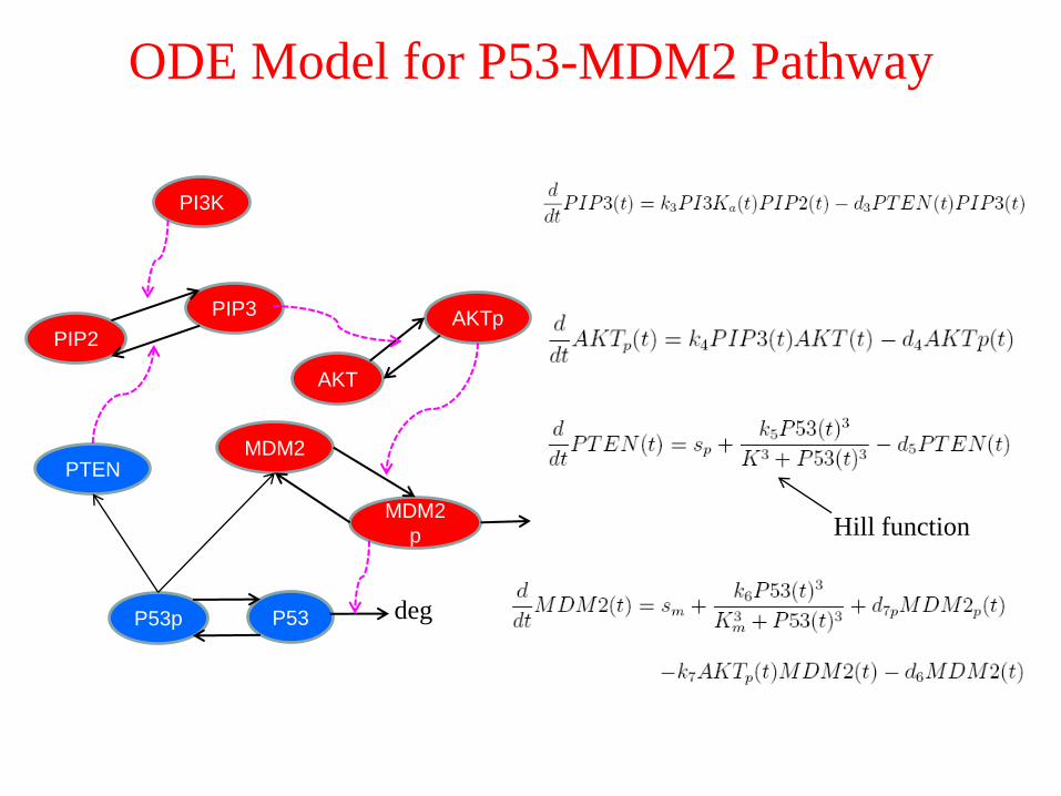
ODE Model for P53-MDM2 Pathway
PTEN
PI3K
MDM2
AKT
P53p
PIP3
MDM2
p
AKTp
P53
PIP2
deg
Hill function
BioNetGen SSA Model
# PI3K phosphorylates PIP2
• PI3K + PIP2 PI3K + PIP3 p1
# PTEN dephosphorylates PIP3
• PTEN + PIP3 PTEN + PIP2 d1
# P53-dependent production of PTEN
• P53(c~p) P53(c~p) + PTEN Hill(d2,K,3)
#PIP3 phosphorylates AKT
• PIP3 + AKT(a~U) PIP3 + AKT(a~p)
• AKT(a~p) + MDM2(b~U) AKT(a~p) + MDM2(b~p) p2
# MDM2p drives P53 degradation
• MDM2(b~p) + P53(c~U) MDM2(b~p) d5
# P53 synthesis
• I() I() + P53(c~U) s0
PTEN
PI3K
MDM2
AKT
P53p
PIP3
MDM2
p
AKTp
P53
PIP2
deg
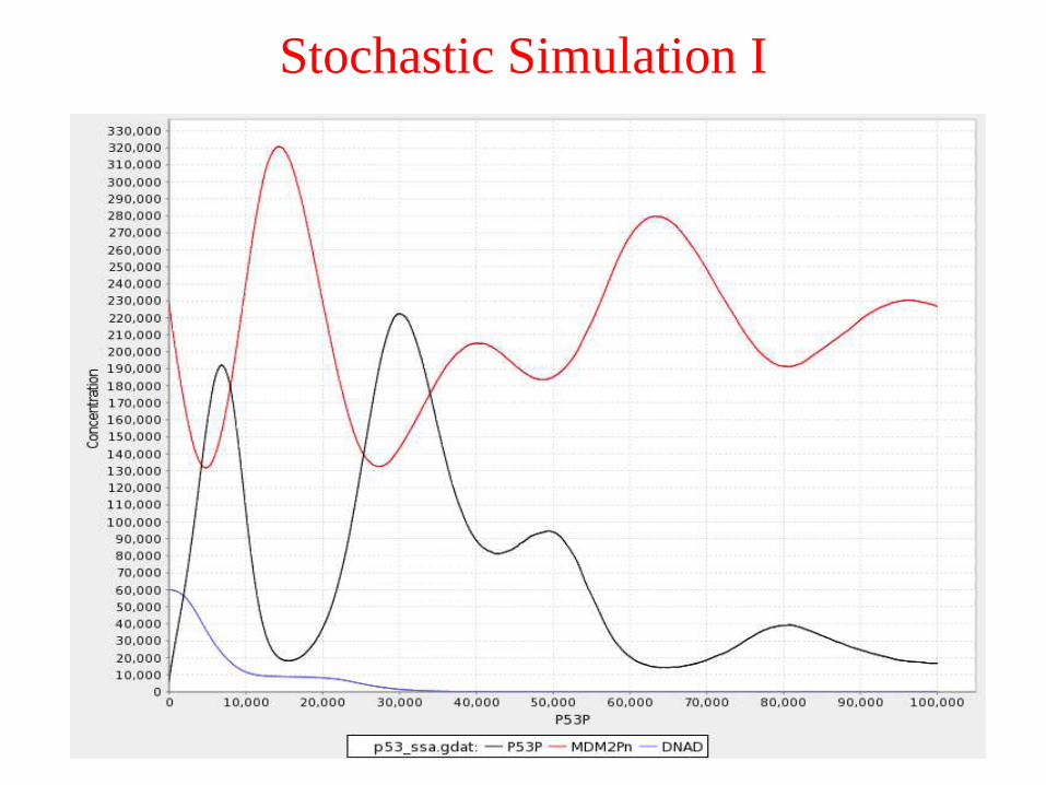
Stochastic Simulation I

Stochastic Simulation II
ARF loss and overexpression of PI3K ARF inhibits MDM2 and P53 accumulates
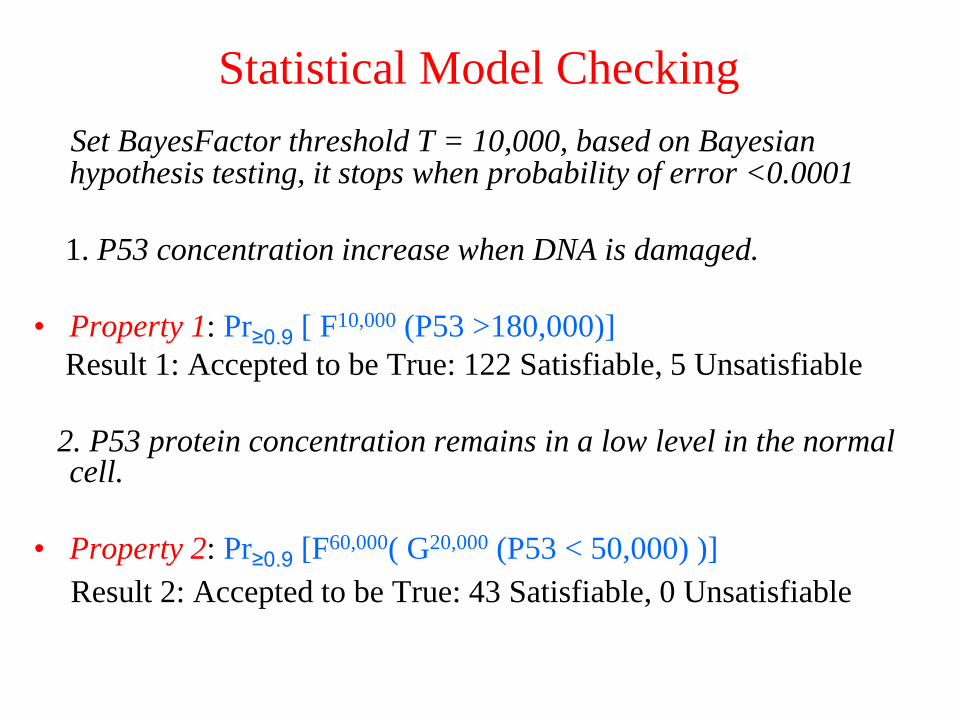
Statistical Model Checking
Set BayesFactor threshold T = 10,000, based on Bayesian hypothesis testing, it stops when probability of error <0.0001
1. P53 concentration increase when DNA is damaged.
• Property 1: Pr≥0.9 [ F10,000 (P53 >180,000)]
Result 1: Accepted to be True: 122 Satisfiable, 5 Unsatisfiable
2. P53 protein concentration remains in a low level in the normal cell.
• Property 2: Pr≥0.9 [F60,000( G20,000 (P53 < 50,000) )]
Result 2: Accepted to be True: 43 Satisfiable, 0 Unsatisfiable
INK4a
HMGB1
PIP3
IRAKsRAC1
MYD88
RAGETLR2/4
RAF
ERK
MEK
RAS
A20
E2F
AP1
NFkB
IKK
Myc
AKT
PI3K
TAB1
Cyclin
E
Cyclin
D
IkB RB
ARF
PTEN
P53
Bcl-
XL
MDM2
BAX
Apoptosis Proliferate
P21
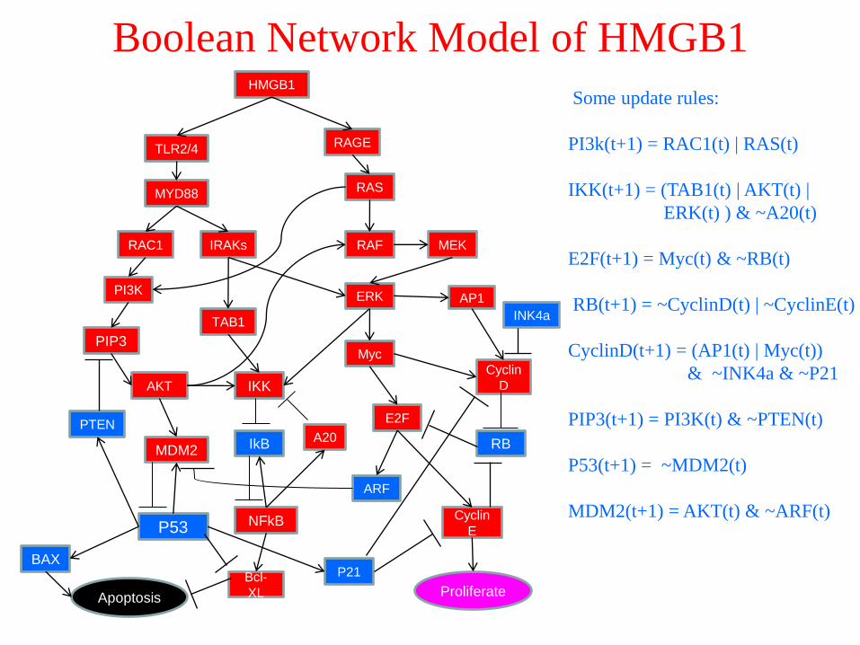
Boolean Network Model of HMGB1
Some update rules:
PI3k(t+1) = RAC1(t) | RAS(t)
IKK(t+1) = (TAB1(t) | AKT(t) |
ERK(t) ) & ~A20(t)
E2F(t+1) = Myc(t) & ~RB(t)
RB(t+1) = ~CyclinD(t) | ~CyclinE(t)
CyclinD(t+1) = (AP1(t) | Myc(t))
& ~INK4a & ~P21
PIP3(t+1) = PI3K(t) & ~PTEN(t)
P53(t+1) = ~MDM2(t)
MDM2(t+1) = AKT(t) & ~ARF(t)
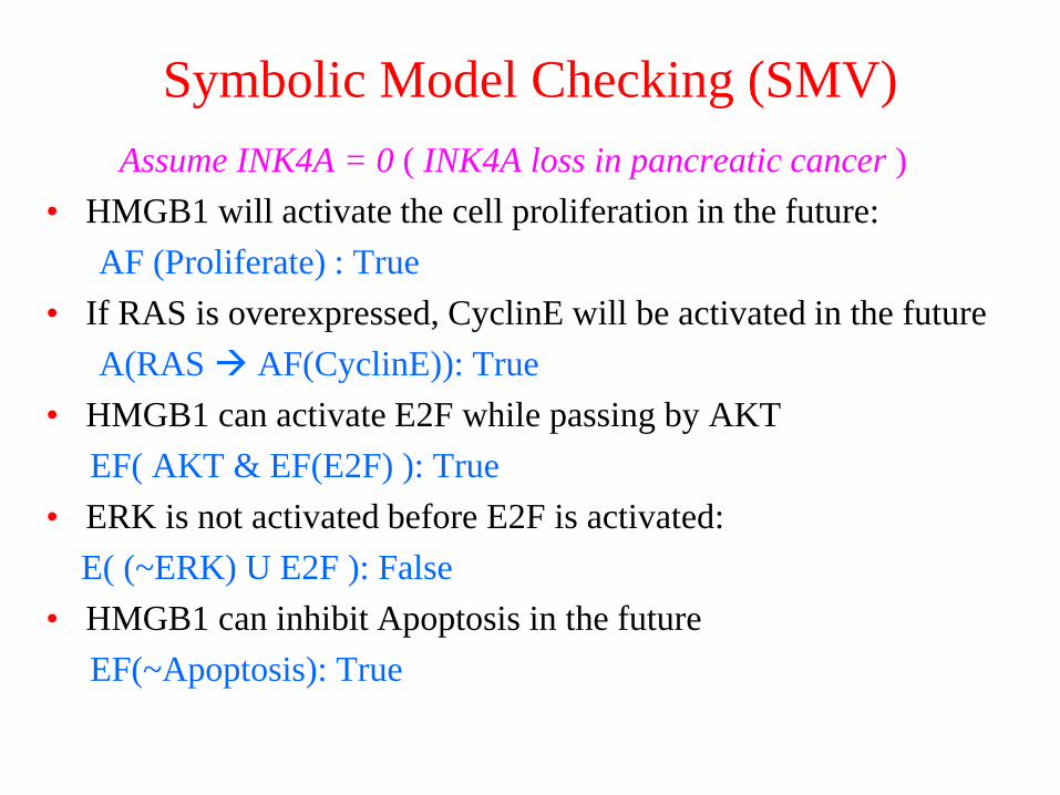
Symbolic Model Checking (SMV)
Assume INK4A = 0 ( INK4A loss in pancreatic cancer )
• HMGB1 will activate the cell proliferation in the future:
AF (Proliferate) : True
• If RAS is overexpressed, CyclinE will be activated in the future
A(RAS AF(CyclinE)): True
• HMGB1 can activate E2F while passing by AKT
EF( AKT & EF(E2F) ): True
• ERK is not activated before E2F is activated:
E( (~ERK) U E2F ): False
• HMGB1 can inhibit Apoptosis in the future
EF(~Apoptosis): True

Inference from Model Checking
Assume INK4A =1 ( NO INK4A mutation )
1. CyclinD = ( Myc | AP1 ) | ~INK4A
• HMGB1 will activate E2F in the future:
AF(E2F): True
HMGB1 and its effectors have a stronger effect than INK4A
2. CyclinD = ( Myc | AP1 ) & ~INK4A
• AF(E2F): False
INK4A has a stronger effect than HMGB1 and its effectors:
HMGB1 can not activate E2F.
Model checking can help to rule out or modify some models
which do not satisfy the properties abstracted from experiment.

Future Work
• Asynchronous Boolean Network Model for HMGB1: protein
mutations occur at different stages of pancreatic cancer
• Apply SMV to larger cell cycle boolean network: e.g., Kohn
map, etc.
• Probabilistic Boolean Network Model and Statistical Model
Checking
• In collaboration with experimental cancer biologist, estimate
important parameters for the Non-boolean models.
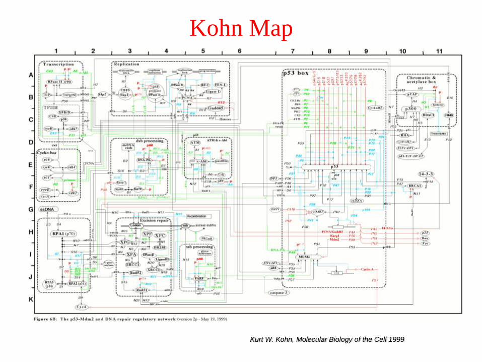
Kohn Map
Kurt W. Kohn, Molecular Biology of the Cell 1999
Acknowledgement
This research is funded by NSF Expeditions in
Computing Program.
• Michael Lotze (UPMC)
• William Buchser (UPMC)
• Kristen Livesey (UPMC)
• Natasa Miskov-Zivanov (Univ. of Pittsburgh)
• Anvesh Komuravelli (CMU)

Thank you!

Appendix

Genetic Progression Model of Pancreatic Cancer
PanINs (Pancreatic intraepithelial neoplasias), represent progressive stages of neoplastic growth
Bardeesy, DePinho, Nature Reviews, 2002

III. RAS Pathways
• Important Proteins
1. RAS
2. RAF
3. MEK
4. MAPK (ERK1/2)
• RAS Pathways
The Protein RAS
• Protein RAS relays signals from outside the cell to the nucleus. Activation of RAS signaling causes cell growth and survival.
• RAS family has three members: HRAS, KRAS, NRAS.
• KRAS mutations increase with disease progression, and are found in more than 90% of pancreatic cancers.
• RAS is activated when it binds to GTP (Guanosine Triphosphate) which is catalyzed by GEFs (Guanine nucleotide Exchange Factors), inactivated if bound to GDP (Guanosine Diphosphate).
• Aberrant signaling through RAS pathways occurs if RAS is mutated or some growth-factor-receptor tyrosine kinases (EGFR, etc.) are over-expressed, or mutations of RAS effectors (RAF, MEK, PI3K).

The Proteins RAF, MEK and MAPK
• RAF protein usually exists in the cytoplasm. When activated by GTP-
RAS protein it relocates to the plasma membrane.
• The activated RAF can phosphorylate and activate the MEK proteins
(mitogen-activated protein kinase kinases (MAPKK)).
• The activated MEKs can phosphorylate and activate protein MAPKs
(ERK1/2)
• The activated ERK can phosphorylate transcription factors such as
ELK1, AP-1 and ETS, which activate the expression of the regulatory
proteins, including Cyclin D protein, that enable progression of the cell
cycle through the G1 phase.
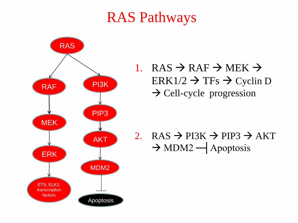
RAS Pathways
1. RAS RAF MEK
ERK1/2 TFs Cyclin D
Cell-cycle progression
2. RAS PI3K PIP3 AKT
MDM2 ─┤Apoptosis
RAF
ERK
MEK
RAS
PIP3
MDM2
AKT
PI3K
ETS, ELK1,
transcription
factors
Apoptosis
IV. NF B Pathway
• Important Proteins
1. NF B
2. I B
3. IKK
• NF B Pathway
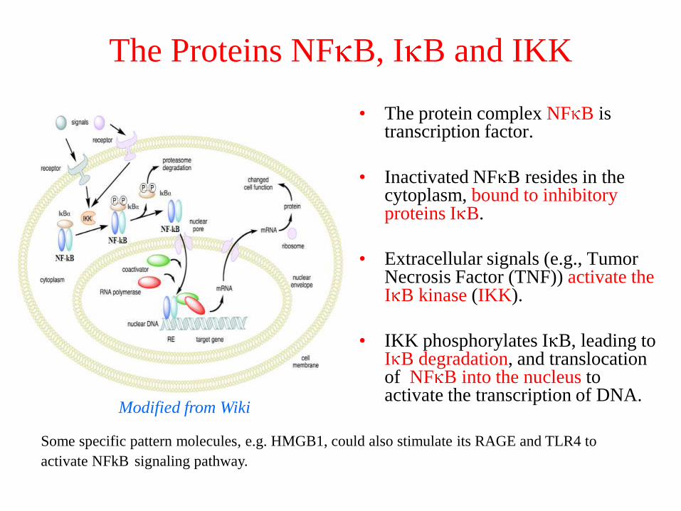
The Proteins NF B, I B and IKK
• The protein complex NF B is transcription factor.
• Inactivated NF B resides in the cytoplasm, bound to inhibitory proteins I B.
• Extracellular signals (e.g., Tumor Necrosis Factor (TNF)) activate the I B kinase (IKK).
• IKK phosphorylates I B, leading to I B degradation, and translocation of NF B into the nucleus to activate the transcription of DNA.
Some specific pattern molecules, e.g. HMGB1, could also stimulate its RAGE and TLR4 to
activate NFkB signaling pathway.
Modified from Wiki
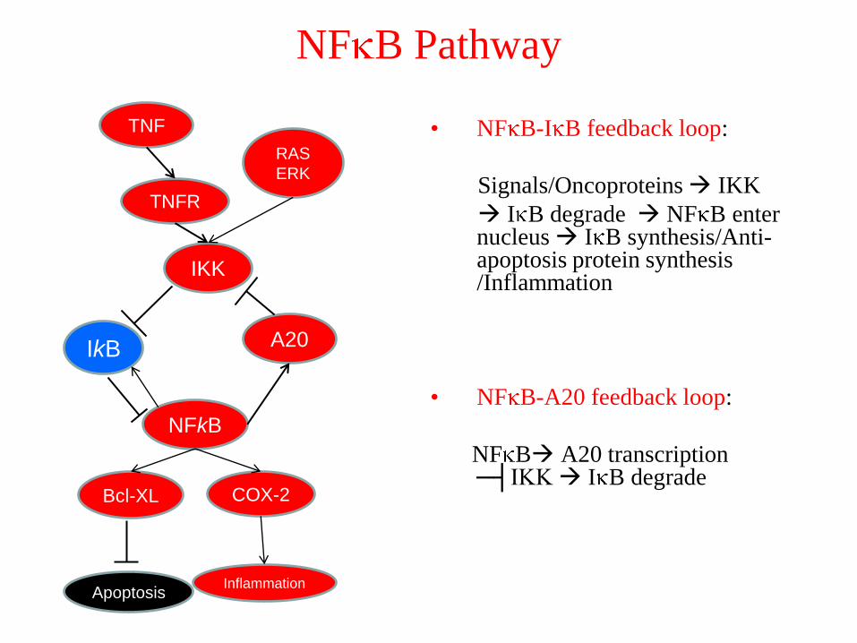
NF B Pathway
• NF B-I B feedback loop:
Signals/Oncoproteins IKK
I B degrade NF B enter nucleus I B synthesis/Anti-apoptosis protein synthesis /Inflammation
• NF B-A20 feedback loop:
NF B A20 transcription ─┤IKK I B degrade
IkB
TNF
A20
IKK
NFkB
TNFR
COX-2Bcl-XL
ApoptosisInflammation
RAS
ERK