signal tranduction
TRANSCRIPT

M.HAMZA KHALID
University Of Lahore
Department Of Pharmacy

SIGNAL TRANSDUCTION

Signal TransductionIt is a process of converting a extracellular signal into a
response.The conversion of this signal
is brought out by the extracellular signaling
molecules known as Ligand.

Ligand:-Substances synthesized and released by signaling cells
and produce a specific response only in target cells that have receptors for the
signaling molecules.

The extra cellular signaling molecule binds with the located on the cell surface or inside the cell and in turn the receptor triggers the biochemical chain of events in the cells which leads to
Cellular Response. Cellular response:-1)Change in gene expression. 2)Cell morphology. 3)Cell movement.


Steps involve in extracellular signaling:-1) Synthesis of extracellular molecule. 2) Release of the signaling molecule by the signaling cell. 3) Transport of the signal to the target cell. 4) Detection of the signal by a specific receptor protein. 5) A change in cellular metabolism, function, or development triggered by the receptor-signal complex . 6) Removal of the signal, which often terminates the cellular response.

Types of Signaling:-ENDOCRINE SIGNALING :-Signaling molecules (hormones) act on target cells distant from their site of synthesis by cells of endocrine organs.PARACRINE SIGNALING :-The signaling molecule (neurotransmitter) released by a cell only affect target cells in close proximity to it. AUTOCRINE SIGNALING :-Cells respond to substances (growth factors) which they themselves release

Membrane Receptors:-In signal transduction the
signaling molecule first binds to the receptors that
my be present on the surface of membrane or
inside the cell.

Receptor:-A region of tissue, or a
molecule in a cell membrane, which responds
specifically to a particular neurotransmitter, hormone, antigen, or other substance.

Receptor can be intra cellular or extracellular
1)Intracellular Receptor:-They are present inside the cell and upon binding with ligand triggers the series of
changes. They are hydrophobic in nature.

2)Extracellular Receptor:-
Cell surface receptor that are specialized are integral trans membrane protein that take part the communication b/t
the cells. Ligand may be harmone,cytokine,growth
factors etc which binds to the receptor but don’t move inside
the cell.


Based on structural and functional similarities, membrane receptors are mainly divided into 3 classes:-1)Ion channel linked receptor 2)Enzyme linked receptor.3)G protein coupled receptor .

Receptors
• INTRACELLULAR RECEPTORS :- 1) Cytoplasmic
2)Nuclear receptors
• CELL SURFACE RECEPTORS:-
ION CHANNEL RECEPTOR(inotropic)
• Ligand gated ion channels
• Controlled by neurotransmitters
• Present in neurons• Eg: Ach cation channel
G -PROTEIN LINKED RECEPTOR(Metabotropic)
• Act via second messengers-cAMP, IP3,
DAG , c GMP
ENZYME LINKED RECEPTOR
• Eg: Protein kinaseTyrosine kinaseTyrosine phosphotaseSerine/threonine kinaseGuanylyl cyclaseHistidine kinase

Ion channel linked receptors :-They are ion-channels (including cation-channels and anion-channels) Na K Cl Ca themselves and constitute a large family of multiple transmembrane proteins. They are involved in rapid signaling events most generally found in electrically excitable cells such as neurons and are also called ligand-gated ion channels. Opening and closing of Ion channels are controlled by neurotransmitters.


Enzyme-linked receptors:-They are either enzymes
themselves, or are directly associated with the enzymes
that they activate. The majority of enzyme-lined receptors are protein kinases, or associate
with protein kinases


G-protein coupled receptors These receptors activate a G protein ligand binding. G-protein is a trimeric protein. The 3 subunits are called α、β and γ. The α subunit can bind with guanosine diphosphate, GDP. This causesphosphorylation of the GDP to guanosine triphosphate, GTP, and activates the α subunit, which then dissociates from the β and γ subunits. The activated α subunit can further affect intracellular signaling proteins or target functional proteins directly.
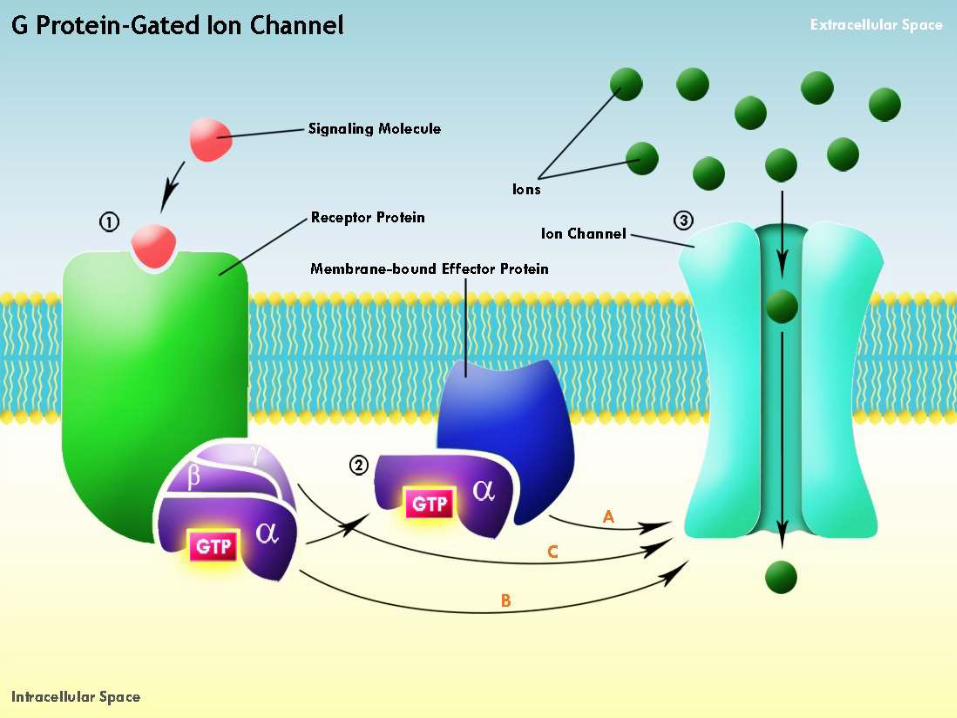

Messenger:-A particular substance that
can carry or transfer an massage which in terms of
biology may be a harmone,protein etc

Primary Messenger:-Transmit the signal from
receptor to the enzyme and activate it to produce secondary messenger.
Example:Gα,Gβ

Secondary Messenger:-Secondary messenger are the
intracellular molecule released by the cell to trigger physiological changes.
After the receptor being activated the secondary play a vital role in s Transmit signals in form of either direct cellular
response eg:cAMP, cGMPOr activate further enzymes to produce
response Eg :IP3,DAG signal
transduction.


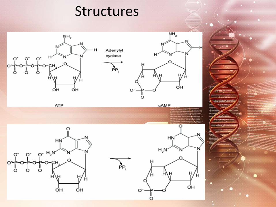
Cyclic Nucleotides
1)cAMP
2)cGMP

Cyclic AMP is synthesized from ATP by the action of the enzyme adenylyl cyclase.Binding of the hormone to its receptor activates G protein which, in turn, activates adenylyl cyclase.The resulting rise in cAMP turns on the appropriate response in the cell by either (or both):Changing the molecular activities in the cytosol, often using Protein Kinase A (PKA) cAMP-dependent protein kinase that phosphorylates target proteins;turning on a new pattern of gene transcription.
Structures

Some of the hormones that achieve their effects through cAMP as a second messenger:1)Adrenaline2)Glucagon3)Luteinizing hormone (LH)

Cyclic GMP serves as the second messenger for:-1) Atrial natriuretic peptide (ANP)2)Nitric oxide (NO)3)The response of the rods of the retina to light.

Inositol trisphosphate (IP3) and Diacylglycerol (DAG):-
As its name suggests, it hydrolyzes phospholipids specifically phosphatidylinositol-4,5-bisphosphate (PIP2) which is found in the inner layer of the plasma membrane. Hydrolysis of PIP2 yields two products:
Diacylglycerol (DAG)DAG remains in the inner layer of the plasma membrane. It recruits Protein Kinase C (PKC) a calcium-dependent kinase that phosphorylates many other proteins that bring about the changes in the cell.

Inositol Triphosphate IP3:-As its name suggests, activation of PKC requires calcium ions. These are made available by the action of the other second messenger — IP3.inositol-1,4,5-trisphosphate (IP3)

Peptide and protein hormones like1)Vasopressin,2)Thyroid-stimulating hormone. 3)Angiotensin.4)Neurotransmitters like GABA. They bind to G protein-coupled receptors (GPCRs) that activate the intracellular enzyme phospholipase C (PLC).


Calcium ions (Ca2+):-As the functions of IP3 and DAG indicate, calcium ions are also important intracellular messengers. In fact, calcium ions are probably the most widely used intracellular messengers.In response to many different signals, a rise in the concentration of Ca2+ in the cytosol triggers many types of events such as:

1)Muscle contraction. 2)Secretion of hormones like insulin.3)Activation of T cells and B cells when they bind antigen with their antigen receptors (TCRs and BCRs respectively).4)Adhesion of cells to the extracellular matrix (ECM).5)Apoptosis. 6)A variety of biochemical changes mediated by Protein Kinase C (PKC).

Getting Ca2+ into (and out of)
the cytosol:-
1) Open in response to a change in
membrane potential, e.g. the
depolarization of an action potential.
2)Skeletal muscle
3)Smooth muscle (These are the channels
blocked by drugs, such as felodipine [Plendil],
used to treat high blood pressure. The influx of
Ca2+ contracts the smooth muscle walls of the
arterioles, raising blood pressure.The drug
block this .

4)When the action potential reaches the presynaptic terminal, the influx of Ca2+ triggers the release of the neurotransmitter.
5)Receptor-operated channels like G-protein-coupled receptors (GPCRs). These are not channels but they trigger a release of Ca2+ from the endoplasmic reticulum as described above.
Ca2+ ions are returned:-
They return to the ECF by active transport using an ATP-driven pump called a Ca2+ ATPase.3 Na+ ions flowing DOWN their concentration gradient to pump one Ca2+ against its gradient and 4 Na+ ions flowing down to pump 1 Ca2+ and 1 K+ ion up their concentration gradients.

Nitric Oxide (NO)Nitric oxide (NO) acts as a second messenger because it is a free radical that can diffuse through the plasma membrane and affect nearby cells.
It is synthesised from arginine and oxygen by the NO synthase.
It activates soluble guanylyl cyclase, which when activated produces another second messenger, cGMP.
It is toxic in high concentrations , but is the cause of many other functions like relaxation of blood vessels, apoptosis etc


Gene Expression

The last step but not least of the signal transduction is
gene expression.
Gene:-
A gene is the sequence of nucleotides in DNA
encoding one polypeptide chain or one mRNA
molecule.

Gene Structure
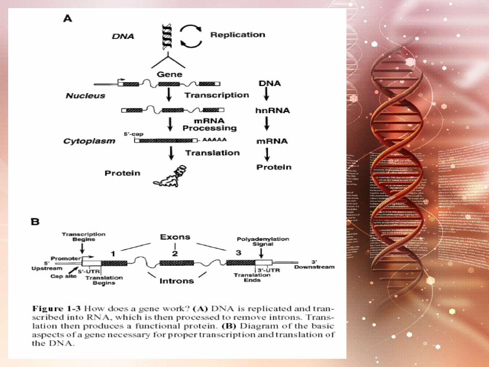
Eukaryotic gene structure: Most eukaryotic genes in contrast to typical bacterial genes , The coding sequence (Exons) are interrupted by noncoding DNA (Introns).
Exons:Expressed sequence.
Introns:Intervening or un expressed sequence

Classification of gene with respect to their Expression:
Constitutive ( house keeping) genes:1- Are expressed at a fixed rate, irrespective to the cell condition.2- Their structure is simplerControllable genes:1- Are expressed only as needed. Their amount may increase or decrease with respect to their basal level in different condition.2- Their structure is relatively complicated with some response elements
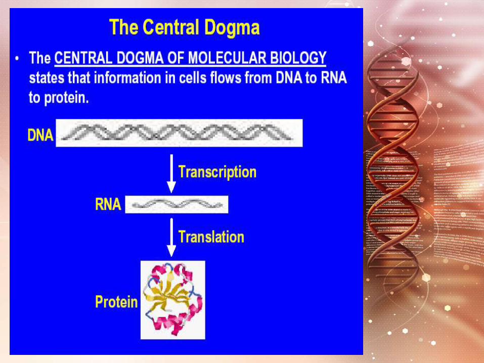
Gene Expression:-It is the process by which information from a gene is used in the synthesis of a functional gene product.These products are often proteins, but in non-protein coding genes such as rRNA genes or tRNA genes, the product is a functional RNA
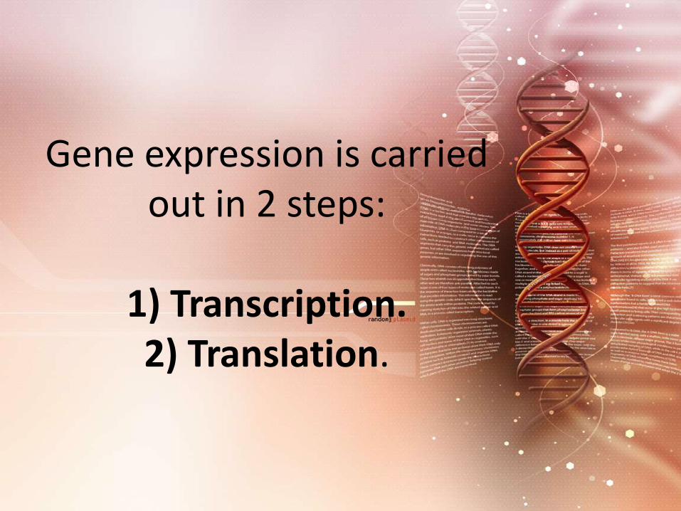
Gene expression is carried out in 2 steps:
1) Transcription. 2) Translation.

Regulation of Gene Expression
Promoters
The region necessary to initiate transcription.
Consists of short nucleotide sequence that serve as the recognition point for binding of RNA polymerase.
Located adjacent to the genes they regulate.

There are significant differences in number , orientation and distance
between promoters in different genes.
Promoters for RNA polymeraseIIinclude:
TATA CAAT
GC

DNA sequences interact with regulatory proteins & increase the efficiency of initiation and transcription and thus increase its rate.
Enhancers

Enhancers:
1)Large up to several hundred bp long). 2)Large Tissue- specific stimulate transcription only in certain tissues

Transcription Factor
They are proteins essential for initiation of the transcription, but
they are not part of RNA polymerase molecule that carry out the
transcription process.

Functions:Each RNA polymerase requires a number of transcription factors which help in:
1. Binding of the enzyme to DNA template.
2. Initiation and maintenance of transcription.
3. Control the rate of gene expression

1) Special TFs:Involved in regulation of heat, light, and hormone inducible genes.They bind to:
a. enhancers.b. Basal TFs.c. RNA polymerase that bind to the gene promoter.Thus special TFs can regulate the transcriptional activity of the gene.

1.Positive regulation:
When the expression of genetic is quantitatively increased by the presence of specific regulatory
element is known as positive regulation.
Element modulating positive regulation is known as activator or
positive regulator

2.Negative regulation
When the expression of geneticinformation diminished by thepresence of specific regulatoryelement.
The element or molecule mediatingthe negative regulation is said to berepressor.


Gene Regulation In
Prokaryotes

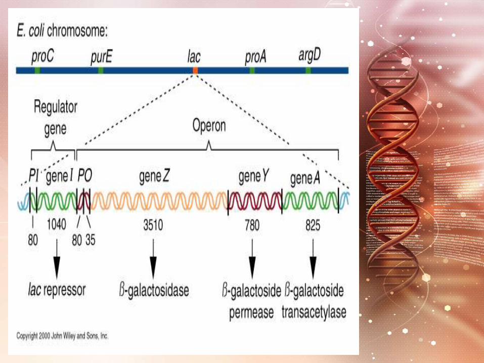
OPERON in gene regulation of prokaryotes
Definition: A few genes that
are controlled collectively by one promoter
Its structure: Each Operon is
consisted of few structural genes( cistrons) and
some cis-acting element such as promoter (P) and operator (O)


Gene Expression In Eukaryotes


Eukaryotic gene regulation at several levels
1.Transcriptional control.
2.RNAprocessing control.
3.RNA transport /localisation control.
4.Translation control.
5.mRNAdegradation control.
6.Protein activator control.


Research Article
Female resistance to pneumonia identifies
lung macrophage nitric oxide synthase-3 as a
therapeutic target

AbstractTo identify new approaches to enhance innate immunity to bacterial pneumonia, we investigated the natural experiment of gender differences in resistance to infections. Female and estrogen-treated male mice show greater resistance to pneumococcal pneumonia, seen as greater bacterial clearance, diminished lung inflammation, and better survival.

AbstractIn vitro, lung macrophages from female mice and humans show better killing of ingested bacteria. Inhibitors and genetically altered mice identify a critical role for estrogen-mediated activation of lung macrophage nitric oxide synthase-3 (NOS3). Epidemiologic data show decreased hospitalization for pneumonia in women receiving estrogen or statins (known to activate NOS3).

Pharmacologic targeting of NOS3 with statins or another small-molecule compound (AVE3085) enhanced macrophage bacterial killing, improved bacterial clearance, and increased host survival in both primary and secondary (post-influenza) pneumonia. The data identify a novel mechanism for host defense via NOS3 and suggest a potential therapeutic strategy to reduce secondary bacterial pneumonia after influenza

ReferencesGuyton & Hall text book of medical physiology.
McGraw Hill physiology
Kuby Immunology
Immunology at a glance( J.H.L Playfair & B.M Chain
Essentials of medical physiology (TAYPEE)

