sigma xi presentation
TRANSCRIPT
Proliferation and Differentiation of Human Neural Stem Cells via
Selective Agonism of AT1 & AT2 Receptors
Brigitte BlancoPine Crest School Research performed at Nova Southeastern University
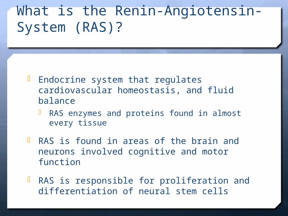
What is the Renin-Angiotensin-System (RAS)?
Endocrine system that regulates cardiovascular homeostasis, and fluid balance RAS enzymes and proteins found in almost every
tissue
RAS is found in areas of the brain and neurons involved cognitive and motor function
RAS is responsible for proliferation and differentiation of neural stem cells
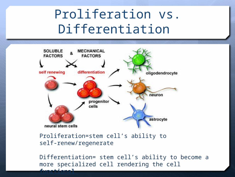
Proliferation vs. Differentiation
Proliferation=stem cell’s ability to self-renew/regenerate
Differentiation= stem cell’s ability to become a more specialized cell rendering the cell functional
Guimond MO, Gallo-Payet N (2012) The Angiotensin II Type 2 Receptor in Brain Functions: An Update. International journal of hypertension 2012: 351758
VasodilationNeurite outgrowth
Cerebral blood flowSpatial memoryDifferentiation Antiremodeling
Neuronal excitability
VasoconstrictionProliferationHypertrophy
Aldosterone releaseOxidative stress
Baroreflex
AT1 receptor AT2 receptor
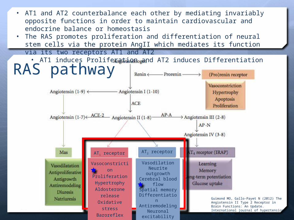
• AT1 and AT2 counterbalance each other by mediating invariably opposite functions in order to maintain cardiovascular and endocrine balance or homeostasis
• The RAS promotes proliferation and differentiation of neural stem cells via the protein AngII which mediates its function via its two receptors AT1 and AT2 • AT1 induces Proliferation and AT2 induces Differentiation
RAS pathway
Application
Selective agonism of AT1 and AT2 receptors could possibly induce proliferation and differentiation in damaged areas of the brain
Therapy for traumatic brain injuries, post-stroke ischemic damage, and chemotherapy
Neurodegenerative diseases and cognitive deficits are associated with neuronal and synaptic dysfunctions Alzheimer’s, Parkinson’s, Huntington’s, mental retardation
Therapeutic treatments for non-proliferating and non-functioning cells associated with neurodegenerative diseases, traumatic brain injuries, and more to proliferate and function again could become a reality
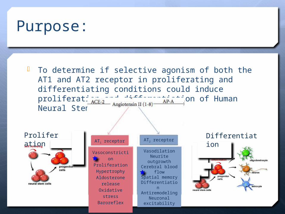
Purpose:
To determine if selective agonism of both the AT1 and AT2 receptor in proliferating and differentiating conditions could induce proliferation and differentiation of Human Neural Stem Cells
Proliferation
Differentiation
VasodilationNeurite outgrowth
Cerebral blood flowSpatial memoryDifferentiation Antiremodeling
Neuronal excitability
VasoconstrictionProliferationHypertrophy
Aldosterone releaseOxidative stress
Baroreflex
AT1 receptor AT2 receptor
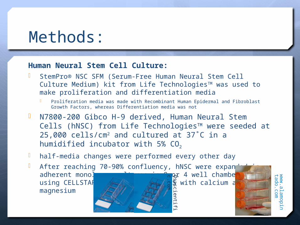
Methods:
Human Neural Stem Cell Culture: StemPro® NSC SFM (Serum-Free Human Neural Stem Cell Culture
Medium) kit from Life TechnologiesTM was used to make proliferation and differentiation media Proliferation media was made with Recombinant Human Epidermal and Fibroblast Growth
Factors, whereas Differentiation media was not
N7800-200 Gibco H-9 derived, Human Neural Stem Cells (hNSC) from Life TechnologiesTM were seeded at 25,000 cells/cm2 and cultured at 37˚C in a humidified incubator with 5% CO2
half-media changes were performed every other day After reaching 70-90% confluency, hNSC were expanded in adherent
monolayer cultures in 8 or 4 well chamber slides using CELLSTARTTM (Gibco), in D-PBS with calcium and magnesium
Usa
scientifi
c.com
ww
w.a
lam
opin
tado.co
m
Methods:
Selective Agonist Drug Treatments
Selective Agonists (below) were administered daily to their respective chamber slides at 10µM AT1 selective agonist (sar1 AngII) AT2R agonist (CGP42112)
AT2 Selective Antagonist administered with AT1 selective agonist AT2 antagonism control (PD123319)
The relationship between these two receptors is unidirectional, meaning that if the AT1 receptor is stimulated, the AT2 receptor will also produce a response. HOWEVER, if the AT2 receptor is stimulated, the AT1 receptor will NOT respond
For this reason, only and AT2 antagonist was added in addition to the AT1 agonist
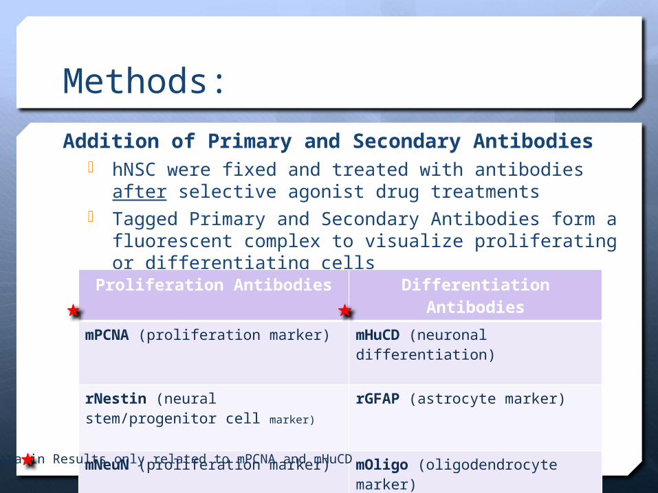
Methods:
Addition of Primary and Secondary Antibodies hNSC were fixed and treated with antibodies after
selective agonist drug treatments Tagged Primary and Secondary Antibodies form a
fluorescent complex to visualize proliferating or differentiating cells
Proliferation Antibodies Differentiation Antibodies
mPCNA (proliferation marker) mHuCD (neuronal differentiation)
rNestin (neural stem/progenitor cell marker)
rGFAP (astrocyte marker)
mNeuN (proliferation marker) mOligo (oligodendrocyte marker)
rs100β (proliferation marker) rDCX (migrating neuroblast marker)
Data in Results only related to mPCNA and mHuCD
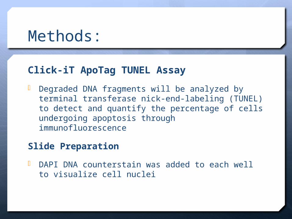
Methods:
Click-iT ApoTag TUNEL Assay
Degraded DNA fragments will be analyzed by terminal transferase nick-end-labeling (TUNEL) to detect and quantify the percentage of cells undergoing apoptosis through immunofluorescence
Slide Preparation
DAPI DNA counterstain was added to each well to visualize cell nuclei
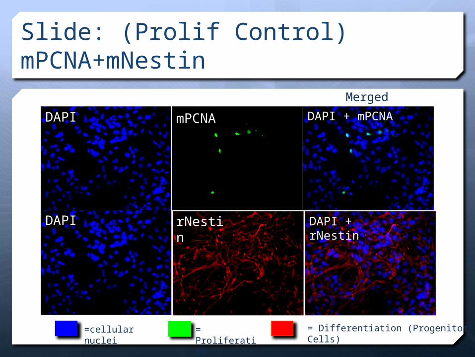
Slide: (Prolif Control) mPCNA+mNestin
DAPI
DAPI
mPCNA
rNestin DAPI + rNestin
DAPI + mPCNA
= Differentiation (Progenitor Cells)= Proliferation
=cellular nuclei
Merged

Results
Treatment Con PD AT1 AT2
4.00
5.00
6.00
7.00
% o
f Im
mu
no
reac
tive
Pro
life
rati
ng
Cel
ls/t
ota
l Im
mu
no
reac
tive
Cel
ls
mPCNA Expression in Proliferation Media
Treatment Key:AT1+ PD=AT1 Selective Agonism & AT2 Antagonism (sar1 AngII + PD123319)
AT2=AT2R agonist (CGP42112)
PD=AT2 antagonism control (PD123319)
+4.84%
-12.42%
-4.35%
AT1 + PD
+/- = increase/decrease compared to control
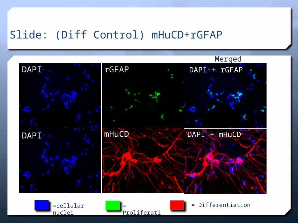
Slide: (Diff Control) mHuCD+rGFAP
DAPI
DAPI
rGFAP
mHuCD
DAPI + rGFAP
DAPI + mHuCD
= Differentiation = Proliferation
=cellular nuclei
Merged

Con PD AT1 AT20
1
2
3
4
5
6
7
% o
f Im
munore
acti
ve P
rolife
rati
ng
Cells/t
ota
l Im
munore
acti
ve C
ells
Con PD AT1 AT20
5
10
15
20
25
30
35
18.3419.40
27.5228.98
Results
mHuCD Expression in Proliferation & Differentiation Media
=Differentiation Media
=Proliferation Media
+50.10%
-7.34%
+58.02%
+78.80%
+5.78%
+37.23%
AT1 + PD
mHuCD Expression in Differentiation MediamHuCD Expression in Proliferation Media
Treatment TreatmentAT1 + PD
*
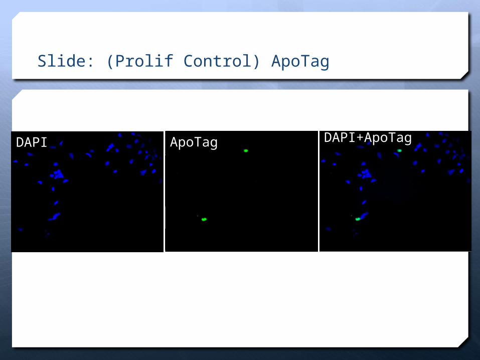
Slide: (Prolif Control) ApoTag
DAPI ApoTag DAPI+ApoTag
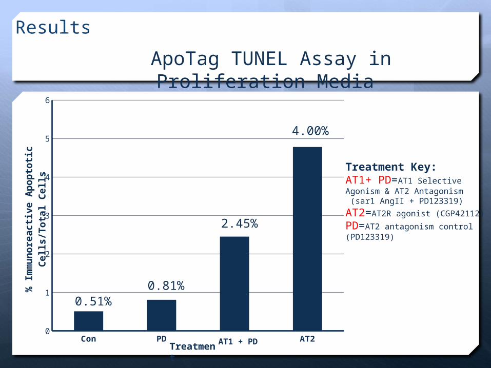
Results
ApoTag TUNEL Assay in Proliferation Media
Con PD AT1 AT20
1
2
3
4
5
6
% I
mm
un
ore
acti
ve A
po
pto
tic
Cel
ls/T
ota
l C
ells
2.45%
4.00%
0.81%0.51%
Treatment Key:AT1+ PD=AT1 Selective Agonism & AT2 Antagonism (sar1 AngII + PD123319)
AT2=AT2R agonist (CGP42112)
PD=AT2 antagonism control (PD123319)
AT1 + PDTreatment
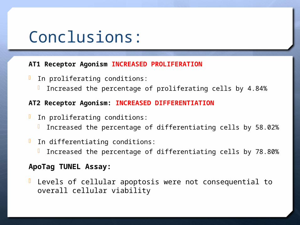
Conclusions:
AT1 Receptor Agonism INCREASED PROLIFERATION
In proliferating conditions: Increased the percentage of proliferating cells by 4.84%
AT2 Receptor Agonism: INCREASED DIFFERENTIATION
In proliferating conditions: Increased the percentage of differentiating cells by 58.02%
In differentiating conditions: Increased the percentage of differentiating cells by 78.80%
ApoTag TUNEL Assay:
Levels of cellular apoptosis were not consequential to overall cellular viability
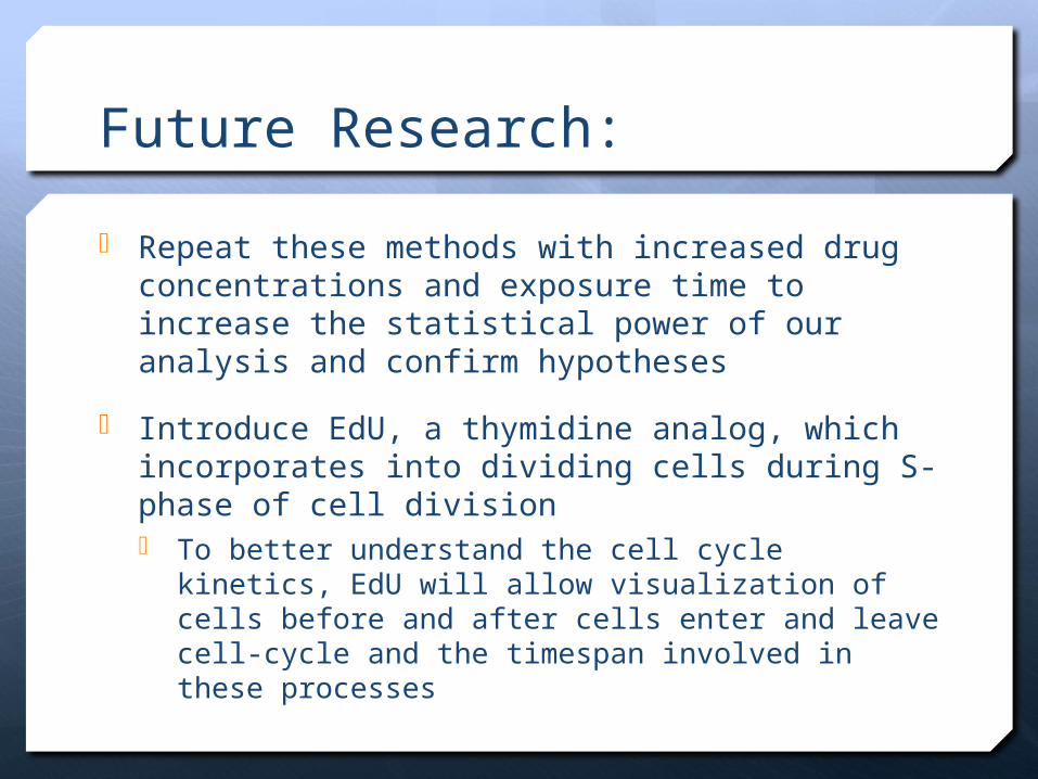
Future Research:
Repeat these methods with increased drug concentrations and exposure time to increase the statistical power of our analysis and confirm hypotheses
Introduce EdU, a thymidine analog, which incorporates into dividing cells during S-phase of cell division To better understand the cell cycle kinetics, EdU will
allow visualization of cells before and after cells enter and leave cell-cycle and the timespan involved in these processes
Bibliography1) Guimond MO, Gallo-Payet N (2012) The Angiotensin II Type 2 Receptor in Brain
Functions: An Update. International journal of hypertension 2012: 3517582) Guimond MO, Gallo-Payet N (2012) How does angiotensin AT(2) receptor activation
help neuronal differentiation and improve neuronal pathological situations? Frontiers in endocrinology 3: 164. doi: 10.3389/fendo.2012.00164.
3) M. O. Guimond, C. Wallinder, M. Alterman, A. Hallberg, and N. Gallo-Payet, “Comparative functional properties of two structurally similar selective nonpeptide drug-like ligands for the angiotensin II type-2 (AT(2)) receptor. Effects on neurite outgrowth in NG108-15 cells,” European Journal of Pharmacology, vol. 699, no. 1–3, pp. 160–171, 2012.
4) Gendron L, Côté F, Payet MD, Gallo-Payet N. Nitric oxide and cyclic GMP are involved in angiotensin II AT2 receptor effects on neurite outgrowth in NG108–15 cells. Neuroendocrinology. 2002;75(1):70–81.
5) Gallo-Payet N, Guimond MO, Bilodeau L, Wallinder C, Alterman M, Hallberg A. Angiotensin II, a neuropeptide at the frontier between endocrinology and neuroscience: is there a link between the angiotensin II type 2 receptor (AT2R) and Alzheimer's disease? Frontiers in Endocrinology. 2011;2(article 17):1–10.
6) Gendron L, Payet MD, Gallo-Payet N. The angiotensin type 2 receptor of angiotensin II and neuronal differentiation: from observations to mechanisms. Journal of Molecular Endocrinology. 2003;31(3):359–372.
7) M. Shum, S. Pinard, M. O. Guimond et al., “Angiotensin II Type 2 receptor promotes adipocyte differentiation and restores adipocyte size in high fat/high fructose diet-induced insulin resistance in rats,” American Journal of Physiology. In press.
8) Guimond MO, Gallo-Payet N (2012) The Angiotensin II Type 2 Receptor in Brain Functions: An Update. International journal of hypertension 2012: 351758
9) Usascientific.com 10) International Rules for Pre-college Science Research: Guidelines for Science and
Engineering Fairs 2012-2013 http://www.societyforscience.org/document.doc?id=398

Thank You!
Research performed at Nova Southeastern University