showkat hamid mentor: dr. mrinalini meesala md, facc. university at buffalo; state university of new...
TRANSCRIPT

Postgraduate Research Presentation
Showkat HamidMentor: Dr. Mrinalini Meesala MD, FACC.
University at Buffalo; State University of New York;Sisters Hospital IMTP
June 12th 2013

Disclosures:None

Topic:Evaluation of electrogenic properties of myocardium in
patients with HFpEF with Tp-e/QT ratio as marker of ventricular repolarization.
Introduction:Nearly one-half of patients presenting with heart failure
have preserved left ventricular ejection fraction 1.
Patients with low ejection fraction are known to be susceptible to arrhythmias and device therapy (ICD/CRT) is a basic tenet to decrease sudden death 2.
Ref:1. Yancy CW, Lopatin M, Stevenson LW, et al. Clinical presentation, management,and in-hospital outcomes
of patients admitted with acute decompensated heart failure with preserved systolic function: a report from ADHERE) Database. J Am Coll Cardiol 2006; 47:76–84.
2. Smith GL, Masoudi FA, Vaccarino V, et al. Outcomes in heart failure patients with preserved ejection fraction: mortality, readmission, and functional decline. J Am Coll Cardiol 2003; 41:1510–1518.

Mortality of patients with HFpEF is not markedly different from patients with decreased ejection fraction.
Ref:1. Yancy CW, Lopatin M, Stevenson LW, et al. Clinical presentation, management,and
in-hospital outcomes of patients admitted with acute decompensated heart failure with preserved systolic function: a report from ADHERE) Database. J Am Coll Cardiol 2006; 47:76–84.

Background:T-wave: a manifestation of ventricular repolarazation.
Tp-e interval corresponds to the dispersion of ventricular repolarization.
Amplification of dispersion of ventricular repolarization is a substrate for ventricular arrhythmias
Ref: Antzelevitch C. T peak-Tend interval as an index of transmural dispersion of repolarization. Eur J
Clin Invest 2001;31:555. Antzelevitch C. The role of spatial dispersion of repolarization in inherited and acquired sudden
cardiac death syndromes. Am J Physiol Heart Circ Physiol 2007. Antzelevitch C. Heterogeneity and cardiac arrhythmias: an overview. Heart Rhythm
2007;4:964.


Ventricular myocardium is comprised of 3 distinct myocardial cell types—
•Epicardial,
•Endocardial, and
•Masonic Midmyocardial Moe cells - M cells.
Cellular basis of T wave and Tp-e Interval:
Ref:Antzelevitch C, Sicouri S, Litovsky SH, et al. Heterogeneity within the ventricular wall: electrophysiology and pharmacology of epicardial, endocardial and M cells. Circ Res 1991;69:1427.
M-cellsHistologically similar; Electrophysiologically
different.
Located in sub-endocardial layer
Longest action potential (APD) than epicardial or endocardial cell at lower rate or in response to action potential prolonging agents.
Ref: Antzelevitch C, Sicouri S, Litovsky SH, et al. Heterogeneity within the ventricular wall:
electrophysiology and pharmacology of epicardial, endocardial and M cells. Circ Res 1991;69:1427

Heterogeneity persists but is less pronounced in intact ventricular wall due to well coupled adjacent myocytes.
Ref: Antzelevitch C, Sicouri S, Litovsky SH, et al. Heterogeneity within the
ventricular wall: electrophysiology and pharmacology of epicardial, endocardial and M cells. Circ Res 1991;69:1427

Ref :Circulation 1998;98:1928, PACE 2006;29:1130, and Heart Rhythm 2008;5:585.
QT interval is specific to species, so-called normal QT interval for that species.
QT interval and Tp-e interval increase linearly with increase in body weight.
Ref: Guo D, Zhou J, Zhao X, et al. Calcium channel recovery kinetics
versusventricular repolarization: preserved membrane-stabilizing mechanism across species. Heart Rhythm 2008;5:271

Adapted from Heart Rhythm 2008;5:271.

Adapted from Heart Rhythm 2008;5:271.

Tp-e/QT ratio is an index of ventricular repolarization that remains constant within a very narrow range of value despite dynamic physiological changes in HR and also evolutionary changes across species.
Ref: Guo D, Zhou J, Zhao X, et al. Calcium channel recovery kinetics
versusventricular repolarization: preserved membrane-stabilizing mechanism across species. Heart Rhythm 2008;5:271

Tp-e interval serves as an index of total dispersion of repolarization (transmural, apicobasal, or global)in vivo.
Changes in this parameter from the baseline value may forecast the risk of arrhythmia.
Ref : Prasad Gupta,Gan-Xin Yan, MD, PhDa, Tp-e/QT ratio as an index of
arrhythmogenesis Journal of Electrocardiology 41 (2008) 567–574

Rationale of the study:Tp-e interval, Tp-e/QT ratio, and Tp-e/QTc ratio are
prolonged in patients with moderate and severe obstructive sleep apnea.
Tp-e interval and Tp-e/QT ratio is increased in patiests with ankylosing spondylitis
Ref : Kilicaslan F, Cebeci BS. Tp-e interval, Tp-e/QT ratio, and Tp-e/QTc ratio are
prolonged in patients with moderate and severe OSA patients. (PACE 2012; 35:966–972)
Acar G, Bozoglan O. Evaluation of Tp-e interval and Tp-e/QT ratio in patients with ankylosing spondylitis. Mod Rheumatol. 2013 Apr 12. [Epub ahead of print]

Left ventricular hypertrophy amplifies the QT, and Tp-e intervals and the Tp-e/QT ratio of left chest ECG
Tp-e/QT ratio may serve as a prognostic predictor of adverse outcomes after successful pPCI treatment in STEMI patients.
Ref : Zhao Z, Yuan Z, Ji Y, Wu Y, Qi Y. Left ventricular hypertrophy amplifies the
QT, and Tp-e intervals and the Tp-e/ QT ratio of left chest ECG J Biomed Res. 2010 Jan;24(1):69-72. doi: 10.1016/S1674-8301(10)60011-5.
Big QuestionWhat happens to Tp-e/QT ratio in
HFpEF ???

Research Hypothesis: “In patients with HFpEF cellular and
metabolic changes in myocytes are associated with changes in electrogenic properties of the ventricular myocardium reflected as prolongation of Tp-e/QT intervals suggesting increased risk of ventricular arrhythmias ”.

Research Design and Methods: Retrospective Study
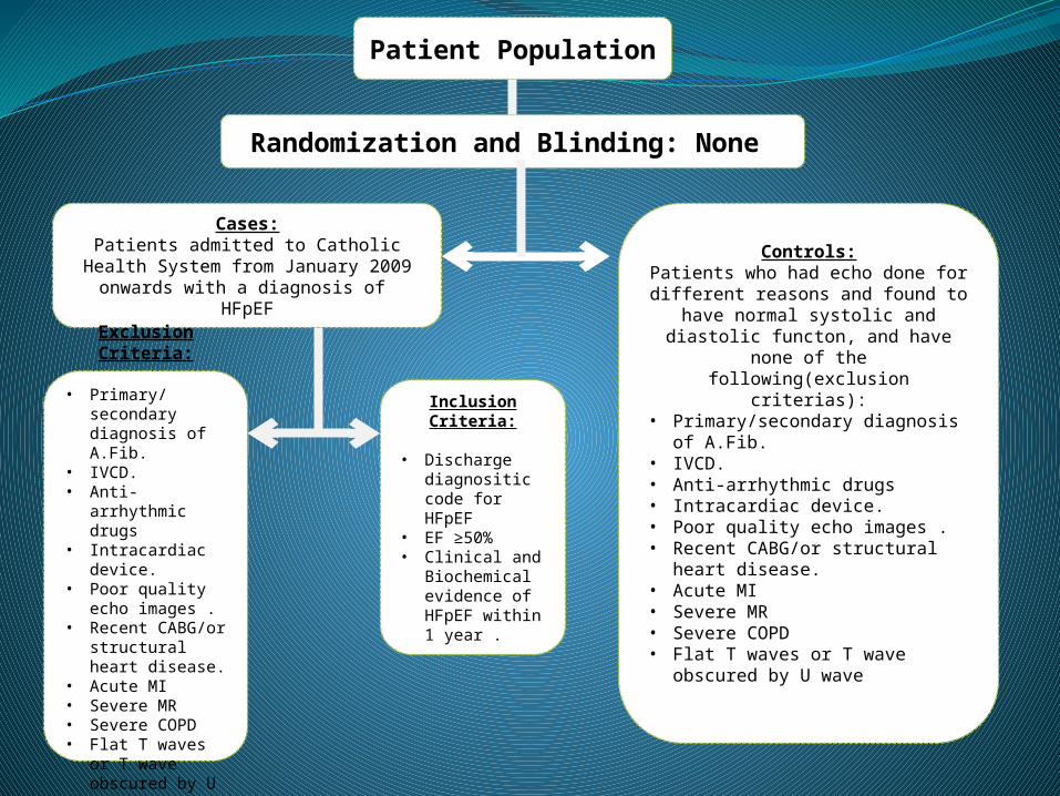
Patient Population
Cases:Patients admitted to Catholic Health System from January 2009 onwards
with a diagnosis of HFpEFControls:
Patients who had echo done for different reasons and found to have
normal systolic and diastolic functon, and have none of the following(exclusion criterias):
• Primary/secondary diagnosis of A.Fib.
• IVCD. • Anti-arrhythmic drugs • Intracardiac device. • Poor quality echo images .• Recent CABG/or structural heart
disease. • Acute MI • Severe MR • Severe COPD • Flat T waves or T wave obscured
by U wave
Inclusion Criteria:
• Discharge
diagnositic code for HFpEF
• EF ≥50% • Clinical and
Biochemical evidence of HFpEF within 1 year .
Exclusion Criteria:
• Primary/secondary diagnosis of A.Fib.
• IVCD. • Anti-arrhythmic
drugs • Intracardiac
device. • Poor quality
echo images .• Recent CABG/or
structural heart disease.
• Acute MI • Severe MR • Severe COPD • Flat T waves or
T wave obscured by U wave
Randomization and Blinding: None

Total (967)
Excluded(828)
Unclassified (999 group)
(16)
Controls (49)
Patients(90)
Included(139)
Grade III(7)
E/A>2DT
<160msAv.
E/e’>13
Grade II(42)
E/A 0.8-<1.5DT 160-200msAv. E/e’ 9-12
Grade I(25)
E/A<0.8DT
>200msAv.
E/e’≤8
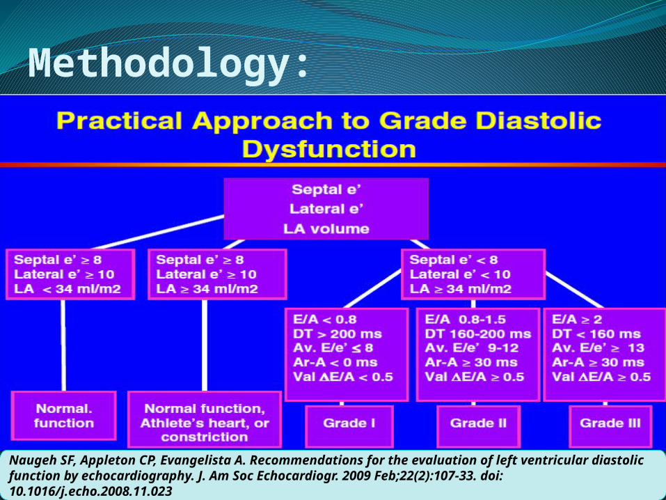
Methodology:
Naugeh SF, Appleton CP, Evangelista A. Recommendations for the evaluation of left ventricular diastolic function by echocardiography. J. Am Soc Echocardiogr. 2009 Feb;22(2):107-33. doi: 10.1016/j.echo.2008.11.023

Methodology cont: EKGs were analyzed for T wave morphology.
T peak and T end interval (Tp-e) were measured by (Standard Tangential Method) identifying two points on isoelectric line: 1) Perpendicular to the isoelectric line from crest
of T wave 2) The point at which the tangent to the down
curve of T wave intersects the isoelectric line.


Results:


Tp-e/QT


Conclusion:There is no significant prolongation of Tp-
e/QT to demonstrate increased risk of
ventricular arrhythmias hence sudden death
in patients with HFpEF in this study.
A decreasing trend in Tp-e/QT ratio with
increasing grade of diastolic dysfunction was
observed which did not achieve statistical
significant due to small cohort of subjects.

Strengths Of the Study:Echo and EKG parameters collected in
different times to avoid observer bias.Internal as well as external comparison were
attemptedPatients with EF>/= 50% strictly were taken
for study. Patients with clinical syndrome of HF along
with biochemical evidence of HF taken.

Limitations of Study:Retrospective Study
Small population size
Extrapolation of results of Wedge Electro-gram to chest ECGs.
Tp-e/QT ratio is a relatively new parameter and not much is known about its significance in HFpEF.

Questions?

Acknowledgements:Dr. K.J.Qazi , MD (Program Director)Dr. Mrinalini Meesala, MD (Research
Mentor)Dr. Micheal Banas, MD (Advisor)Dr. Salim Memon, MBBSDr. Sachitanand MD (Chair IRB)Ms Danielle Casucci (IRB)Staff of Echo Lab Sisters Hospital

Thank you !