short dsrnas with an overhanging 5' ppp-nucleotide, as found in
TRANSCRIPT
1
Short dsRNAs with an overhanging 5’ ppp-nucleotide, as found in arenavirus
genomes, act as RIG-I decoys
Jean-Baptiste Marq, Stéphane Hausmann, Nicolas Veillard, Daniel Kolakofsky* and
Dominique Garcin
Dept of Microbiology and Molecular Medecine,University of Geneva School of Medecine,CMU,
1 rue Michel-Servet,1211 Geneva; Switzerland
*Corresponding author: [email protected]
Arenavirus RNA genomes are initiated
by a “prime and realign” mechanism,
such that the initiating GTP is found as
a single unpaired (overhanging)
nucleotide when the complementary
genome ends anneal to form dsRNA
panhandle structures. dsRNAs
modelled on these structures do not
induce interferon (IFN), as opposed to
blunt-ended 5’ pppdsRNA. This paper
examines whether these viral
structures can also act as decoys, by
trapping RIG-I in inactive dsRNA
complexes. We examined the ability of
various dsRNAs to activate the RIG-I
ATPase (presumably a measure of
helicase translocation on dsRNA)
relative to their ability to induce IFN. We
found that there is no simple
relationship between these two
properties, as if RIG-I can translocate
on short dsRNAs without inducing IFN.
Moreover, we find that 5’ pppdsRNAs with
a single unpaired 5’ ppp-nucleotide can
in fact competitively inhibit the ability
of blunt-ended 5’ pppdsRNA to induce
IFN when co-transfected into cells, and
that this inhibition is strongly
dependent on the presence of the 5’
ppp. In contrast, 5’ pppdsRNAs with a
single unpaired 5’ ppp-nucleotide does
not inhibit poly-I/C induced IFN
activation, which is independent of the
presence of a 5’ ppp group.
The innate immune system senses
RNA virus infections primarily via two
constitutively expressed DExD/H box
helicases, RIG-I and mda-5 (1). These
cytoplasmic sensors act as pattern
recognition receptors that respond to two
RNA pathogen associated molecular
patterns (PAMPs), namely dsRNA (e.g.,
poly-I:poly-C, or poly-I/C) and 5’ pppRNA
(2-4). These RNAs can act as PAMPs
because their presence in the cell
cytoplasm is thought to be primarily
restricted to virus infection. RIG-I, which
senses negative-strand RNA virus (NSV)
infections (5) is a large protein composed
of 3 domains; N-terminal tandem CARDs
http://www.jbc.org/cgi/doi/10.1074/jbc.M110.186262The latest version is at JBC Papers in Press. Published on December 15, 2010 as Manuscript M110.186262
Copyright 2010 by The American Society for Biochemistry and Molecular Biology, Inc.
by guest on April 10, 2018
http://ww
w.jbc.org/
Dow
nloaded from

2
(the effector domain), a central DExD/H
box helicase domain that contains a
dsRNA-dependent ATPase, and a C-
terminal regulatory domain (CTD) that
binds 5’ pppdsRNA,(6;7). The CARD domain
of quiescent RIG-I is thought to be auto-
inhibited by other protein domains, and to
become activated, i.e., available for
interaction with the synonymous domain of
IPS-1 (the central adaptor of the pathway
to IFN activation) via conformational
changes in RIG-I induced upon binding of
PAMP RNAs to the CTD (8).
In the earliest reports, 5’ pppRNA was
thought to act as a PAMP whether the
triphosphate was present on ss or dsRNA.
More recently, good evidence has been
presented that 5’ pppRNA without dsRNA
character is not a PAMP (9-11). In the
latest descriptions of this signalling
pathway, RIG-I is activated and IFN
induced only when the CTD interacts with
dsRNA that also contains a 5’ ppp-end, at
least for relatively short dsRNAs (<100
bp). Both groups found that a stretch of
dsRNA near the 5’ ppp-end, as well as the
5’ ppp-end itself, were essential for RIG-I
to induce IFN. However, these reports
differed about the minimum length of the
dsRNA region required, and more
importantly in the case of arenaviruses,
whether this dsRNA end needed to include
the nucleotide carrying the 5’ ppp.
Arenaviruses (such as Tacaribe and Junin
viruses) contain two ambisense genome
segments whose terminal 19nt are either
fully complementary (large segment), or
contain 2 mis-matches at positions 6 and 8
(12). These mis-matches would create a
symmetrical 3nt bulge when these terminal
sequences anneal to form dsRNA. This
sequence complementarity and the mis-
matches presumably reflect the similarity
and subtle differences of the various
genomic and antigenomic promoters.
Remarkably, arenavirus genome synthesis
initiates with GTP at position +2 of the
template rather than at the precise 3’ end
(position +1), and the initiating 5’ pppG is
then realigned (as 5’ pppGpCOH) before
this primer is extended (13). The net result
of this “prime and realign” mechanism of
genome initiation is that 5’ pppG is found
as an unpaired 5’ nucleotide overhang
when the complementary genome ends
form dsRNA (see fig 5a). Arenavirus RNA
genomes are also unusual in that they are
found in circular nucleocapsids,
presumably due to the annealing of their
complementary terminal sequences (14).
Using 5’ pppRNA made in vitro and purified
so that all dsRNA side-products have been
removed, we determined that both this 5’
nucleotide overhang, as well as the mis-
matches within the dsRNA panhandles,
clearly reduce the ability of these dsRNAs
to induce IFN (15). The presence of this
unpaired 5’ ppp-nucleotide overhang thus
appears to be another way that some
viruses use to avoid activating the IFN
system.
A great deal of attention has been
focused on the RIG-I CTD, as the structure
of this domain was determined at atomic
by guest on April 10, 2018
http://ww
w.jbc.org/
Dow
nloaded from
3
resolution, and structure/function studies
have been carried out (6;7). More recently,
three crystal structures of the CTD bound
to 12-14 bp 5’ ppp-dsRNAs were
determined (16;17). The CTD binds to
both ends of the self-complementary 5’
pppdsRNAs and contacts the A-form
dsRNA primarily through a few nucleotides
at the 5’ end, with the and 5’
phosphates surrounded by multiple lysines
and anchored in the basic cleft via
electrostatic interactions and multiple H-
bonds. The CTD also interacts with the
duplexes via the stacking of Phe853 with
the terminal base pair, providing addition
hydrophobic interaction energy. The
modes of RNA recognition in the three
structures are remarkably similar despite
the very different RNA sequences,
suggesting that 5’ pppdsRNA recognition is
sequence-independent. RNA binding
studies were also carried out, and the
binding affinity of the CTD for 5’ pppdsRNA
was found to be around 15-fold stronger
than for the same 5’ OHdsRNA. The binding
affinity for 5’ pppdsRNA with a single
unpaired 5’ ppp-nucleotide was not
reported.
Although the manner in which the CTD
interacts with blunt-ended 5’ pppdsRNA is
now well documented, and the role of the
N-terminal CARDs in transmitting the
signal to IPS-1 is clear, the role of the
central helicases/ATPase domain in RIG-I
function is less well understood. The
ATPase activity of RIG-I is critical for RIG-I
function, as a mutation of the ATP binding
site (K270A), like the deletion of the
CARDs, eliminates all ability to induce IFN,
(2). The RIG-I helicase/ATPase was
initially found to display typical helicase
activity, i.e., to separate the strands of
dsRNA (6), but more recently RIG-I was
found to translocate on dsRNA without
separating the strands (18) (19).
Moreover, the N-terminal CARDs were
found to strongly suppress this
translocation in the absence of a 5’
triphosphate group, whereas the presence
of 5’ pppRNA (tethered to the dsRNA via a
ssRNA extension) strongly stimulated the
translocation. Although a canonical dsRNA
binding motif is not present in these
helicases, a point mutant in the helicase
domain predicted to establish contacts
with the RNA (Q299A) fails to bind RNA
(20), and residual dsRNA-dependent
ATPase activity of the isolated RIG-I
helicases/ATPase domain points to a
dsRNA-interacting site here (7). It also
seems intuitive that the RIG-I
helicase/ATPase domain binds dsRNA
independently of the CTD, as structural
studies of helicases bound specifically to
double-stranded nucleic acids indicate that
the highly conserved amino acids within
the ATPase core are essential for the
recognition of the double-stranded lattice
(21;22). Thus, one possible scenario for
RIG-I action is that this pattern recognition
receptor initially binds to any region of
dsRNA via its helicase domain binding
site, and then moves in an ATP dependent
fashion to locate and interact with the
by guest on April 10, 2018
http://ww
w.jbc.org/
Dow
nloaded from

4
dsRNA ends. In the most favourable case
for IFN activation induced by relatively
short dsRNAs (<100bp), if a blunt 5’
pppdsRNA end is encountered, this
optimally leads to the conformational
changes that release the CARDs for
interaction with IPS-1, and signalling to the
IFN promoter occurs. However, in this
scenario where RIG-I first binds to dsRNA
independent of their ends, one would
expect the arenavirus dsRNA panhandles
(that are inactive in inducing IFN) to also
act as competitive inhibitors of dsRNAs
that can induce IFN, by sequestering RIG-I
in inactive complexes. This paper
examines the binding of various RNAs to
RIG-I and their ability to stimulate its
ATPase, and then examines whether 5’
pppdsRNAs whose 5’ ppp-nucleotide is
unpaired can in fact competitively inhibit
the ability of blunt-ended 5’ pppdsRNA to
induce IFN.
EXPERIMENTAL PROCEDURES
Plasmids. pβ-IFN-fl-lucter contains the
firefly luciferase gene under the control of
the human IFN-β promoter, was described
previously (23). pTK-rl-lucter, used as a
transfection standard, contains the herpes
simplex virus TK promoter region
upstream of the Renilla luciferase gene
(Promega). pEBS-RIG-I expressed full
length RIG-I.
Immunoblotting. Cytoplasmic extracts
were prepared using 0.5% Nonidet P-40
buffer. Equal amounts of total proteins
were separated by SDS-PAGE and
transferred onto Immobilon-P membranes
by semidry transfer. The secondary
antibodies used were alkaline
phosphatase-conjugated goat anti-mouse
immunoglobulin G (Bio-Rad). The
immobilized proteins were detected by
light-enhanced chemiluminescence
(Pierce) and analyzed in a Bio-Rad light
detector using Quantity One software.
Primary antibody used is a mouse anti-
Rig-I (Alexis).
Transfections. 100,000 cells were plated
into 6-well plates 20 h before transfection
with 1.5 μg of pβ-IFN-fl-lucter, 0.5 μg of
pTK-rl-lucter, and TransIT-LT1 transfection
reagent (Mirus). At 24 h post-transfection,
the cells were (or were not) transfected
with the indicated RNAs using Trans-
messenger transfection reagent
(Qiagen)(a total of 1.2 to 2ug of RNA was
always transfected, the difference always
being made up with tRNA. Transfection of
tRNA by itself was neutral.). Twenty hours
later, cells were harvested and assayed
for firefly and Renilla luciferase activity
(dual-luciferase reporter assay system;
Promega). Relative expression levels were
calculated by dividing the firefly luciferase
values by those of Renilla luciferase.
In Vitro Synthesis of RNA, Purification and
(Annealed) dsRNA. DNA for T7 RNA
polymerase synthesis of 5’ pppJun*61 was
prepared by annealing two oligos :
by guest on April 10, 2018
http://ww
w.jbc.org/
Dow
nloaded from

5
5’
TAATACGACTCACTATAgcgcaccggggatc
ctaggcgattttggttacgctataattgtgactgttt
tctgtttgga (T7 promoter (underlined) and
the -1 to +60 Junin 5’ sequence where U
residues at position +6, +8 and +19 were
replaced by the indicated nucleotides to
allow synthesis of single strand RNA in a
T7 reaction devoid of U residues) and its
complementary oligo 5’
tccaaacagaaaacagtcacaattatagcgtaaccaaa
atcgcctaggatccccggtgcgcTATAGTGAGTC
GTATTA. Transcription was performed
with 100 pmol of gel purified dsDNA using
T7 MEGAshortscript (Ambion) according
to the manufacturer's instructions (no UTP
in the reaction). The total T7 transcripts
were digested with DNase I and then
chromatographed on NucAway Spin
columns (Ambion) to remove
unincorporated nucleotides and DNA
fragments.
DNA for T7 RNA polymerase synthesis of
biotinylated RNA was prepared by PCR
using the partially complementary primers
5′-
TAATACGACTCACTATAgggACACACCA
CAACCAACCCACAAC-3′ (forward) (start
sites are in lowercase type) and 5′-
GAAAGAAAGGTGTGGTGTTGGTGTGGT
TGTTGTGGGTTGGTTGTGG-3′ (reverse).
Transcription was performed with 100
pmol of purified PCR product using T7
MEGAshortscript (Ambion) according to
the manufacturer's instructions.
Biotinylated RNA1 was synthesized using
equal amounts of 5′-biotin-UTP and UTP.
The reaction mix (unpurified 5’ biotinylated
ppp-RNA1) was digested with DNase I
and then chromatographed on NucAway
Spin columns (Ambion) to remove
unincorporated nucleotides and DNA
fragments.
For further purification of biotinylated
5’ppp-ssRNA, slightly radiolabeled ([α-32P]CTP) T7 biotinylated transcripts were
electrophoresed on 10% preparative
denaturing gels. The major 54-nucleotide
band was excised from the gel, and the
RNA was eluted, followed by ethanol
precipitation. For Rnase III treatment, 50
μg of gel-purified RNA1 was digested with
50 unit of RNase III (Ambion) for 60 min at
37 °C in 40 μl of 50 mM NaCl, 10 mM Tris-
HCl, (pH 7.9), 10 mM MgCl2, 1 mM
dithiothreitol. The digestion products were
then phenol/chloroform extracted and
ethanol-precipitated.
(Annealed) dsRNA. T7 5’ pppJun*61 (or
synthetic 5’ OHJun*6) was mixed with the
indicated synthetic complementary 5’OH-
oligoribonucleotides in a molar ratio 1 to 3,
in a final volume of 50 ul (300 mM NaCl,
50 mM tris pH 7.5, 1 mM EDTA), heated 1
min at 90°, and progressively cooled to
room temperature. T7 reaction products
were checked for dsRNA contamination by
RNase III treatment and transfection into
A549 cells (15).
by guest on April 10, 2018
http://ww
w.jbc.org/
Dow
nloaded from

6
Cells. 2fTGH, A549 and A549-RIG-I-kd
(24) cells were grown in Dulbecco's
modified Eagle's medium supplemented
with 10% fetal calf serum.
5’ ppp-RNA beads and RNA pull down.
Streptavidin agarose beads (Fluka 85881)
were pre-equilibrated with Blocking buffer
(Base buffer (20 mM Hepes pH 7.9, 2 mM
EDTA, 15% glycerol, 0.05% NP40, 50 mM
NaCl, 500 unit/ml RNasin (Promega
N2515), 0.02 mg/ml tRNA (Roche
10109495001), 1% protease inhibitor
cocktail (Sigma P8340) and 2 mM DTT)
plus 100 mM NaCl, another 100 unit/ml
RNasin, 0.1 mg/ml glycogen and 2.5
mg/ml BSA, for 2 hours at 4°C.
Biotinylated RNA was bound to the beads
in Base buffer for 2 hours at 4°C. Beads to
which 0.5 ug of RNA were added were
used for each assay. For “in vitro” RNA
pull down, streptavidin beads to which
500ng of biotinylated pppRNA had been
added were incubated with 1 ug of purified
RIG-I (25). After 2 h at 4°, the beads were
washed 3 times with base buffer, SDS
protein sample buffer was added, and the
beads were analyzed by Western blotting
with anti-RIG-I. For “in vivo” RNA pull
down, 5 ug of biotinylated pppRNA were
transfected into 2fTGH cells using trans-
messenger (Qiagen). 4 hours post–
transfection, cells were resuspended in
120ul sonication buffer (50 mM Hepes-
KOH pH7.5, 150mM K-acetate, 5 mM Mg-
acetate, 0.1% digitonin, 2mM DTT and
protease inhibitors) and sonicated. 30ul of
streptavidin agarose beads were added
and the samples treated as described
above.
RESULTS
RNA binding to RIG-I. The binding of
dsRNA to RIG-I was studied primarily
using a pull-down assay. Because
modified nucleosides were shown to
interfere with the ability of 5’ pppRNAs to
induce IFN via RIG-I (4), 54nt-long RNA1
was designed to contain 46 nt of spacer
between the patch of biotinylated uridines
at the 3’ end (that anchor the RNA to the
bead) and the 5’ ppp “business end” of the
RNA, to minimize the effect of these
modified nucleosides on RIG-I interaction.
This approach would not have been
possible with natural RNAs containing
arena-like structures.
5’ pppRNA was prepared in vitro with T7
RNA polymerase (T7) in the presence of
5-biotin-UTP, which was incorporated
uniquely within the 3’ terminal 8
nucleotides of the DNA-encoded 54’mer.
As previously described, the product of
this T7 reaction is mostly double-stranded
(virtually all the product is sensitive to
RNase III digestion), being composed of
the DNA-encoded 54’mer and
complementary RNA fragments (mostly
15-25 nt long), as the DNA-encoded
54’mer itself acts as a template for
complementary RNA synthesis (15). When
transfected into cells, these unpurified T7
transcripts are generally very potent
inducers of IFN. However, when the 54nt
by guest on April 10, 2018
http://ww
w.jbc.org/
Dow
nloaded from
7
ssRNA is purified from the mix, it has lost
virtually all its ability to bind to RIG-I,
stimulate its ATPase, or induce IFN when
transfected into cells, similar to chemically-
made 5’ pppssRNA (9;10;15).
To ensure that this biotinylated,
partially dsRNA bound endogenous RIG-I
under natural conditions in vivo, the
unpurified T7 transcripts were transfected
into 2fTGH cells which had, or had not
been treated with IFN (to increase their
endogenous levels of RIG-I). Cell extracts
were prepared 4h post-transfection,
incubated with streptavidin beads, and the
amount of RIG-I that was bound to the
beads after washing was examined by
Western blotting. As shown in fig 1, IFN
treatment strongly increased the
endogenous level of RIG-I (~10- fold;
lanes 7 and 8 vs lanes 1 and 2), and RIG-I
was efficiently bound to the beads in both
cases only when the biotinylated in vitro
transcripts had been transfected into the
cells (lanes 4 and 6 vs lanes 3 and 5). We
next examined the ability of 5’ pppdsRNA
whose 5’ ppp-nucleotide was base-paired,
or not, to bind to purified, bacterially-
expressed RIG-I in vitro. The T7 DNA-
directed 5’ ppp54’mer (when purified from
the reaction products by denaturing PAGE
and RNase III digestion), did not increase
the binding of RIG-I to the beads over the
background (lane 3 and 4 vs lanes 1 and
2, fig 2), as expected. However, when this
5’ ppp54’mer was annealed to synthetic
18’mers complementary to positions 1-18
(5’ ppp54’mer/1-18), 19-36, or 37-54, all
three 5’ pppdsRNAs clearly bound RIG-I,
albeit 5’ ppp54’mer/1-18 being the most
efficient (lanes 5 vs lanes 6 and 7, fig 2).
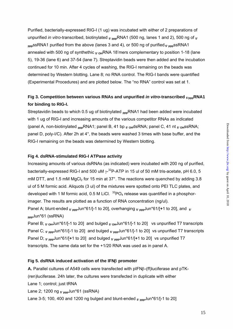
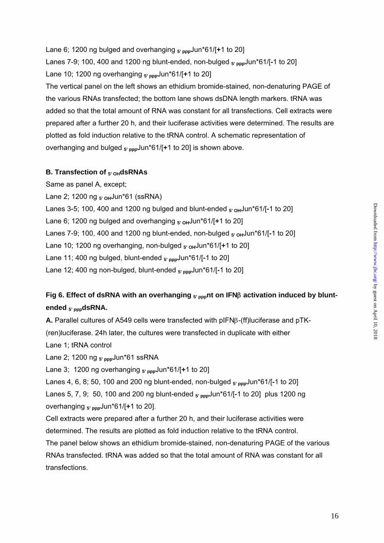
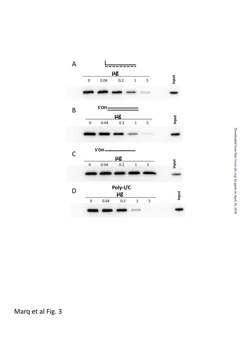
Moreover, in competition pull-down
assays, where non-biotinylated RNAs are
used to compete with the unpurified
biotinylated in vitro transcripts for binding
to RIG-I, synthetic 41bp 5’ OHdsRNA and
poly-I/C were found to compete at least as
well as unpurified non-biotinylated in vitro
transcripts for binding to RIG-I (fig 3).
Thus, all the dsRNAs tested bound to RIG-
I, independent of whether they contained a
5’ ppp. These results are consistent with
the presence of two dsRNA binding sites
on RIG-I; that of the CTD where the
presence of a 5’ triphosphate group also
contributes to dsRNA binding, and that of
the helicases/ATPase domain, where
binding is independent of the RNA ends.
dsRNA-dependent ATPase. To investigate
the effects of a single 5’ pppnt overhang, or
a 3nt bulge, on the ability of 5’ pppdsRNA to
stimulate the RIG-I ATPase, we prepared
5’ pppdsRNAs based on the Junin virus
genome panhandles. These 5’ pppdsRNAs
were composed of a single-stranded 5’
ppp61’mer representing the 5’ end of the
Junin virus large genome segment (except
that the uridines were changed to
adenosines or cytidines; Jun*) and
chemically synthesized RNAs
complementary to positions [-1 to 20] or
[+1 to 20]. Position -1 corresponds to the
overhanging, pseudo-templated 5’ pppG
resulting from the “prime and realign”
by guest on April 10, 2018
http://ww
w.jbc.org/
Dow
nloaded from
8
initiation of genome synthesis; position +1
represents the first nucleotide that is base-
paired with the genome 3’ terminal
nucleotide (see fig 5a). The 5’ ppp61’mer
could be made in vitro as relatively pure
ssRNA due to the absence of UTP in the
T7 reaction, and annealing with a slight
excess of complementary
oligoribonucleotides converted all the 5’
ppp61’mers to dsRNAs (fig 5a and 6a).
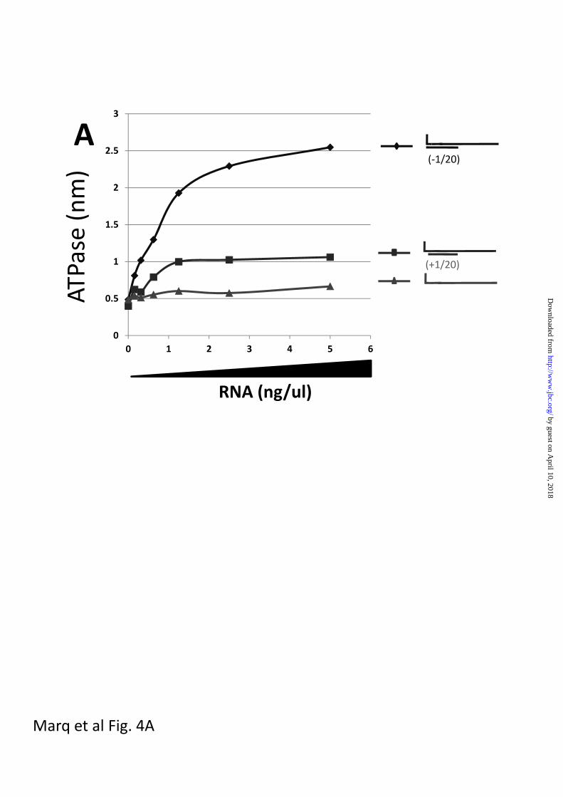
When single-stranded 5’ pppJun*61’mer,
which had little or no ability to stimulate
the RIG-I ATPase by itself (fig 4a), was
annealed with oligoribonucleotides
complementary to positions [-1 to 20] or
[+1 to 20], both dsRNAs stimulated the
ATPase, but the presence of the unpaired
5’ ppp-nucleotide was found to strongly
reduce the stimulation (approximately 4-
fold, fig 4a), consistent with their relative
abilities to activate the IFN promoter
(lane 9 vs lane 10, fig 5a). However, when
5’ pppJun*61/[-1 to 20] and 5’ OHJun*61/[-1 to
20] were compared for their stimulation of
the ATPase activity (relative to unpurified
T7 transcripts), the absence of the 5’
triphosphate group had little no effect on
the ability of this dsRNA to stimulate the
ATPase (figs 4b and 4c). This result is
somewhat surprising, as the affinity of the
isolated CTD domain for blunt-ended
5’pppdsRNAs is much higher than for
5’OHdsRNAs (16), and because 5’
OHJun*61/[-1 to 20] has little or no ability to
activate the IFN promoter (lanes 7-9, fig
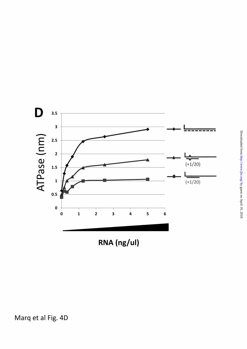
5b). Even more surprisingly, when the
sequences of the complementary
oligoribonucleotides were altered so that a
symmetrical 3 nt bulge was created at
positions 6-8 upon annealing to the 5’
pppJun*61 (similar to the dsRNA
panhandles of arenavirus small genome
segments), the 5’ pppdsRNA containing the
bulge unexpectedly stimulated the ATPase
relative to the perfectly base-paired
dsRNA (approximately 2-fold),
independent of whether the 5’ pppG was
base-paired or unpaired (figs 4c and d).
Note that unpurified T7 transcripts were
also used to stimulate the ATPase in
panels b, c and d, to provide a common
point of reference. Given that this 3nt
bulge reduces the ability of blunt-ended 5’
pppdsRNA to activate the IFN promoter
approx 2-fold (fig 5a), and that 5’ OHdsRNA
is inactive in this respect, there does not
appear to be a simple relationship
between the ability of dsRNAs to stimulate
RIG-I ATPase activity and to induce IFN.
Table I summarizes the interactions of
RIG-I with the various RNAs.
Short dsRNAs with a single, unpaired 5’
ppp-nucleotide end may act as RIG-I
decoys. If blunt-ended 5’ pppdsRNA binds
directly to the CTD of RIG-I to initiate
signalling, 5’ pppdsRNA with a single
unpaired 5’ pppnucleotide would compete
poorly for this interaction (because of its
expected lower CTD affinity), and thus
poorly inhibit the activity of blunt-ended 5’
pppdsRNA. On the other hand, if 5’
pppdsRNA binds first to the helicase
domain and RIG-I then moves along the
by guest on April 10, 2018
http://ww
w.jbc.org/
Dow
nloaded from
9
dsRNA so that the 5’ ends can interact
with the CTD, then 5’ pppdsRNA with a
single unpaired 5’ pppnucleotide might
compete well for this interaction, as this
non-blunt-ended 5’ pppdsRNA does not
induce IFN. In this case, 5’ pppdsRNA with a
single unpaired 5’ pppnucleotide might
sequester RIG-I in an inactive complex.
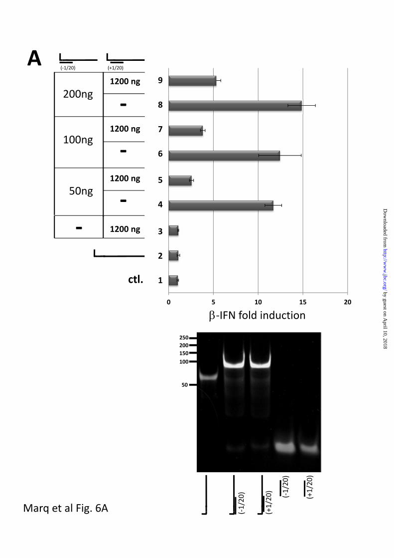
When 1200 ng of single-stranded 5’
pppJun*61’mer alone was transfected into
A549 cells, no activation of the IFN
promoter was detected (fig 6A top, lane 2),
whereas transfection of 50-200 ng of 5’
pppJun*61/[-1 to 20] was clearly active
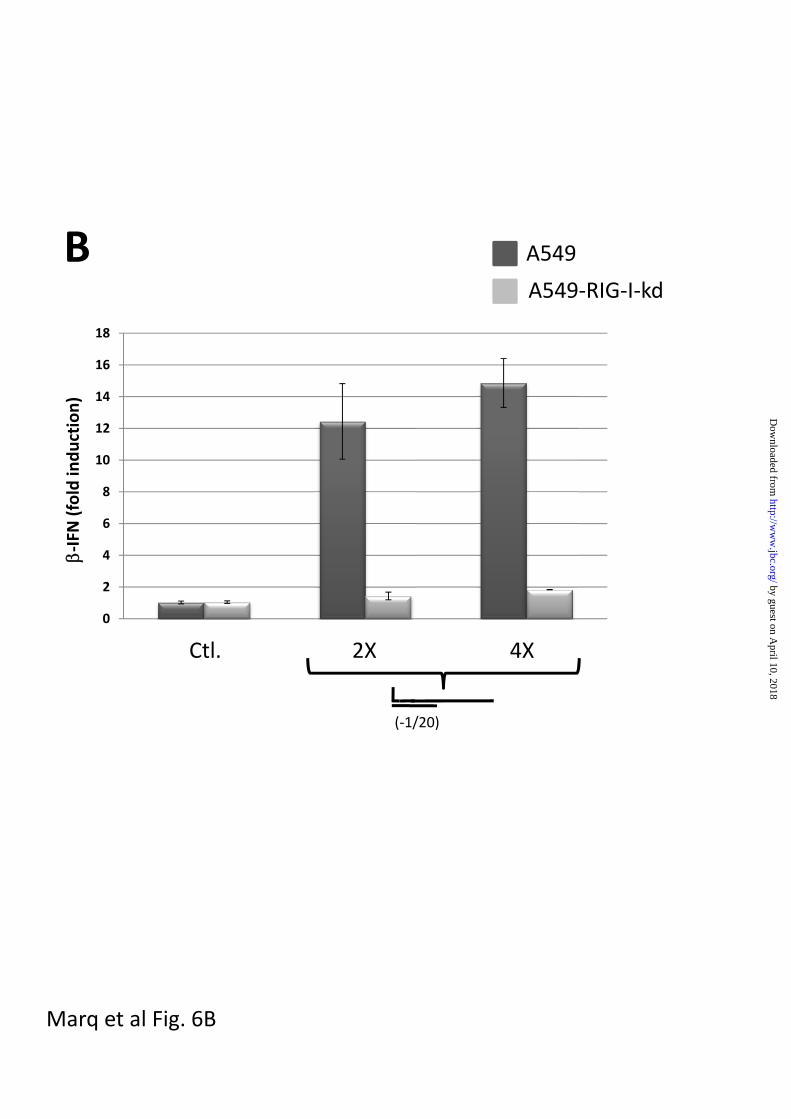
(lanes 4, 6 and 8). This activation of the
IFN promoter did not occur in A549 cells
is which RIG-I levels had been knocked
down (fig 6B). Transfection of 1200 ng of 5’
pppJun*61/[+1 to 20] (whose 5’ pppnucleotide
is unpaired) did not activate the IFN
promoter (lane 3). However, transfection of
1200 ng of 5’ pppJun*61/[+1 to 20] (which
mimics the Junin virus panhandles) along
with 50-200 ng of blunt-ended 5’
pppJun*61/[-1 to 20] dsRNA clearly
inhibited the ability of the latter to activate
the IFN promoter (78% to 64% inhibition;
lane 5,7 and 9). This inhibition did not
appear to be due simply to high (dsRNA)
substrate inhibition, since the transfection
of up to 1200 ng of blunt-ended 5’
pppJun*61/[-1 to 20] continued to increase
IFN promoter activation (lane 9, fig 5A,
and lane 3, fig 7). Thus, 5’ pppdsRNA with a
single unpaired 5’ pppnt inhibits the ability of
blunt-ended 5’ pppdsRNA to activate the
IFN promoter, presumably by competing
for RIG-I and sequestering it in an inactive
complex. In this case, arenavirus genome
5’ pppdsRNA panhandles would not only fail
to induce IFN, they would simultaneously
inhibit RIG-I activity.
Interestingly, in contrast to 5’
pppJun*61/[+1 to 20] dsRNA, transfection
of 1200 ng of non-phosphorylated 5’
OHJun*61/[+1 to 20] dsRNA along with 50
ng of 5’ pppJun*61/[-1 to 20] dsRNA had
little if any ability to inhibit IFN activation
induced by the 5’ ppp blunt-ended dsRNA
(fig 7, lane 5). Moreover, even blunt-ended
5’ OHJun*61/[-1 to 20] dsRNA had only a
modest ability to inhibit 5’ pppJun*61/[-1 to
20] dsRNA induced IFN activation (26%
inhibition, lane 6, vs 68%, lane 4), under
conditions where addition of the same
dsRNA, but bearing a 5’ ppp, roughly
doubles the IFN activation (lane 3, fig 7).
The ability of dsRNA with a single
unpaired 5’ nucleotide to inhibit blunt-
ended 5’ pppdsRNA activity is thus strongly
dependent on the presence of a 5’
triphosphate group. 5’ OHdsRNAs, whether
blunt-ended or containing a single
unpaired 5’ nucleotide, appear to be very
poor competitive inhibitors of blunt-ended
5’ pppdsRNA induced IFN activation.
Poly-I/C induced IFN activation. In contrast
to relatively short dsRNAs which appear to
require a 5’ ppp-blunt end to act as a
PAMP, commercial poly-I/C, which is
unlikely to contain blunt ends and certainly
does not terminate with a 5’ ppp,
by guest on April 10, 2018
http://ww
w.jbc.org/
Dow
nloaded from

10
nevertheless potently induces IFN. This
induction occurs exclusively via RIG-I
when it is several hundred bp long, and,
remarkably, primarily via mda-5 when the
poly-I/C is several thousand bp long
(25;26). Moreover, co-infection with
Sendai viruses that express a transgenic
GFP mRNA plus a mRNA with the
complement of the GFP ORF (that are
expected to form 700bp dsRNA with
ssRNA tails containing a cap at the 5’ end
and a polyA tail at the 3’ end), also
induces IFN via RIG-I (27). These longer
dsRNAs can apparently activate RIG-I
without the need for a 5’ pppdsRNA blunt
end.
Since the ability of dsRNA with a single
unpaired 5’ nucleotide to inhibit blunt-
ended 5’ pppdsRNA activity is strongly
dependent on the presence of its 5’
triphosphate group (fig 7), it was of interest
to determine whether this inhibitor was
equally active against poly-I/C, whose
activity is independent of the presence of a
5’ ppp. As shown in fig 8, transfection of
50 ng of our commercial poly-I/C strongly
induces IFN activation (lane 2), and the
co-transfection of 1200 ng of blunt-ended
5’ pppJun*61/[-1 to 20] dsRNA roughly
doubles the IFN activation (lane3). Co-
transfection of 1200 ng of 5’ pppJun*61/[+1
to 20] dsRNA (with a single unpaired 5’
ppp-nt), in contrast, appears to have no
effect; it neither increases nor decreases
the activation due to the poly-I/C (lane 4). 5’
pppdsRNA with a single unpaired 5’ ppp-nt
thus appears to be a selective inhibitor of
short blunt-ended 5’ pppdsRNA induced IFN
activation.
DISCUSSION
RIG-I is a member of the DExH/D box
family of ATP-dependent motor proteins
(28). Although typically called helicases,
many of these enzymes display different
functions, such as strand annealing and
protein displacement, and in the case of
RIG-I, translocation along dsRNA. In many
cases, their activity is governed by dsRNA-
dependent ATP hydrolysis that modulates
the protein’s conformation, thereby
converting chemical energy into
mechanical movement in a stepwise
manner, or simply altering the
conformation of the protein relative to the
RNA. RIG-I appears to carry out both of
these functions; i) it tracks along the
phophodiester backbone of a single strand
of the duplex, uniquely in the 5’ to 3’
direction, powered by ATP hydolysis
(18;19), and ii) its signalling to the IFN
promoter requires the interaction of the
CTD with a blunt-ended 5’ pppdsRNA (if the
dsRNA is relatively short)(9;15), leading to
a proposed conformational change that
releases the effector CARD domains to
initiate signalling. This latter function again
apparently requires ATP, as the K270A
RIG-I mutant that cannot bind ATP cannot
signal under any conditions, although it
can bind dsRNA (20).
by guest on April 10, 2018
http://ww
w.jbc.org/
Dow
nloaded from
11
This double function of ATP hydrolysis
may explain why there is no simple
relationship between the ability of various
dsRNAs to stimulate the ATPase (fig 4)
and to activate the IFN promoter (fig 5),
e.g., why blunt-ended 5’ pppdsRNA that
contain a bulge increases the ATPase
activity relative to perfectly base-paired 5’
pppdsRNAs, while having the opposite
effect in signalling, and why short 5’
OHdsRNA stimulates the ATPase efficiently
but does not induce IFN. Recent single
molecule studies have found that RIG-I
translocates the entire length of dsRNA in
both directions, in a robust manner (18). If
RIG-I tracks a single strand of the duplex
uniquely in the 5’ to 3’ direction, it must
switch strands at the end of the duplex to
reverse direction. Although highly
speculative, the two strands may come
apart at the end for RIG-I to switch
strands; this would be facilitated by the
bulge, and hence the bulge would
increase ATPase activity. At the same
time, if this “breathing” of the ends
interferes with the conformational change
in RIG-I that releases the CARDs for
downstream signalling, this would
decrease IFN promoter activation.
We have provided evidence that 20 bp-
long 5’ pppdsRNAs with a single unpaired 5’
pppnt (modelled on the Junin virus large
genome segment) inhibits the ability of
blunt-ended 5’ pppdsRNA to activate the
IFN promoter when these dsRNAs are
co-transfected into cells (figs 6 and 7).
Remarkably, this inhibition requires the
presence of the 5’ ppp group; 5’ OHdsRNAs,
whether blunt-ended or containing a single
unpaired 5’ nucleotide, appear to be poor
competitive inhibitors of blunt-ended 5’
pppdsRNA induced IFN activation (fig 7).
The high resolution structures of the CTD
bound to 5’ pppdsRNAs have found that the
CTD interacts with dsRNA in large part
through extensive electrostatic interactions
with the 5’ ppp group, consistent with the
significantly higher affinity of the CTD for 5’
pppdsRNAs than for 5’ OHdsRNAs. Despite
the highly electrostatic nature of CTD/5’
pppdsRNA interactions, both its association
and dissociation are relatively slow
(17;29). The unique slow dissociation
kinetics of this interaction (t1/2 = 327s), as
opposed to that of CTD and 5’ OHdsRNA
(t1/2 = 28s), together with the higher affinity
of the 5’ pppdsRNA, presumably helps
explain why our 5’ pppdsRNA strongly
induces IFN whereas our 5’ OHdsRNA does
not (lane 8, fig 5B). It may also help
explain why our 5’ OHdsRNAs are such poor
competitive inhibitors of 5’ pppdsRNAs
induced IFN activation. If RIG-I binds
anywhere on dsRNA, and then
translocates on the dsRNA to examine its
ends, the association of the CTD with a 5’
OH end may not be sufficiently stable so
that RIG-I remains associated with (or
trapped on) this duplex. One reason why
5’ pppdsRNAs with a single unpaired 5’ pppnt
presumably acts as a competitive inhibitor
of blunt-ended 5’ pppdsRNA (whereas 5’
OHdsRNAs do not), is that this dsRNA has
retained the affinity and half-life of the
by guest on April 10, 2018
http://ww
w.jbc.org/
Dow
nloaded from
12
blunt-ended 5’ pppdsRNA, but that this
interaction is non-productive for whatever
reason (e.g., because this 5’ pppnt is no
longer correctly positioned relative to the
following A-form duplex when Phe857
stacks on a newly-reformed blunt end).
This RIG-I is then trapped on an inactive 5’
pppdsRNA, and is no longer available for
productive signalling. This is possibly
another way that arenaviruses counteract
the innate immune response.
Although the blunt-ended 5’ OHdsRNAs
that we have used have not induced IFN
upon transfection into cells, Lu et al (17)
have reported that a 27 bp blunt-ended 5’
OHdsRNA does induce IFN. More
importantly, they have found that mutation
of one of the four basic residues that
directly interacts with the 5’ ppp (K861E)
abolished the ability of 5’ pppdsRNA to
induce IFN, but had virtually no effect on
that of the 27 bp blunt-ended 5’ OHdsRNA.
RIG-I can thus be activated by more than
one type of dsRNA, and in more than one
way. As mentioned above, poly-I/C, a
dsRNA mimetic, strongly induces IFN via
RIG-I in a 5’ ppp-independent fashion in
A549 cells (24), and this activation is not
inhibited by 5’ pppdsRNAs with a single
unpaired 5’ pppnt (fig 8). The ability of our
arenavirus genome dsRNA mimetic to
inhibit one type of dsRNA but not another,
is further evidence that RIG-I recognizes
different RNA PAMPs in different ways.
Finally, in contrast to our investigations
using defined RNAs based on the known
structures of arenavirus genomes, two
other groups have extracted the RNAs
from PEG precipitates of the supernatants
of lassa virus and LCMV infected cultures,
and both have found that these RNAs
induce IFN when transfected into cells
(30;31). However, given that arenavirions
are known to include cellular components
like ribosomes, they may therefore also
contain some antigenome segments and
mRNAs as well. The precise nature of the
PAMPs present in these extracted RNAs
remains to be determined.
Acknowledgements This work was supported by a grant from the Swiss National Science Fund.
Reference List 1. Takeuchi, O. and Akira, S. (2007) Immunol. Rev. 220, 214-224
2. Yoneyama, M., Kikuchi, M., Natsukawa, T., Shinobu, N., Imaizumi, T., Miyagishi, M., Taira, K., Akira, S., and Fujita, T. (2004) Nat. Immunol. 5, 730-737
by guest on April 10, 2018
http://ww
w.jbc.org/
Dow
nloaded from

13
3. Pichlmair, A., Schulz, O., Tan, C. P., Naslund, T. I., Liljestrom, P., Weber, F., and Reis e Sousa (2006) Science 314, 997-1001
4. Hornung, V., Ellegast, J., Kim, S., Brzozka, K., Jung, A., Kato, H., Poeck, H., Akira, S., Conzelmann, K. K., Schlee, M., Endres, S., and Hartmann, G. (2006) Science 314, 994-997
5. Kato, H., Takeuchi, O., Sato, S., Yoneyama, M., Yamamoto, M., Matsui, K., Uematsu, S., Jung, A., Kawai, T., Ishii, K. J., Yamaguchi, O., Otsu, K., Tsujimura, T., Koh, C. S., Reis e Sousa, Matsuura, Y., Fujita, T., and Akira, S. (2006) Nature 441, 101-105
6. Takahasi, K., Yoneyama, M., Nishihori, T., Hirai, R., Kumeta, H., Narita, R., Gale, M., Jr., Inagaki, F., and Fujita, T. (2008) Mol. Cell
7. Cui, S., Eisenacher, K., Kirchhofer, A., Brzozka, K., Lammens, A., Lammens, K., Fujita, T., Conzelmann, K. K., Krug, A., and Hopfner, K. P. (2008) Mol. Cell 29, 169-179
8. Yoneyama, M. and Fujita, T. (2010) Rev. Med. Virol. 20, 4-22
9. Schlee, M., Roth, A., Hornung, V., Hagmann, C. A., Wimmenauer, V., Barchet, W., Coch, C., Janke, M., Mihailovic, A., Wardle, G., Juranek, S., Kato, H., Kawai, T., Poeck, H., Fitzgerald, K. A., Takeuchi, O., Akira, S., Tuschl, T., Latz, E., Ludwig, J., and Hartmann, G. (2009) Immunity. 31, 25-34
10. Schmidt, A., Schwerd, T., Hamm, W., Hellmuth, J. C., Cui, S., Wenzel, M., Hoffmann, F. S., Michallet, M. C., Besch, R., Hopfner, K. P., Endres, S., and Rothenfusser, S. (2009) Proc. Natl. Acad. Sci. U. S. A 106, 12067-12072
11. Schlee, M. and Hartmann, G. (2010) Mol. Ther. 18, 1254-1262
12. Albarino, C. G., Bergeron, E., Erickson, B. R., Khristova, M. L., Rollin, P. E., and Nichol, S. T. (2009) J Virol 83, 5606-5614
13. Garcin, D. and Kolakofsky, D. (1992) J Virol 66, 1370-1376
14. Palmer, E. L., Obijeski, J. F., Webb, P. A., and Johnson, K. M. (1977) J Gen Virol 36, 541-545
15. Marq, J. B., Kolakofsky, D., and Garcin, D. (2010) J. Biol. Chem. 285, 18208-18216
16. Wang, Y., Ludwig, J., Schuberth, C., Goldeck, M., Schlee, M., Li, H., Juranek, S., Sheng, G., Micura, R., Tuschl, T., Hartmann, G., and Patel, D. J. (2010) Nat. Struct. Mol. Biol. 17, 781-787
17. Lu, C., Xu, H., Ranjith-Kumar, C. T., Brooks, M. T., Hou, T. Y., Hu, F., Herr, A. B., Strong, R. K., Kao, C. C., and Li, P. (2010) Structure. 18, 1032-1043
18. Myong, S., Cui, S., Cornish, P. V., Kirchhofer, A., Gack, M. U., Jung, J. U., Hopfner, K. P., and Ha, T. (2009) Science 323, 1070-1074
19. Myong, S. and Ha, T. (2010) Curr. Opin. Struct. Biol. 20, 121-127
by guest on April 10, 2018
http://ww
w.jbc.org/
Dow
nloaded from
14
20. Plumet, S., Herschke, F., Bourhis, J. M., Valentin, H., Longhi, S., and Gerlier, D. (2007) PLoS. ONE. 2, e279
21. Matranga, C. and Pyle, A. M. (2010) J. Biol. Chem. 285, 25363-25371
22. Durr, H., Korner, C., Muller, M., Hickmann, V., and Hopfner, K. P. (2005) Cell 121, 363-373
23. King, P. and Goodbourn, S. (1994) J. Biol. Chem. 269, 30609-30615
24. Marq, J. B., Hausmann, S., Luban, J., Kolakofsky, D., and Garcin, D. (2009) J. Biol. Chem. 284, 25471-25478
25. Hausmann, S., Marq, J. B., Tapparel, C., Kolakofsky, D., and Garcin, D. (2008) PLoS. ONE. 3, e3965
26. Kato, H., Takeuchi, O., Mikamo-Satoh, E., Hirai, R., Kawai, T., Matsushita, K., Hiiragi, A., Dermody, T. S., Fujita, T., and Akira, S. (2008) J. Exp. Med. 205, 1601-1610
27. Strahle, L., Marq, J. B., Brini, A., Hausmann, S., Kolakofsky, D., and Garcin, D. (2007) J Virol
28. Zou, J., Chang, M., Nie, P., and Secombes, C. J. (2009) BMC. Evol. Biol. 9, 85
29. Zheng, C. and Wu, H. (2010) Structure. 18, 894-896
30. Habjan, M., Andersson, I., Klingstrom, J., Schumann, M., Martin, A., Zimmermann, P., Wagner, V., Pichlmair, A., Schneider, U., Muhlberger, E., Mirazimi, A., and Weber, F. (2008) PLoS. ONE. 3, e2032
31. Zhou, S., Cerny, A. M., Zacharia, A., Fitzgerald, K. A., Kurt-Jones, E. A., and Finberg, R. W. (2010) J Virol 84, 9452-9462
FIGURE LEGENDS
Fig 1. Interaction of RIG-I and in vitro transcribed 5’pppdsRNA1 in vivo
Parallel cultures of 2fTGH cells were treated (or not) with 1000 I.U./ml of IFN for 12h, and
then transfected (or not) with 5 ug of unpurified in vitro transcribed RNA1 containing 5’-biotin-
UMP. After 4h of incubation, cytoplasmic extracts were prepared, and equal amounts of
extracts were incubated with streptavidin beads. After 4 cycles of washing, the RIG-I
remaining on the beads was determined by Western blotting. The amount of RIG-I present in
the extract of 50,000 cells is shown (lanes 1,2 and 7,8), as well as that “pulled down” from
the extract of 250,000 cells (lanes 3-6).
Fig 2. Interaction of RIG-I and purified in vitro-transcribed 5’pppRNA1 in vitro
by guest on April 10, 2018
http://ww
w.jbc.org/
Dow
nloaded from
15
Purified, bacterially-expressed RIG-I (1 ug) was incubated with either of 2 preparations of
unpurified in vitro-transcribed, biotinylated 5’ pppRNA1 (500 ng, lanes 1 and 2), 500 ng of 5’
pppssRNA1 purified from the above (lanes 3 and 4), or 500 ng of purified 5’ pppssRNA1
annealed with 500 ng of synthethic 5’ OHRNA 18’mers complementary to position 1-18 (lane
5), 19-36 (lane 6) and 37-54 (lane 7). Streptavidin beads were then added and the incubation
continued for 10 min. After 4 cycles of washing, the RIG-I remaining on the beads was
determined by Western blotting. Lane 8; no RNA control. The RIG-I bands were quantified
(Experimental Procedures) and are plotted below. The “no RNA” control was set at 1.
Fig 3. Competition between various RNAs and unpurified in vitro-transcribed 5’pppRNA1
for binding to RIG-I.
Streptavidin beads to which 0.5 ug of biotinylated pppRNA1 had been added were incubated
with 1 ug of RIG-I and increasing amounts of the various competitor RNAs as indicated
(panel A, non-biotinylated pppRNA1; panel B, 41 bp 5’ OHdsRNA; panel C, 41 nt 5’ OHssRNA;
panel D, poly-I/C). After 2h at 4°, the beads were washed 3 times with base buffer, and the
RIG-I remaining on the beads was determined by Western blotting.
Fig 4. dsRNA-stimulated RIG-I ATPase activity
Increasing amounts of various dsRNAs (as indicated) were incubated with 200 ng of purified,
bacterially-expressed RIG-I and 500 uM -32P-ATP in 15 ul of 50 mM tris-acetate, pH 6.0, 5
mM DTT, and 1.5 mM MgCl2 for 15 min at 37°. The reactions were quenched by adding 3.8
ul of 5 M formic acid. Aliquots (3 ul) of the mixtures were spotted onto PEI TLC plates, and
developed with 1 M formic acid, 0.5 M LiCl. 32PO4 release was quantified in a phosphor-
imager. The results are plotted as a function of RNA concentration (ng/ul).
Panel A; blunt-ended 5’ pppJun*61/[-1 to 20], overhanging 5’ pppJun*61/[+1 to 20], and 5’
pppJun*61 (ssRNA)
Panel B; 5’ OHJun*61/[-1 to 20] and bulged 5’ OHJun*61/[-1 to 20] vs unpurified T7 transcripts
Panel C; 5’ pppJun*61/[-1 to 20] and bulged 5’ pppJun*61/[-1 to 20] vs unpurified T7 transcripts
Panel D; 5’ pppJun*61/[+1 to 20] and bulged 5’ pppJun*61/[+1 to 20] vs unpurified T7
transcripts. The same data set for the +1/20 RNA was used as in panel A.
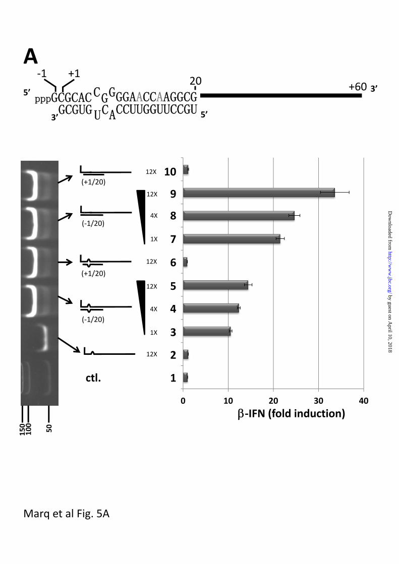
Fig 5. dsRNA induced activation of the IFN promoter
A. Parallel cultures of A549 cells were transfected with pIFN-(ff)luciferase and pTK-
(ren)luciferase. 24h later, the cultures were transfected in duplicate with either
Lane 1; control; just tRNA
Lane 2; 1200 ng 5’ pppJun*61 (ssRNA)
Lane 3-5; 100, 400 and 1200 ng bulged and blunt-ended 5’ pppJun*61/[-1 to 20]
by guest on April 10, 2018
http://ww
w.jbc.org/
Dow
nloaded from
16
Lane 6; 1200 ng bulged and overhanging 5’ pppJun*61/[+1 to 20]
Lanes 7-9; 100, 400 and 1200 ng blunt-ended, non-bulged 5’ pppJun*61/[-1 to 20]
Lane 10; 1200 ng overhanging 5’ pppJun*61/[+1 to 20]
The vertical panel on the left shows an ethidium bromide-stained, non-denaturing PAGE of
the various RNAs transfected; the bottom lane shows dsDNA length markers. tRNA was
added so that the total amount of RNA was constant for all transfections. Cell extracts were
prepared after a further 20 h, and their luciferase activities were determined. The results are
plotted as fold induction relative to the tRNA control. A schematic representation of
overhanging and bulged 5’ pppJun*61/[+1 to 20] is shown above.
B. Transfection of 5’ OHdsRNAs
Same as panel A, except;
Lane 2; 1200 ng 5’ OHJun*61 (ssRNA)
Lanes 3-5; 100, 400 and 1200 ng bulged and blunt-ended 5’ OHJun*61/[-1 to 20]
Lane 6; 1200 ng bulged and overhanging 5’ OHJun*61/[+1 to 20]
Lanes 7-9; 100, 400 and 1200 ng blunt-ended, non-bulged 5’ OHJun*61/[-1 to 20]
Lane 10; 1200 ng overhanging, non-bulged 5’ OHJun*61/[+1 to 20]
Lane 11; 400 ng bulged, blunt-ended 5’ pppJun*61/[-1 to 20]
Lane 12; 400 ng non-bulged, blunt-ended 5’ pppJun*61/[-1 to 20]
Fig 6. Effect of dsRNA with an overhanging 5’ pppnt on IFN activation induced by blunt-
ended 5’ pppdsRNA.
A. Parallel cultures of A549 cells were transfected with pIFN-(ff)luciferase and pTK-
(ren)luciferase. 24h later, the cultures were transfected in duplicate with either
Lane 1; tRNA control
Lane 2; 1200 ng 5’ pppJun*61 ssRNA
Lane 3; 1200 ng overhanging 5’ pppJun*61/[+1 to 20]
Lanes 4, 6, 8; 50, 100 and 200 ng blunt-ended, non-bulged 5’ pppJun*61/[-1 to 20]
Lanes 5, 7, 9; 50, 100 and 200 ng blunt-ended 5’ pppJun*61/[-1 to 20] plus 1200 ng
overhanging 5’ pppJun*61/[+1 to 20].
Cell extracts were prepared after a further 20 h, and their luciferase activities were
determined. The results are plotted as fold induction relative to the tRNA control.
The panel below shows an ethidium bromide-stained, non-denaturing PAGE of the various
RNAs transfected. tRNA was added so that the total amount of RNA was constant for all
transfections.
by guest on April 10, 2018
http://ww
w.jbc.org/
Dow
nloaded from

17
B. Parallel cultures of A549 cells and A549 cells in which RIG-I was knocked down with a
constitutively expressed miRNA (A549-RIG-I-kd cells) were transfected with pIFN-
(ff)luciferase and pTK-(ren)luciferase. 24h later, the cultures were transfected in duplicate
with either 100 (2X) or 200 ng (4X) of non-bulged and blunt-ended 5’ pppJun*61/[-1 to 20]. Cell
extracts were prepared after a further 20 h, and their luciferase activities were determined.
The results are plotted as fold induction relative to the tRNA control.
Fig 7. dsRNA with an overhanging 5’ OHnt does not inhibit IFN activation induced by
blunt-ended 5’ pppdsRNA.
Parallel cultures of A549 cells were transfected with pIFN-(ff)luciferase and pTK-
(ren)luciferase. 24h later, the cultures were transfected in duplicate with either
Lane 1; tRNA control
Lane 2; 50 ng blunt-ended 5’ pppJun*61/[-1 to 20]
Lane 3; 50 ng blunt-ended 5’ pppJun*61/[-1 to 20] and 1200 ng blunt-ended 5’ pppJun*61/[-1 to
20]
Lane 4; 50 ng blunt-ended 5’ pppJun*61/[-1 to 20] and 1200 ng non-blunt-ended 5’
pppJun*61/[+1 to 20]
Lane 5; 50 ng blunt-ended 5’ pppJun*61/[-1 to 20] and 1200 ng non-blunt-ended 5’
OHJun*61/[+1 to 20]
Lane 6; 50 ng blunt-ended 5’ pppJun*61/[-1 to 20] and 1200 ng blunt-ended 5’ OHJun*61/[-1 to
20]
Cell extracts were prepared after a further 20 h, and their luciferase activities were
determined. The results are plotted as fold induction relative to the tRNA control.
tRNA was added so that the total amount of RNA was constant for all transfections.
Fig 8. dsRNA with an overhanging 5’ pppnt does not inhibit IFN activation induced by
poly-I/C.
Parallel cultures of A549 cells were transfected with pIFN-(ff)luciferase and pTK-
(ren)luciferase. 24h later, the cultures were transfected in duplicate with either
Lane 1; tRNA control
Lane 2; 50 ng poly-I/C
Lane 3; 50 ng poly-I/C and 1200 ng blunt-ended 5’ pppJun*61/[-1 to 20]
Lane 4; 50 ng poly-I/C and 1200 ng non-blunt-ended 5’ pppJun*61/[+1 to 20]
Cell extracts were prepared after a further 20 h, and their luciferase activities were
determined. The results are plotted as fold induction relative to the tRNA control.
by guest on April 10, 2018
http://ww
w.jbc.org/
Dow
nloaded from
18
tRNA was added so that the total amount of RNA was constant for all transfections.
Table I. Interaction of RIG-I with various RNAs.
The table lists the names of the various RNAs, the line drawings of their structures, and
their relative abilities to bind to RIG-I, to stimulate the ATPase, and to activate the IFN
promoter upon transfection into cells.
by guest on April 10, 2018
http://ww
w.jbc.org/
Dow
nloaded from

Rig‐I
Cell extr. RNA Pull‐down Cell extr.
‐ IFN + IFN
1 2 3 4 5 6 7 8
Marq et al Fig. 1
by guest on April 10, 2018
http://ww
w.jbc.org/
Dow
nloaded from

Rig‐I
0
1
2
3
4
5
6
7
8
9
1 2 3 4 5 6 7 8
1 2 3 4 5 6 7 8
Marq et al Fig.2
by guest on April 10, 2018
http://ww
w.jbc.org/
Dow
nloaded from
0 0.04 0.2 1 5 Input
0 0.04 0.2 1 5
µg
Input
Input
5’OH
0 0.04 0.2 1 5
Input
0 0.04 0.2 1 5
Poly‐I/C
µg
µg
µg
A
B
C
D
5’OH
Marq et al Fig. 3
by guest on April 10, 2018
http://ww
w.jbc.org/
Dow
nloaded from
(+1/20)
(‐1/20)
Marq et al Fig. 4A
RNA (ng/ul)
ATPase
(nm)
A
0
0.5
1
1.5
2
2.5
3
0 1 2 3 4 5 6
by guest on April 10, 2018
http://ww
w.jbc.org/
Dow
nloaded from

0
1
2
3
4
5
6
0 1 2 3 4 5 6
(‐1/20)p
(‐1/20)
5’OH
ATPase(nm)
0
1
2
3
4
5
6
0 1 2 3 4 5 6
(‐1/20)
(‐1/20)
ATPase
(nm)
5’ppp
B
C
Marq et al Fig 4B and C
RNA (ng/ul)
RNA (ng/ul)
by guest on April 10, 2018
http://ww
w.jbc.org/
Dow
nloaded from
0
0.5
1
1.5
2
2.5
3
3.5
0 1 2 3 4 5 6
Marq et al Fig. 4D
ATPase
(nm)
D
(+1/20)
(+1/20)
RNA (ng/ul)
by guest on April 10, 2018
http://ww
w.jbc.org/
Dow
nloaded from
0 10 20 30 40
(+1/20)
1X
4X
12X
(‐1/20)
ctl.
(+1/20)
(‐1/20)
‐IFN (fold induction)
GCGUGpppGCGCACCGGGGAACCAAGGCG
ACU CCUUGGUUCCGU
5’
5’3’
3’
‐1+6020
1X
4X
12X
12X
12X
12X
50
100
150
Marq et al Fig. 5A
A
1
2
4
3
5
6
8
7
10
9
+1
by guest on April 10, 2018
http://ww
w.jbc.org/
Dow
nloaded from

0 5 10 15 20 25 30
‐IFN (fold induction)
(+1/20)
1X
4X
12X
(‐1/20)
ctl.
(+1/20)
(‐1/20)
1X
4X
12X
12X
12X
12X
4X(‐1/20)
(‐1/20)
4X
50
100
Marq et al Fig. 5B
B
1
2
4
3
5
6
8
7
10
9
11
12
by guest on April 10, 2018
http://ww
w.jbc.org/
Dow
nloaded from
0 5 10 15 20
‐IFN fold induction
(‐1/20)
ctl.
200ng
100ng
50ng
1200 ng
‐1200 ng
‐1200 ng
‐1200 ng‐
(+1/20)
1
2
4
3
5
6
8
7
9
50
100
150
200
250
(+1/20)
(‐1/20)
(+1/20)
(‐1/20)
Marq et al Fig. 6A
A
by guest on April 10, 2018
http://ww
w.jbc.org/
Dow
nloaded from
Marq et al Fig. 6B
B
0
2
4
6
8
10
12
14
16
18
Ctl. 2X 4X
A549
A549‐RIG‐I‐kd
‐IFN (foldinduction)
(‐1/20)
by guest on April 10, 2018
http://ww
w.jbc.org/
Dow
nloaded from

0 5 10 15 20
tRNA 1
2
3
4
5
+
+
+
+
‐
tRNA
(50ng)
Comp.(1200 ng)
+
‐IFN (fold induction)
6
27
Marq et al Fig. 7
by guest on April 10, 2018
http://ww
w.jbc.org/
Dow
nloaded from

Marq et al Fig. 8
0 20 40 60 80
tRNA‐‐IFN (fold induction)
1
2
3
4
Poly‐I/C(50ng)
Comp.(1200 ng)
+
+
+
tRNA by guest on April 10, 2018
http://ww
w.jbc.org/
Dow
nloaded from

RIG‐Ibinding
RIG‐IATPase
IFNinduction
++++
‐
+++
++++
++++ ++++
‐ ‐
‐+++
++++ ++++
1. T7 unpur. RNA1
2. ssRNA1 pur.
++++ ‐
nd+++ ‐
3. RNA1/[1 – 18]
5. RNA1/[19 – 36]
6. RNA1/[37 – 54]
4. RNA1/[2 – 19 or 21]
‐
+
++++
+++++++
++++
++
+++++++
‐
‐
++++
++
‐
‐
7. pppJun*61 ss
8. pppJun*61/[‐1 to 20]
9. pppJun*61/[+1 to 20]
10. pppJun*61/[‐1 to 20] bul.
11. pppJun*61/[+1 to 20] bul.
12. OHJun*61/[‐1 to 20]
13. OHJun*61/[+1 to 20] bul.
‐
nd
nd
nd
nd
nd
nd
nd ‐Marq et al. table 1
by guest on April 10, 2018 http://www.jbc.org/ Downloaded from
Dominique GarcinJean-Baptiste Marq, Stephane Hausmann, Nicolas Veillard, Daniel Kolakofsky and
genomes, act as RIG-I decoysShort dsRNAs with an overhanging 5 prime ppp-nucleotide, as found in arenavirus
published online December 15, 2010J. Biol. Chem.
10.1074/jbc.M110.186262Access the most updated version of this article at doi:
Alerts:
When a correction for this article is posted•
When this article is cited•
to choose from all of JBC's e-mail alertsClick here
by guest on April 10, 2018
http://ww
w.jbc.org/
Dow
nloaded from