short communication morphology, morphometry and ... filethe amazonian manatee spermatozoa were...
TRANSCRIPT

© 2010 Sociedade Brasileira de Zoologia | www.sbzoologia.org.br | All rights reserved.
ZOOLOGIA 27 (6): 1014–1017, December, 2010doi: 10.1590/S1984-46702010000600025
Morphological and ultrastructural analysis of spermato-zoa are useful tools in reproductive biology and phylogeneticstudies of several species, being utilized to contribute charactersfor phylogenetic analysis, evaluate mating strategies, and de-velop new reproductive technologies or improve existing ones(ANDERSON et al. 2005, LUQUE & BÁO 2006, PLÖN & BERNARD 2006).
The Amazonian manatee Trichechus inunguis (Natterer,1883) is a threatened aquatic mammal, endemic of the Amazonbasin, and is the only sirenian that occurs exclusively in freshwater (BEST 1984, ROSAS 1994). It is the smallest sirenian, measur-ing up to 2.8 m and weighing 450 kg (HUSAR 1977, ROSAS 1994).Although certain aspects of Amazonian manatee biology havebeen well studied, there is an absence of basic information aboutthe reproductive anatomy and physiology of this species.
The aim of this study was to contribute to the knowl-edge of the Amazonian manatee reproductive biology present-ing the morphological, morphometric and ultrastructural char-acteristics of its spermatozoon.
The spermatozoa were observed in urine samples collectedfrom a healthy adult T. inunguis.
While spermatozoa were observed in more than one urinesample, only one urine sample was analyzed in this study.
The urine sample was centrifuged (1000 RPM, 10 min),the pellet was washed with saline solution (0,9% NaCl) andcentrifuged again at the same speed and time. A portion of thepellet was fixed with 10% formalin solution for light micros-copy analysis and the other portion was fixed with 2%paraformaldehyde and 2.5% gluteraldehyde in phosphate bufferfor transmission electron microscopy analysis. The formalinfixed sample was analyzed and photographed with a phase-contrast microscope (Zeiss, Oberkochen, Germany). Measure-ments of head length, width and thickness, midpiece length,tail length and total length were taken from 100 spermatozoausing the software Image Pro Express 6.0 (Media CyberneticsInc., Bethesda, MD, USA). The paraformaldehyde/glutaralde-hyde fixed sample was rinsed with a sodium cacodylate buffer(0.1M, pH 7.3), and post-fixed with 1% osmium tetroxide, 0.8%potassium ferricyanide and 5 mM calcium chloride in a so-dium cacodylate buffer. After which, it was dehydrated in aseries of ascending concentrations of acetone solutions (30-
SHORT COMMUNICATION
Morphology, morphometry and ultrastructure of the Amazonianmanatee (Sirenia: Trichechidae) spermatozoa
Rodrigo S. Amaral1, 5; Carolina M. Lucci2; Fernando C. W. Rosas3;Vera M. F. da Silva3 & Sônia N. Báo4
1 Departamento de Reprodução Animal, Faculdade de Medicina Veterinária e Zootecnia, Universidade de São Paulo.05508-270 São Paulo, SP, Brazil.2 Faculdade de Agronomia e Medicina Veterinária, Universidade de Brasília. 70919-970 Brasília, DF, Brazil.3 Laboratório de Mamíferos Aquáticos, Instituto Nacional de Pesquisas da Amazônia. 69083-001 Manaus, AM, Brazil.4 Laboratório de Microscopia Eletrônica, Instituto de Ciências Biológicas, Universidade de Brasília. 70919-970 Brasília,Distrito Federal, Brazil.5 Corresponding author. Email: [email protected]
ABSTRACT. This study describes the morphological, morphometric and ultrastructural characteristics of the Amazonian
manatee Trichechus inunguis (Natterer, 1883) spermatozoon. The spermatozoa were obtained from a urine sample of an
adult T. inunguis kept in captivity. The spermatozoa were analyzed by light and transmission electron microscopy. The
head of Amazonian manatee spermatozoa had a flat oval shape and a well distinguishable midpiece. The mean dimen-
sions of the spermatozoa were: head length, 7.49 ± 0.24 µm; head width, 3.53 ± 0.19 µm; head thickness, 1.61 ± 0.13 µm;
midpiece length, 11.36 ± 0.34 µm; flagellum length, 40.91 ± 1.94 µm; total tail length, 52.16 ± 1.06 µm; total sperma-
tozoon length, 60.08 ± 1.40 µm. The Amazonian manatee spermatozoa were similar in shape to other sirenian sperma-
tozoa; however, presenting a different size. This study describes, for the first time, the morphometric and ultrastructural
characteristics of the Amazonian manatee spermatozoa, and also demonstrates the possible use of spermatozoa re-
trieved from urine samples for biological studies.
KEY WORDS. Anatomy; reproduction; sirenians; spermatozoon; Trichechus inunguis.
1015Morphology, morphometry and ultrastructure of the Amazonian manatee spermatozoa
ZOOLOGIA 27 (6): 1014–1017, December, 2010
100%) and embedded in Spurr epoxy resin. Ultrathin sectionswere cut, stained with uranyl acetate and lead citrate, thenobserved and photographed using a transmission electron mi-croscope (Jeol 1011, Jeol, Tokyo, Japan).
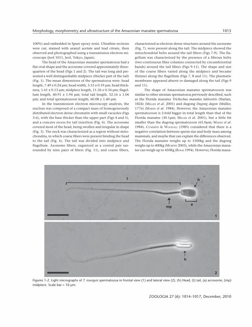
The head of the Amazonian manatee spermatozoa had aflat oval shape and the acrosome covered approximately three-quarters of the head (Figs 1 and 2). The tail was long and pre-sented a well distinguishable midpiece (thicker part of the tail)(Fig. 1). The mean dimensions of the spermatozoa were: headlength, 7.49 ± 0.24 µm; head width, 3.53 ± 0.19 µm; head thick-ness, 1.61 ± 0.13 µm; midpiece length, 11.36 ± 0.34 µm; flagel-lum length, 40.91 ± 1.94 µm; total tail length, 52.16 ± 1.06µm; and total spermatozoon length, 60.08 ± 1.40 µm.
In the transmission electron microscopy analysis, thenucleus was composed of a compact mass of homogeneouslydistributed electron dense chromatin with small vacuoles (Figs3-6), with the base thicker than the upper part (Figs 4 and 5),and a concave recess for tail insertion (Fig. 6). The acrosomecovered most of the head, being swollen and irregular in shape(Fig. 3). The neck was characterized as a region without mito-chondria, in which coarse fibers were present binding the headto the tail (Fig. 6). The tail was divided into midpiece andflagellum. Axoneme fibers, organized as a central pair sur-rounded by nine pairs of fibers (Fig. 11), and coarse fibers,
characterized as electron dense structures around the axoneme(Fig. 7), were present along the tail. The midpiece showed themitochondrial helix around the tail fibers (Figs 7-9). The fla-gellum was characterized by the presence of a fibrous helix(two continuous fiber columns connected by circumferentialbands) around the tail fibers (Figs 9-11). The shape and sizeof the coarse fibers varied along the midpiece and becamethinner along the flagellum (Figs 7, 8 and 11). The plasmaticmembrane appeared absent or damaged along the tail (Figs 9and 11).
The shape of Amazonian manatee spermatozoon wassimilar to other sirenian spermatozoa previously described, suchas the Florida manatee Trichechus manatus latirostris (Harlan,1824) (MILLER et al. 2001) and dugong Dugong dugon (Müller,1776) (MARSH et al. 1984). However, the Amazonian manateespermatozoon is 2-fold bigger in total length than that of theFlorida manatee (30.1µm; MILLER et al. 2001), but a little bitsmaller than the dugong spermatozoon (65.0µm; MARSH et al.1984). CUMMINS & WOODALL (1985) considered that there is anegative correlation between sperm size and body mass amongmammals, and maybe that can explain the differences observed.The Florida manatee weighs up to 1500kg and the dugongweighs up to 400kg (MURPHY 2003), while the Amazonian mana-tee can weigh up to 450Kg (ROSAS 1994). However, Florida mana-
Figures 1-2. Light micrographs of T. inunguis spermatozoa in frontal view (1) and lateral view (2). (h) Head, (t) tail, (a) acrosome, (mp)midpiece. Scale bar = 10 µm.
1
2
h
ht
mp a

1016 R. S. Amaral et al.
ZOOLOGIA 27 (6): 1014–1017, December, 2010
tee spermatozoa described by MILLER et al. (2001), also obtainedfrom a single male, were measured using scanning electronmicroscopy images, thus that size difference needs to be evalu-ated with caution.
In general, the ultrastructure of Amazonian and Floridamanatees’ spermatozoa were similar. However, MILLER et al.(2001) reported that T. m. latirostris spermatozoa show a verypronounced annulus, and this structure was not observed in T.inunguis. On the other hand, the alterations observed (swell-ing and irregular shape of acrosome and the disruption or lossof plasmatic membrane) probably are post-mortem degenera-tion due to osmotic stress by the urine (VIJAYAKUMAR et al. 1986,GRIGGERS et al. 2001). Therefore, the annulus from Amazonianmanatee spermatozoa might have been lost. There is no reportabout dugong spermatozoa ultrastructure to be compared with
Amazonian manatee spermatozoa.This is the first report describing the morphology, mor-
phometry and ultrastructure of Amazonian manatee sperma-tozoa. Although our data are from one individual, apparentlythere are morphometric differences among sirenians sperma-tozoa. This study also shows that morphological analysis ofspermatozoa retrieved from urine samples can be a useful toolin biological studies.
ACKNOWLEDGEMENTS
We thank CNPq, Programa Petrobras Ambiental and Asso-ciação Amigos do Peixe-boi (AMPA) for financial support;PREVET staff for help with sample collection and Renata C.Silva for help with microscopic analysis.
Figures 3-11. Electron micrographs of T. inunguis spermatozoa. (3) Longitudinal section of the head. (pm) plasmatic membrane; (am)acrosomal membrane; (a) acrosome; (n) nucleus; (white arrowhead) vacuole. Note the acrosome swelling. (4-5) Transversal sections ofupper portion and basal portion of the head. (mp) plasmatic membrane; (n) nucleous. (6) Diagonal section of the neck region (ne).Note the mitochondria marking the beginning of the tail. (7-8) Transversal sections of the cranial and caudal portion of midpiece. Notethe difference of size and shape of the coarse fibers (cf). (ax) axoneme; (mt) mitochondria. (9) Tangential section of the tail at thetransition region from midpiece to flagellum. (mt) mitochondria; (fh) fibrous helix. Note the absence of plasmatic membrane. (10-11)Longitudinal and transversal sections of the flagellum. (ax) Axoneme, (fh) fibrous helix, (cf) coarse fibers. Scale bar = 0.5µm.
3 6
4 5
7 8
9 1110

1017Morphology, morphometry and ultrastructure of the Amazonian manatee spermatozoa
ZOOLOGIA 27 (6): 1014–1017, December, 2010
LITERATURE CITED
ANDERSON, M.J.; J. NYHOLT & A.F. DIXTON. 2005. Sperm competitionand the evolution of sperm midpiece volume in mammals.Journal of Zoology 267 (2): 135-142. doi: 10.1017/S0952836905007284.
BEST, R.C. 1984. The aquatic mammals and reptiles of the Amazon,p. 371-412. In: H. SIOLI (Ed.). The Amazon, limnology andlandscape ecology of a mighty tropical river and its basin.Dordhecht, Dr. W. Junk Publishers, 761p.
CUMMINS, J.M. & P.F. WOODALL. 1985. On mammalian spermdimensions. Journal of Reproduction and Fertility 75: 153-175. doi: 10.1530/jrf.0.0750153.
GRIGGERS, S.; D.L. PACCAMONTI; R.A. THOMPSON & B.E. EILTS. 2001.The effects of pH, osmolarity and urine contamination onequine spermatozoal motility. Theriogenology 56 (4): 613-622. doi: 10.1016/S0093-691X(01)00593-3.
HUSAR, S.L. 1977. Trichechus inunguis. Mammalian Species 72(1): 1-4.
LUQUE, M.C.A. & S.N. BÁO. 2006. Structural and ultrastructuralcharacterization of zebu (Bos indicus) spermatozoa. Biocell30 (1): 33-38.
MARSH, H.; G.E. HEINSOHN & T.D. GLOVER. 1984. Changes in the
Submitted: 22.VII.2010; Accepted: 25.IX.2010.Editorial responsibility: Carolina Arruda Freire
male reproductive organs of the dugong, Dugong dugon(Sirenia: Dugondidae) with age and reproductive activity.Australian Journal of Zoology 32 (6): 721-742. doi:10.1071/ZO9840721.
MILLER, D.L.; M.M. DOUGHERTY; S.J. DECKER & G.D. BOSSART. 2001.Ultrastructure of the spermatozoa from a Florida manatee(Trichechus manatus latirostris). Anatomia, Histologia,Embryologia 30 (4): 253-256. doi: 10.1046/j.1439-0264.2001.00330.x.
MURPHY, D. 2003. Sirenia, p. 476-482. In: M.E. FOWLER & R.E. MILLER
(Ed.). Zoo and Wildlife Animal Medicine. Philadelphia, W.B. Saunders Co., 992p.
PLÖN, S. & R.T.F. BERNARD. 2006. A review of spermatozoanmorphology in Cetacea with new data for the genus Kogia.Journal of Zoology 269 (4): 466-473. doi: 10.1111/j.1469-7998.2006.00061.x.
ROSAS, F.C.W. 1994. Biology, conservation and status of theAmazonian manatee Trichechus inunguis. Mammal Review24 (2): 49-59. doi: 10.1111/j.1365-2907.1994.tb00134.x.
VIJAYAKUMAR, R.; O. USUN; R. SWOPE; F. DE LEON & W. HEINE. 1986.Fine structure of spermatozoa retrieved from retrogradeejaculates. Systems Biology in Reproductive Medicine 17(1): 25-33.