shifts in the concentrations of magnesium and calcium in...
TRANSCRIPT

Shifts in the Concentrations of Magnesium and Calcium in Early Porcine andRat Wound Fluids Activate the Cell Migratory ResponseJohn J. Grzesiak and Michael D. PierschbacherLa Jolla Cancer Research Foundation, La Jolla, California 92037
Abstract
Accruing evidence indicates that the levels of extracellularMg2+ and Ca2+ can have a distinct impact on the adhesiveand migratory activities of many cell types. The physiologi-cal relevance of these observations, however, has remainedlargely unexplored. In the present study, wound fluids col-lected throughout the early stages of cutaneous wound re-
pair were examined for possible Mg2" and Ca2+ fluctua-tions. Early in the process, when cell migration into thewound site is initiated, Mg2+ is elevated and Ca2+ is reduced(Mg2+ :Ca2+ = 1). As wound healing progresses, woundfluid concentrations of Mg2+ and Ca2+ begin to return tonormal plasma levels (Mg2+ :Ca2+ = 0.4). When macro-
phages, keratinocytes, fibroblasts, and endothelial cells were
exposed to dialyzed wound fluid, the migration stimulatedby undialyzed wound fluid was lost. Addition back to dia-lyzed wound fluid of 24 h, postinjury concentrations of Mg2+and Ca2+ restored all migratory stimulus. This observedmigration is approximately twofold greater than when nor-mal plasma Mg2+ and Ca2+ concentrations are present.Changes in the levels of Mg2+ and Ca2+ in wound fluid occur
during the same period that inflammatory cells, keratino-cytes, fibroblasts, and neovasculature have been shown tomigrate during wound healing in vivo. Together, these datasuggest that the impact of these changes on integrins andE-cadherin may play a direct role in the activation andmaintenance of the migratory phenotypes of the cells in-volved in the wound healing process. (J. Clin. Invest. 1995.95:227-233.) Key words: divalent cations * integrins * E-cadherin * cell migration - wound healing
Introduction
The process of cutaneous wound healing is a complex andcarefully orchestrated cascade of overlapping events that in-volves changes in extracellular matrix protein composition andcell migration and proliferation in response to numerous cyto-kines ( 1-4). Crucial to the repair process is the migration ofinflammatory cell types such as macrophages and neutrophilsand epithelial cells, fibroblasts, and endothelial cells into thewound site over the course of the first 3 d after injury (1-8),where they have multiple responsibilities resulting, ultimately,
Address correspondence to Dr. M. D. Pierschbacher, La Jolla CancerResearch Foundation, 10901 North Torrey Pines Road, La Jolla, CA92037. Phone: 619-455-6480; FAX: 619-455-0181.
Received for publication 18 February 1994 and in revised forn 8August 1994.
in a healed wound. Integrins, the family of heterodimeric trans-membrane receptors present on the cell surface are responsiblefor mediating much of the interaction of these cells with theextracellular matrix (9-12).
Integrins require divalent cations, such as Mg2+ and Ca2+,to function (13-16). The integrin a subunits contain the puta-tive EF hand cation-binding domains thought to be responsiblefor much of the cation binding capacity of integrins (17, 18).Wehave demonstrated with two Arg-Gly-Asp (RGD)-depen-dent integrins, a,/31 (19, 20), and avl3 (21), both of whichshare the same a subunit, that av,61 binds to ligand only inMg2+ and not in Ca2 , while av,63 binds to ligand in eitherCa2+ or Mg2+ (18). Wealso showed that in the presence of2+2'ehnetelanMg , increasing concentrations of Ca2+ enhance the ligand
binding of av,63 but inhibit the ligand binding of av,61 (18).Recently, we reported that the Mg2+-dependent, a2,l-medi-
ated adhesion and migration of human fibroblasts on type Icollagen substrates could be inhibited by elevated extracellularCa2+. Weobserved, however, that migration could be enhancedtwofold over that seen in Mg2+ alone by providing the twodivalent cations in combination with a Mg2+ to Ca2+ ratioslightly higher than one (22). Keratinocytes are also importantin the cutaneous wound repair process, and the concentrationsof extracellular Mg2+ and Ca2' have been observed to affecttheir a2,l81-mediated migration on type I collagen in a similarfashion.' In addition to integrins, keratinocytes express anotherCa2+-dependent molecule responsible for cell-cell adhesion,E-cadherin (for reviews see references 23 and 24). In the pres-ence of Ca2+, E-cadherin resides principally in the areas of cell-cell contacts. In reduced Ca2+, however, E-cadherin becomessusceptible to proteolytic cleavage and rapidly loses function,eventually disappearing from the cell surface altogether. Recentstudies demonstrate the differential regulation of E-cadherinand the integrin az8, by shifts in the concentrations of extracel-lular Mg2+ and Ca2+, resulting in an "activated" keratinocytephenotype.
Under normal physiologic conditions the concentration ofintracellular Mg2+ in the typical mammalian cell is reported tobe between 15 and 30 mMand that of Ca2+ is only - 1-2 mM(25-28). On the other hand, extracellular levels of these twocations are about 1.0 and 2.5 mM, respectively (28, 29). Wehave speculated, previously, that tissue injury might result in alocal increase in the extracellular Mg2+ level and/or a decreasein the extracellular Ca2+ level due to the spill of Mg2+ fromdamaged tissue and the sequestration of Ca2+ by several possi-ble molecular events including the Ca2+-dependent coagulationcascade (30-32). The migratory phenotype of cells involvedin wound healing might be supported in the local wound envi-ronment and in adjacent surrounding tissue by this diffusiblegradient of altered divalent cations. After the required cells have
1. Grzesiak, J. J., and M. D. Pierschbacher, manuscript submitted forpublication.
Divalent Cation Regulation of the Wound Healing Migratory Response 227
J. Clin. Invest.© The American Society for Clinical Investigation, Inc.0021-9738/95/01/0227/07 $2.00Volume 95, January 1995, 227-233

arrived at the injury site, the extracellular levels of these twocations would be normalized, and the potential for cells to mi-grate might again be reduced.
In the present study we have examined this hypothesis bymeasuring the total extracellular Mg2" and Ca2" levels in por-cine and rat wound fluids during the early phase of cutaneousinjury repair when migration of inflammatory cells, keratino-cytes, fibroblasts, and neovasculature into the wound site havebeen shown to occur in vivo (1-8). Our studies indicate anearly shift in the levels of these two cations, with elevatedextracellular Mg2" and reduced extracellular Ca2". We alsodemonstrate with macrophages on fibrinogen substrates, andkeratinocytes, fibroblasts, and endothelial cells on type I colla-gen substrates that early phase wound fluid stimulates integrin-mediated cell migration in a divalent cation-dependent manner.These are specific integrin-extracellular matrix interactionslikely to be occurring with these cell types in the type I collagenand fibrin(ogen) rich environment of wound healing ( 1-8, 33,34). The inability of dialyzed wound fluid to support suchmigration could be completely restored by adding back Mg2"and Ca2" at the levels observed in wound fluid 24 h after injury.
The influence of divalent cation concentration on integrinand cadherin function is capable of activating the migratoryphenotypes required for the cells involved in wound healing.The ability of wound fluid to achieve this effect has long beenknown but the factor/s responsible have only been partiallyelucidated. In this report we demonstrate that the local shift inthe extracellular divalent cation concentrations may be a criticalpart of the cutaneous repair process.
Methods
Woundfluids. To collect wound fluid three different animal models wereused. In the first, two 2-cm2 partial thickness excisional wounds wereinitiated on the dorsal surface of four 4-mo-old female pigs housed andfed together for 2 wk before surgery. Wound fluid was collected byaspirating the wound bed immediately after wounding, centrifuging col-lected material at 10,000 rpm for 10 min and collecting the supernatant.Citrated plasma was collected from each animal 1 wk before surgeryfor determination of normal plasma Mg2' and Ca2+ levels. In the secondmodel, one 6 mm-diameter-partial-thickness burn wound was generateddorsally on each of two rats (4 mo, male Sprague-Dawley). Necrotictissue was debrided 24 h after wound initiation and wound fluid wascollected at the indicated time points as described in the porcine model.Citrated plasma was collected 1 d before wound initiation. In the thirdanimal model, four 1-cm-full-thickness incisions were initiated dorsallyand 0.25 X 1 x 1 cm sterile polyvinyl alcohol sponges were insertedsubcutaneously under the incision. The wounds were then sutured. Atthe indicated timepoints the sponges were harvested, centrifuged, andthe wound fluid collected as with the other models. Citrated plasma wascollected on the same day just before surgery and again at the indicatedharvest time points. For functional assays, aliquots of wound fluid fromindividual sponges collected from the full-thickness rat wound 24 hafter injury were dialyzed extensively against divalent cation-free Tris-buffered saline at 4°C using dialysis tubing with 6-8 kD molecularmass cutoff.
Magnesium and calcium determination. Atomic absorption spectros-copy was used to evaluate the total Mg2' and Ca2+ levels in porcineand rat wound fluids. Atomic absorption spectroscopy was carried outby C.L. Technology, Inc. (Corona, CA). Magnesium and calcium kitsemploying colorimetric methods (Sigma Chemical Co., St. Louis, MO)were also used to confirm results.
Cells. HaCaT cells, spontaneously transformed nontumorigenic hu-man keratinocytes with phenotypic differentiation characteristics of nor-mal keratinocytes (33), and W138 normal human lung fibroblasts(American Type Culture Collection, Rockville, MD) were cultured in
DMEsupplemented with penicillin (400 U/ml), streptomycin (50 sg/ml), glutamine (300 Mg/ml), and 10% fetal calf serum in a humidifiedatmosphere of 7% CO2 at 370C. Primary cultures of human umbilicalvein endothelial cells (HUVEC) (Clonetics, San Diego, CA), werecultured in endothelial cell growth medium (EGMh) (Clonetics) at370C in a humidified atmosphere of 7% CO2. HUVECcells betweenpassages 3 and 6 were used for migration studies. Bone marrow-derivedmurine macrophages were isolated as described (35) and cultured inDMEcontaining 2 mML-glutamine, 1 mMNa pyruvate, 50 U/mlpenicillin, 50 Mg/ml streptomycin, 20% FCS, and 30% L-cell condi-tioned medium (as a source of macrophage colony stimulating factor)(35) on nontissue culture plastic in a humidified 5%CO2 atmosphere.
Cell migration assays. Migration assays were conducted using themodified Boyden chamber as described (36). Briefly, the chamber con-sists of two compartments separated by a filter, and migration is mea-sured by counting the number of cells crossing the membrane throughpores of defined size. Lower chambers were filled with dialyzed orundialyzed wound fluid collected 24 h after injury. Various CaCl2 orMgCl2 concentrations were then added as indicated. 5- 10-sm porepolycarbonate membrane filters (Poretics Corp., Livermore, CA) thathad been previously coated with 25 Mg/ml bovine type I collagen or 10jig/ml bovine fibrinogen were then placed on top of the lower chambers,and the upper chambers were secured in place. Upper chambers werefilled with medium containing 5.0 X 10⁴ HaCaT keratinocytes, 3.0x 10⁴ WI38 fibroblasts, 3.0 x 10⁴ human umbilical vein endothelialcells, or 5.0 x I0O bone marrow-derived murine macrophages per cham-ber using Ca2", Mg2", and P04-free DMEsupplemented with Ca2"and Mg2+ concentrations consistent with the lower chambers, plus 100Mg/ml BSA. The entire apparatus was then incubated for 4 h at 37°C.After the incubation period, the upper chamber was removed and thefilter was fixed in 3%paraformaldehyde in PBS and stained with 0.5%toluidine blue and 3.7% formaldehyde in PBS. Excess stain was washedaway with water, the attached cells on the upper side of the filter wereremoved and the cells that had migrated to the underside were quanti-tated by counting two high-powered fields (X200) per well using aninverted, light microscope (model CK2; Olympus Corp., Lake Suc-cess, NY).
Protein. Bovine type I collagen was obtained from CollaborativeResearch Inc. (Bedford, MA). Bovine plasma fibrinogen was obtainedfrom Calbiochem (La Jolla, CA).
Results
Extracellular Mg2" and Ca2" concentrations change duringwound healing. Because integrin-mediated leukocyte adhesionand fibroblast, keratinocyte and endothelial cell migration areaffected by the relative concentrations of extracellular Mg2" andCa2" in vitro ( 13, 22, 36-40),' experiments were conducted tomeasure the levels of extracellular Mg2" and Ca2" in an invivo setting known to involve these cells and these integrin-extracellular matrix interactions, i.e., the early stages of cutane-ous injury repair (1-8, 33, 34). Weobserved that the Mg2"concentration in porcine wound fluid from partial-thickness ex-cisional wounds was increased from 0.96 to 1.43 mMimmedi-ately after wounding, while the Ca2+ concentration was reducedfrom 2.53 mMto 1.59 mM(Fig. 1 A). This represents a shiftin the Mg2+ to Ca2' ratio from 0.38 to 0.90 (Fig. 1 B). Theexisting data correlating cell migration activity with extracellu-lar ratios of Mg2+ to Ca2+ indicate that the shift in divalentcation concentrations observed in this wound fluid is sufficientto support nearly maximal migratory activity of human WI38fibroblasts, HaCaT keratinocytes and capillary endothelial cells(18, 36).' To confirm that the observed cation concentrationsin this early wound fluid were altered as a consequence ofwounding, time course studies were conducted. Using partial-thickness burn (Fig. 2, A and B) and full-thickness incisional
228 J. J. Grzesiak and M. D. Pierschbacher

A 3
T2 2-
E
0
01'
Figure 1. Evaluation ofthe extracellular Mg2"and Ca2" levels inwound fluid obtained
WRO immediately after injuryfrom a porcine, partial-thickness model ofwound healing. (A)Mg2+ and Ca2+ levels inwound fluid collectedimmediately afterwounding (to) are
shown. (B) The Mg2+to Ca2' ratio in towound fluid is com-
pared with that of nor-
mal plasma obtained be-fore surgery. The woundfluid results representthe mean±SD of four
animals, each with twowounds. The plasma
values represent the
mean±SD of the four
animals used in theWRO study.
(Fig. 3, A and B) models in the rat for wound fluid collection,we found that the Mg2" to Ca2" ratio observed in porcine woundfluid immediately after injury was maintained in these two mod-els through at least 24 h, with values ranging between 0.85 and0.98. The levels of the two cations overall were somewhat lowerin the rat models than those observed in the porcine model,with concentrations ranging between 0.89 and 1.3 mM. Aswound healing progressed (48 h), the levels of the two cationsappear to begin to return to those normally found in plasma.At the indicated time-points of the full-thickness/PVA spongerat experiments, plasma was also collected. It was determinedthat the plasma levels of Mg2' and Ca2+ were not significantlydifferent from those measured in plasma before injury (notshown).
The altered concentrations of Mg2' and Ca2+ found inwound fluid 24 h after injury promotes maximal macrophage,keratinocyte, fibroblast, and endothelial cell migration. Duringcutaneous injury repair in vivo, the inflammatory cell typesmacrophages and neutrophils arrive at the wound site togetherwithin hours after injury ( 1-4). Keratinocytes have also beenshown to become activated within hours after injury and tomigrate measureably within the first 24 h after injury (1-8).Additionally, fibroblasts and neovasculature have been shown
B
E0
coCM
Cur
+0)
Eca2+Ljmg2+
WFtR4
Figure 2. Time course evaluation of extracellular Mg2' and Ca2+ levelsin wound fluid from a rat, partial-thickness burn model of wound heal-ing. (A) Mg2' and Ca2+ levels in wound fluid are indicated with respectto time throughout a 48-h time course (WFt24 and WFt48) and comparedwith normal plasma levels. (B) The Mg2' and Ca2' ratio in woundfluid throughout the time course is shown and compared with the ratiofound in normal plasma obtained prior to wounding. The data representthe mean values of the cations found in wound fluid derived from singlewounds on each of two rats. In no case did the individual cation concen-
trations differ by > 8%.
to migrate into the wound site from surrounding tissue between1 and 3 d after injury where they begin the process of granula-tion tissue formation (1-8). Fibrin(ogen) is a major compo-nent of the initial clot (1-4). It has been previously demon-strated that the 162 integrin Mac-1 mediates the binding of mac-
rophages and neutrophils to fibrinogen (37, 41-48). It has alsobeen demonstrated that /2 integrin adhesive function is Mg2+-dependent and inhibited by Ca2+ similar to a2,31 (13, 39). Ifthe epithelial basement membrane is disrupted, keratinocytesare directly exposed to type I collagen in the underlying intersti-tium. Fibroblasts and neovasculature migrating into the woundsite from surrounding tissue would likely do so, at least in part,on type I collagen. Additionally, type I collagen is the majornewly synthesized protein during cutaneous injury repair ( 1-
Divalent Cation Regulation of the Wound Healing Migratory Response 229
A
3.0
iEc
0Cu0
B
iE0._o
CM+
0)

extensively against cation-free tris-buffered saline. The dialysistubing had a molecular weight cut-off of 6-8 kD. Wethen
Ca2+ tested these two wound fluid aliquots (dialyzed and not dia-
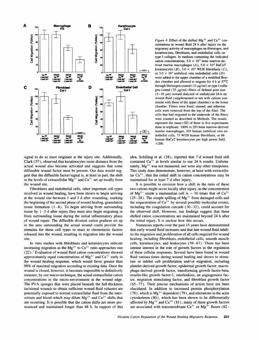
[]Mg2+ lyzed) for their ability to promote the migration of bone mar-row-derived murine macrophages on fibrinogen, and W138 fi-broblasts, HaCaT keratinocytes, and umbilical vein endothelialcells on type I collagen. In the wells that contained dialyzedwound fluid supplemented with divalent cations, maximal mi-gration equivalent to that of the undialyzed wound fluid was
achieved when Mg2' and Ca2+ were added at concentrationsobserved 24 hours after injury (Fig. 4, A-D). Some migrationwas observed in the wells containing normal plasma levels ofMg2+ and Ca2 , but the activity was about half that observedwith wound fluid cation levels observed 24 h after injury. Essen-tially all adhesive and, consequently, migratory activity was lostfor all four cell types when dialyzed wound fluid was used withno added divalent cations.
VRO^8 Discussion
B 1.5
E
0
S..0)
0.0
Plasma WFt12 WFR24 WFt8
Figure 3. Time course evaluation of extracellular Mg2" and Ca2" levelsin wound fluid from a rat, full-thickness/PVA sponge model of woundhealing. (A) Mg2` and Ca2" levels in wound fluid are indicated withrespect to time throughout a 48-h time course (WRt12, WRt24, and WRt48)and compared with normal plasma levels. (B) The Mg2+ to Ca2+ ratioin wound fluid throughout the time course is indicated and comparedto the ratio found in normal plasma obtained before surgery. The resultsrepresent the mean values±SD of the cations found in wound fluidderived from four wounds on each of two animals per time point. Theplasma values (obtained before surgery) represent the mean±SDof thesix animals used in the study.
4, 33, 34). It has been previously demonstrated that the a21Iintegrin, whose divalent cation dependent nature has been welldocumented, is present on these keratinocytes, fibroblasts andendothelial cells in vivo during wound healing, and that thisintegrin mediates the migration of these cells on type I collagen(5, 6, 22, 33, 38).
To test the effect of the shifted divalent cation concentra-tions found in wound fluid on migration of these importantwound healing cell types, wound fluid collected from individualsponges in the full-thickness/PVA sponge rat model 24 h afterinjury was divided into two aliquots. One aliquot was dialyzed
Wedemonstrate that during the early stages of cutaneous injurythe concentrations of extracellular Mg2" and Ca2" in porcineand rat wound fluids are significantly different from those ob-served in normal extracellular fluid. Specifically, Mg2" is ele-vated and Ca2" is reduced. This early wound fluid, after dialysisagainst divalent cation-free Tris-buffered saline, is essentiallyunable to promote the adhesion, and consequently, the migrationof macrophages on fibrinogen, or keratinocytes, fibroblasts andendothelial cells on type I collagen. With the addition of Mg2"and Ca2" to the dialyzed wound fluid in the concentrationsobserved in wound fluid obtained 24 h after injury, however,all migration-promoting activity is restored. This represents a
nearly twofold increase in migration over that observed in re-
sponse to dialyzed wound fluid supplemented with normalplasma levels of the two cations.
During the initial acute inflammatory response to injuryplatelets become activated for aggregation and secretion re-
sponses. It is well established that type I collagen promotesplatelet aggregation and that the a2j31 integrin present on plate-lets responds to Mg2' and Ca2+ in adhesion studies similar tofibroblasts (49). Interestingly, addition of Mg2, to platelets gel-filtered in the absence of Mg2' and Ca2+ promotes optimalaggregation and secretion responses while Ca2+ promotes disag-gregation (50, 51 ). It is possible that an initial burst of Mg2+provided by damaged cells, which contain intracellular Mg2+concentrations in excess of 15 mM(25-28), and exposure totype I collagen could collectively induce the aggregation re-
sponse. Macrophages and neutrophils also become activatedduring this phase, extravasating through the blood vessel wall,surrounding tissue and into the wound site. This activation ischaracterized by a conformational change in 12 integrins thatrenders them functional for ligand binding (9-12). It is interest-ing that this conformational change has been shown to be Mg2+-
dependent (13, 39, 40). These data are in agreement with our
macrophage results which demonstrate enhanced ,62 integrin-mediated migration on fibrinogen when Mg2+ is increased andCa2+ is reduced.
Also occurring during the middle of the inflammatory re-
sponse, by 24 h after injury, keratinocytes also become "acti-
vated" and measurable migration is observed (1-6, 52-57).How this "activation" process occurs has not been clearly de-fined. Mansbridge and Knapp (56), have noted that becausekeratinocytes proliferate and migrate at the injury edge, the
230 J. J. Grzesiak and M. D. Pierschbacher
A
EC
0
0
B
SOIC0
*0
0
tM WF Amzod ia-d d*vd1UMg2+ hWF MWF tM WF1.3v Ca2+ 1.hMeMg2+ ." Mg2+ NO
1.)utdCm2+ 2-5nUCa2+ CAUs
Fib broblastCol I
D 1201
OR so.
00co
240
20
t24hbWF iAd Ad Ad1.3n Mg?2+ bm WF 2WF bW1.3nu Ca2+ 1.1.3I-Mg NOM
1 mSCa2+ 2.5.ddUr+ CATis
KeratinocytoCol I
Endothellal CellCol I
rIB
r-i
IWF jAid Ad1N4 ii07i1 in"M2* i* WF i& WF a VW1.3M C&2+ 1a.M M2 1.Mmg2* Nm
1.3)MCa 25.M C2* CATIONS
Figure 4. Effect of the shifted Mg2" and Ca2+ con-centrations in wound fluid 24 h after injury on themigratory activity of macrophages on fibrinogen, andkeratinocytes, fibroblasts, and endothelial cells ontype I collagen. In medium containing the indicatedcation concentrations, 5.0 x 10⁵ bone marrow-de-rived murine macrophages (A), 5.0 x 10⁴ HaCaTkeratinocytes (B), 3.0 x 10⁴ W138 fibroblasts (C),or 3.0 x 10⁴ umbilical vein endothelial cells (D)were added to the upper chamber of a modified Boy-den chamber and allowed to migrate for 4 h at 370Cthrough fibrinogen-coated (10 Mg/ml) or type I colla-gen-coated (25 ttg/ml) filters of defined pore size(5-10 gm) toward dialyzed or undialyzed 24-h ratwound fluid (supplemented or not with cations con-sistent with those of the upper chamber) in the lowerchamber. Filters were fixed, stained, and adherentcells were removed from the top of the filter. Thecells that had migrated to the underside of the filterswere counted as described in Methods. The resultsrepresent the mean±SD of three to five experimentsdone in triplicate. 100% is 293 bone marrow-derivedmurine macrophages, 103 human umbilical vein en-dothelial cells, 75 W138 human fibroblasts, or 66human HaCaT keratinocytes per high power field.x200.
signal to do so must originate at the injury site. Additionally,Clark (57), observed that keratinocytes some distance from theactual wound also become activated and suggests that somediffusable wound factor must be present. Our data would sug-gest that the diffusible factor/signal is, at least in part, the shiftin the levels of extracellular Mg2" and Ca2" set up locally fromthe wound site.
Fibroblasts and endothelial cells, other important cell typesinvolved in wound healing, have been shown to begin arrivingat the wound site between 1 and 3 d after wounding, markingthe beginning of the second phase of wound healing, granulationtissue formation (1-8). To begin arriving from surroundingtissue by 1-3 d after injury they must also begin migrating infrom surrounding tissue during the initial inflammatory phaseof wound repair. The diffusible divalent cation gradient set upin the area surrounding the actual wound could provide thestimulus for these cell types to react to chemotactic factorsreleased into the wound, resulting in migration into the woundsite.
In vitro studies with fibroblasts and keratinocytes indicateincreasing migration as the Mg2" to Ca2" ratio approaches one(22).1 Evaluation of wound fluid cation concentrations showedapproximately equal concentrations of Mg2' and Ca2' early inthe wound healing response, which would favor greater than90% of maximal migration according to existing data. Once thewound is closed, however, it becomes impossible to definitivelymeasure, by our macro-technique, the actual extracellular cationconcentrations in the micro-environment at the wound edge.The PVA sponges that were placed beneath the full-thicknessincisional wounds to obtain sufficient wound fluid volumes arepotentially exposed to normal extracellular fluid from the inter-stitium and blood which may dilute Mg2' and Ca2+ shifts thatare occurring. It is possible that the cation shifts are more pro-nounced and maintained longer than 48 h. In support of this
idea, Schilling et al. (58), reported that 7-d wound fluid stillcontained Ca2" at levels similar to our 24 h results. Unfortu-nately, Mg2+ was not measured, nor were any other timepoints.This study does demonstrate, however, at least with extracellu-lar Ca2 , that the initial shift in cation concentrations may bemaintained for at least 7 d after injury.
It is possible to envision how a shift in the ratio of thesetwo cations might occur locally after injury, as the concentrationof Mg2+ inside a mammalian cell is - 10 times that of Ca2+(25-28). The simple spilling of Mg2, from damaged cells andthe sequestration of Ca2' by several possible molecular events,including the coagulation cascade (30-32), could account forthe observed shift. However, our findings suggest that theseshifted cation concentrations are maintained beyond 24 h afterthe initial injury. It is unclear how this occurs.
Numerous reports over the past 15 years have demonstratedthat early wound fluid increases and that late wound fluid inhib-its the migration and proliferation of all cells required for woundhealing, including fibroblasts, endothelial cells, smooth musclecells, keratinocytes, and leukocytes (59-67). There has beenintense interest in the role of growth factors in the regulationof these cellular responses. Several have been found in woundfluid various times during wound healing and shown to stimu-late or inhibit cell proliferation and/or migration, includingplatelet-derived growth factor, epidermal growth factor, macro-phage-derived growth factor, transforming growth factor-beta,insulin-like growth factor- 1, interleukins, an angiogenesis fac-tor, migration stimulating factor, and fibroblast growth factor(65-77). Their precise mechanisms of action have not beenelucidated. In addition to increased protein phosphorylation(78), which is Mg2+ dependent (79), and alterations in the actincytoskeleton (80), which has been shown to be differentiallyaffected by Mg2' and Ca2+ (81), many of these growth factorsare associated with transmembrane Ca2+ or Mg2+ fluxes (82-
Divalent Cation Regulation of the Wound Healing Migratory Response 231
A
x
o
m0R0)-
.2_
C 1201
- lo
9
.ca0
2 40
20
-7-1
TI I

84) and could participate in the regulation of divalent cationconcentrations. Despite the presence of these growth factors inwound fluid, however, our data demonstrate that the presenceand concentrations of Mg2" and Ca2" can account for the in-creased ability of cells to migrate. Similar increases in migrationhave been reported using altered concentrations of Mg2" andCa2" with platelet-derived growth factor alone or fetal calf se-rum (22, 35).' It may be that the growth factors found in woundfluid and fetal calf serum provide directional signalling for cellmovement. The alterations in the concentrations of extracellularMg2+ and Ca2+ may act in conjunction with growth factors bycreating conditions that favor increased and directed migration.
Chronic wound fluid from venous stasis and to a lesserdegree, diabetic ulcers show significant breakdown of fibronec-tin and vitronectin along with increased metalloprotease activity(85, 86). The concentrations of Ca2' and Mg2+ in these woundfluids were not measured, however. Interestingly, Sank et al.(87), demonstrated in a guinea pig model that increasing theCa2+ concentration at the wound site resulted in increased colla-genase activity and poor wound closure characteristic of achronic wound. It is possible that the Mg2+ and Ca2+ concentra-tions in a chronic wound are not optimal for proper woundhealing. The resultant increase in protease activity and inhibitionof integrin-extracellular matrix interactions could easily leadto a chronic situation.
In conclusion, local shifts in the concentrations of extracel-lular Mg2+ and Ca2+ appear to occur during wound healing,impacting the function of divalent cation-dependent cell surfacemolecules responsible for cell-cell and cell-extracellular ma-trix interactions. The altered function of these molecules inresponse to these divalent cation fluctuations can account forthe ability of cells to migrate toward the site of a wound. Itmay be important therefore to consider the divalent cation con-centrations when assessing conditions such as chronic wounds.
Acknowledgments
Wewish to thank Dr. S. Reeder and C.L. Technologies (Corona, CA)for expert atomic absorption spectroscopy services; P. Stephan and R.Pieters of H.T.I. Bio-Services, Inc., for technical expertise in the execu-tion of animal studies; Drs. R. Tamura, J. Glass, and J. Polarek forhelpful discussions in the design and implementation of animal studies;Dr. R. Maki for the BMM's; and Drs. E. Pasquale, E. Engvall, K. Vuori,and E. Ruoslahti for helpful discussions and critical readings of themanuscript.
This work was funded by a grant from Telios Pharmaceuticals, Inc.(La Jolla, CA).
References
1. Clark, R. A. F. 1985. Cutaneous tissue repair: basic biologic considerations.J. Am. Acad. Dermatol. 13:701-725.
2. Clark, R. A. F. 1989. Wound repair. Curr. Opin. Cell Biol. 1:1000-1008.3. Clark, R. A. F., and P. M. Henson. 1988. The molecular and cellular biology
of wound repair. Plenum Press, New York. 1-597.4. Woodley, D. T. 1985. Cutaneous wound healing: a model for cell-matrix
interactions. J. Am. Acad. Dermatol. 12:420-433.5. Larjava, H., T. Salo, K. Haapasalmi, R. J. Kramer, and J. Heino. 1993.
Expression of integrins and basement membrane components by wound keratino-cytes. J. Clin. Invest. 92:1425-1435.
6. Juhasz, I., G. F. Murphy, H. Yan, M. Herlyn, and S. M. Albeda. 1993.Regulation of extracellular matrix proteins and integrin cell substratum adhesionreceptors on epithelium during cutaneous human wound healing in vivo. Am. J.Pathol. 143:1458-1469.
7. Schilling, J. A., B. V. Favata, and M. Radakovich. 1953. Studies of fibro-plasia during wound healing. Surg. Gynecol. & Obstet. 96:143-149.
8. Viljanto, J. 1976. Cellstic: a device for wound healing studies in man.Description of the method. J. Surg. Res. 20:115-119.
9. Hemler, M. E. 1990. VLA proteins in the integrin family: structures, func-tions, and their role on leukocytes. Annu. Rev. Immunol. 8:365-400.
10. Hynes, R. 0. 1987. Integrins: versatility, modulation, and signaling in celladhesion. Cell. 69:11-25.
11. Ruoslahti, E. 1991. Integrins. J. Clin. Invest. 87:1-5.12. Springer, T. A. 1990. Adhesion receptors of the immune system. Nature
(Lond.). 346:425-434.13. Dransfield, I., and N. Hogg. 1989. Regulated expression of Mg2' binding
epitope on leukocyte integrin a subunits. EMBO(Eur. Mol. Biol. Organ.) J.8:3759-3765.
14. Ruoslahti, E., and M. D. Pierschbacher. 1987. New perspectives in celladhesion: RGDand integrins. Science (Wash. DC). 238:285-296.
15. Fujimura, K., and D. R. Phillips. 1983. Calcium cation regulation ofglycoprotein fib-mIa complex formation in platelet plasma membranes. J. Biol.Chem. 258:10247-10252.
16. Fitzgerald, L. A., and D. R. Phillips. 1985. Calcium regulation of theplatelet membrane glycoprotein Ilb-IIIa complex. J. Biol. Chem. 260:11366-11374.
17. Argraves, W. S., S. Suzuki, H. Arai, K. Thompson, M. D. Pierschbacher,and E. Ruoslahti. Amino acid sequence of the human fibronectin receptor. J. CellBiol. 105:1183-1190.
18. Kirchhofer, D., J. Grzesiak, and M. D. Pierschbacher. 1991. Calcium asa potential physiological regulator of integrin-mediated cell adhesion. J. Biol.Chem. 266:4471-4477.
19. Vogel, B. E., G. Tarone, F. G. Giancotti, J. Gailit, and E. Ruoslahti. 1990.A novel fibronectin receptor with an unexpected subunit composition (a,3,6). J.Biol. Chem. 265:5934-5937.
20. Bodary, S. C., and J. W. McLean. 1990. The integrin (3, subunit associateswith the vitronectin receptor a, subunit to form a novel vitronectin receptor in ahuman embryonic kidney cell line. J. Biol. Chem. 265:5938-5941.
21. Pytela, R., M. D. Pierschbacher, and E. Ruoslahti. 1985. Identificationand isolation of a 140 kD cell surface glycoprotein with properties expected of afibronectin receptor. Cell. 40:191-198.
22. Grzesiak, J. J., G. E. Davis, D. Kirchhofer, and M. D. Pierschbacher.1992. Regulation of a26,-mediated fibroblast migration on type I collagen byshifts in the concentrations of extracellular Mg2' and Ca2". J. Cell Biol.117:1109-1117.
23. Takeichi, M. 1991. Cadherin cell adhesion receptors as a morphogeneticregulator. Science (Wash. DC). 251:1451-1455.
24. Ranscht, B. 1991. Cadherin cell adhesion molecules in vertebrate neuraldevelopment. Semin. Neurosci. 3:285-296.
25. Polimeni, P. I., and E. Page. 1973. Magnesium in heart muscle. Circ. Res.33:367-374.
26. Henrotte, J. G. 1988. Genetic regulation of blood and tissue magnesiumcontent in mammals. Magnesium. 7(5-6):306-314.
27. Caddell, J. L., and G. F. Reed. 1989. Validity of the parenteral magnesiumload test for mature mammals. Magnesium. 8(2):65-70.
28. Alberts, B., D. Bray, J. Lewis, M. Raff, K. Roberts, and J. D. Watson,editors. 1989. Molecular Biology of the Cell. Garland Publishing, Inc., NewYork.301 pp.
29. Olinger, M. L. 1989. Disorders of calcium and magnesium metabolism.Emerg. Med. Clin. North Am. 7:795-822.
30. Coller, B. S., editor. 1989. Progress in Hemostasis and thrombosis, Vol.9. W. B. Sanders Co., Philadelphia, PA.
31. Cooper, M. S., and M. Schliwa. 1988. Ca-channels and amoeboid cellmovement. In Signal Transduction in Cytoplasmic Organization and Cell Motility.P. Satir, J. S. Condelis, and E. Lazarides, editors. Alan R. Liss, Inc., New York.271-278.
32. Fujimoto, T., K. Fujimura, and A. Kuramoto. 1991. Electrophysiologicalevidence that glycoprotein Ilb-Illa complex is involved in calcium channel activa-tion on human platelet plasma membrane. J. Biol. Chem 266:16470-16375.
33. Scharffetter-Kochanek, K., C. E. Klein, G. Heinen, C. Mauch, T. Schaefer,B. C. Adelmann-Grill, G. Goerz, N. E. Fusenig, T. M. Krieg, and G. Plewig.1992. Migration of a human keratinocyte cell line (Hacat) to interstitial collagentype I is mediated by the a2/,l-integrin receptor. J. Invest. Dermatol. 98:3-11.
34. Woodley, D. T., P. M. Bachmann, and E. J. O'Keefe. 1988. Laminininhibits human keratinocyte migration. J. Cell. Physiol. 136:140-146.
35. Celada, A., and R. A. Maki. 1992. Transforming growth factor-,B enhancesthe M-CSF and GM-CSFstimulated proliferation of macrophages. J. Immunol.148:1102-1105.
36. Banai, S., L. Haggroth, S. E. Epstein, and W. Casscells. 1990. Influenceof extracellular magnesium on capillary endothelial cell proliferation and migra-tion. Circ. Res. 67:645-650.
37. Altieri, D. C. 1991. Occupancy of CDllb/CD18 (Mac-i) divalent ionbinding site(s) induces leukocyte adhesion. J. Immunol. 147:1891-1898.
38. Leavesley, D. I., M. A. Schwartz, M. Rosenfeld, and D. A. Cheresh.1993. Integrin /I3- and 6i3-mediated endothelial cell migration is triggered throughdistinct signalling mechanisms. J. Cell Biol. 121:163-170.
39. Dransfield, I., C. Cabanas, A. Craig, and N. Hogg. 1992. Divalent cationregulation of the function of the leukocyte integrin LFA-1. J. Cell Biol. 116:219-226.
232 J. J. Grzesiak and M. D. Pierschbacher
40. Davis, G. E., and C. W. Camarillo. 1993. Regulation of integrin-mediatedmyeloid cell adhesion to fibronectin: influence of disulfide reducing agents, diva-lent cations and phorbol ester. J. Immunol. 151:7138-7150.
41. Trezzini, C., B. Schuepp, F. E. Maly, and T. W. Jungi. 1991. Evidencethat exposure to fibrinogen or to antibodies directed against Mac-l (CDllb/CD18: CR3) modulates human monocyte effector functions. Br. J. Haematol.77:16-24.
42. Van Strijp, J. A., D. G. Russell, E. Tuomanen, E. J. Brown, and S. D.Wright. 1993. Ligand specificity of purified complement receptor type three(CDllb/CD18, alpha m beta 2, Mac-l). Indirect effects of an Arg-Gly-Asp(RGD) sequence. J. Immunol. 151:3324-3336.
43. Diamond, M. S., and T. A. Springer. 1993. A subpopulation of Mac-l(CD1 lb/CD18) molecules mediates neutrophil adhesion to ICAM-1 and fibrino-gen. J. Cell Biol. 120:545-556.
44. Kaufmann, Y., E. Tseng, and T. A. Springer. 1991. Cloning of the murinelymphocyte function-associated molecule-l alpha-subunit and its expression inCOScells. J. Immunol. 147:369-374.
45. Altieri, D. C., J. Plescia, and E. F. Plow. 1993. The structural motif glycine190-valine 202 of the fibrinogen gammachain interacts with CDI lb/CD18 inte-grin (alpha Mbeta 2, Mac-l) and promotes leukocyte adhesion. J. Biol. Chem.268:1847-1853.
46. Loike, J. D., R. Silverstein, S. D. Wright, J. I. Weitz, A. J. Huang,and S. C. Silverstein. 1992. The role of protected extracellular compartments ininteractions between leukocytes, and platelets, and fibrin/fibrinogen matrices.Ann. NYAcad. Sci. 667:163-172.
47. Elemer, G. S., and T. S. Edgington. 1994. Microfilament reorganizationis associated with functional activation of alpha Mbeta 2 on monocytic cells. J.Biol. Chem. 269:3159-3166.
48. Simon, D. I., A. M. Ezratty, S. A. Francis, H. Rennke, and J. Loscalzo.1993. Fibrin(ogen) is internalized and degraded by activated human monocytoidcells via Mac-l (CDl lb/CD18): a nonplasmin fibrinolytic pathway. Blood.82:2414-2422.
49. Staatz, W. D., S. M. Rajpara, E. A. Wayner, W. G. Carter, and S. A.Santoro. 1989. The membrane glycoprotein Ia-Ia (VLA-2) complex mediatesthe Mg2+-dependent adhesion of platelets to collagen. J. Cell Biol. 108:1917-1924.
50. Mustard, J. F., D. W. Perry, R. L. Kinlough-Rarthbone, and M. A. Pack-ham. 1975. Factors responsible for ADP-induced release reaction of human plate-lets. Am. J. Physiol. 228:1757-1763.
51. Lages, B., M. C. Scrutton, and H. Holmsen. 1975. Studies on gel-filteredhuman platelets: isolation and characterization in medium containing no addedCa2", Mg2" or K+. J. Lab. Clin. Med. 85:811-817.
52. Grinnell, F. 1990. The activated keratinocyte: up regulation of cell adhe-sion and migration during wound healing. J. Trauma. 30:S144-S149.
53. Grinnell, F. 1992. Wound repair, keratinocyte activation and integrinmodulation. J. Cell Sci. 101: 1-5.
54. Guo, M., K-I. Toda, and F. Grinnell. 1990. Activation of human keratino-cyte migration on type I collagen and fibronectin. J. Cell Sci. 96:197-205.
55. Grinnell, F., K-I. Toda, and A. Takashima. 1987. Activation of keratino-cyte fibronectin receptor function during cutaneous wound healing. J. Cell Sci.Suppl. 8:199-209.
56. Mansbridge, J. N., and A. M. Knapp. 1987. Changes in keratinocytematuration during wound healing. J. Invest. Dermatol. 89:253-263.
57. Clark, R. A. F. 1990. Fibronectin matrix deposition and fibronectin receptorexpression in healing and normal skin. J. Invest. Dermatol. 94:128S-134S.
58. Schilling, J. A., L. E. Milch, and Cardiovascular Research Group. 1955.Fractional analysis of experimental wound fluid. Proc. Soc. Exp. Biol. Med.89:189-192.
59. Mills, C. D., V. E. Pricolo, J. E. Albina, and M. D. Caldwell. 1991.Concomitant macrophage activation and fibroblast/lymphocyte inhibition bywound fluid: the "arginine-deficiency of inflammation" is a partial explanation.In Clinical and Experimental Approaches to Dermal and Epidermal Repair: Nor-mal and Chronic Wounds. A. Barbul, editor. Wiley-Liss, Inc., New York. 193-203.
60. Greenburg, G. B., and T. K. Hunt. 1978. The proliferative response invitro of vascular endothelial and smooth muscle cells exposed to wound fluidsand macrophages. J. Cell. Physiol. 97:353-360.
61. Jalkanen, M., T. Haapanen, A.-M. Lyytikainen, and H. Larjava. 1983.Wound fluids mediate granulation tissue growth phases. Cell Biol. Int. Rep. 7:745-753.
62. Orredson, S. U., D. R. Knighton, H. Scheuenstuhl, and T. K. Hunt. 1983.
A quantitative in vitro study of fibroblast and endothelial cell migration in responseto serum and wound fluid. J. Surg. Res. 35:249-258.
63. Alper, J. C., L. L. Tibbetts, and A. A. Sarazen, Jr. 1985. The in vitroresponse of fibroblasts to the fluid that accumulates under a vapor-permeablemembrane. J. Invest. Dermatol. 84:513-515.
64. Pricolo, V. E., M. D. Caldwell, B. Mastrofrancesco, and C. D. Mills.1990. Modulatory activities of wound fluid on fibroblast proliferation and collagensynthesis. J. Surg. Res. 48:534-538.
65. Dvonch, V. M., R. J. Murphey, J. Matsuoka, and F. R. Grotendorst. 1992.Changes in growth factor levels in human wound fluid. Surgery (St. Louis).112:18-23.
66. Katz, M. H., A. F. Alvarez, R. S. Kirsner, W. H. Eaglstein, and V. Falanga.1991. Humanwound fluid from acute wounds stimulates fibroblast and endothelialcell growth. J. Am. Acad. Dermatol. 25:1054-1058.
67. Chen, W. Y. J., A. A. Rogers, and M. J. Lydon. 1992. Characterizationof biologic properties of wound fluid collected during early stages of woundhealing. J. Invest. Dermatol. 99:559-564.
68. Banda, M. J., D. R. Knighton, T. K. Hunt, and Z. Werb. 1982. Isolationof a nonmitogenic angiogenesis factor from wound fluid. Proc. Natl. Acad. Sci.USA. 79:7773-7777.
69. Grotendorst, G. R., Y. Soma, K. Takehara, and M. Charette. 1989. EGFand TGF-alpha are potent chemoattractants for endothelial cells and EGF-likepeptides are present at sites of tissue regeneration. Cell Physiol. 139:617-623.
70. Baird, A., and P. A. Walicke. 1989. Fibroblast growth factors. BritishMedical Bulletin. 45:438-452.
71. Cromack, D. T., M. B. Sporn, A. B. Roberts, M. J. Merino, L. L. Dart,and J. A. Norton. 1987. Transforming growth factor beta levels in rat woundchambers. Surg. Res. 42:622-628.
72. Ford, H. R., R. A. Hoffmann, E. J. Wing, D. M. Magee, L. McIntyre, andR. L. Simmons. 1989. Characterization of wound cytokines in the sponge matrixmodel. Arch. Surg. 124:1422-1428.
73. Spencer, E. M., G. Skover, and T. K. Hunt. 1988. Somatomedins: do theyplay a pivotal role in wound healing? Prog. Clin. Biol. Res. 266:103-116.
74. Hunt, T. K. 1991. Wound fluid: the growth environment. In Clinical andExperimental Approaches to Dermal and Epidermal Repair: Normal and ChronicWounds. Wiley-Liss, Inc., New York. 223-230.
75. Picardo, M., A-M. Grey, M. McGurk, I. Ellis, and S. L. Schor. 1992.Detection of migration stimulating activity in wound fluid. Exp. Mol. Pathol.57:8-21.
76. Soma, Y., V. Dvonch, and G. R. Grotendorst. 1992. Platelet-derivedgrowth factor AA homodimer is the predominant isoform in human platelets andacute human wound fluid. FASEB (Fed. Am. Soc. Exp. Biol.) J. 6:2996-3001.
77. Matsuoka, J., and G. R. Grotendorst. 1989. Two peptides related to platelet-derived growth factor are present in human wound fluid. Proc. Natl. Acad. Sci.USA. 86:4416-4420.
78. Ek, B., and C.-H. Heldin. 1984. Use of an antiserum against phosphotyro-sine for the identification of phosphorylated components in human fibroblaststimulated by platelet-derived growth factor. J. Biol. Chem. 259:11145-11152.
79. Lardy, H. A. 1951. The influence of inorganic ions on phosphorylationreaction. In Phosphorous Metabolism. Vol. 1. W. D. McElroy and 0. Glass,editors. Johns Hopkins University Press, Baltimore, MD. p. 477.
80. Kadowaki, T., S. Koyasu, E. Nishida, T. Kadooka, H. Fukami, H. Sakai, F.Takaku, and M. Kasuga. 1987. Insulin-like growth factors, insulin, and epidermalgrowth factor cause rapid cytoskeletal reorganization. J. Biol. Chem. 261:16141 -
16147.81. Orlova, A., and E. H. Egelman. 1993. A conformational change in the actin
subunit can change the flexibility of the actin filament. J. Mol. Biol. 232:334-341.82. Tashjian, A. H., Jr., E. F. Voelkel, W. Lloyd, R. Derynck, M. E. Winkler,
and L. Levine. 1986. Actions of growth factors on plasma calcium. J. Clin. Invest.78:1405-1409.
83. Ives, H. E., and T. 0. Daniel. 1987. Interrelationship between growthfactor-induced pH changes and intracellular Ca2 . Proc. Natl. Acad. Sci. USA.84:1950-1956.
84. Gow, C. B., M. Wilkinson, M. J. Silvapulle, and G. P. Moore. 1992. FluidBalance, electrolyte profiles and plasma parathyroid hormone concentrations inewes treated with epidermal growth factor. J. Endocrinol. 135:91-101.
85. Wysocki, A. B., and F. Grinnell. 1990. Fibronectin profiles in normal andchronic wound fluid. Lab. Invest. 63:825-831.
86. Grinnell, F., C. H. Ho, and A. Wysocki. 1992. Degradation of fibronectinand vitronectin in chronic wound fluid: analysis by cell blotting, immunoblotting,and cell adhesion assays. J. Invest. Dermatol. 98:410-416.
87. Sank, A., M. Chi, T. Shima, R. Reich, and G. R. Martin. 1989. Increasedcalcium levels alter cellular and molecular events in wound healing. Surgery (St.Louis). 106:1141-1148.
Divalent Cation Regulation of the Wound Healing Migratory Response 233