sglt-1: a new therapeutic strategy to maintains … · tutor: prof. cristiano rumio coordinatore:...
TRANSCRIPT

UNIVERSITA’ DEGLI STUDI DI MILANO
Facoltà di Medicina e Chirurgia
Dipartimento di Scienze Biomediche per la Salute
Scuola di Dottorato in Scienze Morfologiche e Fisiologiche
XXV ciclo
SGLT-1: A NEW THERAPEUTIC STRATEGY
TO MAINTAINS INTESTINAL EPITHELIAL INTEGRITY
AND BARRIER FUNCTION
Settore Disciplinare BIO16
Tutor: Prof. Cristiano Rumio
Coordinatore: Prof.ssa Laura Vizzotto
Tesi di Dottorato di Ricerca di
Diego Cardani
Matr. R08679
Anno Accademico 2012-2013

2
INDEX
1 INTRODUCTION……………………………………………………………………3
2 MATHERIALS AND METHODS…………………………………………………21
3 RESULTS…………………………………………………………………………..31
4 DISCUSSION………………………………………………………………………53
5 CONCLUSIONS…………………………………………………………………....60
6 REFERENCES……………………………………………………………………..61

3
1. INTRODUCTION
1.1 EPITHELIAL BARRIER: THE IMPORTANCE OF BEING IN TACT
Epithelial barrier and its role in intestinal homeo stasis maintenance
Pluricellular organisms interface with their external environments at multiple sites,
including mucosae of the airway, oral cavity, digestive tract and genitourinary tract, and the
skin. Although the skin is the most visible site of interface, the combined area of the
mucosal surface is much greater than that of the skin. Epithelial cells that define the
interface between the organism and external world forms barriers that are essential to life.
This is particularly true in the intestine, where the epithelial barrier supports nutrient and
water transport while preventing microbial contamination of the interstitial tissue.
Gut epithelial surfaces are composed of cells lineages that arise from a pluripotent stem
cell progenitor: absorptive enterocytes, globet cells, enteroendocrine cells and Paneth
cells (1). All cellular types contribute in a unique way to mucosal defense and the
maintenance of barrier integrity. Goblet cells, both the small and large intestines, secrete
mucus, which is composed mainly of highly glycosylated proteins that form a protective
layer on the epithelium surface. Mucus acts trapping the vast majority of antigens that, in
the intestinal tract, are cleared by peristalsis. By keeping microbes moving, these physical
factors limit the time available for adherence to the epithelia and thus restrict invasion. The
importance of mucus gel hydration is shown by cystic fibrosis, in which the production of
hyper-viscous mucus contributes to pulmonary, pancreatic and intestinal disease (2).
Defective mucus production has also been reported in various immune-mediated
diseases, and spontaneous colitis develops in mice that lack specific mucin genes (3).
In the small intestine, Paneth cells are the key effectors of antimicrobial defense. These
specialized epithelial cells are situated at the base of small intestinal crypts and harbor
secretory granules containing several microbicidal proteins including defensins, lysozyme,
and phospholipase A2. Paneth cells sense bacterial proximity and react by discharging

4
their microbicidal granule contents into the gut lumen (4). Only the microbes that can by-
pass innate defenses have the ability to infect and cause disease.
The epithelial and sub-epithelial region of mucosal surfaces contains abundant
immunocytes of many varieties. Indeed, in a healthy human adult, the mucosal immune
system contributes almost 80% of all immunocytes (5). The number of immunoglobulin
(Ig)-secreting cells in this population exceeds by several fold the number of Ig-secreting
cells in all other lymphoid organs (spleen, bone marrow, and lymph nodes) combined (6).
Importantly, these cells also have the ability to directly sense danger through expression of
surface receptors specific for conserved microbial products. Under the influence of signals
from the epithelium, stimulation through these receptors induces these cells to secrete
cytokines and other modulatory factors. These factors work in concert with the epithelial
signals to alert and dictate adaptive responses (7). In this way, cooperation of leukocytes
and epithelial cells extends the definition of the innate immune system to include all cells
of the body (8).
Enteroendocrine cells (EEC) form the largest endocrine system in the body. They secrete
multiple regulatory molecules which control physiological and homeostatic functions,
particularly postprandial secretion and motility. Their key purpose is to act as sensors of
luminal contents, either in a classical endocrine fashion, or by a paracrine effect on
proximate cells, notably vagal afferent fibers (9). They also play a pivotal role in the control
of food intake, and emerging data add roles in mucosal immunity and repair.
The primary responsibility for mucosal barrier function resides in the epithelial cell plasma
membrane, which is impermeable to most hydrophilic solutes (in the absence of specific
transporters). Accordingly, direct epithelial cell damage, such as that induced by mucosal
irritants or cytotoxic agents, including surfactants and some drugs used for cancer
chemotherapy, results in a marked loss of barrier function. However, in the presence of an
intact epithelial cell layer, the paracellular pathway between cells must be sealed or
5
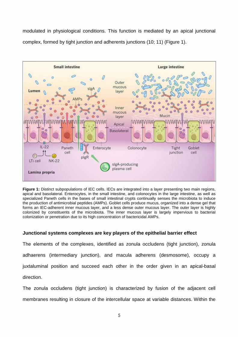
modulated in physiological conditions. This function is mediated by an apical junctional
complex, formed by tight junction and adherents junctions (10; 11) (Figure 1).
Figure 1: Distinct subpopulations of IEC cells. IECs are integrated into a layer presenting two main regions, apical and basolateral. Enterocytes, in the small intestine, and colonocytes in the large intestine, as well as specialized Paneth cells in the bases of small intestinal crypts continually senses the microbiota to induce the production of antimicrobial peptides (AMPs). Goblet cells produce mucus, organized into a dense gel that forms an IEC-adherent inner mucous layer, and a less dense outer mucous layer. The outer layer is highly colonized by constituents of the microbiota. The inner mucous layer is largely impervious to bacterial colonization or penetration due to its high concentration of bactericidal AMPs.
Junctional systems complexes are key players of the epithelial barrier effect
The elements of the complexes, identified as zonula occludens (tight junction), zonula
adhaerens (intermediary junction), and macula adherens (desmosome), occupy a
juxtaluminal position and succeed each other in the order given in an apical-basal
direction.
The zonula occludens (tight junction) is characterized by fusion of the adjacent cell
membranes resulting in closure of the intercellular space at variable distances. Within the

6
closed region, the dense outer leaflets of the adjoining cell membranes converge to form a
single intermediate line. A diffuse layer of dense cytoplasmic material is often associated
with this junction and its development varies from one epithelium to another.
The zonula adhaerens (intermediate junction) is characterized by the presence of an
intercellular space (~200 Å) occupied by homogeneous, apparently amorphous material of
low density; by strict parallelism of the adjoining cell membranes over distances of 0.2 to
0.5 µm; and by conspicuous bands of dense material located in the subjacent cytoplasmic
matrix.
The desmosome or macula adhaerens is also characterized by the presence of an
intercellular space (~240 Å) which, in this case, contains a central disc of dense material;
by discrete cytoplasmic plaques disposed parallel to the inner leaflet of each cell
membrane; and by the presence of bundles of cytoplasmic fibrils converging on the
plaques (12-14) (Figure 2).
Figure 2: scheme of adherens junctions main components

7
Tight junctions are multi-protein complexes composed of transmembrane proteins,
peripheral membrane (scaffolding) proteins and regulatory molecules including kinases
(Figure 3).
Tight junctions limit solute flux along the paracellular pathway, which is typically more
permeable than the transcellular pathway. Tight junctions have also the role of rate-limiting
step in transepithelial transport and are the principal determinant of mucosal permeability.
Thus, it is important to understand the specific barrier properties of the tight junction, which
can be defined in terms of size selectivity and charge selectivity.
A second pathway is characterized by small pores that are thought to be defined by tight
junction-associated claudin proteins, which are also primary determinants of charge
selectivity. These pores have a radius that excludes molecules larger than 4 Å.
The expression of claudins varies between organs and even within different regions of a
single organ and, as detailed below, can be modified by external stimuli, such as
cytokines. Thus, tight junctions show both size selectivity and charge selectivity, and these
properties may be regulated individually or jointly by physiological or pathophysiological
stimuli.

8
Figure 3: Tight Junctions structure and mayor components
At present, the most well-studied tight junction proteins are the claudins, a large family that
includes at least 24 members (15-16). These proteins have four transmembrane helices
with a very short intracellular amino-terminal sequence and a somewhat longer C-terminal
tail. The first extracellular loop is approximately 50 residues long, although there is some
variation among claudin family members. More importantly, sequence variation within the
first extracellular loop determines tight junction charge selectivity (17), consistent with the
view that the array of claudin proteins expressed in a given cell type defines the
paracellular pore (18; 19) through which ions and perhaps nonionic solutes travel. The
second extracellular loop of claudins is smaller than the first, ranges from 16 to 33 amino
acids in length, and is poorly characterized. The cytoplasmic C-terminal tail varies in length
from 21 to 63 residues and is the least conserved region of claudin proteins, suggesting
that the tail may also be a site of functional regulation. Importantly, almost all claudin
proteins present a three-aminoacid terminal motif that binds to PDZ domains (Figure 4).
Tight junction proteins panel include ZO-1 as well as the related proteins ZO-2 and ZO-3
(20); each of these proteins contains three PDZ domains within the amino-terminal portion

9
of the protein. PDZ domains, which were initially identified in PSD95, DlgA, and ZO-1, are
specialized for protein interactions. In the case of ZO-1, ZO-2, and ZO-3, the most
aminoterminal PDZ domain binds to the C-terminal tail of claudins (21). This is functionally
critical because deletion of the PDZ-binding motif prevents efficient claudin targeting to the
tight junction. Moreover, ZO-1 and ZO-2 are each able to direct claudins to developing
tight junctions (22); simultaneous elimination of ZO-1 and ZO-2 expression blocks claudin
recruitment and tight junction formation with consequent lost of barrier function (23).
Several other groups of proteins, including transmembrane, cytoskeletal, and signaling
proteins (24) complete the tight junction complex; in particular actin and myosin are
essential structural elements of the tight junction (25).
Figure 4: PDZ domain interactions and the regulation of EMT and Wnt signalling. Schematic diagram of some of the critical regulators involved in the regulation of the canonical and non-canonical Wnt signalling pathways. Interlinked with this are PDZ interactions important in the regulation of EMT. Proteins possessing PDZ domains are shown in red and those possessing PBMs are shown in blue. β-CAT, β-catenin; CK2, casein kinase 2; GSK-3β, glycogen synthase kinase 3β; PCP, planar cell polarity; TCF, T-cell factor; WGEF, weak-similarity GEF.

10
The best-characterized example of tight junctions regulation in response to physiological
stimuli is represented by the reversible increase in intestinal paracellular permeability
induced by apical Na+/nutrient co-transport (26; 27); increase in paracellular permeability is
thought to allow passive paracellular flux of nutrients, along with water. Ultrastructural
analyses of tight junctions showed that activation of Na+/nutrient co-transport induced
condensation of perijunctional microfilaments (28), interpreted as an indicator of
actomyosin contraction. Unfortunately no tools needed to study these biochemical events
in intact tissue are still developed. To solve problem, an in vitro model that simulate
Na+/glucose co-transport–induced tight junction regulation was developed through use of
cultured intestinal epithelial cell monolayers (29). Based on the ultrastructural data,
phosphorylation of myosin II regulatory light chain (MLC), a trigger for actomyosin
contraction, was examined and shown to increase following activation of Na+/glucose co-
transport (30). Moreover, MLC-kinase (MLCK) inhibition prevented both MLC-
phosphorylation and barrier regulation induced by Na+/glucose co-transport in cultured
monolayers and also blocked barrier regulation in intact intestinal mucosa (31). These data
suggest that MLCK is a critical physiological regulator of tight junction permeability.
Furthermore, in vitro studies of epithelial cells with inducible expression of constitutively
active MLCK demonstrate that enzymatic MLCK activation is sufficient to trigger
downstream events necessary for barrier regulation (32).
Immune stimulation and barrier regulation: the fund amental role of apoptotic
signals
The ability of cytokines, such as TNF and IFNγ, to regulate the function of the tight junction
(33) barrier was first described 20 years ago. Since then, increased tight junction protein
transcription, vesicular removal of proteins from the tight junction, tight junction protein
degradation, kinase activation and cytoskeletal modulation have all been proposed to
mediate cytokine-induced loss of tight junction barrier function (34). Although extensive

11
apoptosis of epithelial cells may also cause barrier loss, the relevance of single-cell
apoptosis to barrier dysfunction remains controversial owing to differing results in diverse
experimental systems.
TNF and IFNγ modify tight junction barrier function in intestinal, renal, pulmonary and
salivary gland epithelia as well as between endothelial cells. The effects of TNF on barrier
integrity have been best studied in the gut (35), where this cytokine has a central role in
many diseases associated with intestinal epithelial barrier dysfunction, including
inflammatory bowel disease, intestinal ischaemia and graft-versus host disease. For
example, although the effect of therapy with TNF-specific antibodies may be largely due to
the overall reduction in inflammation. Increased mucosal TNF production may also
contribute to increased intestinal permeability and susceptibility to colitis in mice with
defective mucin biosynthesis. MLCK has been shown to have a central role in TNF
induced epithelial and endothelial barrier deregulation, both in vitro and in vivo (36).
Similar to Na+/nutrients co-transport, TNF-induced MLCK activation seems to increase
paracellular flux through the leak pathway. Similarly, MLCK expression and activity are
increased in intestinal epithelial cells of patients with inflammatory bowel disease.
1.2 BARRIER BREAKDOWN: THE PANDORA'S BOX OPENED
Chemotherapy-induced gastrointestinal mucositis
Many anti-cancer therapeutic strategies based on radiotherapy and chemotherapy are
effective in the treatment of malignant disease but, conversely, present a plethora of side
effects including oral and gastrointestinal mucositis. There is no approved therapy to
prevent or treat chemotherapy-induced mucositis at present and the development of an
effective intervention is seen as a high priority in oncological supportive care (37-39).
Severe mucositis is especially common among patients who receive aggressive
myeloablative chemotherapy and in patients who receive radiation therapy as treatment for
cancers of the oral cavity, oropharynx, nasopharynx and salivary glands. Patients with

12
severe mucositis develop ulcerations that penetrate fully into the submucosa and cause
severe pain, which routinely necessitates narcotic analgesia.
In addition to the symptoms of mucositis and its impact on quality of life, mucositis
adversely affects a variety of other health and economic outcomes. Among patients who
have received chemotherapic treatments, the presence of mucositis results in lengthened
hospital stays, increased use of resources and higher costs. Mucositis also threatens the
efficacy of treatment plans by necessitating breaks in chemotherapy, reductions in doses
of drugs used in chemotherapy and modifications in the selection of anti-neoplastic agents.
Anthracyclines like doxorubicin are among the most widely used and effective
chemotherapeutic agents but also the use of which often causes the onset of mucositis
(especially in combination with 5-fluorouracil) (40-42).
One of the most important event that compromise the intestinal epithelium biological
functions (like nutrients absorptions and barrier effect against pathogens invasion) is the
lack of the correct permeability caused by alterations of the junctional systems (43; 44) of
epithelial cells in response to apoptotic events promoted by chemotherapeutic agents,
events like apoptosis and inflammation, which affect the tunica mucosa, the vascular
endothelium and the whole body, contribute to the onset of mucositis. One of the most
important apoptotic pathways involves caspases; in particular caspase-3 that is connected
with junctional damages; two target proteins for Tight Junctions and Adherent Junctions,
zonula occludens-1 (ZO-1) and beta-catenin respectively, are down-regulated in presence
of chemotherapy-induced apoptosis. (45; 46) Because the gastrointestinal tract present a
vast array of microorganisms, loss of epithelial integrity, especially when patients undergo
myeloablation, markedly increases the risk of bacteraemia, fungaemia and sepsis (47; 48)
Inflammatory Bowel Diseases (IBD)
Idiopathic IBDs such as Crohn disease and ulcerative colitis occur in clinically
immunocompetent individuals whose characteristic symptoms and signs arise from a

13
robust, cytokine-driven (yet noninfectious) inflammation of the gut (49). Crohn disease is
associated with excess IL-12/IL-23 and IFN-γ/IL-17 production that affects the small bowel
and colon with discontinuous ulceration and full thickness bowel wall inflammation often
including granulomas. Patients report gastrointestinal symptoms of abdominal pain,
diarrhea, and rectal bleeding as well as systemic symptoms of weight loss, fever, and
fatigue. Crohn disease patients can also develop obstructing structures of the bowel and
inflammatory connections (fistulae) (Figure 5) between segments of bowel or between the
bowel and skin and other organs. In comparison, ulcerative colitis is associated with
excess IL-13 production, primarily affecting the colon, with a continuous inflammation of
the mucosa nearly always involving the rectum and extending proximally (50).
It has been proposed that three main features should be present for IBD to occur: a
genetically susceptible mucosal immune system; an antigen, or pro-inflammatory
compound, which reaches the gut and can trigger the susceptible immune system; and an
alteration in gut barrier function which allows antigens to have contact with the mucosal
immune system (51).
Since the studies about Crohn's disease have shown immunological factors underlying
onset of this disease, the conventional treatments for IBD as corticosteroids, mesalamine,
and immunosuppressants, provide mostly to block downstream inflammatory events.
Medical therapy relies on classic anti-inflammatory and immunosuppressant drugs:
corticosteroids, mesalamine compounds, azathioprine, and derivatives of the latter. (52-
54). Newer biological drugs such as anti–TNF-α antibodies targeting the general
inflammatory cytokine, TNF-α, have added greatly to our ability to control IBD, but even
this therapy is limited by lack or loss of efficacy and associated toxicities (55). Emerging
therapies for IBD are focusing on major effector cytokines as they are identified in ongoing
investigations, for instance using an anti–IL-12p40 antibody to neutralize the effects of IL-
12 and IL-23 in Crohn disease (56).

14
Increased paracellular permeability was reported in patients with Crohn’s disease (CD)
over 25 years ago (57). Although such barrier loss may be caused by erosions that occur
in active disease, some approaches used to measure permeability partially excluded this
possibility by normalizing absorption of the inert sugar lactulose to that of the smaller, and
more easily absorbed, inert sugar mannitol (58) .
In recent years, several lines of evidence suggested that an increased intestinal
permeability play a central role in the pathogenesis of IBD. In different animal models of
Crohn's disease, an increased small intestinal permeability has been shown, even before
disease expression, and the reversal of this barrier defect can attenuate the disease,
implying that the increased permeability is not simply an epiphenomenon but rather is an
important etiological event (59-61). In the human condition it is also evident that increased
small intestinal permeability is commonly observed in populations at high risk of
developing Crohn's disease (62; 63). This would suggest that there is a subtle alteration of
function, independent of inflammation, which can be visible as an increase in paracellular
permeability. This raises the possibility that restoration of barrier function may be
therapeutic in CD Consistent with this hypothesis, emerging data indicate that inhibition of
cytoskeletally mediated barrier dysfunction may be able to prevent disease progression.
Barrier restoration may, therefore, represent a non-immunosuppressive approach to
achieving or maintain disease remission.

15
Figure 5: Classical representation of Crohn Disease lesions
1.3 SODIUM-DEPENDENT GLUCOSE TRANSPORTER-1
SGLT1: classical transporter and novel receptor. Tw o faces of the same medal
The primary functions of the gastrointestinal (GI) tract have traditionally been perceived to
be limited to the digestion and absorption of nutrients and electrolytes and to water
homeostasis but the intestinal epithelial barrier also controls the equilibrium between
tolerance and immunity to non-self-antigens by regulating antigen trafficking both through
the transcellular and paracellular pathways.
Luminal content influences intestinal epithelium function and the components of diet
certainly play a role in the modulation of enterocytes response. The large surface of the
gut is a wide area to protect and constitutes an easy way to the whole organism for
pathogen bacteria. On the other hand a so large line on external environment is necessary
to an appropriate absorption of the nutrients. Often the absorption is a transport against
gradient because the concentration of nutrient molecules is lower in the lumen than inside
the cells. For this reason a wide range of transporters are evolved in order to guarantee

16
the absorption of water and electrolytes, minerals and vitamins, sugars, fatty acids, amino-
acids and small peptides. The same molecules probably exert different effects on the role
of IECs in mucosal immunity. D-glucose, in particular, is prevalently considered as
energetic substrate and other possible roles are poorly analyzed.
Glucose is among the more abundant monosaccharide in the diet, and its absorption in the
intestine is mediated by the high-affinity, sodium-dependent glucose cotransporter (SGLT-
1), located in the apical membrane of enterocytes.
Recently SGLT-1 has been described as involved in the repair of plasma membrane
integrity and tight junction (TJ) integrity injured by heat stress or cisplatin chemo-treatment
(45; 64) In our recent papers we demonstrate the protective effect of the activation of
sodium-dependent glucose transporter-1 (SGLT-1) on damages induced by TLRs ligands
in intestinal epithelial cells, in a murine model of septic shock and in LPS-induced liver
injury, as well as liver injury and death induced by an overdose of acetaminophen (65; 66).
Intestinal glucose absorption and the role of SGLT- 1
D-glucose is the major source of energy in mammals and it is absorbed by the epithelial
cells lining the surface of the small intestine. The transport of glucose from lumen to
enterocytes in the small intestine is principally mediated by Na+-dependent glucose co-
transporters (SGLTs, members of a larger family of Na+-dependent transporters, gene
name SLC5A) that use the Na+-electrochemical gradient at the generated by the Na+/K+-
ATPase to transport glucose into cells (Figure 6)
SGLT-1 is the most important active glucose transporter at intestinal level and it is found in
brush border membrane of mature enterocytes in the small intestine. During the process of
intestinal sugar transport, on the luminal side of the brush border membrane, two Na+ ions
bind to SGLT-1 and produce a conformational change that permits sugar binding. Another
conformational change allows the substrates to enter the enterocyte. The sugar, followed
by the Na+, dissociates from SGLT-1 because the affinity of the cytosolic sites is low, and

17
also because the intracellular concentration of Na+ is low (10 vs 140 mEq/L). The Na+/K+-
ATPase in the basolateral membrane is responsible for maintaining the Na+ and K+
electrochemical gradients across the cell membrane (67-70).
Figure 6: schematic model of intestinal sugar transport. SGLT1 is the sodium dependent glucose/galactose transporter on the brush border membrane. The Na+K+-ATPase on the basolateral membrane maintains the gradient necessary for the functioning of SGLT1. GLUT2 transports glucose, galactose and fructose out of the cell. IF images of SGLT-1 in villus brush border.
SGLT-1 as a new player in epithelial homoeostasis m aintenance. Engagement with
natural and synthetic molecules.
Since 2005 SGLT-1 assumes a novel physiological role. SGLT-1 activation loads to a local
and systemic anti-inflammatory and cytoprotective response in different inflammatory
conditions. Firstly the effect of glucose has been analysed in enterocytes exposed to
agonists of TLR4 and TLR9. The encouraging in vitro results prompted us to analyze the
effects of glucose on a systemic inflammatory response syndrome (endotoxic shock) in
mice (Figure 7).

18
Figure 7: schematic representation of SGLT-1 activation pathways in different cases of tissue damage
Oral ingestion of glucose was found to protect 100% of mice from lethal endotoxic shock
induced by intraperitoneal LPS administration (65); protection was only observed when
glucose was administered orally, not by i.p. route, suggesting the important role of
intestinal epithelial cells. Subsequently the study investigated the possibility that orally
administered D-glucose exerts a systemic anti-inflammatory activity on hepatic liver failure.
In this study (66) that D-glucose prevents LPS-induced liver injury, as well as liver injury
and death induced by acetaminophen overdosing. In both of these models, physiological
liver morphology is maintained and organ protection was confirmed also by unchanged
levels of circulating markers of hepatotoxicity, such as ALT or LDH. In addition, D-glucose
was found to protect liver from alpha-amanitin intoxication. In this case, however, a second
signal had to be present in addition to glucose in order to achieve protective efficacy.
In addition, we observed that the in vivo protection depends from a systemic increase of
anti-inflammatory cytokine IL- 10. The milestone of the observed immunomodulatory
effects resides in activation of SGLT-1; in fact, the glucose analogue 3-OMG, which
induces the transporter activity but is not metabolized, exerted the same effects as glucose
both in vitro and in vivo. Unfortunately 3-OMG is active at the same high concentration of

19
D-glucose and in order to solve this problem we decided to screen a library of synthetic
glucose analogues as ligands for our experiments.
BLF501: origin of the synthetic compound
In the search for non-metabolizable glucoderivatives able to “activate” the SGLT-1
transporter at pharmacological concentrations, the chemical researcher team generated a
small library of naphthyl C-glycoside and glycitol derivatives, mostly without the use of
protecting groups. We tested in vitro anti-inflammatory activity of different compounds,
among them, we have selected a synthetic SGLT-1 ligand, named BLF501 (figure 8),
which binds to the transporter with high potency, i.e. at concentrations 5 orders of
magnitude lower than glucose.
O
HO
HO
OH
HO
NH
S OO
NMe2
OOMe
HO
HO
OH
HO
O
HO
HO
O
HO
OH
O
HO
HO
OH
HO
NH2
1 2
BLF501
a b c
1
23
4
5
6
1'
2'
Figure 8: Synthesis and Structure of BLF501
BLF501, a member of the C-glycoside class, was designed to resist glucose metabolism
and to retain hydrolytic stability in the acidic and basic conditions found in the
gastrointestinal tract. Stability of BLF501 in different pH conditions was tested in samples
prepared by dissolving 2.3 mg in 550 µl of D2O and adjusting the pH to 1.2 through DCl
addition (acidic conditions) or by dissolving 3 mg of BLF501 in 550 µl of D2O and adjusting
the pH to 12.5 through NaOD addition (basic conditions). Both samples were kept at room
temperature until 1H-NMR spectra were recorded at 10 min, 4.5 h and 24 h. As shown in
Figure 2, neither sample evidenced degradation in 24 h. BLF501 matches with the
fundamental characteristic of absence of metabolic activity. In fact in the field of C-
glycosides like BLF501 no relevant bioactivity has been reported until now (71), allowing
us to consider BLF501 a non-metabolizable glucose derivate.

20
3 mg pH 12.5 (D20, NaOD) 2.3 mg pH 1.2 (D20, DCl)
Figure 9. 1H-NMR spectra of BLF501 at pH 12.5 and at pH 1.2.
1.4 AIM OF THE STUDY:
The aim of this work is to demonstrate that SGLT-1 engagement via orally administered
ligands, in particular the synthetic one BLF501, could activate protective mechanisms at
intestinal epithelial level, preserving correct epithelial morphology and permeability and
accelerating recovery of homeostatic conditions.
ppm (f1)
2.03.04.05.06.07.08.0
10 min
4.5 h
24 h
ppm (f1)
2.03.04.05.06.07.08.0
10 min
4.5 h
24 h
ppm (f1)
2.03.04.05.06.07.08.0
10 min
4.5 h
24 h
ppm (f1)
2.03.04.05.06.07.08.0
10 min
4.5 h
24 h

21
2. MATERIALS AND METHODS:
2.1 SGLT-1 ACTIVATION IN GIM MODELS:
Chronic and acute in vivo experiments were performed on mice strain BALB/C purchased
from Charles River Italy. The mice used for the different treatments were housed in
specific aseptic conditions, with a constant temperature and humidity and received food
and water ad libitum. The animals were divided randomly into groups and assigned to two
treatment protocols.
Chronic treatment:
15 animals formed the control group; 15 animals formed the group treated with doxorubicin
and 5-fluorouracil, 15 animals formed the group treated with doxorubicin and 5-fluorouracil
and BLF501 25 µg/Kg; 15 animals formed the group treated with doxorubicin and 5-
fluorouracil and BLF501 2.5 µg/Kg; 15 animals formed the group treated with doxorubicin
and 5-fluorouracil and BLF501 0.25 µg/Kg and 15 animals formed the group treated with
BLF501 25 µg/Kg only. Administration of doxorubicin (DXR) 7 mg/kg (adriablastina, Pfizer)
and 5-fluorouracil (5-FU) 100 ng / kg (fluorouracil; TEVA), dissolved in saline solution for a
final volume of 300 µl, was carried out weekly for a total duration of three weeks by
intraperitoneal injection. The administration of BLF501 (25, 2.5 and 0.25 µg/kg in saline for
100µl total) occurred simultaneously with the chemotherapic agents by gavage using a
gastric tube.
Acute treatment:
Administration of DXR (20 mg/kg), dissolved in saline solution for a final volume of 300 µl,
was carried out at day 0 intraperitoneal injection. The administration of BLF501 (25 µg/kg
in saline for 100 µl total) occurred simultaneously with the chemotherapic agent by gavage
using a gastric tube
7 animals formed the control group; 14 animals formed the group treated with DXR; 14
animals formed the group treated with DXR and BLF501; 14 animals formed the group

22
treated with BLF501. Seven mice from all treated groups were sacrificed after 48 and 72h.
Control group was sacrificed after 72h. All the animals of acute treatment received, an
hour prior to sacrifice, an intraperitoneal injection of bromodeoxyuridine (BrdU) to perform
evaluation of cell proliferation with immunofluorescence reaction.
Chemotherapy simulation:
Chemotherapy simulation in vivo experiments was performed on mice strain SKH-1 nude
mice purchased from Charles River Italy. The mice used for the different treatments were
housed in specific aseptic conditions, with a constant temperature and humidity and
received food and water ad libitum. A431 tumoral cells were implanted subcutaneously to
32 mice and when the tumor weight reached a mean value of 240 mg mice were divided
randomly into groups and assigned to the treatment protocols. 8 animals formed the
control group; 8 animals formed the group treated with doxorubicin 6 mg/Kg 8 animals
formed the group treated with doxorubicin 6 mg/Kg plus BLF501 25 µg/Kg; 8 animals
formed the group treated with BLF501 25 µg/Kg only. Tumor weight was registered daily.
At day 26 of treatment mice were sacrificed.
Processing of samples
After collection, the samples were fast separated in small pieces and fixed in 10% formalin
with 2% sucrose for 4 hours at 4 ° C. Then we proce eded with 2 rinses in PBS half-hour
after each and were placed in 70% ethanol for at least 12 hours, finally the samples were
dehydrated by successive steps in an ascending scale of alcohols, placed in xylene for 2
hours and finally embedded in paraffin.
Staining with hematoxylin and eosin
Hematoxylin-eosin staining was performed on sections obtained from paraffin-embedded
samples. The samples were deparaffinized in xylene, rehydrated through descending
scale of alcohol, left for 10 minutes in hematoxylin, washed under running water and then
placed in eosin for 3 minutes, finally dehydrated through ascending scale of alcohols and

23
xylene in then mounted with coverslips using Entellan (Merck, Darmstadt, Germany). The
observation of the samples and photographs were taken with a microscope Nikon Eclipse
80 with a digital camera Nikon DS-L1.
ZO-1 Immunofluorescence
Samples sections were deparaffinized in oven at 60 ° C for 30 minutes, placed in xylene
and rehydrated. It was then performed a quick rinse with 0.1 M Tris/HCl pH 7.4 after which
the sections were incubated with proteinase K 20 g/ml Tris/EDTA buffer pH 8 for 15
minutes at 37 ° C. It was subsequently made a furth er washing with Tris/HCl after the
sections were permeabilized with 1% Triton X-100 in Tris/HCl for 5 minutes at room
temperature. Slides were washed with Tris/HCl and then sections were blocked with a
solution containing 1mM Hepes, 2% goat serum, 1x HBSS and 0.5% Triton X-100 for one
hour at room temperature. Sections were incubated with primary antibody α-ZO-1
(Invitrogen, Camarillo, CA) 1 5 g / ml in Tris/HCl overnight at 4 ° C. Samples were washed
for three times with Tris/HCl and 0.01% Triton X-100. After washing, the sections were
incubated with secondary antibody goat anti-rabbit conjugated to tetramethyl-rhodamine
isothiocyanate (TRITC) diluted 1:1000 in Tris/HCl for 45 minutes at room temperature.
We proceeded with further washes for 5 minutes with Tris/HCl + 0.01% Triton X-100, then
the sections were incubated with 20µl of enhancer per slice for 30 minutes at room
temperature. 2 more washes were carried out for 5 minutes with Tris/HCl and 0.01% Triton
X-100. Sections were incubated with DAPI 1:10000 in Tris/HCl for 5 minutes at room
temperature and then subjected to 3 washes with Tris/HCl and 0.01% Triton X-100. Slides
were mounted with Mowiol. The observation of the samples and photographs were taken
with a microscope Nikon Eclipse 80, with a digital camera Nikon DS-L1.
Beta-catenin immunohistochemistry.
The sections on slides were deparaffinized in oven at 60 ° C for 30 minutes, placed in
xylene and rehydrated It was then performed using two antigen unmasking steps

24
interspersed with 5 minutes 1 minute pause, in a microwave oven with citrate buffer pH 6 =
0.005 M. The sections were cooled for about an 30 minutes and then washed with 0.1 M
Tris/HCl pH 7.4 + 0.025% Triton X-100 (three washes of 5 minutes each). Then was
carried out the endogenous peroxidase inhibition with a solution of 3% H2O2 in 0.1 M
Tris/HCl pH 7.4 for 20 minutes, after which the sections were washed with 0.1 M Tris/HCl
pH 7.4 + 0.025% Triton X- 100 (three washes of 5 minutes each). Subsequently
nonspecific sites were blocked with HHG solution (1 mM Hepes, 2% goat serum, 1X
HBSS, 0.5% Triton X-100) in Tris/HCl for 1 hour at RT. Sections were incubated with
primary antibody anti-beta-catenin (Abcam, Cambridge, UK) 1:500 in Tris/HCl + 1% BSA
for 2 hours at RT, followed by 3 washes for 5 minutes with Tris/HCl, 0.1% Triton X-100.
After washing, the sections were incubated with biotinylated secondary antibody goat anti-
rabbit 1:1000 diluted in 0.1 M Tris/HCl pH 7.4 + 0.025% Triton X-100 for 45 minutes at
room temperature. After washes with 0.1 M Tris/HCl pH 7.4 + 0.025% Triton X-100
sections were incubated with ABC-kit and DAB (Vector, Burlingame, U.S.), the
development has been blocked in distilled H2O. Sections were counterstained with
hematoxylin for 10 minutes RT and finally dehydrated through ascending scale of alcohols
and xylene, then mounted with coverslips using Entellan (Merck, Darmstadt, Germany).
The observation of the samples and photographs were taken with a microscope Nikon
Eclipse 80th, with a digital camera Nikon DS-L1.
Immunofluorescence for BrdU
The sections on slides are deparaffinized in oven at 60 ° C for 30 minutes, placed in
xylene and rehydrated sections were rinsed in PBS and incubated with 2N HCl for 30
minutes at RT and in Na2B4O7 for 1o minutes. Sections were incubated with PBS/3% BSA
for 20 minutes at RT and then with proteinase K 20 g/ml in TrisEDTA buffer pH 8 for 15
minutes at 37°C. After three washes in PBS/BSA 3% s ections were incubated with primary
antibody α-BrdU (Novocastra) 1:200 in PBS/3% BSA for 1 hour at RT, washed with

25
PBS/3% BSA and then incubated with secondary antibody goat anti- mouse Alexa Fluor
488 1:500 in PBS/BSA 3%. Slides were washed with PBS/BSA 3% and with
Tris/HCl/0.01% Triton X-100. Sections were incubated with 1:2500 DAPI in Tris/HCl for 5
minutes at room temperature and then subjected to 3 washes with PBS / BSA 3%. Slides
were mounted with Mowiol.
Real-Time polymerase-chain reaction (PCR)
Real-Time PCR experiments were performed in accordance with manufacturer’s
instructions using a 7900HT Fast Real Time PCR System (Applied Biosystems, Foster
City, CA, US). Primers for SI; TFF3; DLL1, and beta-actin were purchased from Applied
Biosystems.
Western Blots
The expression of Akt/P-Akt, Caspase-3 and Ezrin was assessed by analyzing the total
protein extract from small intestine samples taken from differently treated animals. The
protein extracts were quantified using the BCA method (BCA Protein Assay Kit, Pierce,
Rockford, IL). Proteins (30 µg) were fractionated on a polyacrylamide gel (BIO-RAD Labs,
Hercules, CA) containing 0.1% to 8% of SDS and transferred onto a nitrocellulose filters
(American Biosciences, Buckinghamshire, England) by electroblotting. Filters were
incubated for 1 hour in TBS (8% NaCl, 3% Tris) containing 1% Tween-20 and 5% milk
powder to block nonspecific binding sites. For the various reactions following antibodies
were used:
Protein Primary Ab Primary Ab dilution
Solvent solution Reaction conditions
Akt monoclonal anti-mouse Akt
1:1000 TBS + 1% Tween-20 +5% milk O/N at 4°C
P-Akt monoclonal anti-mouse P-Akt
1:1000 TBS + 1% Tween-20 +5% milk O/N at 4°C
Caspase-3 policlonal rabbit anti-mouse caspase-3
1:500 TBS O/N at 4°C
Ezrin monoclonal anti-mouse Ezrin
1:1000 TBS O/N at 4°C
Beta-actin policlonal rabbit anti-mouse beta-actin
1:1000 TBS + 1% Tween-20 +5% milk O/N at 4°C
Table 1: primary antibodies used to perform WB assays

26
After three rinses with TBS containing 1% Tween-20, the filters were incubated for 1 hour
at room temperature with secondary antibody: rabbit anti-peroxide (Vector Laboratories)
diluted 1:1000 in TBS with Tween-20 to 0.1% and 5% milk. Following three rinses were
performed with TBS containing 1% Tween-20 and a rinse in TBS. The bands were finally
visualized using the ECL ™ Western Blotting Detection Reagents and plates by
autoradiography (American Biosciences).
Cell cultures
Cell lines Caco-2/bbe and Caco-2/bbe permanently transfected with SGLT-1 (kind gift of
Prof. Mark Donowitz, MD, Johns Hopkins University School of Medicine, Baltimore) were
cultured in DMEM high-glucose medium (4,5g/L) (Euroclone, Pero, Italy) supplemented
with 10% FBS (Euroclone), 1% glutamine (Euroclone), 15 nM sterile HEPES solution
(Euroclone). For Caco-2/bbe/SGLT1 cells, 250 µg/ml G418 gentamicin bisulfate salt
solution (Sigma-Aldrich) was added as antibiotic agent; for normal Caco-2/bbe
penicillin/streptamicin solution (Euroclone) was added.
A431 cells were cultured in RPMI 1640 medium (Euroclone) supplemented with 10% FBS
(Euroclone), 1% glutamine (Euroclone) and 1% penicillin/streptamicin solution (Euroclone).
Cell treatments
For viability assays 1 x 10⁵ Caco-2 cells were plated on 96 flat-bottom well plates and
treated after 24 hours with doxorubicin (100 µM) with or without BLF501 11.4 µM. Cell
viability was evaluated after 48 h with Neutral Red assay.
2.2 SGLT-1 ACTIVATION IN IBD MODELS
Mice and in vivo treatments
C57BL/6 female mice were purchased from Charles River, Italy (Head Office Wilmington,
MA). Mice were housed under specific pathogen-free conditions, maintained at constant
temperature and humidity, with food and water given ad libitum, and used at 8-12 weeks of
age. Experimental protocols were approved by the Ethics Committee for Animal

27
Experimentation of Istituto Nazionale Tumori, Milano, and carried out according to
guidelines of the United Kingdom Co-ordinating Committee on Cancer Research for
animal welfare in experimental neoplasia (1998).
Induction of acute and chronic colitis
Mice weighting 20-22 g received 2% dextran sodium salt (m. m. 40 kD, MP Biomedicals,
Irvine, CA) ad libitum in filter-purified drinking water for 7 days. For treatments studies
mice undergoing DSS from day 4 at day 7 received oral administration of BLF501 250
µg/kg or 25 µg/kg or 2.5 µg/kg once day. BLF501 was administrated with a sterile gastric
tube In other mice groups received only BLF501 250 µg/kg or 25 µg/kg or 2,5 µg/kg or
glucose 2,5 g/kg. One group received only DSS 2% in drinking water.
Day 1 Day 2 Day 3 Day 4 Day 5 Day 6 Day 7 Day 8 DSS 2% DSS 2% DSS 2% DSS 2%
+BLF501 DSS 2% +BLF501
DSS 2% +BLF501
DSS 2% +BLF501
Sacrifice
Table 2 . Schematic presentation of different treatments in the time. Mice received DSS 2% in drinking water. BLF501 250 µg/kg or 25 µg/kg or 2,5 µg/kg were administrated (diluted in physiological solution) once per day at day 4, 5, 6, 7 and at 8th mice were sacrificed. Groups Treatment 1 Untreated 2 DSS 2% only 3 DSS 2% + BLF501 250 µg/kg 4 DSS 2% + BLF501 25 µg/kg 5 DSS 2% + BLF501 2,5 µg/kg 6 BLF501 250 µg/kg 7 BLF501 25 µg/kg 8 DSS 2% + physiological solution
Table 3. Schematic representation mice groups. 5 mice/groups.
To induce chronic colitis four groups of 10 mice/groups mice received three cycles of DSS
2%. Each cycle consists of 2% DSS dissolved in filter-purified drinking water for 7 days,
followed by a 14 days interval with normal water administration. After ten days from the
last DSS cycle we started different treatments.
The first group of animals (control group) received normal drinking water only, during the
whole time of the experiment. The second group of 12 mice received only three cycles of

28
treatment with DSS, as previously described, and no any further treatment. Four mice of
this group were sacrificed one week after DSS cycle completion; four mice of this group
were sacrificed two weeks after DSS cycle completion; and 4 mice were sacrificed after
three weeks.
After DSS cycles completion, third mice group received oral administration of 25 µg/kg
BLF501 four times/week for one week; forth received 25 µg/kg BLF501 four times/week for
two consecutive weeks; and fifth group received 25 µg/kg BLF501 four times/week for
three consecutive weeks.
Groups Treatments 1 Untreated 2 DSS 2% +physiological solution 3 DSS 2%+ BLF501 25 µg/kg for 1 week 4 DSS 2% + BLF501 25 µg/kg for two week 5 DSS 2% + BLF501 25 µg/kg for three weeks
Table 4. Schematic representation mice groups. 12 mice in group 1. In goups 2, 3, 4 and 5 10 mice/group.
TREAT.
DSS
H2O
DSS
H2O
DSS
H2O
BLF501 25 µg/kg
Death Group3/2
BLF501 25 µg/kg
Death Group4/2
BLF501 25 µg/kg
Death Group5/2
N days 7 14 7 14 7 10 7 X 7 X 7 X
Table 5 Schematic presentation of different treatments time. N days is for number days of water DSS 2% added or water only administration. After ten days of the last DSS cycle BLF501 was orally administrated 4 times/week for 1 or 2 or 3 consecutive week. X is for sacrifice day.
Evaluation of intestinal inflammation in acute and chronic colitis
Acute colitis was scored daily using standard parameters that includes evaluation of body
weight and stool consistence. Body weight of mice was registered daily at 5.00 p.m.. Stool
consistence was evaluated daily, but comparative analysis between different groups was
conducted at the end of experiments.
Score Stool consistence Blood
0 Normal No visibile 1 Soft but still formed No visibile 2 Very soft Blood traces in stool visible 3 Diarrea Rectal bleeding
Table 6. Scoring system for the comparative analysis of stool consistence.

29
At the end of treatments the colon of mice was cut close to the ileo-cecal valve and
rectum, and the length was measured with and .for acute colitis experiment photos were
captured.
Ussing Chamber analysis..
In order to measure the colon permeability in 5 mice/group, organ segments were
mounted between the two chambers of a Ussing System (0.125 cm2 opening). Two
calomel voltage-sensitive electrodes and two Ag-AgCl current passing electrodes (EVC-
4000 World Precision Instrument Inc., Sarasota, FL) were connected to the Ussing
chamber via agar bridges. Both the mucosal and serosal sides of the chamber were
connected to sterilized circulating reservoirs containing 10 ml of oxygenated Krebs buffer
(115 mM NaCl, 8 mM KCl, 1.25 mM CaCl2, 1.2 mM MgCl2, 2 mM KH2PO4, and 225 mM
NaHCO3; pH 7.35). The buffers were maintained at 37°C by a heated water jacket and will
circulate by a gas lift column of 95% oxygen/5% CO2. Glucose (5.5 mM) was added to the
serosal and mucosal sides. Colonic membrane mounted in the Ussing chamber, the
system was allowed to stabilize for 20 minutes, in order to test the system functionality and
the integrity of the colonic mucosal membrane. Trans-epithelial electrical potential
difference in millivolt across the mucosal membrane was measured directly, while the
trans-membrane resistance was calculated indirectly as ohms x cm2, using Ohm's law.
Histological evaluation of colitis severity.
Murine colon specimens of 5 mice/group will be fixed in 10% neutral buffered formalin,
embedded in paraffin, sectioned at 4 µm and collected on xilanized slides.
Histopathological analysis using hematoxylin-eosin-stained sections of distal colon
samples of mice were performed. Samples were observed with a Nikon Eclipse 80i
microscope equipped with a digital Nikon DS-L1 camera. To quantify/evaluate acute and
chronic colitis-associated histological alteration in the colon we used a scoring system,
provided in table 7.

30
Score Histologic changes 0 No evidence of inflammation 1 Low level of inflammation with scattered infiltranting mononuclear cells 2 Moderate inflammation with multiple foci 3 High level of inflammation with increate vascular density and marked wall thickening 4 Maximal severity of inflammation with transmural leukocyte infiltration and loss of globet cells.
Table 7. Scoring system for inflammation associated histological changes in the colon
Immunofluorescence analysis.
Murine colon specimens were fixed in 10% neutral buffered formalin, embedded in
paraffin, sectioned at 4 µm and collected on xilanized slides; samples were deparaffinized,
rehydrated, and incubated for 10 minutes at 37 °C i n a humidified chamber with protease
type XIV 2 mg/ml (from Sigma) in Tris/HCl; samples were then incubated with glycine 0,1
M for 20 minutes at room temperature, washed with Tris/HCl + Triton X-100 0.01%,
incubated with NaBH4 0.5mg/ml for 20 minutes at room temperature, washed with Tris/HCl
+ Triton X-100 0.01%, incubated with Image-IT FX signal enhancer (Invitrogen) for 30
minutes, blocked with 2% goat serum for 20 minutes at room temperature and incubated
with rabbit anti-occludin or anti-ZO-1 antibody 4 µg/ml (both from Invitrogen). Incubation
with secondary antibody 546 Alexa conjugated goat anti-rabbit and staining of the nuclei
with DAPI were performer. Samples were observed with a Nikon Eclipse 80i microscope
equipped with a digital Nikon DS-L1 camera.

31
3 RESULTS
3.1 SGLT-1 activation exerted by BLF501 accelerates recovery from the alterations
of the small intestine induced by a single administ ration of DXR.
The effect of BLF501 on alterations of the small intestine induced by DXR was evaluated
in mice treated with DXR alone (20mg/kg i.p., n=14); DXR plus BLF501 (25ug/kg BLF501
n=12); BLF501 alone (n=14) or left untreated (n=7). Half of the mice were sacrificed after
48 hours and the others after 72 hours.
Macroscopic examination of small intestine samples collected at 48 or 72 hours did not
reveal morphological alterations. On the other hand, determination of the proliferation rate
of crypt cells rate, performed by immunofluorescence BrdU assay, revealed a significant
reduction of proliferation in DXR-treated mice (1.3 ± 0.2 % at 48 h; 0.8 ± 0.6 % at 72h,
untreated 2.3 ± 2.2 % (72h); p=0.0086 and p=0.0229 respectively), that was absent in
mice treated simultaneously with DXR and BLF501 (DXR + BLF501 3.3 ± 0.5 % and 2.2 ±
0.5 % at 48 and 72 h respectively; p=0.004 and p=0.028 vs DXR at 48h and 72h,
respectively) (Figure 10). No modification of the proliferation of intestinal epithelial cells
was observed in samples from mice treated with BLF501 alone.

32
Figure 10: BrdU immunofluorescence for evaluation of cellular proliferation (A) UNTR, untreated; (B) BLF501 25 µg/Kg (C) DXR 48h; (D) DXR + BLF501 25 µg/Kg 48h; (E) DXR 72h (F) DXR + BLF501 25 µg/Kg 72h. Statistical analysis: DXR+BLF501 25 µg/Kg 48h vs DXR 48h ** P=0.004; DXR+ BLF501 25 µg/Kg 72h vs DXR 72h * P=0.0282
Treatment with BLF501 was also found to restore the expression of beta-catenin, a unique
intracellular protein functioning as an integral component of the cell-cell adhesion complex
and a principal signaling protein mediating canonical Wnt signaling linked with cellular
proliferation. In fact, expression of beta-catenin was reduced at 48 hours after DXR
administration in the villi and at 72 hours in both villi and crypts (Figure 11).

33
Figure 11: Immunohistochemistry for the expression of beta-catenin in a model of acute mucositis. (A-B ) UNTR, untreated; (C-D) BLF501 25 µg/Kg (E-F) DXR+5-FU 48h; (G-H) DXR+5-FU + BLF501 25 µg/Kg 48h; (I-L) DXR+5-FU 72h (M-N) DXR+5-FU +BLF501 25 µg/Kg 72h. Bars = 20 µm.
The protective effect of BLF501 was then investigated by analyzing the expression of
different genes implicated in the early response to tissue injury. In particular, we analyzed
the expression of DLL-1, a marker for crypt cells that are in active proliferation like stem
cells; TFF-3 and beta-actin, which are components of the mucus layer and cytoskeleton,
respectively, and whose reduced expression mirrors decreased mucin production and
alteration of cytoskeletal structure, respectively; sucrose isomaltase, a marker for brush
border integrity and a key enzyme in carbohydrate metabolism, whose reduced expression
mirrors epithelial damage and malabsorption of nutrients. Expression of all of these
markers was reduced 48 hours after DXR treatment, but was similar to control mice in
animals that had been co-administered BLF501 (Figure 12).

34
Figure 12: Real-Time PCR analysis of changes in gene expression (A) DLL-1: (B) TFF-3; (C) Beta-actin; (D) Sucrase Isomerase;
3.2 BLF501-induced recovery from injuries to the mu cosa of the small intestine
induced by repeated treatments with DXR and 5-FU.
In the following, we evaluated the effect of BLF501 in a model of DXR and 5-FU-induced
mucositis that mimics medium-term, chemotherapy-induced effects on the intestinal
mucosa. Intestinal epithelia from mice treated with the two chemotherapeutics were
extensively damaged (Figure x). In particular, villi were atrophic, fused and reduced in
height (-36.15 ± 2.56 % vs untreated); epithelial cells were hyperplastic and brush borders
had large areas of erosion; focal ectasia of chyliferous vessels was detectable, numbers of
goblet cells were decreased (DXR+5-FU 2.56 ± 1.28 %; untreated 7.13 ± 0.64 %), cells
undergoing mitosis and cellular infiltrates rich in lymphocytes and plasma cells were also
observed. Mice treated also with 25µg/Kg BLF501 showed substantial recovery from

35
chemotherapy-induced injury to the intestinal mucosa (villus eight: -12.31 ± 1.58 %; p =
0.0014 vs untreated; globet cells: 6.93 ± 0.63 %; p = 0.0383 vs untreated ). At 2.5 µg/Kg,
BLF501 improved morphological parameters (villus height: -16.82 ± 1.33 % , p = 0.0026 vs
untreated), but was ineffective against loss of globet cells (2.2 ± 1.72 %) and appearance
of mitotic cells; the 0.25 µg/Kg dosage of BLF501 was ineffective on all investigated
parameters. Samples from the intestinal epithelia of mice treated with BLF501 alone were
identical to those of control mice. A synopsis of these results is shown in Figure 13.
Figure 13: Hematoxilin/eosin staining of jejunum samples: (A) UNTR, untreated; (B) DXR+5-FU; (C) DXR+5-FU + BLF501 2.5 µg/Kg; (D) DXR+5-FU +BLF501 25 µg/Kg; (E) BLF501 25 µg/Kg; (F) DXR+5-FU luminal bacterial content. (G) Synoptic panel of evaluated parameters. Statistical analysis: villus length DXR+5-FU +BLF501 25 µg/Kg vs DXR+5FU ** p=0.0014. DXR+5FU +BLF501 2.5 µg/Kg vs DXR+5-FU ** p=0.0026. Percentage of goblet cells: DXR+5-FU +BLF501 25 µg/Kg vs DXR+5-FU * p=0.0383. Damage score legend: +++= SEVERE; ++= MILD; += LIGHT; 0= ABSENT. Bars = 20 µm.

36
We also analyzed the junctional systems of the intestinal epithelia of mice treated with the
chemotherapeutics alone or with BLF501. In particular, we investigated the expression of
ZO-1, which mirrors the integrity of tight junctions and beta-catenin, a component of
adherens junctions. Immunofluorescent and Immunohistochemical staining of junctional
systems revealed respectively that ZO-1 and beta-catenin were less expressed and had
an altered distribution in intestinal samples from DXR/5-FU-treated mice. In contrast,
samples from mice that were co-administered with BLF501 at 25 µg/Kg showed the typical
honeycomb distribution of ZO-1 and expression/ distribution of beta-catenin similar to
control mice. The 2.5 µg/Kg BLF501 dose was less effective. BLF501 (25 µg/Kg) alone did
not alter the expression or distribution of either ZO-1 or beta-catenin (Figure 14)
Figure 14: ZO-1 immunofluorescence assay: (A) UNTR, untreated; (B) DXR+5-FU; (C) BLF501 25 µg/Kg; (D) DXR+5-FU +BLF501 2.5 µg/Kg; (E) DXR+5-FU + BLF501 25 µg/Kg. Magnification 60X Immunohistochemistry reaction for beta-catenin expression in a model of chronic mucositis. (F) UNTR, untreated; (G) DXR+5-FU; (H) DXR+5-FU + BLF501 2.5 µg/Kg; (I) DXR+5-FU +BLF501 25 µg/Kg; (L) BLF501 25 µg/Kg.

37
3.3 BLF501 reduces the expression of specific marke rs for epithelial damages
connected with chemotherapy-induced mucositis.
It’s possible to appreciate that BLF 501 could restore the proper expression of a panel of
proteins involved in cellular response after treatment with chemotherapic agents like
doxorubicin and 5-fluorouracil. Akt is involved in cellular response events after a generic
damage and, in the specific case, after treatment with 5-FU. Western Blots for Akt and P-
Akt indicated that this protein is strongly expressed after treatments with doxorubicin and
5-fluorouracil. BLF501 treatment reduced Akt/ P-Akt levels in a dose-dependent manner.
Caspase-3 is a marker for apoptosis and its expression is increased by chemotherapic
treatment. BLF501 co-administration only at 25 µg/Kg dosage was effective to reduce
Caspase 3 expression. Ezrin is a protein involved in tight junctions and adherent junctions
remodelling and functional management; over-expression of Ezrin induces TJ and AJ
opening and an alteration of intercellular permeability. Doxorubicin and 5-fluorouracil
chronic administration induced Ezrin over-expression and BLF501 restored low levels of
this protein in dose-dependent manner (Figure 15). BLF501 at 0.25 µg/Kg dose was
completely ineffective (data not shown). Beta-actin assay was performed to assess
uniformity of proteins concentration.

38
Figure 15 : western blot assay for epithelial damage markers.
3.4 Oral administration of BLF501 does not interfer e with the antineoplastic activity
of DXR.
Overexpression of SGLT-1 is one survival strategy put in place by several tumoral cellular
types, in particular EGFR-positive ones (31). To evaluate whether oral administration of
BLF501 could interfere with the antitumor activity of the chemotherapeutics, we performed
experiments in athymic mice that were injected subcutaneously with A431 cells, which
strongly express both EGFR and SGLT-1 (32). Tumor growth rate in mice co-administered
with DXR and BLF501 was similar to that in mice treated with DXR alone (p=0.1836). This
shows that oral administration of BLF501 does not interfere with the antitumor activity of
DXR (Figure 7). Interestingly, while DXR-treated mice showed an average reduction of 1g
of body weight at the end of the experiment, a slight increase in the weight (~0.4 g) was
observed in the group treated with DXR and BLF501. No differences in tumor growth rate
were observed between untreated mice and mice treated with BLF501 alone.

39
Figure 16. Evaluation of tumor growth rate and body weight rate in simulated chemotherapy experiments.
3.5 SGLT-1 activation in stably transfected Caco-2/ bbe cells is fundamental for
cytoprotection after doxorubicin plus BLF501 challe nge.
BLF501 is a selective ligand for SGLT-1 (71) and in vivo experiments confirm the
biological activity of the synthetic molecule. Despite this animal model is a complex system
and BLF501 interaction with others targets could be not excluded. In vitro experiments
were performed to assess SGLT-1 activation ability to maintain normal cells viability rates
after DXR treatment and to further demonstrate the unique and selective functional
interaction between SGLT-1 and BLF501. Caco-2/bbe cells stably transfected with SGLT-1
were treated with DXR with or without BLF501 co-administration for 48 hours.
Dose-response Neutral Red cell viability assays were performed in order to determine
DXR cytotoxic dose and BLF501 effective concentration (data not shown). Selected doses
for DXR and BLF501 were 100 µM and 11.5 µM respectively and they maintained used for
all in vitro experiments. Not-transfected Caco-2/bbe cells were used as negative control, in
order to confirm and clarify the pivotal role of SGLT-1 in protective action.

40
Neutral red viability assay was performed 48 hours after treatments; not-transfected Caco-
2 cells resulted sensitive to DXR (viability rate reduced of 74% vs untreated cells) and
BLF501 co-administration was ineffective to preserve cells viability (viability rate reduced
of 75% vs untreated cells). Stably transfected Caco-2/bbe cells resulted sensitive to DXR
treatment (viability rate reduced of 29% vs untreated cells) and BLF501 co-administration
preserves cells viability.
Figure 17. Neutral Red viability assays on transfected and not-transfected Caco2 cells

41
3.6 BLF501 protection against inflammatory bowel di sease in vivo. Oral
administration of BLF-501 protects mice from DSS-in duced diarrhoea.
In vitro and vivo results in GIM model lead us to hypothesize that SGLT-1 may be an
important target in intestinal diseases involving alteration of epithelial barrier effect. We
tested BLF501’s activity in a chemically induced mouse model of acute and chronic
intestinal inflammation choosing a dextran sodium sulphate (DSS)-induced colitis model
for its simplicity and reproducibility of the colonic lesions. DSS polymers are directly toxic
to the gut epithelial cells, affecting firstly the integrity of the mucosal barrier and then
stimulating local inflammation as a secondary phenomenon. For this reason we can
improve BLF.501 activity in intestinal barrier protection, which is thought to be the initial
inciting event in many intestinal disorders, including IBD.
A DSS concentration of 2% (w/v) addicted in the drinking water for 7 days induced strong
colitis. To evaluate the effects of BLF-501 in this model, mice (10/group) were treated in
the drinking water for 7 days with DSS 2% concomitantly or not with oral administration of
BLF-501 (250 or 25 µg/kg) once a day in the last four days (as described in materials and
methods). DSS intake did not differ in different groups of mice. Colitis in the DSS model is
usually associated with wasting disease. Determination of weight every day to get a rough
idea of colitis severity and is often indicative of differences in colitis development between
experimental groups. Body weight significantly decreased in DSS group compared to
control mice. The oral treatment with BLF-501 at doses of 250 µg/kg and 25 µg/kg
recovers DSS-induced weight loss; the 2.5 µg/kg dose appeared to be ineffective.

42
Figure 18. Weight rate after DSS treatment with or without co- treatment with BLF501
The DSS-induced damages are generally located in the distal colon, whose length
decreases as the disease develops. Colon mean length of mice group treated only with
DSS decrease of 32% compared to untreated ones. BLF-501 at 250 µg/kg and 25 µg/kg
oral treatments of mice significantly reduced colonic shortening induced by DSS. Group of
mice treated with BLF501 2.5 µg/kg and DSS showed colon length similar to DSS group.
About body weight and colon length results load us to consider 25 µg/kg the lower
effective dose of our analogue glucose in this animal model.

43
Figure 19. Colon length evaluation after DSS treatment with or without co- treatment with BLF501
After evaluation of colon length, colitis level of different group was scored using stool
consistence as a standard parameter. DSS treated animals group have very soft stools
with a 1,9 mean score, whereas untreated group shows normal stools with mean score 0
These results showed that oral treatment with BLF501 250 or 25 µg/kg inhibit water loss,
maintaining normal stool consistence.
* *

44
Figure 20. Stool score after DSS treatment with or without co- treatment with BLF501
The most important damage DSS-induced is characterized by changes in colon epithelium
showing loss of crypts and reduction of globet cells, signs of surface epithelial
regeneration, focal ulcerations, infiltration of inflammatory cells into the mucosa, and
oedema in the sub-mucosa. Histopathological analysis using hematoxylin-eosin staining of
colon section showed that DSS treatment induced crypt damage, ulceration, and infiltration
of inflammatory cells in the distal colon. A histological colitis score was calculated for 10
slides for each mouse of different groups; score increasing has associated with
inflammation develop (mean score of DSS group = 3.5 vs untreated = 0). Scoring system
of colon section of DSS-treated group plus BLF-501 250 or 25 µg/kg shows protective role
of SGLT-1 in this animal model of acute colitis.
*
*

45
A
B
a b c
A
A

46
Figure 21. (A) Rapresentative colonic H&E section from mice receiving water without DSS . a) Lamina mucosa; b)Lamina submucosa; c)Circular and longitudinal mussle. In magnification arrow shows colon crypte with normal morphology and black arrowhead indicates globet cells. (B) Colon histology from DSS-treated group. DSS induces thickening of the colon wall, globet cells and crypt loss in large areas (in magnification arrow). Infiltration reaching the lamina submucosa (withe arrow). (C) H&E of BLF501 250 µg/kg and (D) BLF501 25 µg/kg, histologically comparable with control group.
C
D

47
Figure 22. Histological scores of the colon section stained with H&E. Results are representative of 10 section for each animal of different groups. DSS mean score shows high level of inflammation with transmurall leukocite infiltration and loss of globet cells. BLF501 250 and 25 µg/kg oral treatment prevents and protects from DSS-induced mucosal damage.
DSS is directly toxic to gut epithelial cells of mucosal epithelium and induces a alteration of
the mucosal barrier, with increase of antigens from colon lumen to lamina propria. In this
animal model intestinal permeability alteration is the first step in colitis aetiology/onset.
Previous data have already described that increased colon permeability is associated with
colitis development after administration of DSS. Ussing Chamber analysis of the colon of
DSS-treated mice presented very low transmembrane resistance (25 ± 2,24 Ωcm2 vs 53.4
±2,73 Ωcm2 for untreated mice), indicating an increased permeability, whereas in mice
group treated with DSS and oral administration of glucose and BLF501 250 and 25 µg/kg
the transmembrane resistance remained similar to untreated mice (respectively 49,6 ±
1,52 Ω cm 2 or 50,4 ± 1,52 or 50,2 ± 1,2 vs 53.4 ±2,73 Ω cm2 in untreated mice). This
suggests that the increase of colon permeability DSS-induced is reverted by BLF501 oral
administration.

48
Figure 23. Colon resistence. Values represent the mean of 5 animals/group. Data are the means (±SD). *, p < 0,01 vs DSS.
3.7 BLF501 oral administration protects against che mically-induced mouse model of
chronic intestinal inflammation.
SGLT-1.activation by BLF501 protects against DSS-induced acute colitis with important
recovery of epithelial barrier intestinal barrier function. Several data in literature show that
in different animal model of Crohn disease increased intestinal permeability has been
shown before inflammation expression and the reversal of this defect can attenuate the
disease, implying that the increased permeability is not simply epiphenomena but rather is
a important etiological event. The administration of DSS at a concentration of 2% in the
drinking water for three cycles is a validated chemically-induced mouse model of chronic
intestinal inflammation. To evaluate the effect of BLF-501 in this model we valuated
different parameters with attention for epithelial permeability function and morphology.
After chronic colitis induction in DSS administered mice the colon length was significantly
reduced if compared to samples from untreated mice. A significant decrease in the
* *

49
shortening of the colon length was observed in mice that had received BLF501 almost for
3 week compared with DSS-administered mice (Figure 24).
Figure 24. Distal colon length. Values represent the mean of 5 animals/group. Data are the means (±SD). *, p < 0,01 vs DSS
From Histopathological analysis of hematoxylin/eosin-stained colon section is observable
in DSS-treated mice samples the loss of crypts and reduction of globet cells, focal
ulcerations, moderate infiltration of inflammatory cells to the mucosa, and oedema in
submucosa. A significant decrease in histological score was observed in DSS plus
BLF501 treated mice compared with those who received only DSS (Figure 25)
*
A

50
Figure 25. Hystological analisis colon section stained H&E (A) UNTR; (B) DSS; (C) DSS+BLF501 25µg/Kg 3W
The intestinal epithelial barrier function is compromised in chronic inflammation,
manifested by an increase in intestinal epithelial permeability. We analyzed the intestinal
permeability by Ussing Chamber analysis, in order to determine if BLF501 is able to
restore the intestinal barrier function. Segments of colon were mounted in Ussing
chambers and electrophysiologic parameters were measured. As shown in figure 26 DSS
treatment significantly reduced the barrier function of colon tissue, leading to an increase
of TEER, while mice BLF501 presented a substantially normal mean value of resistance.
B
C
A

51
Figure 26. Colon resistance values in chronic DSS model.
Data are the means (±SD). *, p < 0,01 vs DSS.
The decreased barrier function in colon of DSS-treated mice led us to determine if
alteration in tight junction (TJ) structure accompanied this pathological alteration. Colon
sections of different treatment were examined Immunohistochemically. In the colon of
untreated mice, the transmembrane protein occludin and ZO-1 were precisely localized to
the epithelial TJ. In well-oriented sections this is easily appreciated at low magnification as
a regular series of bright red spots at the apical aspect of cell junctions. Examination at
higher magnification revealed occludin and ZO-1 co-localization with the apical
perijunctional actomyosin ring colonic epithelial of control. In contrast, DSS-treated mice
show loss of crypts morphology and loss of bright red spots, occludin and ZO-1, at the
apical aspect of cell junctions. Co-treatment with BLF501 after 3 weeks restores normal
expression and distribution of the evaluated proteins.
*

52
Figure 27. Immunofluorescent staining for ZO-1 and Occludin Colon section of untreated mice, (A) UNTR; (B) DSS;( C-D) DSS+BLF501 25µg/Kg 3W
ZO-1 OCCLUDIN
C C
A A
B B
D D

53
4. DISCUSSION
The intestinal epithelial cells must integrate two different fundamental functions. They
guarantee the absorption of nutrients transporting, often actively, a variety of substances
from the intestinal lumen to the blood flow and, on the other hand, these cells form a
physical barrier that results the first mechanism of protection that the innate immunity
response executes against pathological agents that reach the mucosa through water and
food ingestion.
The physical barrier represented by intestinal epithelium must not be considered as an
unchanging entity, resistant to external stimuli. We highlight the ability of the intestinal
epithelium of reacting to stimuli of different nature, physiological or harmful, and in
particular we attribute to SGLT-1 a key role in the management of homeostasis of the
intestinal barrier.
The classic role of SGLT-1 is to absorb D-glucose against a concentration gradient
together with Na. The expression of SGLT-1 on the apical membrane of enterocytes, the
cells that line the gut and overlook to the intestinal lumen, is fundamental in order to obtain
the maximum D-glucose absorption from the digested alimentary bolo that transits through
the gut.
Our researches and recent results published by other groups help us to shed new light on
the “not-canonical role” of SGLT-1.
Recent papers outline the protective effect of active-type glucose transporter-1 (SGLT-1)
engagement with high oral doses of D-glucose and non metabolizable 3-O-methyl-D-
glucopyranose (3OMG) on damages induced by TLRs ligands in intestinal epithelial cells,
in a murine model of septic shock and in LPS-induced liver injury, as well as liver injury
and death induced by an overdose of acetaminophen (65; 66). This was found being due
to glucose-induced down-regulation of systemic production of inflammatory cytokines, and

54
parallel enhanced production of anti-inflammatory cytokines. However, the high amounts
of D-glucose and 3OMG necessary to induce anti-inflammatory effects represent a serious
limitation for an hypothetical therapeutic application. To exploit SGLT-1 as a
pharmacological target, we synthesized a library of glycomimetics based on a glucose and
galactose scaffold structure. The aim was to identify agonists with much higher activity
than glucose that does not undergo its metabolism.
From the screening of the library we identified a glucose derivative named BLF501,
exerting anti-inflammatory activity at a molar concentration five orders of magnitude lower
than glucose (71).
In parallel studies SGLT-1 has been described to be involved in anti-inflammatory and
anti-apoptotic signalling, in the repair of plasma membrane integrity and tight junction (TJ)
integrity injured by heat stress or cisplatin chemo-treatment (64).
The findings herein reported suggest the use of the synthetic D-glucose analogue BLF501
for the treatment of chemotherapy-induced mucositis. We showed that engagement of
SGLT-1 by BLF501 accelerated recovery of intestinal epithelial structures from injuries
induced by DXR and 5-FU. This effect is independent from glucose metabolism, since
BLF501 is a C-glycoside, and as such does not to enter the metabolic pathways of D-
glucose (71). The ability of SGLT-1 agonists that do not enter glucose metabolic pathways,
to protect the intestinal mucosa upon oral administration had already been observed in a
model of LPS-induced injury. Thus, oral administration of 3-OMG protected from LPS-
induced injury similarly to D-glucose (65).
It is noteworthy that the dose of BLF501 necessary to protect from DXR-induced injury
was much lower than doses of D-glucose or 3-OMG to protect from LPS-induced injury (25
µg/Kg vs. 2.5 g/Kg). This capacity of BLF501 to act at very low doses is presumably due to
its high affinity for SGLT-1. Although we have no direct data on the binding of BLF501 to
SGLT-1, this possibility is supported by our previous finding that, in vitro, both D-glucose

55
and BLF501 blocked LPS-induced release of IL-8, but BLF-501 was active at a dose about
105-106-fold lower than D-glucose (71).
Regarding the mechanism underlying its therapeutic effect, orally administered BLF501 did
not directly protect the intestinal epithelium from chemotherapy-induced injury but, rather,
anticipated and facilitated the recovery, which was detectable at 48 hours and almost
complete at 72 hours after a single administration of DXR.
Regarding the different modifications that were observed upon co-administration of
BLF501 with chemotherapeutics, we found the reduced expression of caspase-3 in the
intestines of these mice compared to those that were administered only
chemotherapeutics. This is likely to reflect a reduction of apoptotic events and is in
accordance with recent findings that glucose administration reduces LPS-induced
apoptotic events in enterocytes both in vitro and in vivo. (72, 64).
We observed also restoration of the integrity of tight junctions in intestinal epithelia upon
administration of BLF501 to mice that were treated also with DXR, or DXR and 5-FU.
Overall, these results are consistent with those obtained by others showing that in the
presence of heat- or chemical-induced sub-lethal stress conditions, activation of SGLT-1
restores the integrity of tight junctions. This has been shown in vitro experiments in which
the integrity of tight junctions had been disrupted by cisplatin or heat shock proteins (64;
45). In particular, samples of the small intestine from mice treated with DXR and 5-FU
showed altered expression and distribution of the junctional proteins ZO-1 and beta-
catenin. On the other hand, in mice that had been administered also BLF501, ZO-1 and
beta-catenin were normally expressed and distributed. Moreover, Ezrin, that has a
physiological role in the remodelling of junctional mechanisms (30; 73-77), and was over-
expressed upon treatment of DXR and 5-FU treatment causing the opening of junctional
systems, returned to normal levels in mice that had been administered also BLF501.

56
On the other hand we did not observe any effect of orally administered BLF501 on the
antitumor activity of the chemotherapeutics. No difference was observed between DXR-
and DXR plus BLF501-treated mice whereas, as expected, tumours in untreated mice
grew significantly faster that tumours in mice that had been administered DXR.
We believe that our findings are very relevant in view of a potential therapeutic use of
BLF501 in the setting of chemo- or radiotherapy-induced mucositis. In fact,
chemotherapeutics induce apoptotic cell death and inhibit proliferation in rapidly dividing
epithelia like those in the intestine (78). The consequence is mucosal atrophy and the
reduction of the absorptive capacity of the intestine. This leads to a further deterioration of
the general conditions of patients that are already heavily compromised.
In our mucositis models, BLF501 has been found to restore proliferation and integrity of
junctional systems, as well as reduce the expression of a marker of apoptosis. These
positive consequences on the overall integrity of the epithelium were confirmed by our
histological analyses. These findings suggest the use of BLF501 as a unique therapeutic
device for the maintenance of the integrity of the intestinal epithelium in case of
chemotherapy-induced injury. There are currently different therapeutic approaches for
patients suffering from mucositis, but all of them have serious side effects and/or act only
on to reduce symptoms. These approaches include opioids with their potent anti-
nociceptive activity, the use of Lactobacillus, KGF, hyperbaric chamber, glutamine, and 5-
acetyl salicylic acid (79-81)
The role of in vivo SGLT-1 activation by BLF501 is fundamental for the observed goals in
GIM treatment. However, the complexity of in vivo models makes difficult to demonstrate
clearly the interaction between SGLT-1 and BLF501. To establish a more direct link
between receptor and substrate appropriate in vitro assays were performed. In WT Caco-
2/bbe cells DXR treatment has a dramatic effect on cell viability and administration of
BLF501 in absence of SGLT-1 transfection was totally ineffective and no protection was

57
observed. Transfection with SGLT-1 changed radically the situation described above. First
of all surviving rate of cells treated with only DXR was higher in confront with not-
transfected Caco-2 cells in presence of DXR, even though significantly reduced if
compared with untreated cells. This result lead us to attribute to SGLT-1 an intrinsic
protective role exerted by simple expression of this protein; furthermore SGLT-1 activation
via BLF501 simultaneous administration enhanced cytoprotection maintaining a control-
like growth rate. Results of this panel of in vitro experiments lead us to attribute a key role
to SGLT-1 expression and activation, confirming the specificity of the ligand and the
essential role of the interaction between BLF501 and SGLT-1.
In conclusion, our results show that oral administration of the non-metabolizable glucose
analogue BLF501 leads to accelerated recovery of the intestinal mucosa from injuries
induced by chemotherapeutic drugs. This suggests a possible prophylactic and/or
therapeutic use of BLF501 for the prevention or reduction of severity of chemotherapy-
induced mucositis.
In parallel with the good results obtained in the protection of the structural integrity of the
intestine in GIM model with SGLT-1 activation, we performed similar experiments in a
model of IBD based on DSS treatment.
Crohn’s disease is a chronic inflammatory process that affects the small intestine and
colon and is associated with a "leaky gut", characterized by an increase in intestinal
epithelial permeability. Actual pharmacological therapies vary in their ability to induce and
maintain a sufficient control of symptoms balancing their activity with an intrinsic inevitable
toxicity. Our data obtained from the present study suggest that the activation of SGLT-1
with BLF501 may constitute a new approach for an effective treatment of IBD.
DSS-colitis models share many clinical and pathological features of human ulcerative
colitis with regard to ulceration and loss of barrier function. Feeding mice for several days
with DSS polymers in the drinking water induces an acute colitis characterized by bloody

58
diarrhea, intestinal ulcerations and infiltrations with granulocytes. It is generally believed
that DSS is directly toxic to gut epithelial cells of the basal crypts and affects the integrity
of the mucosal barrier. The administration of DSS at a concentration of 2% in the drinking
water for three cycles will result in development of chronic colitis. DSS, with is surfactant
action on epithelial monolayer, has been shown to increase mucosal permeability in mice
(82) and reduce TEER intestinal epithelium (83). For these reasons DSS-colitis model has
been widely used to study mucosal healing (84). The DSS-colitis model is effective in
mimic pathological features of human IBDs; although the exact action of DSS is not fully
understood, it is believed that DSS causes mucosal injury and disrupts the barrier function,
leading to inflammation. In the human condition it is evident that increased intestinal
permeability is commonly observed in population at high risk of developing Crohn’s
disease. Increased permeability is observed in the absence of symptoms of disease,
suggesting that it is not merely an early manifestation of Crohn’s disease (Alteration in
intestinal permeability).
In our previous work our data obtained by Ussing Chamber analysis of colon samples
have shown that the increase in colon permeability, which may be observed in an
enterocolitis mouse model, is avoided when animal are concomitantly treated with oral
glucose. This result leads us to hypothesize that SGLT-1 activation exerted by BLF501
may protect intestinal mucosal barrier in mouse model of chronic and acute inflammatory
diseases and DSS-based murine model is appropriate to investigate the effect of oral
administration of BLF501.
Using this model, we found that mice with chronic colitis BLF501-treated not presents
typical mucosal injury, shows a weight recovery and not develops severe clinical
symptoms, including bleeding and dehydratation. Histopathological analysis of
hematoxylin & eosin stained samples from diverse groups of animals confirmed these
findings from an anatomophatological point of view. DSS-treated mice, both in chronic and

59
acute models present an altered macroscopical appearance of the colon epithelium; in
particular an abnormal organization of the IECs monolayer is reported. Co-treatment with
BLF501 restores an untreated-like condition in a dose dependent manner in mice treated
in acute model; evidence confirmed in the chronic one at BLF501 the single dosage.
In GIM model the macroscopic alteration of the epithelial structure was probably the most
impressive result of the treatment with chemotherapic agents. In DSS models the most
important evaluated aspect is the permeability alteration. We investigated the capacity of
BLF501 oral administration to control barrier intestinal permeability in vivo and data
obtained by Ussing chamber analysis shown that acute and chronic treatment with DSS
leads to increased colon permeability compared with control mice, oral administration of
BLF501 induce in an intestinal permeability recovery in a dose-dependent manner.
TEER evaluation is an important direct parameter to test intestinal permeability but an
analysis of specific proteic markers linked with junctional systems expression and
distribution. Tight junctions seal the paracellular space and regulate the permeability of the
mucosal barrier and are the highly ordered structure formed by multiprotein complexes
consisting of transmembrane proteins, as occludin and nonmembrane proteins, as ZO-1.
The TJ protein has been reported to be deregulated in IBD; this is the fundamental
background event underlying TEER reduction. We evaluated occludin and ZO-1 only in
chronic model because their altered expression is more evident that in acute model.
Selected junctional proteins expression and distribution in colon tissue samples of different
treatments was evaluated with specific immunostainings assays and we evaluated that
DSS treatment alters both expression and localization of occludin and ZO-1. BLF501
exerts its protective action restoring normal quantitative expression and distribution of both
proteins linking intestinal permeability recovery observed with Ussing Chamber analysis
with TJ protection.

60
These results suggest that the BLF501-mediated action involves stabilization of epithelial
junction complex. SGLT-1 activation plays an important role in restoring mucosal integrity
after DSS treatment and this reparative mechanism could reestablish intact barrier function
and structure.
5 CONCLUSIONS
In this work it was decided to evaluate the role of activation of SGLT-1 as a key factor in protecting
the intestinal epithelium in the presence of potential sources of damage and the choice of two very
different types of damage and numerous techniques for the assessment of the effects was dictated
by the desire to have the broadest possible look at the potential of SGLT-1 activation by the
synthetic molecule BLF501.
All the collected data describe different aspects linked with SGLT-1 activation:
1. SGLT-1 presence is fundamental for the protective action exerted by BLF501
2. SGLT-1 is activated by a very low amount of a synthetic molecule: an important aspect for
pharmacologic and pharmacotherapeutic studies of ligands and receptor.
3. SGLT-1 activation has a direct effect on expression and distribution of member proteins of
adherents and tight junctions, contributing to the correct formation and action of junctional
systems.
4. SGLT-1 activation maintains the correct proliferation rate of intestinal epithelial cells.
5. SGLT-1 activation restores epithelial homeostasis balancing pro-apoptotic and anti-
apoptotic pathways in presence of damage.
From these studies we can affirm that SGLT-1 has a key role in the management of events related
to the structural integrity of the intestinal epithelium with particular attention to the aspects of
maintaining the correct macroscopic structure, barrier function and tissue self-renewal ability after
damages of different nature. SGLT-1 may constitute a target for new therapeutic approaches both
for GIM and IBDs and, at the same time BLF501 could become a lead compound for SGLT-1-
active ligands interaction studies.

61
6 REFERENCES
1. Pinto D, Clevers H. Wnt control of stem cells and differentiation in the intestinal
epithelium. Exp Cell Res. 2005 Jun 10;306(2):357-63.
2. Pilewski, J. M. & Frizzell, R. A. Role of CFTR in airway disease. Physiol. Rev. 79,
S215–S255 (1999).
3. Heazlewood, C. K. et al. Aberrant mucin assembly in mice causes endoplasmic
reticulum stress and spontaneous inflammation resembling ulcerative colitis. PLoS
Med. 5, e54 (2008).
4. Ayabe T, Satchell DP, Wilson CL, Parks WC, Selsted ME, Ouellette AJ.Secretion of
microbicidal alpha-defensins by intestinal Paneth cells in response to bacteria. Nat
Immunol. 2000 Aug;1(2):113-8.
5. Gewirtz AT, Rao AS, Simon PO Jr, Merlin D, Carnes D, Madara JL, Neish AS.
Salmonella typhimurium induces epithelial IL-8 expression via Ca(2+)-mediated
activation of the NF-kappaB pathway. J Clin Invest. 2000 Jan;105(1):79-92.
6. Hooper LV, Stappenbeck TS, Hong CV, Gordon JI. Angiogenins: a new class of
microbicidal proteins involved in innate immunity. Nat Immunol. 2003 Mar;4(3):269-
73
7. Barton GM, Medzhitov R. Control of adaptive immune responses by Toll-like
receptors. Curr Opin Immunol. 2002 Jun;14(3):380-3
8. Matzinger P. The danger model: a renewed sense of self. Science. 2002 Apr
12;296(5566):301-5
9. Dockray GJ. Luminal sensing in the gut: an overview. J Physiol Pharmacol. 2003
Dec;54 Suppl 4:9-17
10. Suzuki T. Regulation of intestinal epithelial permeability by tight junctions. Cell Mol
Life Sci. 2012 Jul 11
11. Assimakopoulos SF, Papageorgiou I, Charonis A. Enterocytes' tight junctions: From

62
molecules to diseases. World J Gastrointest Pathophysiol. 2011 Dec 15;2(6):123-37
12. Schneeberger EE, Lynch RD. The tight junction: a multifunctional complex. Am J
Physiol Cell Physiol. 2004 Jun;286(6):C1213-28
13. Shin K, Fogg VC, Margolis B. Tight junctions and cell polarity. Annu Rev Cell Dev
Biol. 2006;22:207-35
14. Keiper T, Santoso S, Nawroth PP, Orlova V, Chavakis T. The role of junctional
adhesion molecules in cell-cell interactions. Histol Histopathol. 2005 Jan;20(1):197-
203
15. Amasheh S, Fromm M, Günzel D. Claudins of intestine and nephron - a correlation
of molecular tight junction structure and barrier function. Acta Physiol (Oxf). 2011
Jan;201(1):133-40
16. Markov AG, Veshnyakova A, Fromm M, Amasheh M, Amasheh S. Segmental
expression of claudin proteins correlates with tight junction barrier properties in rat
intestine. J Comp Physiol B. 2010 Apr;180(4):591-8
17. Angelow S, Yu AS. Structure-function studies of claudin extracellular domains by
cysteine-scanning mutagenesis. J Biol Chem. 2009 Oct 16;284(42):29205-17
18. Veshnyakova A, Krug SM, Mueller SL, Piontek J, Protze J, Fromm M, Krause G.
Determinants contributing to claudin ion channel formation. Ann N Y Acad Sci. 2012
Jun;1257:45-53
19. Will C, Fromm M, Müller D. Claudin tight junction proteins: novel aspects in
paracellular transport. Perit Dial Int. 2008 Nov-Dec;28(6):577-84
20. González-Mariscal L, Quirós M, Díaz-Coránguez M. ZO proteins and redox-
dependent processes. Antioxid Redox Signal. 2011 Sep 1;15(5):1235-53
21. Van Itallie CM, Colegio OR, Anderson JM. The cytoplasmic tails of claudins can
influence tight junction barrier properties through effects on protein stability. J
Membr Biol. 2004 May 1;199(1):29-38

63
22. Wu J, Yang Y, Zhang J, Ji P, Du W, Jiang P, Xie D, Huang H, Wu M, Zhang G, Wu
J, Shi Y. Domain-swapped dimerization of the second PDZ domain of ZO2 may
provide a structural basis for the polymerization of claudins. J Biol Chem. 2007 Dec
7;282(49):35988-99
23. Sabath E, Negoro H, Beaudry S, Paniagua M, Angelow S, Shah J, Grammatikakis
N, Yu AS, Denker BM. Galpha12 regulates protein interactions within the MDCK
cell tight junction and inhibits tight-junction assembly. J Cell Sci. 2008 Mar
15;121(Pt 6):814-24
24. Citi S, Pulimeno P, Paschoud S. Cingulin, paracingulin, and PLEKHA7: signaling
and cytoskeletal adaptors at the apical junctional complex. Ann N Y Acad Sci. 2012
Jun;1257:125-32
25. Ivanov AI. Actin motors that drive formation and disassembly of epithelial apical
junctions. Front Biosci. 2008 May 1;13:6662-81
26. Turner JR. Show me the pathway! Regulation of paracellular permeability by Na(+)-
glucose cotransport. Adv Drug Deliv Rev. 2000 Jun 30;41(3):265-81
27. Uhing MR. Effect of sodium ion coupled nutrient transport on intestinal permeability
in chronically catheterised rats. Gut. 1998 Jul;43(1):22-8
28. Kovbasnjuk ON, Szmulowicz U, Spring KR. Regulation of the MDCK cell tight
junction. J Membr Biol. 1998 Jan 1;161(1):93-104
29. Turner JR, Black ED, Ward J, Tse CM, Uchwat FA, Alli HA, Donowitz M, Madara
JL, Angle JM. Transepithelial resistance can be regulated by the intestinal brush-
border Na(+)/H(+) exchanger NHE3. Am J Physiol Cell Physiol. 2000
Dec;279(6):C1918-24
30. Turner JR, Rill BK, Carlson SL, Carnes D, Kerner R, Mrsny RJ, Madara JL.
Physiological regulation of epithelial tight junctions is associated with myosin light-
chain phosphorylation. Am J Physiol. 1997 Oct;273(4 Pt 1):C1378-85

64
31. Clayburgh DR, Rosen S, Witkowski ED, Wang F, Blair S, Dudek S, Garcia JG,
Alverdy JC, Turner JR. A differentiation-dependent splice variant of myosin light
chain kinase, MLCK1, regulates epithelial tight junction permeability. J Biol Chem.
2004 Dec 31;279(53):55506-13
32. Berglund JJ, Riegler M, Zolotarevsky Y, Wenzl E, Turner JR. Regulation of human
jejunal transmucosal resistance and MLC phosphorylation by Na(+)-glucose
cotransport. Am J Physiol Gastrointest Liver Physiol. 2001 Dec;281(6):G1487-93
33. Li Q, Zhang Q, Wang M, Zhao S, Ma J, Luo N, Li N, Li Y, Xu G, Li J. Interferon-
gamma and tumor necrosis factor-alpha disrupt epithelial barrier function by altering
lipid composition in membrane microdomains of tight junction. Clin Immunol. 2008
Jan;126(1):67-80. Epub 2007 Oct 26.
34. Kobayashi K, Umezawa K, Yasui M. Apoptosis in mouse amniotic epithelium is
induced by activated macrophages through the TNF receptor type 1/TNF pathway.
Biol Reprod. 2011 Feb;84(2):248-54
35. Watson AJ, Hughes KR. TNF-α-induced intestinal epithelial cell shedding:
implications for intestinal barrier function. Ann N Y Acad Sci. 2012 Jul;1258:1-8
36. Wang F, Graham WV, Wang Y, Witkowski ED, Schwarz BT, Turner JR. Interferon-
gamma and tumor necrosis factor-alpha synergize to induce intestinal epithelial
barrier dysfunction by up-regulating myosin light chain kinase expression. Am J
Pathol. 2005 Feb;166(2):409-19
37. Scardina GA, Pisano T, Messina P. Oral mucositis. Review of literature. NY State
Dent J. 2010 Jan;76(1):34-8. Review.
38. Sonis ST. Regimen-related gastrointestinal toxicities in cancer patients. Curr Opin
Support Palliat Care. 2010 Mar;4(1):26-30. Review.
39. Sonis ST. Pathobiology of mucositis. Semin. Oncol. Nurs. 2004; 20:11–5
40. Carvalho C, Santos RX, Cardoso S, Correia S, Oliveira PJ, Santos MS, Moreira PI.

65
Doxorubicin: the good, the bad and the ugly effect. Curr Med Chem.
2009;16(25):3267-85. Epub 2009 Sep 1. Review.
41. Honda M, Miura A, Izumi Y, Kato T, Ryotokuji T, Monma K, Fujiwara J, Egashira H,
Nemoto T. Doxorubicin, cisplatin, and fluorouracil combination therapy for
metastatic esophageal squamous cell carcinoma. Dis Esophagus. 2010 Jun 10.
42. Jones JA, Avritscher EB, Cooksley CD, Michelet M, Bekele BN, Elting LS.
Epidemiology Peterson DE, Bensadoun RJ, Roila F. Management of oral and
gastrointestinal mucositis: ESMO clinical recommendations. ESMO Guidelines
Working Group. Ann Oncol. 2009 May;20 Suppl 4:174-7. Review.
43. Melichar B, Dvorák J, Hyspler R, Zadák Z. Intestinal permeability in the assessment
of intestinal toxicity of cytotoxic agents. Chemotherapy. 2005 Oct;51(6):336-8. Epub
2005 Oct 13.
44. Blijlevens NM, van't Land B, Donnelly JP, M'Rabet L, de Pauw BE. Measuring
mucosal damage induced by cytotoxic therapy. Support Care Cancer. 2004
Apr;12(4):227-33. Epub 2004 Jan 30.
45. Ikari A, Nakano M, Suketa Y, Harada H, Takagi K. Reorganization of ZO-1 by
sodium-dependent glucose transporter activation after heat stress in LLC-PK1 cells.
J Cell Physiol. 2005 Jun;203(3):471-8.
46. Dekaney C. et al. Regeneration of intestinal stem/progenitor cells following
doxorubicin treatment of mice. Am J Physiol Gastrointest Liver Physiol 297: G461–
G470, 2009
47. Van Vliet MJ, Harmsen HJ, de Bont ES, Tissing WJ. The role of intestinal
microbiota in the development and severity of chemotherapy-induced mucositis.
PLoS Pathog. 2010 May 27;6(5):e1000879.
48. Jones JA, Avritscher EB, Cooksley CD, Michelet M, Bekele BN, Elting LS.
Epidemiology of treatment-associated mucosal injury after treatment with newer

66
regimens for lymphoma, breast, lung, or colorectal cancer. Support Care Cancer.
2006 Jun;14(6):505-15. Epub 2006 Apr 7.
49. Podolsky DK. Inflammatory bowel disease. N Engl J Med. 2002 Aug 8;347(6):417-
29.
50. Fuss IJ, Heller F, Boirivant M, Leon F, Yoshida M, Fichtner-Feigl S, Yang Z, Exley
M, Kitani A, Blumberg RS, Mannon P, Strober W. Nonclassical CD1d-restricted NK
T cells that produce IL-13 characterize an atypical Th2 response in ulcerative colitis.
J Clin Invest. 2004 May;113(10):1490-7.
51. Arrieta MC, Bistritz L, Meddings JB. Alterations in intestinal permeability. Gut. 2006
Oct;55(10):1512-20
52. Feagan BG. Maintenance therapy for inflammatory bowel disease. Am J
Gastroenterol. 2003 Dec;98(12 Suppl):S6-S17.
53. Sandborn WJ. Treatment of ulcerative colitis with oral mesalamine: advances in
drug formulation, efficacy expectations and dose response, compliance, and
chemoprevention. Rev Gastroenterol Disord. 2006 Spring;6(2):97-105.
54. Siegel CA, Sands BE. Review article: practical management of inflammatory bowel
disease patients taking immunomodulators Aliment Pharmacol Ther. 2005 Jul
1;22(1):1-16.
55. Billioud V, Sandborn WJ, Peyrin-Biroulet L. Loss of response and need for
adalimumab dose intensification in Crohn's disease: a systematic review. Am J
Gastroenterol. 2011 Apr;106(4):674-84. Epub 2011 Mar 15.
56. Melgar S, Karlsson L, Rehnström E, Karlsson A, Utkovic H, Jansson L, Michaëlsson
E. Validation of murine dextran sulfate sodium-induced colitis using four therapeutic
agents for human inflammatory bowel disease. Int Immunopharmacol. 2008
Jun;8(6):836-44
57. Dawson DJ, Lobley RW, Burrows PC, Notman JA, Mahon M, Holmes R. Changes

67
in jejunal permeability and passive permeation of sugars in intestinal biopsies in
coeliac disease and Crohn's disease. Clin Sci (Lond). 1988 Apr;74(4):427-31
58. Vilela EG, Torres HO, Ferrari ML, Lima AS, Cunha AS. Gut permeability to
lactulose and mannitol differs in treated Crohn's disease and celiac disease patients
and healthy subjects. Braz J Med Biol Res. 2008 Dec;41(12):1105-9
59. Madsen KL, Doyle JS, Jewell LD, Tavernini MM, Fedorak RN. Lactobacillus species
prevents colitis in interleukin 10 gene-deficient mice. Gastroenterology. 1999
May;116(5):1107-14
60. Olson TS, Reuter BK, Scott KG, Morris MA, Wang XM, Hancock LN, Burcin TL,
Cohn SM, Ernst PB, Cominelli F, Meddings JB, Ley K, Pizarro TT. The primary
defect in experimental ileitis originates from a nonhematopoietic source. J Exp Med.
2006 Mar 20;203(3):541-52
61. Resta-Lenert S, Smitham J, Barrett KE. Epithelial dysfunction associated with the
development of colitis in conventionally housed mdr1a-/- mice. Am J Physiol
Gastrointest Liver Physiol. 2005 Jul;289(1):G153-62. Epub 2005 Mar 17.
62. Hollander D, Vadheim CM, Brettholz E, Petersen GM, Delahunty T, Rotter JI.
Increased intestinal permeability in patients with Crohn's disease and their relatives.
A possible etiologic factor. Ann Intern Med. 1986 Dec;105(6):883-5
63. Munkholm P, Langholz E, Hollander D, Thornberg K, Orholm M, Katz KD, Binder V.
Intestinal permeability in patients with Crohn's disease and ulcerative colitis and
their first degree relatives. Gut. 1994 Jan;35(1):68-72
64. Ikari A, Nagatani Y, Tsukimoto M, Harada H, Miwa M, Takagi K. Sodium-dependent
glucose transporter reduces peroxynitrite and cell injury caused by cisplatin in renal
tubular epithelial cells. Biochim Biophys Acta. 2005 Nov 30;1717(2):109-17.
65. Palazzo M, Gariboldi S, Zanobbio L, Selleri S, Dusio GF, Mauro V, Rossini A,
Balsari A, Rumio C. Sodium-dependent glucose transporter-1 as a novel

68
immunological player in the intestinal mucosa.J Immunol. 2008 Sep 1;181(5):3126-
36. Erratum in: J Immunol. 2008 Nov 15;181(10):7428.
66. Zanobbio L, Palazzo M, Gariboldi S, Dusio GF, Cardani D, Mauro V, Marcucci F,
Balsari A, Rumio C. Intestinal glucose uptake protects liver from lipopolysaccharide
and D-galactosamine, acetaminophen, and alpha-amanitin in mice.Am J Pathol.
2009 Sep;175(3):1066-76.
67. Wright EM, Loo DD, Hirayama BA. Biology of human sodium glucose transporters.
Physiol Rev. 2011 Apr;91(2):733-94.
68. Sala-Rabanal M, Hirayama BA, Loo DD, Chaptal V, Abramson J, Wright EM.
Bridging the gap between structure and kinetics of human SGLT1. Am J Physiol
Cell Physiol. 2012 May 1;302(9):C1293-305.
69. Raja MM, Kinne RK. Interaction of C-terminal loop 13 of sodium-glucose
cotransporter SGLT1 with lipid bilayers. Biochemistry. 2005 Jun 28;44(25):9123-9.
70. Tyagi NK, Puntheeranurak T, Raja M, Kumar A, Wimmer B, Neundlinger I, Gruber
H, Hinterdorfer P, Kinne RK. A biophysical glance at the outer surface of the
membrane transporter SGLT1. Biochim Biophys Acta. 2011 Jan;1808(1):1-18.
71. La Ferla B, Spinosa V, D’Orazio G, Palazzo M, Balsari A, Foppoli AA, Rumio C,
Nicotra F. Dansyl C-glucoside as a novel agent against endotoxic shock.
ChemMedChem. 2010 Oct 4;5(10):1677-80.
72. Gorboulev V, Schürmann A, Vallon V, Kipp H, Jaschke A, Klessen D, Friedrich A,
Scherneck S, Rieg T, Cunard R, Veyhl-Wichmann M, Srinivasan A, Balen D, Breljak
D, Rexhepaj R, Parker HE, Gribble FM, Reimann F, Lang F, Wiese S, Sabolic I,
Sendtner M, Koepsell H. Na(+)-D-glucose cotransporter SGLT1 is pivotal for
intestinal glucose absorption and glucose-dependent incretin secretion. Diabetes
2012;61(1):187-96.

69
73. Lin R, Murtazina R, Cha B, Chakraborty M, Sarker R, Chen TE, Lin Z, Hogema BM,
de Jonge HR, Seidler U, Turner JR, Li X, Kovbasnjuk O, Donowitz M. D-glucose
acts via sodium/glucose cotransporter 1 to increase NHE3 in mouse jejunal brush
border by a Na+/H+ exchange regulatory factor 2-dependent process.
Gastroenterology 2011;140(2):560-71.
74. Shenolikar S, Weinman EJ. NHERF: targeting and trafficking membrane proteins.
Am J Physiol Renal Physiol 2001;280(3):F389-95.
75. Fouassier L, Duan CY, Feranchak AP, Yun CH, Sutherland E, Simon F, Fitz JG,
Doctor RB. Ezrin-radixin-moesin-binding phosphoprotein is expressed at the apical
membrane of rat liver epithelia. Hepatology 2001;33(1):166-76.
76. Hu Z, Wang Y, Graham WV, Su L, Musch MW, Turner JR. MAPKAPK-2 is a critical
signaling intermediate in NHE3 activation following Na+-glucose cotransport. J Biol
Chem 2006;281(34):24247-53.
77. Wu KL, Khan S, Lakhe-Reddy S, Jarad G, Mukherjee A, Obejero-Paz CA,
Konieczkowski M, Sedor JR, Schelling JR. The NHE1 Na+/H+ exchanger recruits
ezrin/radixin/moesin proteins to regulate Akt-dependent cell survival. J Biol Chem
2004;279(25):26280-6.
78. Keefe DM, Brealey J, Goland GJ, Cummins AG. Chemotherapy for cancer causes
apoptosis that precedes hypoplasia in crypts of the small intestine in humans. Gut
2000;47(5):632-7.
79. Gibson RJ, Keefe DM. Cancer chemotherapy-induced diarrhoea and constipation:
mechanisms of damage and prevention strategies. Support Care Cancer
2006;14(9):890–900
80. Sonis ST. Mucositis as a biological process: a new hypothesis for the development
of chemotherapy-induced stomato- toxicity. Oral Oncol 1998;34(1):39–43) ,

70
81. Gibson RJ, Keefe DM, Lalla RV, Bateman E, Blijlevens N, Fijlstra M, King EE,
Stringer AM, van der Velden WJ, Yazbeck R, Elad S, Bowen JM; For The Mucositis
Study Group of the Multinational Association of Supportive Care in
Cancer/International Society of Oral Oncology (MASCC/ISOO). Systematic review
of agents for the management of gastrointestinal mucositis in cancer patients.
Support Care Cancer 2012 Nov 10.
82. Zhao H, Zhang H, Wu H, Li H, Liu L, Guo J, Li C, Shih DQ, Zhang X. Protective role
of 1,25(OH)2 vitamin D3 in the mucosal injury and epithelial barrier disruption in
DSS-induced acute colitis in mice. BMC Gastroenterol. 2012 May 30;12:57.
83. Feighery LM, Smith P, O'Mahony L, Fallon PG, Brayden DJ. Effects of Lactobacillus
salivarius 433118 on intestinal inflammation, immunity status and in vitro colon
function in two mouse models of inflammatory bowel disease. Dig Dis Sci. 2008
Sep;53(9):2495-506. Epub 2007 Dec 20.
84. Rose WA 2nd, Sakamoto K, Leifer CA. Multifunctional role of dextran sulfate
sodium for in vivo modeling of intestinal diseases. BMC Immunol. 2012 Aug
1;13:41. doi: 10.1186/1471-2172-13-41.