severe open fracture tibia the relationship … · open tibial fractures treated with external...
TRANSCRIPT
SEVERE OPEN FRACTURE TIBIA
THE RELATIONSHIP BETWEEN EXTERNAL
FIXATION AND NONUNION
BY
DR. SANUSI AZNI AB. GHANI
M.D. (UKM)
. . ,-, f '\ '. : ( . '
~ , • I ! ' • ·,
Submitted In Partial Fulfillment Of The Requirement
For The Degree Of Master Of Medicine
(ORTHOPAEDICS)
MAY2000.
SCHOOL OF MEDICAL SCIENCES UNIVERSITY SAINS MALAYSIA.
Acknowledgement.
Bismillahirrahmanirahim.
In the name of Allah who is the most loving and merciful. I pray for thankfulness
and the strength he gives me to complete this dissertation.
My warmest gratitude to my supervisor Dr. Nordin Simbak: for his endless advice ,
guidance and his encouragements throughout the preparation of this dissertation.
Many thanks also to Assoc. Prof. Dr. Devnani, Dr. Zulmi Wan, Dr. Abd. Halim, Dr.
Iskandar Md. Amin and to all my friends for their ideas and support which had been
the motivation for me to prepare this dissertation.
My thanks are also to my friends for their statistical help.Also I would like to thank
the staffs at medical record office of HUSM for their assisstance.
Finally my sincere thanks to my family especially my father, my mother, my sister
and brother, for their support.
Thank you.
11
CONTENTS.
1 INTRODUCTION.
2 LITERATURE REVIEW.
3 BLOOD SUPPLY OF TIBIA.
4 EXTERNAL FIXATOR.
5 OPEN FRACTURE TIBIA.
6 NONUNION.
7 DEFINITION OF TERMS.
8 OBJECTIVE OF STUDY.
9 CRITERIA OF INCLUSION AND EXCLUSION.
10 MATERIALS AND METHODS.
11 ILLUSTRATED CASE HISTORY.
12 RESULTS AND ANALYSIS.
13 DISCUSSION.
14 CONCLUSION.
15 REFERRENCES.
iii
PAGES
1-2
3-10
11-12
13-42
43-54
55-56
57-60
61
62-63
64
86-87
88-97
65-79
List of Figures
Figure Title Page
1 Sex distribution of the patients with severe open 66
tibial fractures treated with external fixator.
2 Age distribution of patients with severe open tibial 67
fractures treated with external fixator.
3 Distribution mode of injury in patients with severe 68
open tibial fractures treated with external fixator.
4 Degree of comminution of severe open tibial 70
fracture treated with external fixation.
5 Degree of severity of open tibial fracture treated 71
with external fixator.
IV
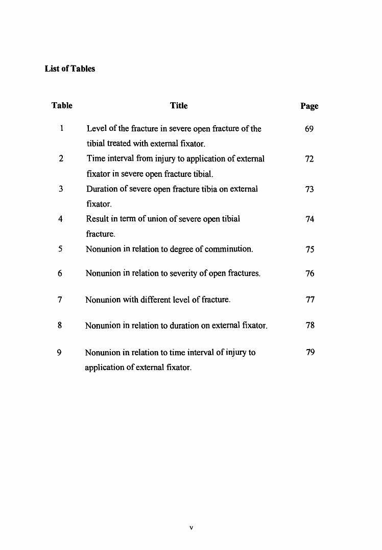
List of Tables
Table Title Page
1 Level of the fracture in severe open fracture of the 69
tibial treated with external fixator.
2 Time interval from injury to application of external 72
fixator in severe open fracture tibial.
3 Duration of severe open fracture tibia on external 73
fixator.
4 Result in term of union of severe open tibial 74
fracture.
5 Nonunion in relation to degree of comminution. 75
6 Nonunion in relation to severity of open fractures. 76
7 Nonunion with different level of fracture. 77
8 Nonunion in relation to duration on external fixator. 78
9 Nonunion in relation to time interval of injury to 79
application of external fixator.
v
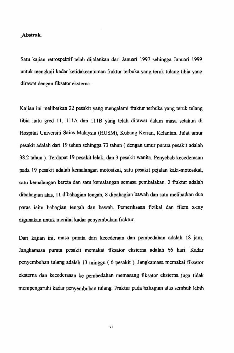
_Abstrak.
Satu kaj ian retrospektif telah dijalankan dari J anuari 1997 sehingga J anuari 1999
untuk mengkaji kadar ketidakcantuman fraktur terbuka yang teruk tulang tibia yang
dirawat dengan fiksator ekstema.
Kajian ini melibatkan 22 pesakit yang mengalami fraktur terbuka yang teruk tulang
tibia iaitu gred 11, 111A dan lllB yang telah dirawat dalam masa setahun di
Hospital Universiti Sains Malaysia (HUSM), Kubang Kerian, Kelantan. Julat umur
pesakit adalah dari 19 tahun sehingga 73 tahun ( dengan umur purata pesakit adalah
38.2 tahun ). Terdapat 19 pesakit lelaki dan 3 pesakit wanita. Penyebab kecederaaan
pada 19 pesakit adalah kemalangan motosikal, satu pesakit pejalan kaki-motosikal,
satu kemalangan kereta dan satu kemalangan semasa pembalakan. 2 fraktur adalah
dibahagian atas, 11 dibahagian tengah, 8 dibahagian bawah dan satu melibatkan dua
paras iaitu bahagian tengah dan bawah. Pemeriksaan fizikal dan filem x-ray
digunakan untuk menilai kadar penyembuhan fraktur.
Dari kajian ini, masa purata dari kecederaan dan pembedahan adalah 18 jam.
J angkamasa purata pesakit memakai fiksator ekstema adalah 66 hari. Kadar
penyembuhan tulang adalah 13 minggu ( 6 pesakit ). J angkamasa memakai fiksator
ekstema dan kecederaaan ke pembedahan memasang fiksator ekstema juga tidak
mempengaruhi kadar penyembuhan tulang. Fraktur pada bahagian atas sembuh lebih
vi

cepat dari fraktur pada bahagian bawah dan tengah, walaubagaimanapun ia tidak
bermakna secara statistik. Fraktur terbuka tulang tibia gred 11 sembuh lebih cepat
dari gred lllA dan gred lllB. Fraktur tulang yang remuk sembuh lebih perlahan
dari fraktur mudah ( Winquist 4: 28 minggu), walau bagaimanapun ia tidak
bermakna secara statistik.
vii
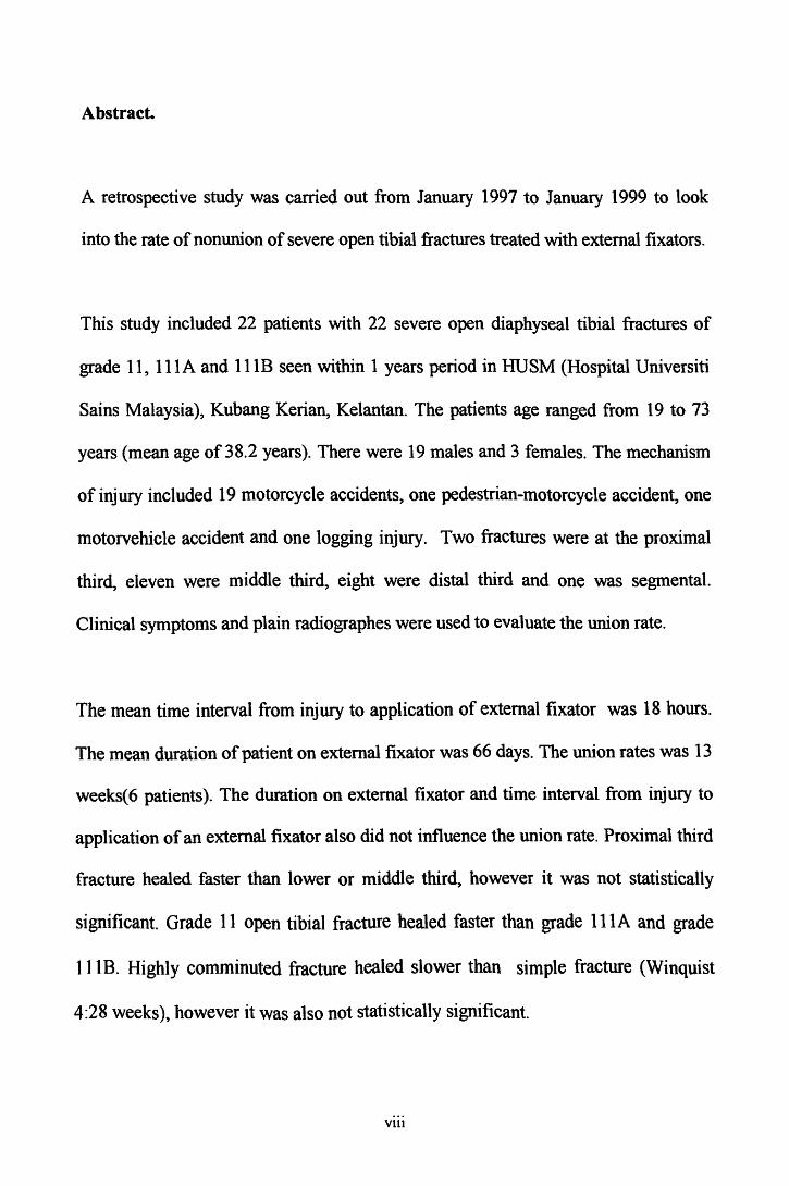
Abstract.
A retrospective study was carried out from January 1997 to January 1999 to look
into the rate of nonunion of severe open tibial fractures treated with external fixators.
This study included 22 patients with 22 severe open diaphyseal tibial fractures of
grade 11, lilA and lllB seen within 1 years period in HUSM (Hospital Universiti
Sains Malaysia), Kubang Kerian, Kelantan. The patients age ranged from 19 to 73
years (mean age of38.2 years). There were 19 males and 3 females. The mechanism
of injury included 19 motorcycle accidents, one pedestrian-motorcycle accident, one
motorvehicle accident and one logging injury. Two fractures were at the proximal
third, eleven were middle third, eight were distal third and one was segmental.
Clinical symptoms and plain radiographes were used to evaluate the union rate.
The mean time interval from injury to application of external fixator was 18 hours.
The mean duration of patient on external fixator was 66 days. The union rates was 13
weeks(6 patients). The duration on external fixator and time interval from injury to
application of an external fixator also did not influence the union rate. Proximal third
fracture healed faster than lower or middle third, however it was not statistically
significant. Grade 11 open tibial fracture healed faster than grade 111 A and grade
111 B. Highly comminuted fracture healed slower than simple fracture (Winquist
4:28 weeks), however it was also not statistically significant.
viii
1.0 Introduction.
The tibia is particularly prone to severe open injuries because of its location,
structural anatomy and sparse anterior coverage by soft tissue. (Caudle & Stern,
1987). An open fracture of the tibia with additional severe soft tissue damage is one
of the most difficult problem in traumatology. (Med. et al., 1983).
The treatment of open fractures of the tibial shaft remains controversial. (Blachut et
al., 1990). The value of primary stable osteosynthesis with bone plates is questioned
because of the additional damage to soft tissues and blood supply. The vast majority
of authors strongly object to the use of intramedullary nails for stabilization of such
fractures. Currently, the treatment of choice in these cases is external fixation.( Med.
et al., 1983). It facilitates stabilization of bony and soft tissue lesions at a distance
from the injury site, appear ideally suited for the initial treatment of open diaphyseal
fractures.(Behrens et al.,1983).
With the introduction of better designs and exacting postoperative management,
many authors have reported high rate of success with external fixation. The fracture
can be reduced and stabilised without sacrificing access to the injured soft tissues
and without burying foreign material next to the fracture or under damaged
skin.(Edge & Denham , 1981 ).
Stable external fixation of open tibial fractures promotes healing of skin and soft
tissue damage, reduces the risk of infection, and facilitates the treatment of patients
with multiple injury. Functional end results after a stable external fixation compare
favorably with the results of internal fixation with Arbeitsgemeinschaft fur
Osteosynthesefragen (AO) plates.(Karlstrom & Olerud ,1983)
However, despite many refinements in this technique, it has been associated with
numerous complications, including problems at the sites of the pins, non-union,
delayed union, malunion and infection.(Blachut et al.,l990). Although early
removal of fixator is frequently recommended to prevent delayed union and
nonunion, little or no data have been found to support this suggestion.(Thomas &
Rae ,1983).
2
2.0 Literature review
Non union and delayed union following severe open tibial fracture is well known.
The rate of non-union has been high (20 to 30 percent), reflecting the characteristics
of the fracture~ a high energy injury with severe loss of soft tissue and
comminution.( Court-Brown et al., 1990).
Velazco et al (1983) in a prospective study of 40 consecutive patients with open
type 11 or 111 tibial fractures who were treated with external fixator noted five
nonunions ( 12 %) at 18-month follow-up which had healed after bone graft.
Kimmel ( 1982) in their study of severe open diaphyseal fractures of the tibial in 19
patients where 50 % were classified as grade 111. Overall, three non unions( 13%)
resulted. Forty-five percent of patients required a bone graft for eventual union.
Chan et al(1984) in a retrospective study of extensive type 111 open tibial fractures,
found a delayed union 60 percent and infected nonunion 30 percent.
The cause of nonunion in open tibial fracture is multifactorial.It is commonly
assumed that prolonged external fixator is a cause of nonunion.However, the high
incidence of nonunion noted with external fixation is not due to prolonged use of the
3
external fixation. In fact, it is the failure of the fracture to unite that causes the
surgeon to continue the use of the fixator.
Thomas & Rae (1983) in a series of 20 open tibial fractures treated with external
fixators, demonstrated an association between nonunion and prolonged use of the
ftxator, but no cause-and-effect relation was shown. However, there was no
association was found between the development of nonunion and degree of soft
tissue injury, delay in fixator application, or diaphyseal versus metaphyseal fracture.
Chatziyiannakis et a1.(1997) found that the type of open fracture, comminution of
the fracture and extension of the original wound for satisfactory reduction, played an
important role in the development of nonunion.
Aho et al.(1983) studied on the advantages of external fixation in the treatment of
severe open tibial fractures (grade 11 and 111) in 79 patients during the period from
1971 to 1978 noted that the risk of delayed union or nonunion and secondary bone
atrophy was due not only to the severity, comminution, soft tissue injury and/ or
infection of the fracture but also to the prolonged use of the external fixators and the
long nonweight-bearing time.
4
Rommens ( 1992) in his prospective study of the significance of soft tissue trauma
for fracture healing on 70 severe open tibial shaft fractures observed that the more
severe the soft tissue injury, the more difficult the fracture healing will be.
Augeneder et al. ( 1989) in their study of 50 severe open tibial fractures treated with
external fixator which was published in Germany journal noted that on average,
fracture healing took 6.7(4-15) months, significantly correlating with the severity of
soft-tissue lesion.
With an external fixator in place in a severely comminuted fractures with defects,
stable bridging callus may require a long time to form, even with repeated bone
grafting. If there are no signs of beginning bony union after about 10 to 12 weeks and
if the soft tissue is healed, Med et al (1983) recommended changing to internal
fixation with simultaneous cancellous bone grafting. Combined secondary plate
osteosynthesis and bone grafting accelerate bony union in cases of delayed healing.
Fracture healing is theoretically enhanced by anatomic reduction, preservation of
blood supply, and sufficiently stable internal and external immobilization.(Philip &
Jack, 1983)
Sufficient stability and adequate revascularization are essential for fracture union.
The motion at a fracture site is dependent on both the load placed on it and the
5
rigidity of the fracture fixation. The rigidity of the fracture fixation depends on both
the biologic tissues present normally and those developed in the process of healing,
as well as on the orthopaedic stabilizing devices used, whether a plaster cast, external
skeletal fixation, or internal fixation.
In 1955 Y amagishi and Yoshimura demonstrated an association between increasing
callus and decreasing stability with external skeletal fixation of fractures in rabbits.
White et al.( 1977) demonstrated an early increase in stiffness and a later increase in
strength of externally compressed rabbit tibial fractures. The interfragmentary strain
that develops during fracture healing seems to be associated with the type of tissue
found at the fracture site.
Granulation tissue between the bone ends will tolerate a strain of 100 % before
failure. Cartilage will fail with 10 % strain and bone with only 2 %. The greater the
motion that occurs, the more likely is fibrous tissue to form. The motion between the
fracture ends must be reduced to a minimum before bone tissue can bridge the
fracture gap.
Fracture healing cannot occur without adequate vascularization. The injury itself
produces vascular injury, both to bone and surrounding soft tissues, that must be
repaired. Continued gross motion at a fracture site impedes healing process. Given
6
these well founded principles, it is difficult to conceive of prolonged use of_ external
fixation causing nonunion.
Proper management of fractures by external skeletal fixation obviously includes
avoidance of distraction. Removal of the external fixator and application of a plaster
cast would result in a decrease in fracture site stability at a critical time in fracture
healing. There is neither theory nor evidence to support the concept that decreasing
fracture site stability at this critical period will enhance union.
Not only must overdistraction be scrupiously avoided, but fracture site compression
should also be sought. This will avoid fracture site motion under both compressive
loading and torsion. Regardless of the technique utilized, fracture site motion will be
greater than with rigid internal fixation if weight-bearing is not limited. Shear
displacement at the fracture site may well be best controlled by "minimal internal
fixation" by interfragmentary screws or short plates, as suggested by Philip and Jack,
(1983).
The other major requirement for fracture healing is adequate vascularity to supply
the required energy, nutrition, and cells. In fractures that fail to heal due to excessive
motion, radiographic evaluation usually reveals abundant quantities of callus
surrounding each fragment but persistence of the fracture gap.
7
Thomas & Rae (1983) noted fractures that failed to heal were characterized by a
lack of radiographic callus, supporting the concept of inadequate vascularity as a
cause of nonunion rather than excessive motion. They reported their experience with
the use of bone scanning to evaluate fracture healing. The static two-hour
postinjection bone scan was found to be of little value. In contrast, the 7.5-15.0-
minute uptake of bone-seeking isotope discriminated well between normally healing
fractures, delayed unions, and nonunions. Three months after injury normally healing
fractures showed a net uptake of 15 %, whereas the nonunions showed a flat line
during the 7.5-15.0-minute period. The high rate of success with posterolateral bone
grafting in tibial nonunions also supports this concept.
Union by a bridge of external callus has definite advantages, especially in severe
injuries such as those treated using external fixation. It can bridge gaps due to
missing bone or devitalised fragments, and is the most rapid variety of bone healing.
(Edge & Denham, 1981).
The significance of external callus in monitoring the healing of tibial fractures
treated by external fixation is supported by several recent reports. In contrast, Lawyer
and Lubber (1980) proposed that union may develop without callus formation,
suggesting primary union by external fixation. All of these results suggest a great
need for a quantitative technique for measuring fracture healing. Bending moments
8
rather than axial loads are more appropriate and should be measured, as well as
angular deformation at the fracture site.
Thomas &Rae (1983) recommended meticulous initial debridement, irrigation, and
preparation of a soft tissue envelope. Early application of rigid external fixation will
expedite wound healing and decrease the development of osteomyelitis.
They also recommmend bone grafting in fractures that are unstable at four months,
with or without removal of the external fixation system. Loosening of the fixation
clamps allows manipulation of the fracture site and clinical evaluation of motion.
Despite all the complications, external skeletal fixation remains an ideal treatment
for complicated extremity fractures in that it provides an increase in fracture site
stability as compared with plaster cast technique without the increased risks
associated with internal fixation.
In addition to provide adequate immobilization of the fracture, this method allows
free access to the wounds and optimal care of the critical soft tissue conditions.It is
also versatile and easy to apply with minimun operative trauma.
9

Thakur and Patankar ( 1991) treated 79 open tibial fractures with unilateral uniplanar
tubular external fixators found that combined with early bone grafting, external
fixation is an excellent method for the management of open tibial fractures.
Bach et al ( 1989) in their prospective study of 59 patients with Grade 11 or 111
open tibial shaft fractures compared internal and external fixation. They note that
external fixation should be regarded as a primary method of stabilization for grades
11 and Ill open tibial shaft fractures compared to plate.
10
3.0 Blood supply of tibia
The blood supply of the tibial is derived from three vessels systems: Nutrient
vessels, Metaphyseal vessels and Periosteal vessels.
The nutrient artery of the tibia arises from the posterior tibial artery and enters the
posterolateral cortex of the bone at the origin of the soleus muscle just below the
oblique line of the tibia posteriorly. The artery divides into three ascending branches
and only one main descending branches, which give off smaller branches to the
endosteal surface. It responsibles for the perfusion of the marrow and the inner two
thirds of the diaphyseal cortex. (Trueta J, 1974).
Periosteal vessels arise from branches of the anterior tibial artery as it courses down
the interosseus membrane. It supplies the outer one-third of the cortical bone.
Metaphyseal vessels arises from the periarticular vascular plexus (i.e geniculate
arteries). It provides numerous anastomoses with the branches of the nutrient artery.
It is capable of maintaining the perfusion of the marrow and the inner one-half of the
cortex following division of the nutrient artery.
The role of each source in fracture healing is controversial. Rhinelander (1974)
believe that the periosteal blood supply plays a relatively minor role in supplying the
11
normal adult tibial cortex. He also stated that the intramedullary vascular supply is
the most important in nonnal bone~ however, after an injury that disrupts the
intramedullary vascular pattern, the periosteal blood vessels increase their
contribution and become prominent in the formation of new bone.Macnab and Haas
(1974) found that the periosteal vessels were especially important in the distal third
tibial fractures but found no difference in intramedullary supply between proximal
and distal regions.
In experimental animals the nutrient artery can be ligated or the metaphyseal
anastomoses can be interrupted without any apparent circulatory deficiency.
Interruption of both nutrient artery and the metaphyseal intercommunications lead to
extensive necrosis of the inner two-thirds of the cortical bone. (Trueta, 1974)
Experimental studies by Rhinelander ( 1974) and others have shown the enormous
capacity of the medullary and periosteal blood supply to regenerate after injury or
osteotomy in laboratory animals. Complete revascularization of the cortex occurs
relatively early in the fracture-healing process.
12
The two basic frame types, unilateral and bilateral, can be applied in one or two
plane configurations. The one-plane configurations are less obstructive and
generally suffice for most injury situations.
Two-plane frames are more effective in neutralization multi-directional bending and
torsional movements ( Behren et al. 1983; Behrens and Johnson 1985). However,
they are only needed when dealing with severe comminuted fracture or with bone
loss (Behrens and Searls 1986), and for arthrodesis as well as osteotomies.
4.3 External f"'txator component mechanics.
4.3.1 Fasteners.
External fixation frames are fastened to bone using Schantz screws. In principle, the
most significant parameter that affects the stability of an external fixation system is
the radius of the screw. The bending stiffness of the screw increases as a function of
the fourth power of the radius of that implant. (Burstein et al., 1972)
Care must be taken to limit the diameter of any screw hole to no greater than 30 % of
the diameter of the diaphysis. To exceed this significantly weakens the bone,
effectively creating an open section. It has been shown that a hole greater than 30 o/o
14
of the diameter markedly increases the risk of fracture. Burstein and colleagues
(1972) demonstrated that a screw hole equal to 30% of the diameter weakened the
torsional strength of that bone by 45 %. Over 6 to 8 weeks, the bone will remodel
about the implant, restoring its strength. However, upon removal of the screw, the
weakening recurs until the bone has remodeled once more. (Burstein et al., 1972)
Screw design has concentrated on the development of implants with a greater core or
root diameter to increase rigidity. Because of an increased rate of implant loosening,
current recommendations favor stiffer, larger root-diameter screws with bicortical
threads for better purchase and decreased loosening characteristics.
Bending preload technique would theoretically reduce pin loosening by allowing the
elastically loaded implant to maintain three-point bending contact. Animal studies,
however, have suggested that the bending preload technique actually accelerates
loosening because of rapid pressure necrosis on the compression side of the preload
pin. Therefore, bending preload is discouraged.( James VN, 1996)
Hydahl and coworkers ( 1991) recommends radial preload to improve screw fixation
and prevent loosening. This can be achieved either by first drilling a pilot hole
slightly smaller than the root diameter screw design to produce a radial preload as
the screw is introduce. They demonstrated in animal studies that radial preload is
preferable to bending preload techniques. (Hydahl et al., 1991)
15
Enlarging the shank diameter of screws has been shown to increase the overall
rigidity of the implant which, in turn, results in lower bending stresses at the entry
site cortex and in a diminished rate of osteolysis and loosening. (Chao & Aro, 1991)
4.3.2 Reduction.
The composite stability of the bone-fixator construct is the most important factor in
treating fractures with external fixation. Fracture configuration and reduction
profoundly affect stresses at the screw/bone interface. End-on-end transverse or other
stably reduced fracture constructs maintained with external fixation have been shown
in both in vitro and in vivo testing to reduce stresses at the screw/bone interface. This
results in decreased rates of pin loosening. As demonstrated by Chao and Aro
(1991), bone-fixator constructs without bone-end contact, and those with very
oblique(unstable) fracture patterns, tend to have increased rates of screw loosening
and a longer healing time when compared with stably reduced transverse fractures
with good bone-end contact.
4.3.3 Insertion technique.
The insertion technique may have mechanical effects on the initial screw purchase as
well as a profound biological influence on the maintenance of the implant-bone
16
interface. The insertion of self-drilling screws has been associated with microfracture
and high temperatures at the bone implant interface. Thennal necrosis often the end
results. To reduce the potential for both thermal necrosis and the premature
loosening associated with it, an alternative technique that involves predrilling pilot
holes for all screw sites is now recommended to minimize microfracture and avoid
excessive increase in bone temperatures. (Matthews et al., 1984).
4.3.4 Screw/Pin Materials.
Chao and Aro ( 1991) advocate the use of high modulus materials in screw design to
minimize bending and thereby decrease loosening. Others, however, believe that
using more isoelastic materials, such as titanium alloy, improves loosening rates.
This issue remains controversial.
Previous metals, such as gold and silver, have long been known to inhibit bacterial
growth. It has been suggested that coating :fixator screws with gold or silver might
prevent pin-track infection. Limited laboratory and clinical studies have indicated
decreased pin-track infection rates when coated pins are used. However, such
implants are not clinically available.
17
4.3.5 Threaded Versus Smooth Fasteners.
Smooth implants may allow bone to translate on fixator pins. Fixation with a smooth
implant is therefore less stable than fixation with a threaded implant of the same
diameter. (James VN, 1996)
4.3.6 Number of Screws/Pins/Wires
In any system of external fixation, stability is improved by increasing the number of
fixation devices to bone. Ideally, to achieve maximal effect, these additional screws
should be evenly distributed across the greatest possible area of the major fragments
to be stabilized. Many larger body/frame external fixation systems can achieve more
than adequate stability for most applications by using two closely set screw clusters
that are remote from the fracture to be stabilized.
4.3.7 Frame geometry.
Traditional early bar and clamp fixators, such as the ASIF or Hoffmann-Vidal-Ad.rey
systems, offer considerable versatility in exchange for stability. For simple fracture
patterns with good fracture reduction, the simple uniplanar frame geometry of these
systems provides adequate stability. Any unilateral application can be made more
resilient to the forces experienced in any one direction or plane by applying a like
18

frame that opposes that plane of instability. When the frames are connected, they
may constitute a so-called "delta configuration" or triangular frame. Comparative
stability studies have shown that larger unilateral systems offer a degree of stability
comparable to the more complex multiplanar configurations of more traditional
systems.
Excessive rigidity can have a negative influence on healing.lnherently stronger large ..
body unilateral systems should almost never be applied in multi planar configurations
for routine fractures because they may inhibit callus formation.
4.3.8 Bone Frame Distance
Another technique used to significantly increase the stability of any given construct
involves placing the frame component as close to the bone as clinically possible. The
closer the frame is to the bone, the more stable the construct will be. This principle
can also be used by clinicians who choose to produce cyclic dynamization in a frame
by moving the fixator bars or body further out on the fixation pins or screws. This
creates a more flexible construct. This method of decreasing frame rigidity while
maintaining reduction was one of the earliest techniques of dynamization.
19
4.3.9 Implant-Clamp Fixation.
In hatf ... screw fixator systems, implants are held by individual or grouped coupling
clamps that affix screws to the frame. To improve fixation of the half ... pins to the
frame, screw coupling clamps can be made wider to achieve broader, more stable
fixation to the shaft of the half-screw. Fixators with smaller single ... coupling clamps
can be used to achieve similar stability by "double stacking'' them. This is another
way to broaden the fixation of the screw to the frame and improve construct stability.
4.3.10 Biomechanics: summary.
In summary, an "ideal" degree of osseous stability for managing each clinical
situation treated with external fixation is ill defined. Any fixation technique should
attempt to match the biomechanical requirement of the clinical situation with the
stability of the overall construct.
The following frame characteristic have been shown to increase the stiffness of an
applied frame and to diminish motion at the fracture site:
1. Increasing the diameter of the schanz screws.
2. Increasing the number of schanz screws in each bony fragment.
3. Increasing schanz screw spread within each main bony fragment.
4. Using multiplanar fixation.
20
5. Placement of the principal frame in the sagittal plane.
6. Reducing the distance between fixator frame and bone.
7. Predrilling all half-pin screw sites whenever possible, and irrigating drills and
implants on insertion with a cool saline solution to avoid thennal necrosis and
associated premature loosening.
8. Applying radial preload techniques to half-screw fixation.
9. Pre loading of schanz screws-automatically done by slightly oversizing ( +0.2
mm) the core.
10. Improving fastener-frame fixation with improved clamp technology or double
stacking.
11. Reducing fractures with fragment contact improving stability and permitting "off
loading" of implants (without use of adjunctive lag screws).
12. Dynamizing frames when possible to allow for bone-end contact, a reduction in
screw stress and decrease in the tendency to loosen.
4. 4 Clinical indication of external fiXation.
External fixation is usually relegated to the stabilization of more severe complex
injuries, especially those with associated soft-tissue wounds that are not amenable to
other techniques. In instances of severe intra-articular fractures, external fixation
may be used as a portable traction device, offering a means of reduction and
stabilization through ligamentotaxis. Finally , external fixation can be an alternative
21
method for temporary stabilization of long bone and pelvic injuries in multiply
traumatized patients when the blood loss and operative time associated with
definitive internal fixation are considered undesimble.
4.5 Principles of external fixation.
To be safe and effective, an applied fixator should have a low rate of serious
complications, be nonobstructive, be stiff enough to maintain alignment under
adverse loading situation, facilitate full weight-bearing, and be adaptable to a wide
variety of injury and patient conditions.
Experience accumulated over the past decade has shown that these are best achieved
by adhering to four basic principles(Behrens and Searls, 1986) which demand that
the applied frame optimally accomodates the vital limb anatomy, access for
debridement and secondary procedures, mechanical demands of patient injury, and
patient comfort.
4.6 Fracture healing following external fixation.
There are basically two types of fracture healing- union through external periosteal
callus and primary bone healing also known as in situ fracture remodelling.
22
Fracture callus forms in reaction to the disruption of the periosteum and endosteum
combined with the interfragmentary strain or motion associated with bone injury.
Callus bridges the fracture fragments and acts as both a stabilizing structural
framework and the biological substrate that provides the cellular material for union
and remodelling.
Rigid fixation will not only preclude the development of callus; it will also typically
result in a protracted, biomecbanical dependency of the bone-hardware construct on
the fixation system itself before adequate remodelling of the bone allows for safe
removal of the implant. This principle has an important bearing on the external
fixation technique.
In an attempt to reproduce plate-like stability, early external fixation systems
stressed the need for increasingly rigid frames in multiplanar configurations.
Adjunctive interfragmentary screws were often used to increased construct stability.
Although these rigid constructs did occasionally yield an anatomic rtestoration, it is
now known that these techniques may have actually delayed or prevented union.
While early treatment algorithms taught that external fixators were to be removed 6
weeks after application to avoid the complication of pin-track infection, the early
shift from a rigid fixator construct (promoting primary healing) to a cast or brace
23
presupposed that the fracture would have the intrinsic stability to sustain functional
load bearing of the limb.
Because of the absence of callus fonnation, primary bone healing, whether promoted
through rigid plating or rigid external fixation, requires the fracture to be supported
and protected until the bone achieves sufficient strength to prevent refracture or
angulation when it is once again subjected to functional stress. Before adequate
fracture remodelling, refracture may occur with a loss of reduction. A rigid external
ftxator that eliminates micromotion must be kept in place longer and necessarily
requires prolonged maintenance of the fixator pin/bone interface.
At the time any external fixator is applied, a "race" begins between fracture healing
and fixator pin failure (due to infection, loosening, etc.) External fixation depends, of
course, on proper fixation of the screws to the bone. Techniques that rely on frame
constructs that are too rigid, and therefore require prolonged pin fixation and frame
maintenance, will often fail because the fracture cannot adequately remodel by the
time the pins loosen and the fixator must be removed.
In light of contemporary interfragmentary strain theories about fracture healing,
current external fixation systems have been designed to allow micromotion at the
fracture site to promote callus formation. Stable yet less rigid systems of external
24