session six thigh (compartments) posterior compartment anterior compartment medial compartment
TRANSCRIPT

Session SixThigh (Compartments)
•Posterior Compartment•Anterior Compartment•Medial Compartment

Thigh by deep fascia
( fascia lata ) divided in
to three compartments
and form anatomical
spaces (femoral triangle
and adductor canal ).
Thigh

Thigh
ANTERIOR
COMPARTMENT
POSTERIOR
COMPARTMENT
MEDIAL
COMPARTMENT

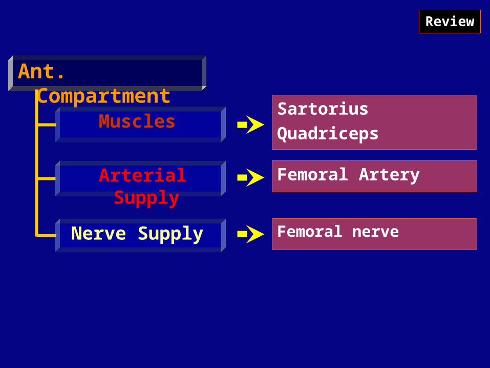
Ant. Compartment
Muscles
Arterial Supply
Nerve Supply
Femoral Artery
Femoral nerve
Femoral nerve is formed of (L2, L3, L4) greater branches of lumbar plexus
Femoral artery is continuation of the external iliac artery
Psoas Major
Iliacus
Sartorius
Quadriceps:1. Rectus femoris
2. Vastus medialis
3. Vastus lateralis
4. Vastus intermedius

Back
Anterior Compartment
Muscles

Back
Muscles of the anterior compartment of thigh
Muscle Origin Insertion Innervation Function
Psoas major Posterior abdominal wall (lumbar transverse processes, intervertebral discs, and adjacent bodies from T12 toL5
Lesser trochanter of femur
Anterior rami L1,L2,L3
Flexes the thigh at the hip joint
Iliacus Posterior abdominal wall (iliac fossa)
Lesser trochanter of femur
Femoral nerve [L2,L3]
Flexes the thigh at the hip joint
Sartorius Anterior superior iliac spine
Anterior surface of tibia just inferolateral to tibial tuberosity
Femoral nerve [L2,L3]
Flex, Abd, & Lat.rotation the thigh at the hip joint and flexes the leg at the knee joint

Back
Anterior Compartment
Muscles

Back
Muscles of the anterior compartment of thigh (continue)
Muscle Origin Insertion Innervation Function
Rectus femoris
Straight head originates from the anterior inferior iliac spine; reflected head originates from the ilium just superior to the acetabulum
Quadriceps femoris tendon
Femoral nerve [L2,L3,L4]
Flexes the thigh at the hip joint and Extends the leg at the knee joint
Vastus medialis
Medial part of intertrochanteric line, Spiral line, medial lip of the linea aspera, medial supracondylar line of femur
Quadriceps femoris tendon and medial border of patella
Femoral nerve [L2,L3,L4]
Extends the leg at the knee joint
Vastus intermedius
Upper two-thirds of anterior and lateral surfaces of femur
Quadriceps femoris tendon and base of patella
Femoral nerve [L2,L3,L4]
Extends the leg at the knee joint
Vastus lateralis
Lateral part of intertrochanteric line, margin of greater trochanter, lateral margin of gluteal tuberosity, lateral lip of the linea aspera
Quadriceps femoris tendon and lateral margin of patella
Femoral nerve [L2,L3,L4]
Extends the leg at the knee joint

Back
Ant. Compartment
Muscles
Arterial Supply
Nerve Supply
Sartorius
Quadriceps
Femoral Artery
Femoral nerve
Review

Post. Compartment
Muscles
Arterial Supply
Nerve Supply
Hamstring:1. Semitendinosus
2. Semimembranosus
3. Biceps femorisProfonda femoris:Branch of femoral artery
Sciatic nerve
Sciatic nerve is formed of (L4, L5, S1, S2, S3) branches of sacral plexus
Back
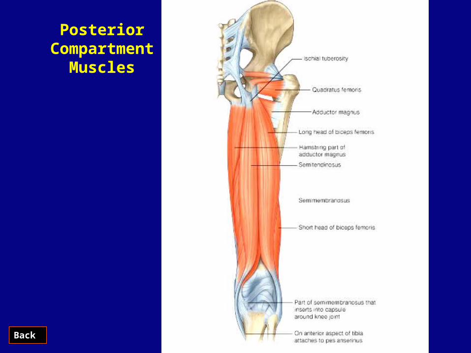
Posterior Compartment
Muscles

Back
Muscles of the posterior compartment of thigh
Muscle Origin Insertion Innervation Function
Biceps femoris
Long head-inferomedial part of the upper area of the ischial tuberosity; short head-lateral lip of linea aspera
Head of fibula Sciatic nerve [L5 to S2]
Flexes leg at knee joint; extends and laterally rotates thigh at hip joint and laterally rotates leg at knee joint
Semitendinosus
Inferomedial part of the upper area of the ischial tuberosity
Medial surface of proximal tibia
Sciatic nerve [L5 to S2]
Flexes leg at knee joint and extends thigh at hip joint; medially rotates the thigh at the hip joint and leg at the knee joint
Semimembranosus
Superolateral impression on the ischial tuberosity
Groove and adjacent bone on medial and posterior surface of medial condyle of tibia
Sciatic nerve [L5,S1,S2]
Flexes leg at knee joint and extends thigh at hip joint; medially rotates thigh at the hip joint and leg at the knee joint
Back

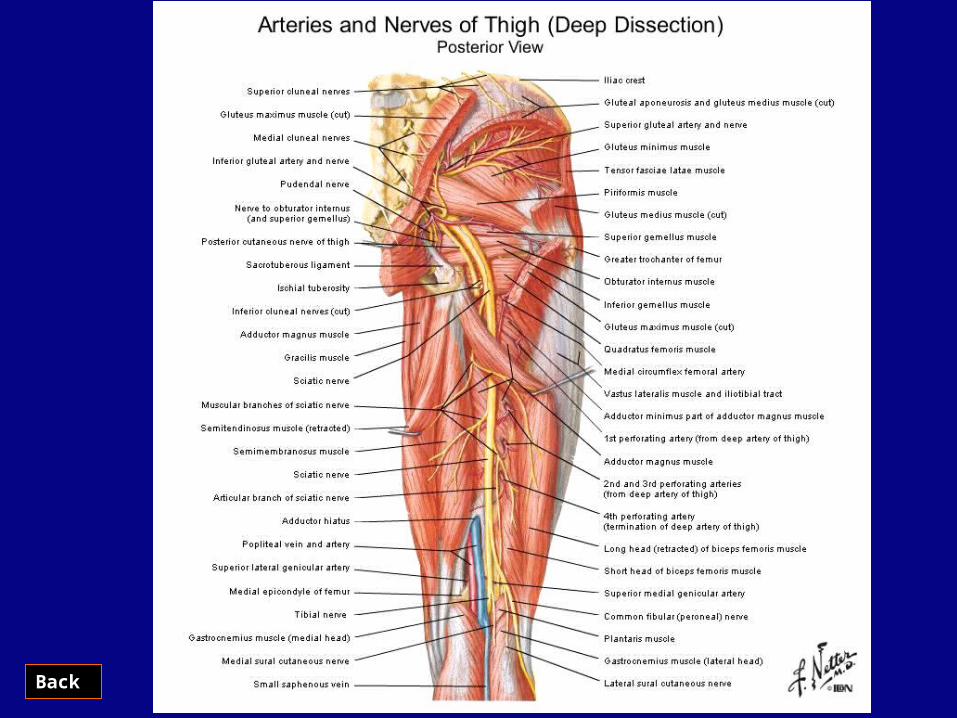
Post. Compartment
Muscles
Arterial Supply
Nerve Supply
Hamstring:1. Semitendinosus
2. Semimembranosus
3. Biceps femoris
Profonda femoris
Femoral nerveBranch of femoral artery
Review
Sciatic nerve is formed of (L4, L5, S1, S2, S3) branches of sacral plexus
