session: innovative technologies · pasquale de blasio ... mounted on a microscope slide ... brest,...
TRANSCRIPT

Session: Innovative Technologies
Abstract Title: Tissue Microarray Technology:
New opportunity for Tissue Banks to contribute
to Cancer Research and Diagnostics.
Pasquale De Blasio • Founding President of ESBB
• Member of Marble Arch Int. Biobanking Working Group
• President of ISENET-USA LLC

Combines tens to hundreds of paraffin-
embedded tissue specimens into a
single paraffin block.
Tissue Microarray Technology
Pathology archives PPFT Tissue Blocks & Slides Tissue Microarrayer
TMA Block

• It is possible to slice up to 200 consecutive
sections of 4-5μm from each TMA block,
mounted on a microscope slide
• The TMA slide can be processed like
ordinary tissue sections with a wide range
of techniques: Histo
staining, Immuno
histo
Immuno
fluorescent.
FISH
Tissue Microarray Technology

“Advantages to use
Pathology Archives”
• widest collection of clinical tissues
available with the entire clinical
heterogeneity range
• Tissue samples are related to clinical
records and have follow-up and
outcome information
• Are under the responsibility of
pathologists that have access to all the
information and are bound by
professional secrecy
• It is now possible to perform many
types of molecular analysis on this
type of tissues.

Tissue Microarrays
Validation In Real Patient Samples
In Vitro
Screening
Methods
Clinical
Trials
Epidemiology
Based Studies
Multi-tumor
Systems
Micro-Arrays Other
Methods
TMAs in Biomarker Development
TMAs advantages: • the speed of molecular analyses is increased by more than 100-fold.
• precious tissues are not destroyed
• large number of molecular targets can be analysed from consecutive
TMA sections (Cost reduction). Compliments of S. Hewitt, NCI TARP Laboratory

First scientific pubblications on TMA

From Tissue To Arrays
• Develop Hypothesis
• Identify Tissue
• Prepare Tissue
• Array Tissue
• Section Arrays
• Perform Experiment
• Analyze Data
• Publish
• Repeat
Compliments of S. Hewitt, NCI TARP Laboratory

Quality of Tissue is everything
1
2
3
4
5
6
7
Tissue Quality
Histology
Proteins
Nucleic Acids
Diagnostic Reporting
Clinical Annotation
Storage Of Biomaterial
Compliments of S. Hewitt, NCI TARP Laboratory

Challenges in the
Construction of a TMA
• Identifying Adequate/Appropriate
Material
• Negotiating MTA/IRB Approval
• Specimens Present Special
Challenges
– Biopsy & Small Specimens Are Hard
To Array
– Prior Use of Tissue for diagnostic
• Diversity of Specimen Sources
– Regional Differences In Diagnosis &
Treatment
– Differences In Specimen Handling
• Informatics
• Managing Expectations
– Pathologist Vs Scientist
TRACEABILITY

Integrated Tissue MicroArray
Pathology Platform “workflow”
Clinical Database
Quantitative Digital
Pathology
Digital Scanners
Fish Fluorescence Histo Immuno
Tissue Microarrayer
Platform

Summary
Harvest and re-suspend cells in agarose
Embed cells in paraffin donor blocks
Pick cell or tissue cores from donor blocks
FFPETs donor blocks
Spot cell/tissue cores in recipient paraffin blocks
Immunohisitochemical/ immunofluorescemnce
analysis
DNA and RNA extraction
Place cell/tissue cores in specific
vessels

• Tissue microarrays (TMAs) have become a
mainstay in preclinical and translational research,
especially for the development of biomarker assays
for characterization of disease.
• They allow for analysis of extremely small amounts
of tissue, thus preserving valuable tissue blocks.
• At the same time, they dramatically increase the
efficiency and cost–effectiveness of performing
tissue-based studies by enabling the examination of
10s to 100s of different patient samples on the same
slide.
Summary

Summary
• A block of Samples from
hundreds of Tissue Blocks
– Multiple Samples (3-4) from
Paraffin Embedded or Frozen
Tissue blocks
– Arranged in an organized way
Platform For High-Throughput Pathology
Immuno Histo Fluorescence Fish
Compliments of S. Hewitt, NCI TARP Laboratory

Summary
• A Protein Array with Retained
Histomorphology
– Cell Type Localization
– Cell Type Quantification
– Subcellular Localization
Platform For High-Throughput Proteomics
Immuno Histo Fluorescence Fish
Compliments of S. Hewitt, NCI TARP Laboratory

H&E IHC IHC (75 cases, dupl)
LABOR INDIRECT
COSTS
MATERIALS
SINGLE SLIDES TMA
€ 2
5
€ 2
5
€ 1
,88
8
€ 3
8
€ 3
8
€ 2
,81
3
€ 2
71
€ 4
€ 2
71
€ 0.00
€ 500.00
€ 1,000.00
€ 1,500.00
€ 2,000.00
€ 2,500.00
€ 3,000.00
Total cost per unit Cost per patient Cost per 75 patients
H&E
IHC
IHC on TMA
Conclusion
Compliments from G. Cattoretti, UNIMIB- Italy

Summary
Brest, PgR (DAB),
Pos Rat (N, A)
Brest, Gult1, IHC,
Membrane (A, I)
Connectivity
Melanoma
KI-67 + MART1
(DAB, AEC) Proliferation

Courtesy of: Giorgio Cattoretti
Associate Professor, Universitá degli Studi Milano-Bicocca
Director, Division of Surgical Pathology, Cytology, Medical Genetics and Nephropathology
Azienda Ospedaliera San Gerardo, Monza
This material may be copyrighted. Do not diffuse without authorization.
http://www.tma.cattoretti.it/
Conclusion
Compliments from G. Cattoretti, UNIMIB- Italy
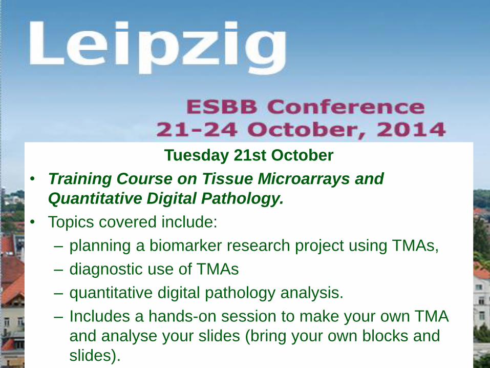
Tuesday 21st October
• Training Course on Tissue Microarrays and
Quantitative Digital Pathology.
• Topics covered include:
– planning a biomarker research project using TMAs,
– diagnostic use of TMAs
– quantitative digital pathology analysis.
– Includes a hands-on session to make your own TMA
and analyse your slides (bring your own blocks and
slides).

Acknowledgments
Maurizio Andrea Aikaterini Tatiana Davide
Monica Simona Alberto Alessandria Valeria