session 3-part 2: skeletal muscleefs.efslibrary.net/certificateprograms/pft/course 2-intro... ·...
TRANSCRIPT

Session 3-Part 2: Skeletal Muscle
Course: Introduction to Exercise Science-Level 2 (Exercise Physiology)
Presentation Created byKen Baldwin, M.ED, ACSM-H/FI
Copyright © EFS Inc. All Rights Reserved.

Skeletal Muscle
Human body contains over 400 skeletal muscles– 40-50% of total body weight
Functions of skeletal muscle– Force production for locomotion and breathing– Force production for postural support– Heat production during cold stress

Structure of Skeletal Muscle: Connective Tissue Covering
Epimysium– Surrounds entire muscle
Perimysium– Surrounds bundles of muscle fibers
• Fascicles
Endomysium– Surrounds individual muscle fibers

Connective Tissue Covering of Muscle

Structure of Skeletal Muscle: Microstructure
Sarcolemma– Muscle cell membrane
Myofibrils– Threadlike strands within muscle fibers– Actin (thin filament)
• Troponin• Tropomyosin
– Myosin (thick filament)

Microstructure of Skeletal Muscle

Structure of Skeletal Muscle: The Sarcomere
Further divisions of myofibrils– Z-line– A-band– I-band
Within the sarcoplasm– Sarcoplasmic reticulum
• Storage sites for calcium– Transverse tubules– Terminal cisternae
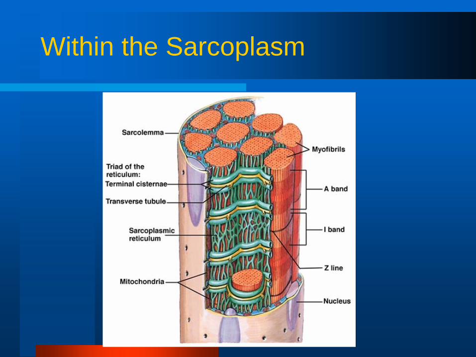
Within the Sarcoplasm

The Neuromuscular Junction
Site where motor neuron meets the muscle fiber– Separated by gap called the neuromuscular cleft
Motor end plate– Pocket formed around motor neuron by
sarcolemmaAcetylcholine is released from the motor neuron– Causes an end-plate potential (EPP)
• Depolarization of muscle fiber

Illustration of the Neuromuscular Junction

Muscular Contraction
The sliding filament model– Muscle shortening occurs due to the
movement of the actin filament over the myosin filament
– Formation of cross-bridges between actin and myosin filaments
– Reduction in the distance between Z-lines of the sarcomere

The Sliding Filament Model of Muscle Contraction

Cross-Bridge Formation in Muscle Contraction
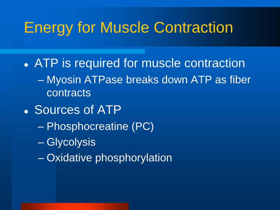
Energy for Muscle Contraction
ATP is required for muscle contraction– Myosin ATPase breaks down ATP as fiber
contractsSources of ATP– Phosphocreatine (PC)– Glycolysis– Oxidative phosphorylation

Sources of ATP for Muscle Contraction
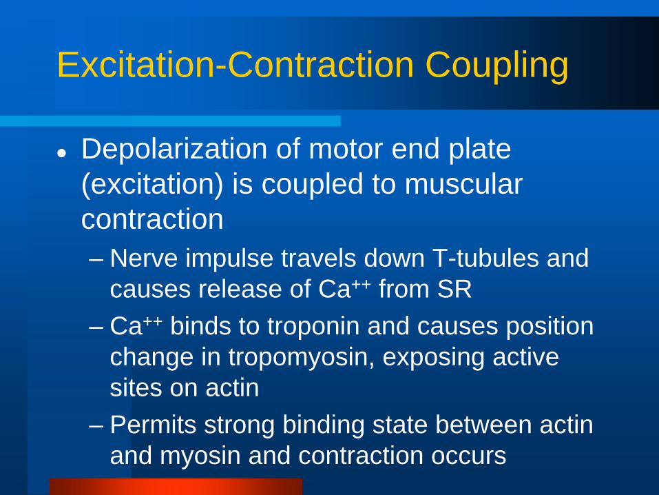
Excitation-Contraction Coupling
Depolarization of motor end plate (excitation) is coupled to muscular contraction– Nerve impulse travels down T-tubules and
causes release of Ca++ from SR– Ca++ binds to troponin and causes position
change in tropomyosin, exposing active sites on actin
– Permits strong binding state between actin and myosin and contraction occurs

Illustration of the Steps of Excitation-Contraction Coupling

Steps Leading to Muscular Contraction

Properties of Muscle Fibers
Biochemical properties– Oxidative capacity– Type of ATPase
Contractile properties– Maximal force production– Speed of contraction– Muscle fiber efficiency
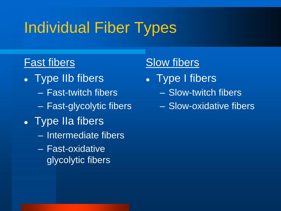
Individual Fiber Types
Fast fibersType IIb fibers – Fast-twitch fibers– Fast-glycolytic fibers
Type IIa fibers– Intermediate fibers– Fast-oxidative
glycolytic fibers
Slow fibersType I fibers– Slow-twitch fibers– Slow-oxidative fibers

Muscle Fiber Types
Fast fibers Slow fibers
Characteristic Type IIb Type IIa Type I
Number of mitochondria Low High/moderate High
Resistance to fatigue Low High/moderate High
Predominant energy system Anaerobic Combination Aerobic
ATPase activity Highest High Low
Vmax (speed of shortening) Highest Intermediate Low
Efficiency Low Moderate High
Specific tension High High Moderate

Comparison of Maximal Shortening Velocities Between Fiber Types
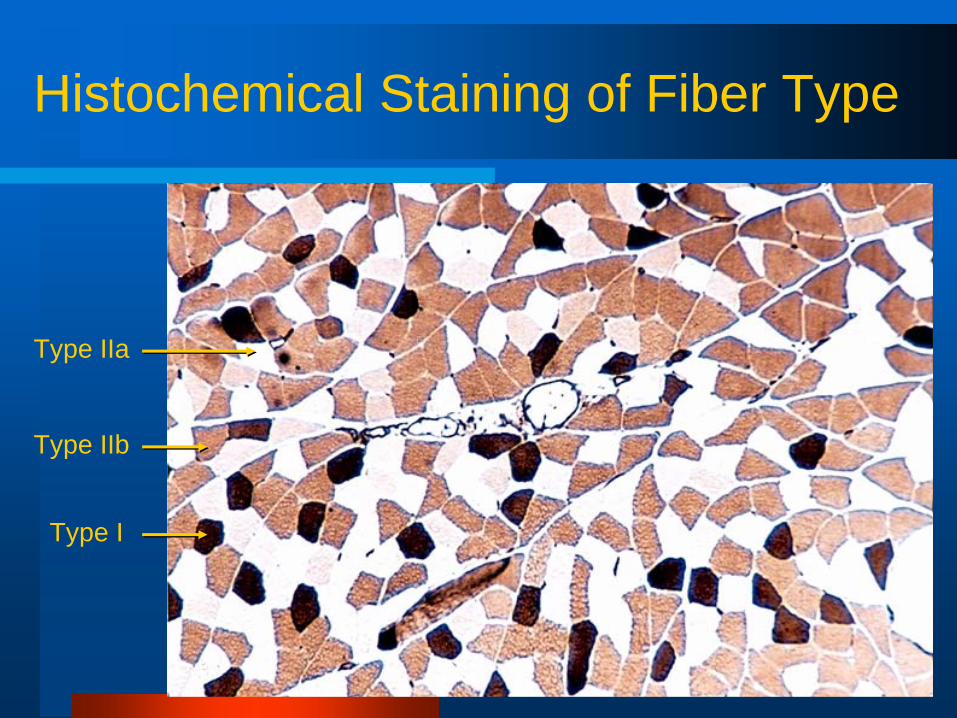
Histochemical Staining of Fiber Type
Type IIa
Type IIb
Type I

Fiber Types and Performance
Power athletes—75% FT; 25% ST – Sprinters– Possess high percentage of fast fibers
Endurance athletes—75% ST; 25% FT– Distance runners– Have high percentage of slow fibers
Others– Weight lifters and nonathletes– Have about 50% slow and 50% fast fibers

Alteration of Fiber Type by Training
Endurance and resistance training– Cannot change fast fibers to slow fibers– Can result in shift from Type IIb to IIa fibers
• Toward more oxidative properties

Age-Related Changes in Skeletal Muscle
Aging is associated with a loss of muscle mass– Rate increases after 50 years of age
Regular exercise training can improve strength and endurance– Cannot completely eliminate the age-
related loss in muscle mass
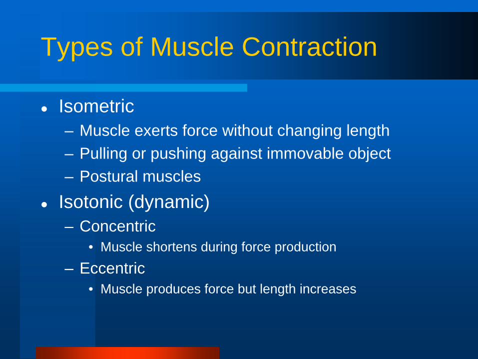
Types of Muscle Contraction
Isometric– Muscle exerts force without changing length– Pulling or pushing against immovable object– Postural muscles
Isotonic (dynamic)– Concentric
• Muscle shortens during force production
– Eccentric• Muscle produces force but length increases

Isotonic and Isometric Contractions

Force Regulation in Muscle
Types and number of motor units recruited– More motor units = greater force– Fast motor units = greater force
Initial muscle length– “Ideal” length for force generation
Nature of the motor units neural stimulation– Frequency of stimulation
• Simple twitch, summation, and tetanus

Relationship Between Stimulus Frequency and Force Generation
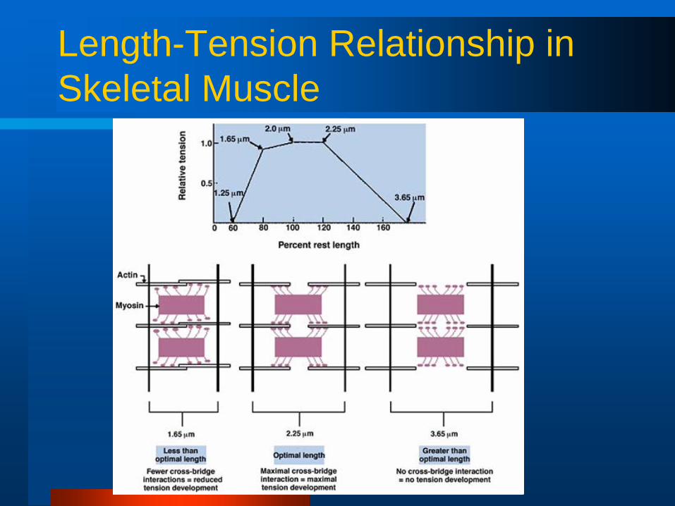
Length-Tension Relationship in Skeletal Muscle

Simple Twitch, Summation, and Tetanus

Receptors in Muscle
Muscle spindle
Golgi tendon organ (GTO)

Muscle Spindle
Muscle spindle– Detect dynamic and
static changes in muscle length
– Stretch reflex• Stretch on
muscle causes reflex contraction

Golgi Tendon Organ
Monitor tension developed in muscle
Prevents damage during excessive force generation
– Stimulation results in reflex relaxation of muscle

Physiological Effects of Strength Training
Strength training results in increased muscle size and strengthNeural factors– Increased ability to activate motor units– Strength gains in initial 8-20 weeks
Muscular enlargement– Mainly due enlargement of fibers (hypertrophy)– Long-term strength training