servizio sanitario regionale emilia-romagna uoc … · algie in fossa iliaca dx. alvo chiuso da...
TRANSCRIPT

Patologia ischemica ileo-colica
SERVIZIO SANITARIO REGIONALE
EMILIA-ROMAGNA
UOC Radiologia Interaziendale Ferrara
Massimo Tilli Maria Teresa Cannizzaro AOU S. Anna (Ferrara)

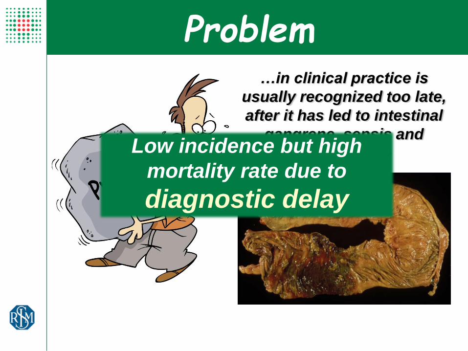
…in clinical practice is
usually recognized too late,
after it has led to intestinal
gangrene, sepsis and
organ failure Low incidence but high
mortality rate due to
diagnostic delay
Problem

Blood supply

-Between the SMA and IMA
“Watershed” areas
-Between IMA and rectal arteries
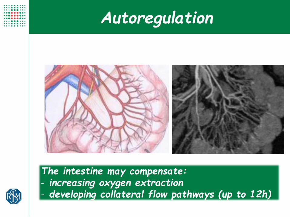
Autoregulation
The intestine may compensate: - increasing oxygen extraction - developing collateral flow pathways (up to 12h)
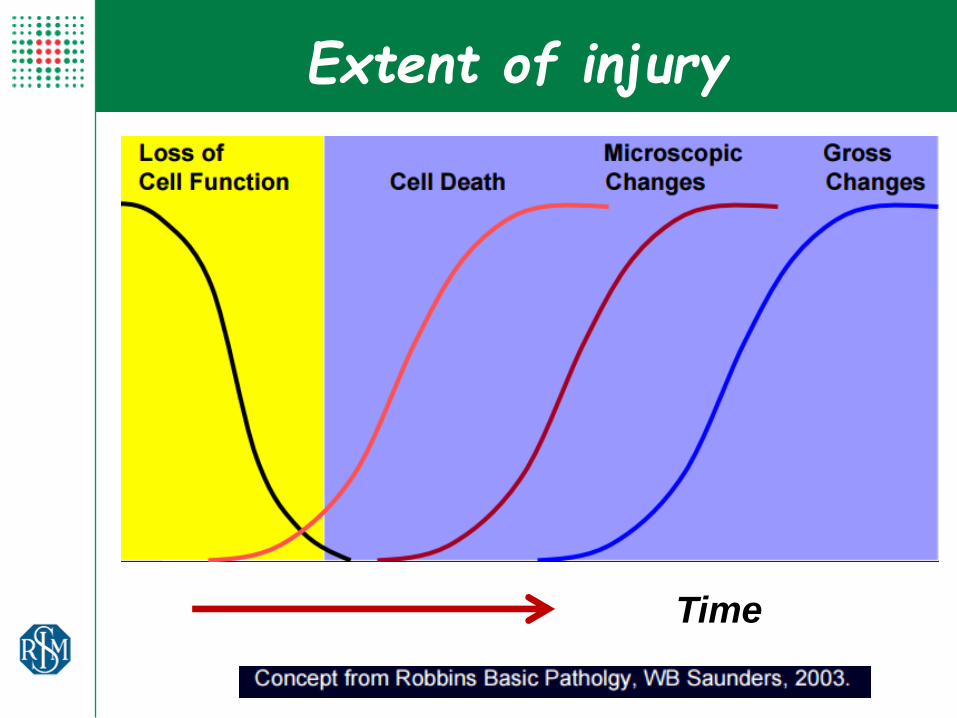
Extent of injury
Time
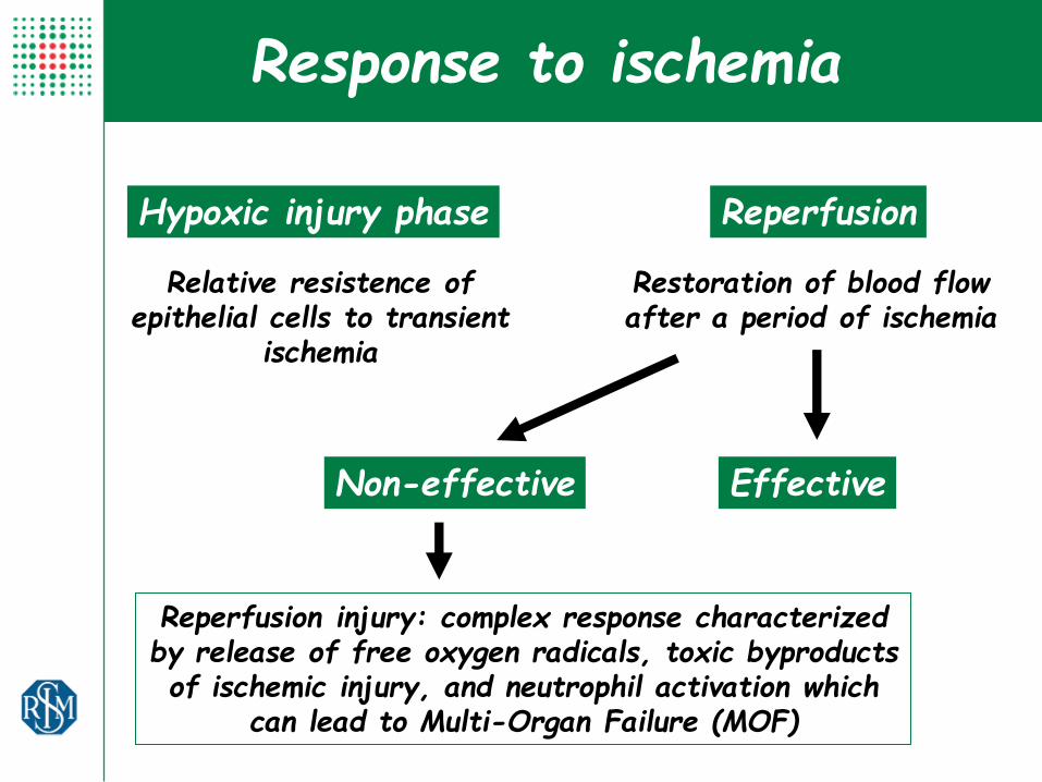
Response to ischemia
Hypoxic injury phase Reperfusion
Restoration of blood flow after a period of ischemia
Relative resistence of epithelial cells to transient
ischemia
Effective Non-effective
Reperfusion injury: complex response characterized by release of free oxygen radicals, toxic byproducts of ischemic injury, and neutrophil activation which
can lead to Multi-Organ Failure (MOF)

Prolonged ischemia generates
vasoconstriction in afferent bed and reduces
collateral supply
Wall infarction
Autoregulation failure
Extent of injury
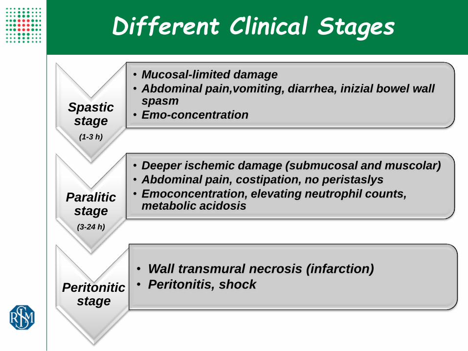
Spastic stage
(1-3 h)
• Mucosal-limited damage
• Abdominal pain,vomiting, diarrhea, inizial bowel wall spasm
• Emo-concentration
Paralitic stage (3-24 h)
• Deeper ischemic damage (submucosal and muscolar)
• Abdominal pain, costipation, no peristaslys
• Emoconcentration, elevating neutrophil counts, metabolic acidosis
Peritonitic stage
• Wall transmural necrosis (infarction)
• Peritonitis, shock
Different Clinical Stages

Acute or Chronic
Arterious or Venous
Occlusive or Non-occlusive
Small or Large bowel
Classification
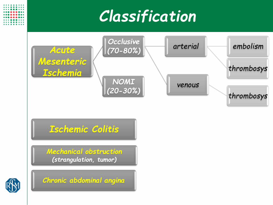
Acute Mesenteric Ischemia
Occlusive (70-80%)
arterial embolism
thrombosys
venous
thrombosys
NOMI (20-30%)
Mechanical obstruction (strangulation, tumor)
Ischemic Colitis
Chronic abdominal angina
Classification
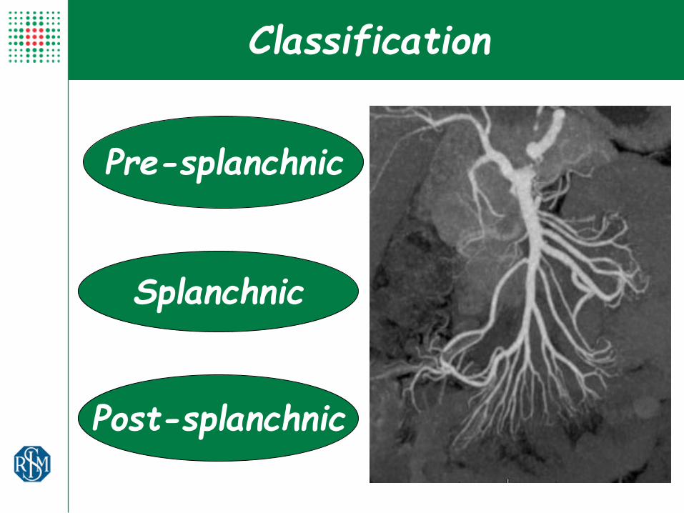
Pre-splanchnic
Splanchnic
Post-splanchnic
Classification
Pre-splanchnic
Non Occlusive Mesenteric Ischemia (NOMI)
-15-20% of acute mesenteric ischemia
-Secondary vasoconstriction of mesenteric arteries (low-flow states, hypotension, sepsis or heart failure)
-Ischemia is distributed over a wide area of the bowel in a non-consecutive manner

Splanchnic
Acute mesenteric arterial Embolism
(AMAE)
Acute mesenteric arterial Thrombosis
(AMAT)
-25% of acute mesenteric ischemia
-Associated with chronic atherosclerotic disease
-Well-developed collateral circulation
-50-55% of acute mesenteric ischemia
-Generally involves the distal aspect of the vessel
-Absence of associated collateral vessels.

Splanchnic
Ischemic Colitis (IC)
Chronic ischemia
-Atherosclerotic involvement of splanchnic vessels (>2-3)
-Well-developed collateral circulation
-Post-prandial abdominal pain and weight loss
- Secondary to acute or chronic decrease of bowel blood supply - Most common causes are low-flow conditions associated with chronic atherosclerotic disease
- Second most frequent cause of lower GI bleeding
Post-splanchnic
Mesenteric Venous Thrombosis (MVT )
-15-20% of acute mesenteric ischemia -Common risk factors:
- Hypercoagulable states - Portal hypertension - Recent surgery

AMI is underdiagnosed without clinical suspicion
(81% vs 97% in detecting crucial findings of AMI)
Are we confident..???
Radiologic diagnosis

- No CT detection of vessel occlusion - Low specificity of indirect signs
- Significant inter-observer variability
Radiologic diagnosis
Problems

What have we learned?

First phase
• Vessels occlusion (emboli or thrombi)
• Spastic reflex ileo
• Poor wall enhancement
Mesenteric ischemia

Second phase
• “Paper thin” wall
• Ipotonic ileo
• Gas filled loops
• Decrease wall enhancement
• Small account of fluid
Mesenteric ischemia

• Degree of wall enhancement (wall infart or necrosis or thickening in case of riperfusion)
• Paralitic ileo
• Ascites
• Pneumatosis (?)
Third
phase
Mesenteric ischemia

Correct Reporting Approach
Vessels patency
Wall thickness
Wall enhancement
Pneumatosis
Mesentery
Ascites
Luminal dilation

Vessel patency

Vessel patency

*Michael Macari et Emil J. Balthazar, AJR 2001
Bowel wall thickening
Cut-off : 3-5 mm
More common finding
Least specific finding (depends on the
degree of distension)
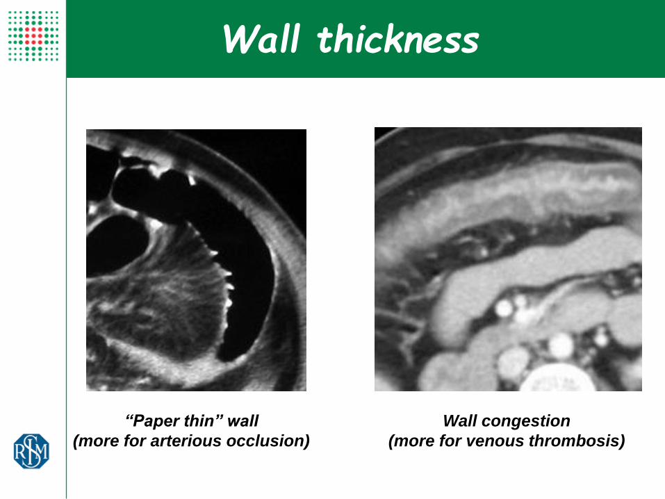
Wall thickness
“Paper thin” wall
(more for arterious occlusion)
Wall congestion
(more for venous thrombosis)

Length of involvement
Wall thickness
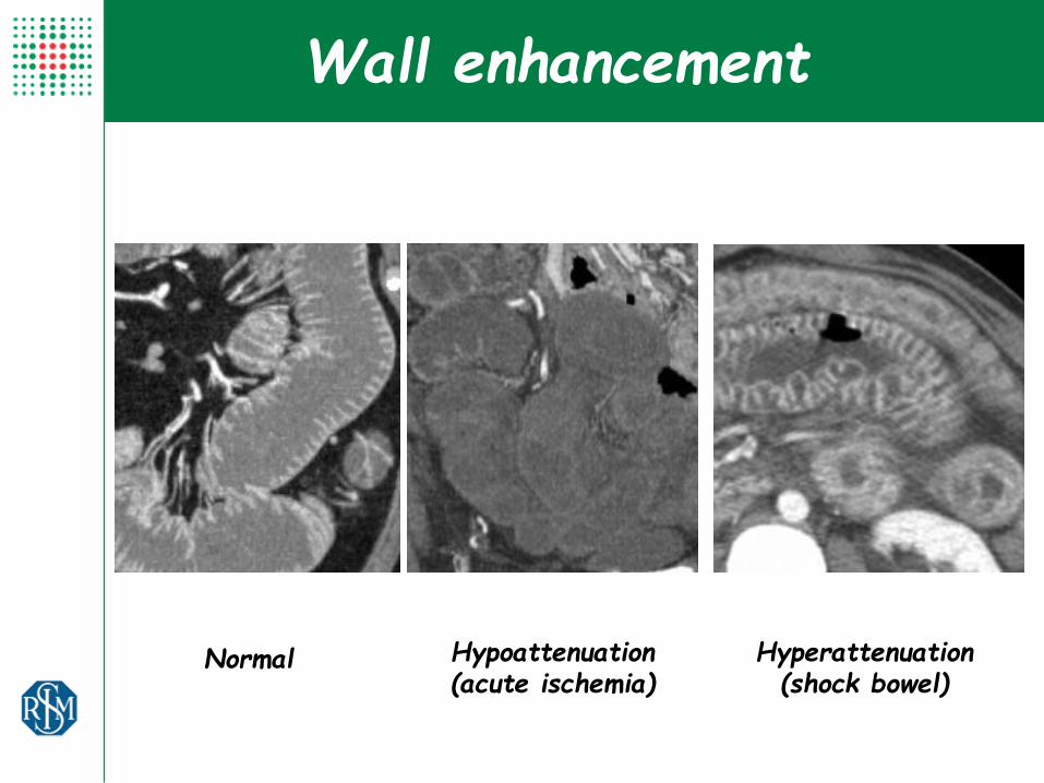
Normal Hypoattenuation (acute ischemia)
Hyperattenuation (shock bowel)
Wall enhancement

Mucosal
enhancement
Wall enhancement
Sub-mucosal
congestion Poor enhancement
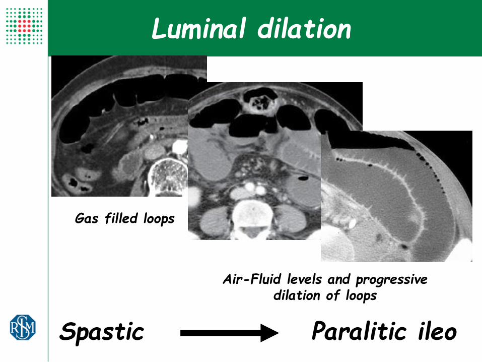
Luminal dilation
Gas filled loops
Air-Fluid levels and progressive dilation of loops
Spastic Paralitic ileo

Mesenteric congestion
Edema and venous engorgement (more frequent in MVT or in case of riperfusion failure)

Ascites
Index of severity only in case of mesenteric arterial occlusion
(not for MVT and IC)

Pneumatosis

Pneumatosis
Pneumatosis Intestinalis in the Adult: Benign to Life-Threatening Causes Lisa M. et al. AJR 2007
Secondary to transmural bowel infarction (wall air dissection)
Association with portomesenteric venous gas !!!

Pseudopneumatosis
Gas bubbles are trapped between the fecal debris and the bowel wall
String of pearls sign: Entrapment of air bubbles between the folds
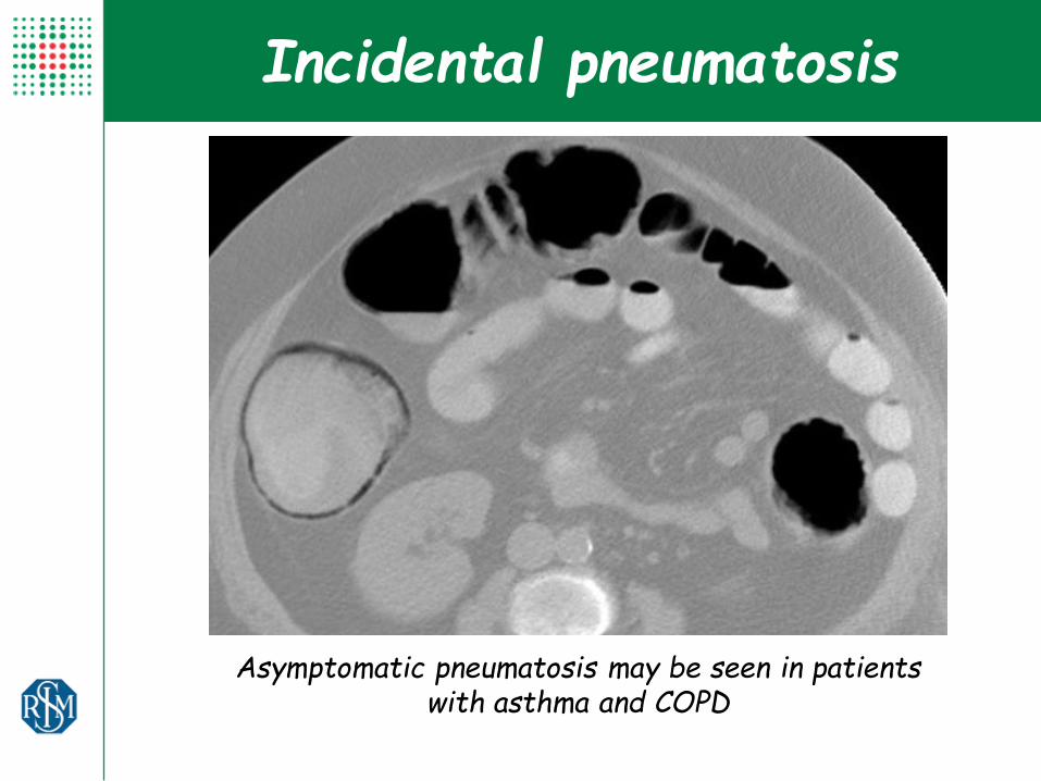
Asymptomatic pneumatosis may be seen in patients with asthma and COPD
Incidental pneumatosis
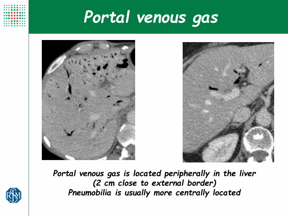
Portal venous gas is located peripherally in the liver (2 cm close to external border)
Pneumobilia is usually more centrally located
Portal venous gas

Pz di 84 aa
Algie addominali con diarrea e vomito
Leucocitosi (20.96) e aumento PCR, mioglobina e LDH
Case 1
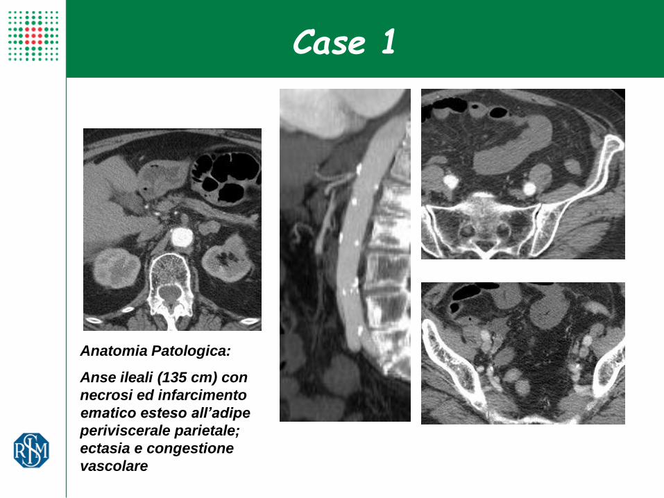
Anatomia Patologica:
Anse ileali (135 cm) con
necrosi ed infarcimento
ematico esteso all’adipe
periviscerale parietale;
ectasia e congestione
vascolare
Case 1
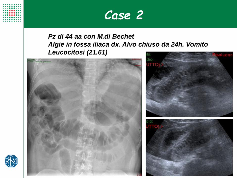
Pz di 44 aa con M.di Bechet
Algie in fossa iliaca dx. Alvo chiuso da 24h. Vomito
Leucocitosi (21.61)
Case 2

Terapia conservativa
Progressiva ricanalizzazione parziale della TVP ai controlli
CEUS e TC.
Case 2

Pz di 48 aa
Addome acuto con diarrea. Peristalsi assente.
Case 3
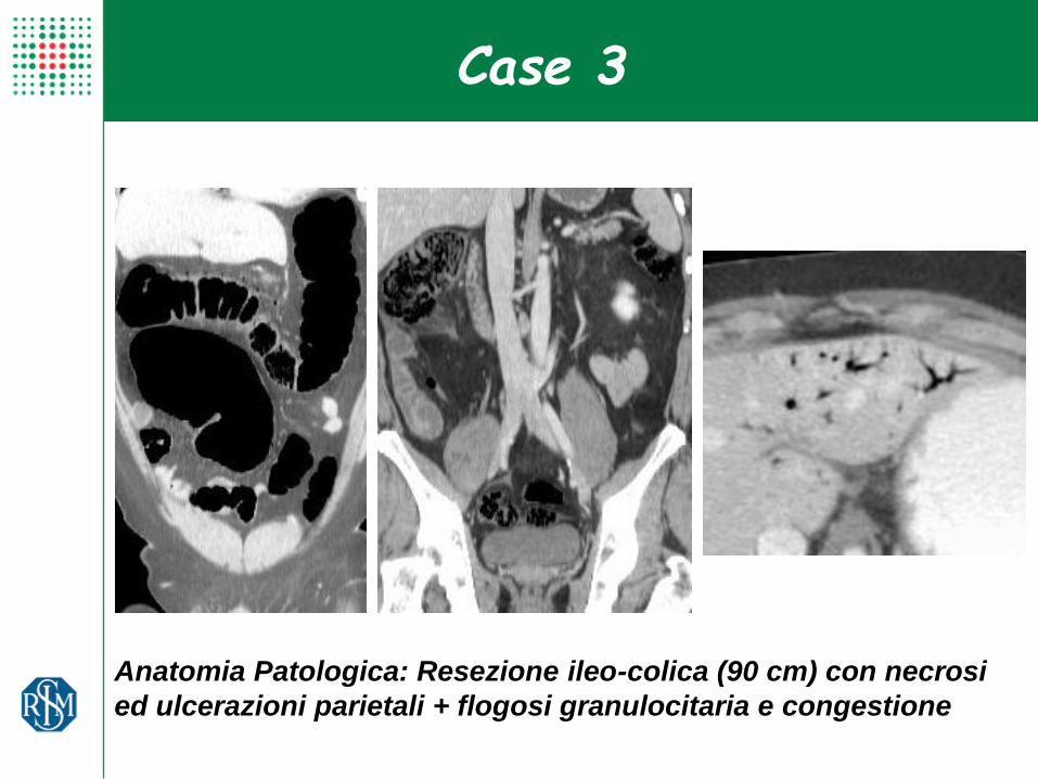
Anatomia Patologica: Resezione ileo-colica (90 cm) con necrosi
ed ulcerazioni parietali + flogosi granulocitaria e congestione
Case 3
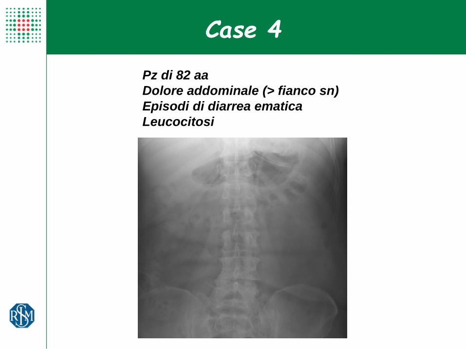
Case 4
Pz di 82 aa
Dolore addominale (> fianco sn)
Episodi di diarrea ematica
Leucocitosi
Case 4

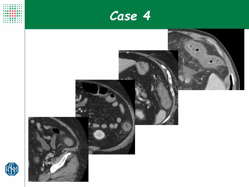
Mucosa colica congesta con aspetti microerosivi, facilmente sanguinante
Flessura splenica
Terapia conservativa
Case 4

What about Ischemic Colitis ?
What about Ischemic Colitis ?

Severe gangrenous (20%)
Ischemic colitis (IC)
Non-gangrenous (80%)
Acute
Sub-acute
Chronic
Reversible Irreversible
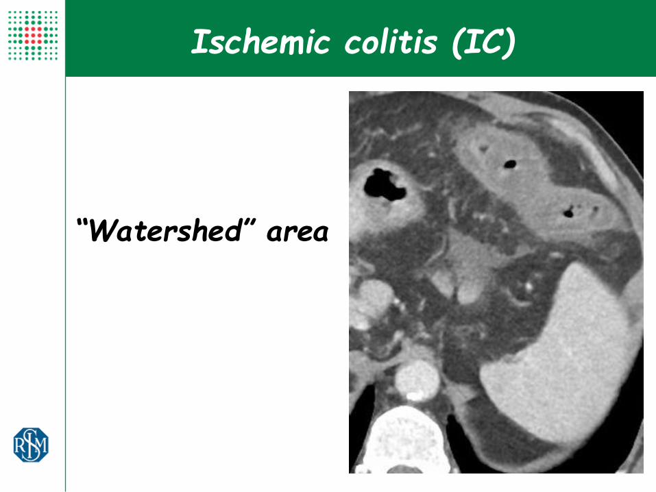
“Watershed” area
Ischemic colitis (IC)
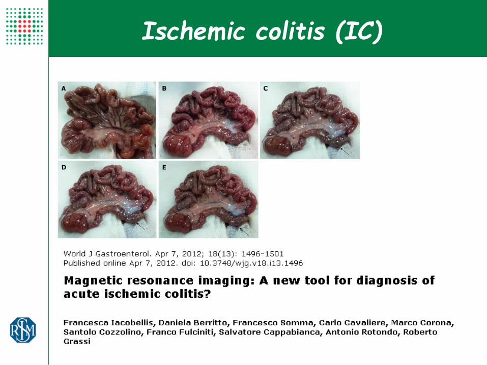
Ischemic colitis (IC)

Ischemic colitis (IC)
Acute
Pericolic fluid
“Paper-thin wall”
Hypotonic wall
CT Findings in Acute, Subacute, and Chronic Ischemic Colitis: Suggestions for Diagnosis F.Iacobellis, D.Berritto, D. Fleischmann, G.Gagliardi, A.Brillantino, M.A Mazzei, and R.Grassi
BioMed Res INT 2014

Ischemic colitis (IC)
Uneffective Reperfusion
Bowel wall remains hypodense, unthickened or thinned Fluid increases
Sub-acute
CT Findings in Acute, Subacute, and Chronic Ischemic Colitis: Suggestions for Diagnosis F.Iacobellis, D.Berritto, D. Fleischmann, G.Gagliardi, A.Brillantino, M.A Mazzei, and R.Grassi
BioMed Res INT 2014

Ischemic colitis (IC)
Sub-acute
Peritoneal fluid
resorption
Wall thickening
(target sign)
CT Findings in Acute, Subacute, and Chronic Ischemic Colitis: Suggestions for Diagnosis F.Iacobellis, D.Berritto, D. Fleischmann, G.Gagliardi, A.Brillantino, M.A Mazzei, and R.Grassi
BioMed Res INT 2014
Effective Reperfusion

Ischemic colitis (IC)
Chronic
No pericolic fluid
Wall fibrotic thickening
BioMed Res INT 2014
CT Findings in Acute, Subacute, and Chronic Ischemic Colitis: Suggestions for Diagnosis F.Iacobellis, D.Berritto, D. Fleischmann, G.Gagliardi, A.Brillantino, M.A Mazzei, and R.Grassi

Imaging findings often suggest advanced ischemia (second or third stage)
Need of new tests for better and earlier detection of ischemia
Future trends
A 24-h delay on diagnosys decreases survival rates by 20%

Bowel enhancement ??
Mayo Clin Proc. Jan 2016
Quantitative MDCT measurements of bowel wall attenuation
(Enhancing Ratio)
Low Enhancing Ratio value predicts the presence of an ischemic bowel
segment.

Dual-energy CT significantly improved the conspicuity of the ischemic bowel
compared with conventional CT
Dual-energy CT

….and MRI ??
Overall sensitivity and specificity were 100% and 95%
Risk of overgrading
MRA is useful in evaluation of pts with
suspected mesenteric ischemia.

….and MRI ??
High concordance CT/MRA for vascular evaluation. Reviewed CT scans were sufficient to assess patency of the mesenteric vasculature, but vascular findings were not reported in most cases. A direct description within the report may have obviated the request for further MR imaging. MRA adds little value after portal venous CT in assessing bowel ischemia.

MEMRI: able to distinguish between normal and ischemic
small bowel for AMI.
MEMRI at a low dose of Mn2+ reveals differences of
relaxivity between normal and ischemic small intestines after
occlusion of SMA.
….and MRI ??
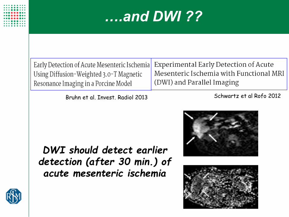
….and DWI ??
DWI should detect earlier detection (after 30 min.) of acute mesenteric ischemia
Bruhn et al. Invest. Radiol 2013 Schwartz et al Rofo 2012

Acute Mesenteric Ischemia Biomarkers like fatty acid binding protein (I-FABP) or procalcitonin (PCT)
shows promise for detecting vascular ischaemia
….biomarkers ??