sequence and structural determinants of human …d-scholarship.pitt.edu/29420/1/art%3a10.1186... ·...
TRANSCRIPT

Mitra et al. Retrovirology (2015) 12:3 DOI 10.1186/s12977-014-0130-8
RESEARCH Open Access
Sequence and structural determinants of humanAPOBEC3H deaminase and anti-HIV-1 activitiesMithun Mitra1,6, Dustin Singer1, Yu Mano2, Jozef Hritz3,4,7, Gabriel Nam1, Robert J Gorelick5, In-Ja L Byeon3,4,Angela M Gronenborn3,4, Yasumasa Iwatani2 and Judith G Levin1*
Abstract
Background: Human APOBEC3H (A3H) belongs to the A3 family of host restriction factors, which are cytidinedeaminases that catalyze conversion of deoxycytidine to deoxyuridine in single-stranded DNA. A3 proteins containeither one (A3A, A3C, A3H) or two (A3B, A3D, A3F, A3G) Zn-binding domains. A3H has seven haplotypes (I-VII)that exhibit diverse biological phenotypes and geographical distribution in the human population. Its singleZn-coordinating deaminase domain belongs to a phylogenetic cluster (Z3) that is different from the Z1- andZ2-type domains in other human A3 proteins. A3H HapII, unlike A3A or A3C, has potent activity against HIV-1.Here, we sought to identify the determinants of A3H HapII deaminase and antiviral activities, using site-directedsequence- and structure-guided mutagenesis together with cell-based, biochemical, and HIV-1 infectivity assays.
Results: We have constructed a homology model of A3H HapII, which is similar to the known structures of otherA3 proteins. The model revealed a large cluster of basic residues (not present in A3A or A3C) that are likely to beinvolved in nucleic acid binding. Indeed, RNase A pretreatment of 293T cell lysates expressing A3H was shownto be required for detection of deaminase activity, indicating that interaction with cellular RNAs inhibits A3Hcatalytic function. Similar observations have been made with A3G. Analysis of A3H deaminase substrate specificitydemonstrated that a 5′ T adjacent to the catalytic C is preferred. Changing the putative nucleic acid bindingresidues identified by the model resulted in reduction or abrogation of enzymatic activity, while substitutingZ3-specific residues in A3H to the corresponding residues in other A3 proteins did not affect enzyme function.As shown for A3G and A3F, some A3H mutants were defective in catalysis, but retained antiviral activity againstHIV-1vif (−) virions. Furthermore, endogenous reverse transcription assays demonstrated that the E56A catalyticmutant inhibits HIV-1 DNA synthesis, although not as efficiently as wild type.
Conclusions: The molecular and biological activities of A3H are more similar to those of the double-domainA3 proteins than to those of A3A or A3C. Importantly, A3H appears to use both deaminase-dependentand -independent mechanisms to target reverse transcription and restrict HIV-1 replication.
Keywords: HIV-1, APOBEC3H, Homology model, Deaminase activity, Antiviral activity, Deaminase-independentrestriction, Reverse transcription
BackgroundThe human APOBEC3 (A3) family consists of seven cyti-dine deaminases that catalyze the conversion of deoxycy-tidine (dC) to deoxyuridine (dU) in single-stranded (ss)DNA, thereby inducing G-to-A hypermutation in double-stranded DNA [1-5]. A3 proteins play an important role
* Correspondence: [email protected] on Viral Gene Regulation, Program in Genomics of Differentiation,Eunice Kennedy Shriver National Institute of Child Health and HumanDevelopment, National Institutes of Health, Bethesda, MD 20892-2780, USAFull list of author information is available at the end of the article
© 2015 Mitra et al.; licensee BioMed Central. TCommons Attribution License (http://creativecreproduction in any medium, provided the orDedication waiver (http://creativecommons.orunless otherwise stated.
in the innate immune defense system by inhibiting abroad range of exogenous viruses such as human im-munodeficiency virus type 1 (HIV-1) (reviewed in refs.[6-13]), human T-lymphotropic virus type 1 (HTLV-1)[14,15], and hepatitis B virus (HBV) [16,17] as well asendogenous retrotransposons such as LINE-1 and Aluelements (reviewed in refs. [7,18]). These proteins con-tain either one (A3A, A3C, and A3H) or two (A3B,A3D (formerly known as A3D/E), A3F, and A3G) Zn-binding domains with the conserved motif HX1EX23-24
CX2-4C (X is any amino acid) [19] (reviewed in refs.
his is an Open Access article distributed under the terms of the Creativeommons.org/licenses/by/4.0), which permits unrestricted use, distribution, andiginal work is properly credited. The Creative Commons Public Domaing/publicdomain/zero/1.0/) applies to the data made available in this article,

Mitra et al. Retrovirology (2015) 12:3 Page 2 of 15
[20,21]). The histidine and two cysteines coordinate aZn ion, while the glutamic acid residue is thought toact as a proton shuttle during catalysis [6,22]. Based onphylogenetic analysis, the Zn-binding domains werefurther classified into the following groups: Z1 (A3Aand C-terminal domains (CTD) of A3B and A3G), Z2(A3C, N-terminal domains (NTD) of A3B and A3G,and both NTD and CTD of A3D and A3F), and Z3(A3H) [23,24].A3H is the most divergent member of the A3 family
and has a single Zn-binding domain that belongs tothe unique Z3 group [24,25]. The A3H message under-goes alternative splicing to generate variants containingdistinct C-terminal regions [26,27]. Furthermore, unlikeother A3 genes, A3H is present in the human popula-tion as different haplotypes containing functional poly-morphisms. At present, seven haplotypes of A3H (HapI-VII) have been identified that differ in their antiviralactivities: only Hap II, Hap V, and Hap VII are stablyexpressed and are able to restrict Vif-deficient HIV-1[26-29]. Interestingly, the distribution of A3H haplotypesin the human population is correlated with geographicallocation [26,29]. For example, a higher frequency of HapIIis present in Africa, compared to Europe and Asia, pos-sibly due to a greater selection pressure against pathogensendogenous to that region [26].HIV-1 Vif, which counteracts antiviral activity by pro-
moting proteasomal degradation of A3C, A3D, A3F, andA3G, exhibits different degrees of potency against theindividual A3H haplotypes [12,29-32]. In cell-based as-says, the sensitivity of antiviral A3H HapII towards Vifwas shown to be dependent upon the Vif subtype [33,34]and a remarkable study involving recently infectedHIV-1 patients revealed adaptive changes in viral Vifsequences that were attributed to the presence of thedifferent antiviral A3H haplotypes [32]. These observa-tions provide strong evidence for a significant role ofA3H as an antiviral defense protein.A3 proteins deaminate dC residues in a sequence-
specific manner. For example, A3A exhibits a greaterpreference for the dC in the center of a TCA target[35-42], while A3G specifically deaminates the dC in aCCC motif [21,43-45]. Evaluation of the structural basisof the sequence specificity suggested that it is deter-mined by the architecture of the active site and sur-rounding amino acids, in particular, residues in loop 7[41-46] (reviewed in refs. [21,47]). Although, A3H isknown to deaminate dC in TC motifs of HIV-1 minus-strand DNA [27,48], a detailed investigation of the nu-cleotide context immediately 5′ and 3′ of the dC onsequence-specific deamination has not been reported.In addition, the amino acid residues that are importantfor A3H deaminase activity and the role of structure indictating biological function have not been investigated.
In the present study, we focus on the biochemical andstructural determinants of A3H HapII (to be referred toas “A3H”) deaminase and antiviral activities, using site-directed and structure-guided mutagenesis. We haveconstructed a homology model of A3H and find that theA3H structure, as expected, is similar to the knownstructures of A3A [41], A3C [49], A3G-CTD [43,50-53],and A3F-CTD [54,55], with differences mainly in flexibleloop regions. Our model resembles the ones generatedby (i) MODELLER [56], based on the A2 and A3G-CTDstructures [57], and (ii) the automated structure-homology-modeling server, SWISS-MODEL, using the A2 structure[13], although details may be different. Interestingly, ourmodel also reveals a large cluster of basic residues, whichis not present in other A3 deaminase-active domains, andis consistent with the observation that deaminase activityin cell-free extracts is detected only after removal of RNAby treatment with RNase A. In addition, we have evalu-ated the deaminase and antiviral activities of a series ofA3H mutants. Although these activities can be correlatedin most cases, a significant number of mutants lacking en-zymatic activity are still able to inhibit HIV-1 replication,albeit at a lower efficiency than wild type (WT). Thisresult raises the possibility that A3H restricts HIV-1 bycatalytic-dependent and -independent mechanisms. In-deed, assays of endogenous reverse transcription (ERT)support this hypothesis. Taken together, our findingsprovide new insights into the role of A3H as a naturallyoccurring human restriction factor and should contrib-ute to continuing efforts to combat HIV infection inthe African human population.
ResultsSequence- and structure-based design of A3H mutantsIn this work, we set out to investigate the determinantsof A3H cytidine deaminase and antiviral activities, usinga mutagenic approach. Given A3H's unique Z3-type Zn-binding domain [24], we initially carried out a sequencecomparison of the Z3 domain of A3H with the Z1 andZ2 domains of other A3 proteins to identify conservedand distinct regions in A3H (Additional file 1: Figure S1).Sequence identities range from 28-43% and the Z3 do-main shares the greatest identity with the Z2 domains ofA3C and A3F-CTD and the least with A3D-NTD. The se-quence alignment also identified four residues unique tothe Z3 domain: T81, L102, S109, and V135, which are re-placed by S, V, A, and I in other Z domains.A more extensive sequence alignment was performed by
comparing the residues in the complete A3H protein withthe sequences of A3 proteins whose three-dimensionalstructures have been solved at high resolution, i.e.,A3A [41], A3C [49], A3F-CTD [54,55] and A3G-CTD[43,50-53] (Figure 1). The overall sequence identity bet-ween A3H and each of these proteins is very similar and
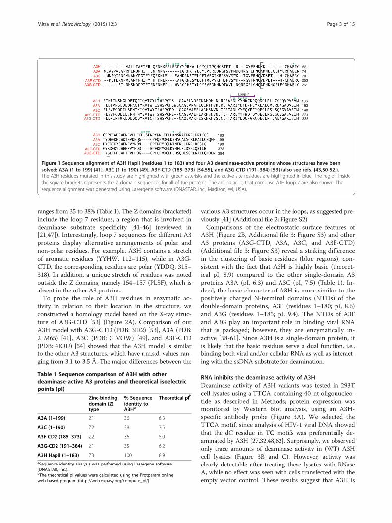
Figure 1 Sequence alignment of A3H HapII (residues 1 to 183) and four A3 deaminase-active proteins whose structures have beensolved: A3A (1 to 199) [41], A3C (1 to 190) [49], A3F-CTD (185–373) [54,55], and A3G-CTD (191–384) [53] (also see refs. [43,50-52]).The A3H residues mutated in this study are highlighted with green asterisks and the active site residues are highlighted in blue. The region insidethe square brackets represents the Z domain sequences for all of the proteins. The amino acids that comprise A3H loop 7 are also shown. Thesequence alignment was generated using Lasergene software (DNASTAR, Inc., Madison, WI, USA).
Mitra et al. Retrovirology (2015) 12:3 Page 3 of 15
ranges from 35 to 38% (Table 1). The Z domains (bracketed)include the loop 7 residues, a region that is involved indeaminase substrate specificity [41-46] (reviewed in[21,47]). Interestingly, loop 7 sequences for different A3proteins display alternative arrangements of polar andnon-polar residues. For example, A3H contains a stretchof aromatic residues (YYHW, 112–115), while in A3G-CTD, the corresponding residues are polar (YDDQ, 315–318). In addition, a unique stretch of residues was notedoutside the Z domains, namely 154–157 (PLSF), which isabsent in the other A3 proteins.To probe the role of A3H residues in enzymatic ac-
tivity in relation to their location in the structure, weconstructed a homology model based on the X-ray struc-ture of A3G-CTD [53] (Figure 2A). Comparison of ourA3H model with A3G-CTD (PDB: 3IR2) [53], A3A (PDB:2 M65) [41], A3C (PDB: 3 VOW) [49], and A3F-CTD(PDB: 4IOU) [54] showed that the A3H model is similarto the other A3 structures, which have r.m.s.d. values ran-ging from 3.1 to 3.5 Å. The major differences between the
Table 1 Sequence comparison of A3H with otherdeaminase-active A3 proteins and theoretical isoelectricpoints (pI)
Zinc-bindingdomain (Z)type
% Sequenceidentity toA3Ha
Theoretical pIb
A3A (1–199) Z1 36 6.3
A3C (1–190) Z2 38 7.5
A3F-CD2 (185–373) Z2 36 5.0
A3G-CD2 (191–384) Z1 35 6.2
A3H HapII (1–183) Z3 100 8.9aSequence identity analysis was performed using Lasergene software(DNASTAR, Inc.).bThe theoretical pI values were calculated using the Protparam onlineweb-based program (http://web.expasy.org/compute_pi/).
various A3 structures occur in the loops, as suggested pre-viously [41] (Additional file 2: Figure S2).Comparisons of the electrostatic surface features of
A3H (Figure 2B, Additional file 3: Figure S3) and otherA3 proteins (A3G-CTD, A3A, A3C, and A3F-CTD)(Additional file 3: Figure S3) reveal a striking differencein the clustering of basic residues (blue regions), con-sistent with the fact that A3H is highly basic (theoret-ical pI, 8.9) compared to the other single-domain A3proteins A3A (pI, 6.3) and A3C (pI, 7.5) (Table 1). In-deed, the basic character of A3H is more similar to thepositively charged N-terminal domains (NTDs) of thedouble-domain proteins, A3F (residues 1–180; pI, 8.6)and A3G (residues 1–185; pI, 9.4). The NTDs of A3Fand A3G play an important role in binding viral RNAthat is packaged; however, they are enzymatically in-active [58-61]. Since A3H is a single-domain protein, itis likely that the basic residues serve a dual function, i.e.,binding both viral and/or cellular RNA as well as interact-ing with the ssDNA substrate for deamination.
RNA inhibits the deaminase activity of A3HDeaminase activity of A3H variants was tested in 293Tcell lysates using a TTCA-containing 40-nt oligonucleo-tide as described in Methods; protein expression wasmonitored by Western blot analysis, using an A3H-specific antibody probe (Figure 3A). We selected theTTCA motif, since analysis of HIV-1 viral DNA showedthat the dC residue in TC motifs was preferentially de-aminated by A3H [27,32,48,62]. Surprisingly, we observedonly trace amounts of deaminase activity in (WT) A3Hcell lysates (Figure 3B and C). However, activity wasclearly detectable after treating these lysates with RNaseA, while no effect was seen with cells transfected with theempty vector control. These results suggest that A3H is
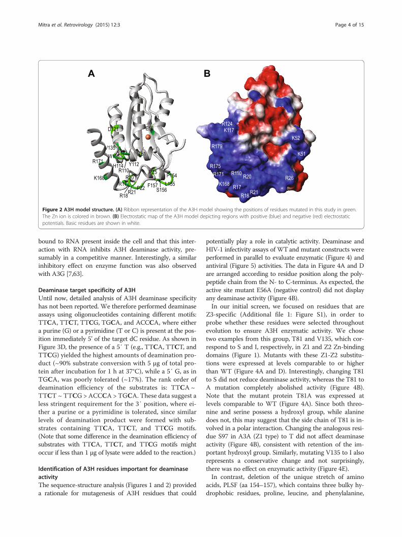
Figure 2 A3H model structure. (A) Ribbon representation of the A3H model showing the positions of residues mutated in this study in green.The Zn ion is colored in brown. (B) Electrostatic map of the A3H model depicting regions with positive (blue) and negative (red) electrostaticpotentials. Basic residues are shown in white.
Mitra et al. Retrovirology (2015) 12:3 Page 4 of 15
bound to RNA present inside the cell and that this inter-action with RNA inhibits A3H deaminase activity, pre-sumably in a competitive manner. Interestingly, a similarinhibitory effect on enzyme function was also observedwith A3G [7,63].
Deaminase target specificity of A3HUntil now, detailed analysis of A3H deaminase specificityhas not been reported. We therefore performed deaminaseassays using oligonucleotides containing different motifs:TTCA, TTCT, TTCG, TGCA, and ACCCA, where eithera purine (G) or a pyrimidine (T or C) is present at the pos-ition immediately 5’ of the target dC residue. As shown inFigure 3D, the presence of a 5′ T (e.g., TTCA, TTCT, andTTCG) yielded the highest amounts of deamination pro-duct (~90% substrate conversion with 5 μg of total pro-tein after incubation for 1 h at 37°C), while a 5′ G, as inTGCA, was poorly tolerated (~17%). The rank order ofdeamination efficiency of the substrates is: TTCA ~TTCT ~ TTCG > ACCCA > TGCA. These data suggest aless stringent requirement for the 3′ position, where ei-ther a purine or a pyrimidine is tolerated, since similarlevels of deamination product were formed with sub-strates containing TTCA, TTCT, and TTCG motifs.(Note that some difference in the deamination efficiency ofsubstrates with TTCA, TTCT, and TTCG motifs mightoccur if less than 1 μg of lysate were added to the reaction.)
Identification of A3H residues important for deaminaseactivityThe sequence-structure analysis (Figures 1 and 2) provideda rationale for mutagenesis of A3H residues that could
potentially play a role in catalytic activity. Deaminase andHIV-1 infectivity assays of WTand mutant constructs wereperformed in parallel to evaluate enzymatic (Figure 4) andantiviral (Figure 5) activities. The data in Figure 4A and Dare arranged according to residue position along the poly-peptide chain from the N- to C-terminus. As expected, theactive site mutant E56A (negative control) did not displayany deaminase activity (Figure 4B).In our initial screen, we focused on residues that are
Z3-specific (Additional file 1: Figure S1), in order toprobe whether these residues were selected throughoutevolution to ensure A3H enzymatic activity. We chosetwo examples from this group, T81 and V135, which cor-respond to S and I, respectively, in Z1 and Z2 Zn-bindingdomains (Figure 1). Mutants with these Z1-Z2 substitu-tions were expressed at levels comparable to or higherthan WT (Figure 4A and D). Interestingly, changing T81to S did not reduce deaminase activity, whereas the T81 toA mutation completely abolished activity (Figure 4B).Note that the mutant protein T81A was expressed atlevels comparable to WT (Figure 4A). Since both threo-nine and serine possess a hydroxyl group, while alaninedoes not, this may suggest that the side chain of T81 is in-volved in a polar interaction. Changing the analogous resi-due S97 in A3A (Z1 type) to T did not affect deaminaseactivity (Figure 4B), consistent with retention of the im-portant hydroxyl group. Similarly, mutating V135 to I alsorepresents a conservative change and not surprisingly,there was no effect on enzymatic activity (Figure 4E).In contrast, deletion of the unique stretch of amino
acids, PLSF (aa 154–157), which contains three bulky hy-drophobic residues, proline, leucine, and phenylalanine,
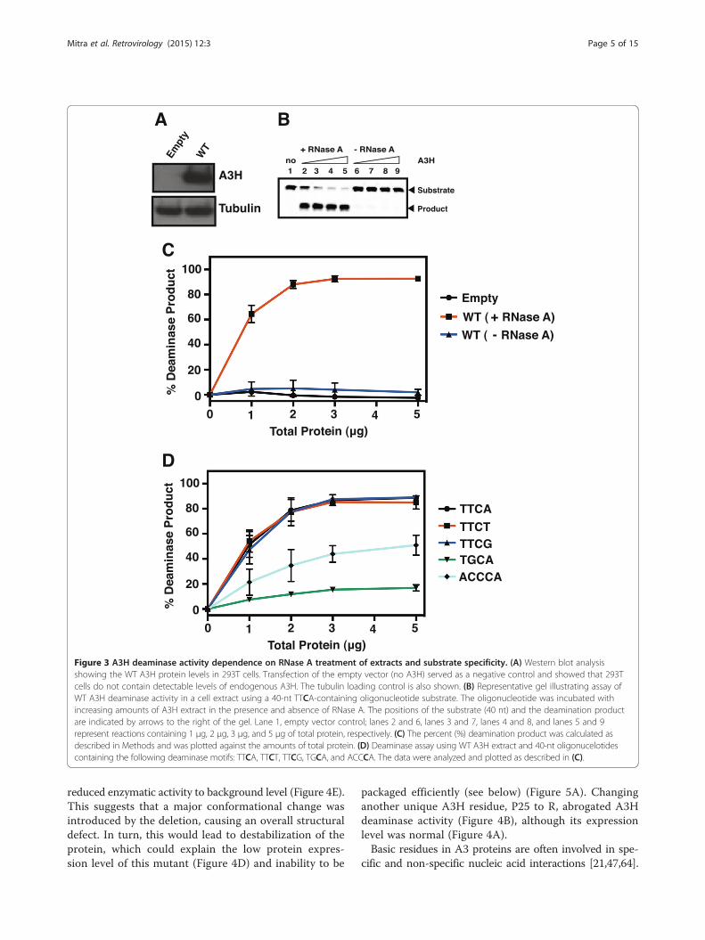
Figure 3 A3H deaminase activity dependence on RNase A treatment of extracts and substrate specificity. (A) Western blot analysisshowing the WT A3H protein levels in 293T cells. Transfection of the empty vector (no A3H) served as a negative control and showed that 293Tcells do not contain detectable levels of endogenous A3H. The tubulin loading control is also shown. (B) Representative gel illustrating assay ofWT A3H deaminase activity in a cell extract using a 40-nt TTCA-containing oligonucleotide substrate. The oligonucleotide was incubated withincreasing amounts of A3H extract in the presence and absence of RNase A. The positions of the substrate (40 nt) and the deamination productare indicated by arrows to the right of the gel. Lane 1, empty vector control; lanes 2 and 6, lanes 3 and 7, lanes 4 and 8, and lanes 5 and 9represent reactions containing 1 μg, 2 μg, 3 μg, and 5 μg of total protein, respectively. (C) The percent (%) deamination product was calculated asdescribed in Methods and was plotted against the amounts of total protein. (D) Deaminase assay using WT A3H extract and 40-nt oligonucelotidescontaining the following deaminase motifs: TTCA, TTCT, TTCG, TGCA, and ACCCA. The data were analyzed and plotted as described in (C).
Mitra et al. Retrovirology (2015) 12:3 Page 5 of 15
reduced enzymatic activity to background level (Figure 4E).This suggests that a major conformational change wasintroduced by the deletion, causing an overall structuraldefect. In turn, this would lead to destabilization of theprotein, which could explain the low protein expres-sion level of this mutant (Figure 4D) and inability to be
packaged efficiently (see below) (Figure 5A). Changinganother unique A3H residue, P25 to R, abrogated A3Hdeaminase activity (Figure 4B), although its expressionlevel was normal (Figure 4A).Basic residues in A3 proteins are often involved in spe-
cific and non-specific nucleic acid interactions [21,47,64].

Figure 4 Deaminase activities of A3H WT and mutants. (A) and (D) Western blot analysis showing the amounts of A3H WT and mutants andA3A S97T in 293T cells. (B, C, E, F) Deaminase assays using 293T cell extracts expressing A3H WT and mutants and A3A S97T. The deaminaseassays were performed as described in Methods.
Mitra et al. Retrovirology (2015) 12:3 Page 6 of 15
To examine the role of these residues in A3H deaminaseactivity (Figure 4C and F) and to determine whether thelocation of these residues in the A3H model structure isrelated to their function, we focused on residues in the“basic patch” (Figure 2B): R17, R18, R20, R21, R110, K168,and R171. Note that R18, R20, and R21 are not present inother A3 proteins (Figure 1). We constructed a single mu-tant with an R→D change (R110D) as well as double mu-tants R17D/R18D, R20D/R21D, and K168A/R171A. Withthe exception of R17D/R18D and R110D, the mutant pro-teins were efficiently expressed (Figure 4A and D), but al-most all lacked deaminase activity (Figure 4C, 4E, and F),even at high amounts of total protein. Two mutants, del(20–22) (Figure 4C) and K168A/R171A (Figure 4E), dis-played greatly reduced, but measurable activity (~15%and ~11% product, respectively, at 5 μg total protein).These results suggest that positive charges are necessaryfor binding of A3H to the ssDNA substrates and thatthe introduction of a single negative charge in the basic
patch disrupts the favorable charge-charge interaction.The change to the non-polar alanine or deletion of resi-dues 20–22, which are present only in A3H as a uniqueinsertion, is less detrimental. Taking all of the abovedata together, it appears likely that these basic residuesform part of the A3H nucleic acid binding interface.Another region of interest, loop 7 (Figure 1), which in other
A3 proteins has been shown to be important for substratebinding and recognition [41-46] (reviewed in refs. [21,47])was also subjected to mutagenesis. Several of the mutant pro-teins e.g., W115A, W115F, del(115), and Y113D/H114D/W115Q (YHW/DDQ) were expressed at low levels comparedto WTA3H (Figure 4D), possibly resulting from reduced pro-tein stability due to removal of the large tryptophan sidechain. Changing residues Y112, Y113, and H114, e.g., Y112A,Y113D/H114D (Figure 4F), and YHW/DDQ, which intro-duces polar residues from the A3G-CTD loop 7 (Figure 4E),led to the complete loss of deaminase activity. A similar resultwas obtained when W115 was deleted (del115) or changed to

0.1
1
100
% In
fect
ivit
y
10
B
WTVector Y113D/H114D
W115A W115F del(115)
0.1
1
100
% In
fect
ivit
y
10
C
WTVector
R17D/R18D
R20D/R21D
del(20-22)
0.1
1
100
% In
fect
ivit
y
10
D
WTVector P25R E56AT81S T81A
A3H
CA (p24)
A
Empt
yW
TR17
D/R18
Dde
l(20-
22)
R20D/R
21D
Y113
D/H11
4DW
115A
W11
5Fde
l(154
-157
)K16
8A/R
171A
E56A
T81A
Empt
yW
TA3H
HIV-1 vif(-) No
A3H
Gag (Pr55 )
Tubulin
gag
Virion
Cell
del(154-157)
V135I Y112A K168A/R171A
Figure 5 Antiviral activities of A3H WT and mutants. (A) Western blot analysis showing the amounts of A3H WT and mutants in HIV-1vif (−)virions and 293T cells. 293T cells were transfected with HIV-1vif (−) and A3H WT or mutant plasmids at a 1:1 ratio (1 μg each for determination ofexpression in cell extracts and 8 μg each for determination of A3H in viral lysates). Cell extracts (10 μg of total protein) as well as viral lysates(8 μl of viral pellet resuspended in 200 μl of loading buffer) were subjected to Western blot analysis. Viral lysates were probed with antibodies tothe N-terminal FLAG tag of A3H and HIV-1 CA; cell extracts were probed with antibodies to the N-terminal FLAG tag of A3H, HIV-1 Gag (Pr55gag),and tubulin. Controls: Left side, EMPTY refers to HIV-1vif (−) and empty vector (pTR600); Right side, EMPTY refers to empty vector alone; WT, refersto A3H plasmid DNA alone. (B-D) Antiviral activity was determined as described in Methods. The gray and black bars respresent transfection with0.1 μg or 1 μg of the indicated A3H plasmid, respectively.
Mitra et al. Retrovirology (2015) 12:3 Page 7 of 15
alanine (W115A) (Figure 4F). However, changing trypto-phan to another aromatic residue, phenylalanine (W115F)(Figure 4F), led to only partial loss of deaminase activity,suggesting that these aromatic residues could be involvedin base stacking interactions with the nucleic acids. Finally,we tested the deaminase activity of W115A, del115,Y113D/H114D, YHW/DDQ, and R110D at 10 μg total pro-tein, but again, no activity was observed (data not shown).Collectively, these results suggest that the residues in A3Hloop 7 are also likely to participate in specific interactionswith nucleic acids.
Role of A3H residues in A3H antiviral activityInhibition of HIV-1vif (−) replication by A3F and A3G is me-diated by both deaminase-dependent and -independent mech-anisms [20,61,65-79]. In an early study, it was concluded thatA3H antiviral activity is deaminase-independent [80], but otherreports indicated that this activity is dependent on catalysis[25,27]. It was therefore of interest to evaluate whether thepresence or absence of deaminase activity (Figure 4) could becorrelated with A3H antiviral activity (Figure 5) in our system.To determine whether the inability of certain mu-
tants to restrict HIV-1 replication was due to a defect
Mitra et al. Retrovirology (2015) 12:3 Page 8 of 15
in A3H packaging, Western blot analysis was performed(Figure 5A). Virions were probed for capsid protein (CA)as well as for A3H. For comparison, expression levels ofA3H and tubulin in cells were also measured and the datawere similar to the results in Figure 4A and D. Interest-ingly, several mutants that were expressed poorly in cells,packaged little or no A3H in virions (Figure 5A). Thesemutants include: R17D/R18D, Y113D/H114D, W115A,and del(154–157). Although W115F exhibited lower levelsof protein than WT (Figures 4D and Figure 5A), a signifi-cant amount of A3H was encapsulated (Figure 5A).Single-cycle infectivity assays of virions produced in
cells expressing WT and mutant A3H proteins were per-formed using two different amounts of A3H plasmid(0.1 μg and 1 μg). Under conditions where the 0.1 μgdose was used, the antiviral activities of the mutantscould be divided into three groups: (1) mutants thatshowed little or no deaminase or antiviral activities (i.e.,having values similar to the empty vector control), suchas R17D/R18D, R20D/R21D, del(20–22) (low level of de-aminase activity), Y113D/H114D, W115A, W115F (re-duced level of deaminase activity), del(115), and del(154–157) (Figure 5B); (2) mutants with WT levels ofdeaminase activity and appreciable antiviral activity, al-beit lower than that of WT, such as T81S and V135I(Figure 5C); and (3) mutants completely lacking (P25R,E56A, T81A, Y112A) or having reduced levels (K168A/R171A) of deaminase activity that retain antiviral activity(Figure 5D).The results obtained with mutants in groups 1 and 2
are consistent with deaminase-dependent antiviral activity,since group 1 mutants have neither activity and group 2mutants have both. With the exception of R20D/R21D,del (20–22), and W115F, all of the group 1 mutants thatwere analyzed by Western blot exhibited packaging de-fects (see above), which would account for their lack ofvirion-associated deaminase and anti-HIV activities. Inter-estingly, the results with group 3 mutants were discordantand suggest that A3H may also utilize a deaminase-independent mechanism for HIV-1 restriction. Note thatthe levels of antiviral activity for these deaminase-negativemutants were still lower than WT values (0.1 μg condi-tion), suggesting that deaminase activity is indeed requiredfor maximal activity.The antiviral activities of WT and a majority of the
mutants increased upon increasing the transfected plas-mid amount to 1 μg. Surprisingly, the antiviral activitiesof T81S and Y112A were similar to WT levels under thiscondition. The behavior of Y112A in our study differedfrom that of A3H Hap VII Y112A, which although ex-pressed efficiently in cells, was poorly packaged andexhibited a very low level of anti-HIV-1 activity [29].The explanation for this difference is not clear. We alsoperformed a side-by-side comparison of the activities
of the deaminase-negative catalytic mutants of A3G(E259Q) and A3H (E56A) as well as the respective WTs(Additional file 4: Figure S4) and found that both A3Hand A3G WT and mutant samples displayed dose-dependent inhibition of HIV-1 infectivity. This suggeststhat A3G and A3H utilize a common mechanism forantiviral activity.
Mechanism of A3H antiviral activityA3G and A3F deaminase-independent anti-viral ac-tivity targets nascent DNA synthesis during reversetranscription [20,68-79]. To determine whether A3Hdeaminase-independent inhibition of HIV-1 infectivityis also associated with a reduction in viral DNA syn-thesis, we performed ERT assays using WT A3H andthe active site mutant E56A.In our assays, HIV-1vif (−) and A3H plasmids were
transfected at two different ratios: 10:1 or 3:1, respectively(see Methods). Synthesis of R-U5 DNA (minus-strandstrong-stop DNA) and R-5’UTR DNA (plus-strand DNAsynthesized after plus-strand transfer) was measured overa 4-h time interval (Figure 6A and B). With WT A3Husing the 10:1 condition, the levels of R-U5 were de-creased to about 40% of the minus A3H control (100%) at2 h (Figure 6A). The levels were drastically reduced forWT (3:1 condition) (~15% at 2 h) and the time courseshowed no appreciable change in level over the 4-h win-dow. The E56A mutant, which lacks deaminase activity,was also capable of reducing the R-U5 levels to about 60%at 2 h (10:1 condition). At the higher dose (3:1 condition),R-U5 levels were further reduced relative to the control(~25% at 2 h), but the inhibition did not saturate evenafter 4 h, indicating partial inhibition. Similar trends forthe WT and E56A mutant were also observed whensynthesis of R-5′UTR (plus-strand synthesis) was moni-tored (Figure 6B). These results demonstrate that A3Hdeaminase-independent HIV-1 restriction involves in-hibition of viral DNA synthesis.
DiscussionIn this work, we use sequence- and structure-guidedmutagenesis to provide a detailed analysis of A3H deam-inase activity and to correlate enzymatic function withantiviral activity. In addition, we identify the A3H struc-tural elements associated with these activities. We alsoshow that A3H deaminase activity in cell extracts is sup-pressed by binding to cellular RNAs (Figure 3), consist-ent with an earlier report indicating that A3H interactsstrongly with 7SL, Y1, Y3, and Y4 RNAs in 293T cells[57]. Interestingly, extensive mutagenesis studies dem-onstrate that the determinants of deaminase and anti-viral activities are not necessarily the same, as a numberof deaminase-negative mutants retain antiviral activity(Figures 4 and 5). Thus, A3H appears to inhibit HIV-1

Figure 6 ERT assays of virions produced following transfection of 293T cells with HIV-1vif (−) and WT A3H or the E56A active sitemutant. (A and B) Kinetics of DNA synthesis in ERT assays measuring the levels of R-U5 (A) and R-5′UTR (B). The assays were performed asdescribed in Methods. Note that synthesis of R-U5 DNA was more efficient than synthesis of R-5’UTR DNA in the presence of WT A3H under boththe 10:1 and 3:1 conditions: the time required for 50% inhibition was 2 h for R-U5 and 3 h for R-5’UTR over the time course of the analysis.
Mitra et al. Retrovirology (2015) 12:3 Page 9 of 15
infectivity via deaminase-dependent and -independentmechanisms.To assess the molecular and structural properties of
A3H Hap II, we generated a homology model based onthe crystal structure of A3G-CTD (PDB: 3IR2) [53].The electrostatic surface potential of the A3H modelidentified a “large basic patch” (Figure 2B, blue region),containing a cluster of basic residues that could be po-tentially involved in specific binding to ssDNA sub-strates as well as binding to cellular RNAs. While thismanuscript was under review, a paper by Shandilya et al.[81] appeared, presenting homology models of the indi-vidual domains of several A3 proteins, including A3H.An electrostatic map of this A3H model also revealed a
large basic patch. In fact, comparison of the two structuresdid not show any significant structural differences.The basic nature of A3H is important and impacts
catalytic function. Thus, deaminase activity is stronglyinhibited upon mutating a subset of the basic residues(R17, R18, R20, R21, Figure 4C; R110, Figure 4F; K168,R171, Figure 4E), suggesting reduced binding to the 40-nt nucleic acid substrate. Arginine mutants that weretested also lacked antiviral activity (Figure 5B). However,the basic residues may not all function in the same man-ner, since the double mutant K168A/R171A retainedsome deaminase (Figure 4E) as well as restriction activity(Figure 5D). Interestingly, the region corresponding toR17 to P22 in A3H (containing four basic residues) is

Mitra et al. Retrovirology (2015) 12:3 Page 10 of 15
part of an alpha helix (Figure 2), but is not conserved inother A3 domains (Figure 1). In A3A, this region iscompletely missing, while in A3C, A3G-CTD, and A3F-CTD, there is a loss of four (A3C, A3G-CTD) or two(A3F-CTD) basic residues. Deletion of A3H residuesR20, R21, and P22 (the del(20–22) mutant) results insignificant loss, but not complete abolition of deaminaseactivity (Figure 4C), possibly due to perturbation of thelocal structure in the mutant protein. Although this mu-tant is efficiently packaged into virions (Figure 5A), itcannot restrict HIV-1 (Figure 5B).Residues in A3H that are unique to its Z3 domain e.g.,
T81 and V135 are replaced in other active deaminasedomains by S and I respectively, which are chemicallysimilar (Figure 1 and Additional file 1: Figure S1). Thisimplies similarity in function and is consistent with ourfinding that the A3H mutants T81S and V135I, retainenzymatic (Figure 4B and E, respectively) as well as re-striction activity (Figure 5C). However, the T81A mutantin which the hydroxyl group common to T and S, is re-moved, has only background levels of deaminase activity(Figure 4B). This suggests an important structural rolefor the hydroxyl group at this position, possibly partici-pation in hydrogen bonding interactions with a nearbyside chain. In fact, the Oγ atom of S284, the correspond-ing residue in A3G, is within hydrogen bonding distanceto the Y219 HN backbone atom and is also observed inother A3 proteins [41,43,49,54]. It is interesting that des-pite lacking enzymatic activity, the T81A mutant exhibitssignificant antiviral activity (Figure 5D).Although the loop 7 element of A3 proteins plays an
essential role in determining deaminase specificity, eachprotein possesses a unique sequence (Figure 1) and isaffected differently when subjected to mutation. Forexample, as indicated by modeling the A3A/ssDNAcomplex, the A3A loop 7 cluster of residues (127-ARIY-DYDPL-135) interacts with the T (D133) and A (D131,Y132, and D133) nucleotides in the TCA motif, whileD131 makes contacts with the central C [41]. Substitu-tions in all four cluster residues lead to complete loss ofA3A deaminase activity [42]. In the case of the A3G-CTD loop 7 (313-RIYDDQGR-320), mutation of theonly aromatic residue, Y315, negatively impacts deami-nase activity [43]. Additionally, changing both D316 andD317 to R [43] or replacing D317 with the correspon-ding A3A residue (Y132) [46] results in altered deami-nase substrate specificity, as manifested by preferentialdeamination of the central C in the motif CCC ins-tead of the 3′ C (CCC). Exchanging the A3H loop 7(109-SRLYYHWCK-118) cluster residues YHW withthe corresponding A3G-CTD residues, DDQ (YYHW→YDDQ), abrogates deamination activity (Figure 4E). Thiscould reflect a change in substrate specificity or in the de-tails of the nucleic acid binding mode due to a protein
conformational change. Interestingly, a similar switch insequence in the case of A3A (YDYD→YDDD) also re-duces activity to background levels [42].The importance of the A3H W115 residue in loop 7 is
underscored by our finding that mutation of W115 to Aresults in low cellular protein levels (Figures 4D and5A), blocks packaging into virions (Figure 5A), and elim-inates deaminase (Figure 4F) and antiviral (Figure 5B)activities. Wang et al. [29] were unable to detect expres-sion of (Hap VII) W115A in 293T cells, while Zhen et al.[57] reported that (Hap II) W115A is present at lowlevels in cell lysates. However, even when expression isequivalent to that of WT (by transfecting cells with ahigh amount of the Hap II mutant plasmid), W115A isbarely detectable in viral lysates, does not restrict HIV-1,and binds cellular RNAs with greatly reduced efficiency[57]. Collectively, these results lend further support tothe conclusion that the identity of loop 7 residues is crit-ical for enzyme function.Surprisingly, although A3H is highly basic and has
only one domain, it more closely resembles the double-domain proteins A3D, A3F, and A3G than the single-domain A3A and A3C proteins with respect to severalimportant biological properties. For example, like A3D,A3F, and A3G [53,64,82-92], A3H associates in solutionin the absence of nucleic acid [93] and forms multimersand high molecular weight ribonucleoprotein complexesin cells [92,94]. In addition, it restricts the infectivity ofHIV-1vif (−) virions (Figure 5) [26,27,62,80,94]. The anti-HIV-1 activity of A3 proteins A3D, A3F, A3G, and A3Hinvolves (i) packaging of the proteins into the cores ofnascent virions in the producer cell [28,36,95,96]; and(ii) inhibition of viral replication in the target cell(reviewed in ref. [9]). These parameters are linked tosubcellular localization, which varies among the A3proteins. However, in this case too, just as A3D, A3F,and A3G are located in the cytoplasm [82-84,86,92,97-99],A3H haplotypes that have antiviral activity (e.g., Hap II)are predominantly cytoplasmic [28,100].The packaging of antiviral A3 proteins inside the HIV-1
core allows them to interact directly with viral nucleicacids (genomic RNA and nascent DNA synthesized dur-ing reverse transcription) in the presence of nucleocapsidprotein and reverse transcriptase (RT). Here we showthat A3H inhibits viral infectivity by both deaminase-dependent and -independent mechanisms (Figure 5),although it is likely that deaminase-dependent activityis dominant (compare data for WT and the catalyticmutant E56A in Figure 5D, Figure 6, Additional file 4:Figure S4). For A3G, we have proposed a “roadblock”mechanism that is based on its tight nucleic acid bind-ing [61,101] and that is independent of catalytic activity[69,74]. We reasoned that since A3G displays slow on-off binding kinetics [69,79], RT is unable to traverse the

Mitra et al. Retrovirology (2015) 12:3 Page 11 of 15
template when A3G is bound and consequently, RT-catalyzed polymerization is blocked. Recent single mol-ecule stretching studies in support of this mechanismshowed that A3G can transform from a fast enzyme (re-quired for deamination) to a slow enzyme (causing a road-block) during the course of protein oligomerization [79].The highly basic character of the A3H protein (Figure 2B,
Table 1) (like the NTDs of A3D, A3F, and A3G) stronglysuggests that it also binds tightly to viral RNA and DNA,in agreement with studies of A3H binding to cellular RNA[57]. Moreover, the observed reduction in minus- andplus-strand DNA synthesis by RT, even in the absence ofdeaminase activity (Figure 6) and the fact that A3H canmultimerize [92,93] suggest that a roadblock mechanismmight also be relevant to A3H deaminase-independentHIV-1 restriction (Figure 6). Interestingly, although A3Hmediates hypermutation of HIV-1 [27,48,62] and HBV [17]DNA, the antiviral activity of A3H against HTLV-1 doesnot involve editing [15].In summary, A3H as well as A3D, A3F, and A3G con-
stitute a group of A3 proteins used by the human innateimmune system in its arsenal against HIV-1. The select-ive pressure to maintain expression of antiviral haplo-types of A3H in certain populations warrants greaterunderstanding of this protein in terms of its molecularproperties. Here, we present such an analysis and correl-ate enzyme function and antiviral activity. AlthoughA3H is a single-domain protein and the most divergentmember of the A3 family, we show that it utilizes strat-egies similar to those used by other antiviral double-domain A3 proteins (A3D, A3F, and A3G) to counteractHIV-1 infectivity. Knowledge of A3H structure-functionrelationships should be invaluable for the design ofdrugs to modulate A3H deaminase activity and augmentits anti-HIV effect. Furthermore, since A3H is a single-domain protein and contains a unique Z3 domain, itmay present a more specific target than the double-domain antiviral A3 proteins. Thus, taken together, A3Hclearly provides a molecular paradigm to explore theantiviral response of A3 proteins to retroviral pathogens.
ConclusionsBased on a homology model of A3H Hap II and the resultsof extensive mutagenesis, we have identified structural ele-ments and key residues associated with A3H deaminaseand anti-HIV-1 activities. In addition, we provide evidencethat A3H restriction of HIV-1 replication and inhibitionof reverse transcription occur by deaminase-dependentand -independent mechanisms.
MethodsMaterialsDNA oligonucleotides labeled with AlexaFluor 488® wereobtained from Integrated DNA Technologies (Coralville,
IA). The concentration of each oligonucleotide was de-termined by measuring its absorbance at 260 nm, usingthe extinction coefficients provided by the manufacturer.RNase A (endonuclease-free) was purchased from QiagenInc. (Germantown, MD). Escherichia coli uracil DNA gly-cosylase (UDG) was obtained from New England Biolabs(Beverly, MA). Gel loading buffer and nuclease-freewater were purchased from Ambion® (Life Technologies,Grand Island, NY). Anti-A3H sera (p3A3 or p1H6), anti-HIV-1 CA sera (for ERT experiments), and TZM-bl cells(from John C. Kappes, Xiaoyun Wu, and Tranzyme Inc.)[102-104] were obtained from the AIDS Research and Ref-erence Reagent Program (Division of AIDS, NIAID, NIH).An anti-HIV-1 p24 monoclonal antibody was purchasedfrom ZeptoMetrix (Franklin, MA) and was used for detec-tion of CA and Pr55gag in virions and cell lysates, respect-ively (see Figure 5). A monoclonal antibody (Anti-FLAGM2) against a FLAG tag was obtained from Sigma-Aldrich (St. Louis, MO). Anti-tubulin antibody was pur-chased from Abcam (Cambridge, MA).
Construction of the A3H HapII homology modelThe initial A3H Hap II homology model was construc-ted without a Zn atom and was based on the A3G-CTDcrystal structure (PDB: 3IR2) [53] by using MODELLERversion 9v8 [56] and the sequence alignment shown inFigure 1. The Zn atom was subsequently added to thestructure at a position equivalent to that in the structureof A3G-CTD. This was followed by energy minimizationwhere only the Zn ion and the H54, C85, C88 side-chainswere allowed to move. During the energy minimization,the lengths of the coordination bonds were kept as fol-lows: 1.90 Å for the bond between the Zn ion and theH54 Nδ1 atom; and 2.25 Å for the bond between the Znion and the C85 or C88 Sγ atom. The charge of individualatoms and their radius parameters based on an amberforce field [105] were generated by the pdb2pqr program[106]. Partial charges of atoms within the Zn coordinationsite were manually adjusted to +1e for the Zn ion, −0.6efor the Sγ atom and +0.1e for the Cβ atom of the cyste-ines to account for the approximate redistribution ofthe charges between the two reduced cysteines and Znion through the coordination bonds. All structure andelectrostatic potential map figures were generated withMOLMOL [107]. Sequence identity was determined withDNAStar (http://www.dnastar.com/megalign_help/index.html#!Documents/calculationofpercent.htm).
Construction of A3H mutantspTR600 mammalian expression plasmids without an in-sert [108] or containing the A3H HapII coding sequence[27] were a generous gift from Viviana Simon (MountSinai School of Medicine, New York, NY) and had eitherno tag or an N-terminal Flag tag. A3H mutants were all

Mitra et al. Retrovirology (2015) 12:3 Page 12 of 15
constructed in the pTR600 A3H-Flag vector using theQuikChange Lightning Site-Directed Mutagenesis Kit(Agilent Technologies, Santa Clara, CA) or the TagmasterSite-Directed Mutagenesis Kit (GM Biosciences, Rockville,MD). For primer design, the on-line QuikChange PrimerDesign Program provided by the manufacturer or the de-sign guidelines provided with the Tagmaster kit were used.Large-scale plasmid preparations were obtained using theHiSpeed Plasmid Maxi Prep kit (Qiagen, Inc.). The A3Hsequence in each plasmid was verified by DNA sequen-cing performed by ACGT (Wheeling, IL).
Preparation of mammalian cell extractsPropagation of 293T cells, transfection procedure, prep-aration of cell extracts, and determination of proteinconcentration were performed as detailed in Mitra et al.[42]. The expression levels of A3H were estimated bysubjecting ~20 μg of total protein in the extract to Westernblot analysis using the Western Breeze chemiluminescentWestern blot kit (Life Technologies). The primary anti-bodies used for this analysis were anti-A3H or anti-tubulin(loading control).
Deaminase assayPrior to performing the deaminase assay, the 293T cellextract (20 μg) was treated with RNase A (Qiagen, finalconcentration 1 μg/μl) in a 20-μl reaction volume andincubated at 37°C for 15 min, unless indicated otherwise.Details of the deaminase assays and polyacrylamide gelanalysis are described in Mitra et al. [42]. A list of oligo-nucleotides used for the deaminase assays is given inAdditional file 5: Table S1. Throughout the text, the dCresidue that is deaminated is highlighted in bold (C). A40-nt Alexa-Fluor 488-labeled ssDNA (JL913) containingthe TTCA deaminase motif was used as the substrate, un-less specified otherwise. To calculate the percent deami-nase product, the product signal intensity for each lanewas divided by total signal intensity and multiplied by 100.The data presented represent the average of two determi-nations from two independent transfections. Note that theFLAG tag did not interfere with deaminase activity, asuntagged and tagged WT A3H showed similar levels ofactivity (Figure 4C).
HIV-1 infectivity assayTo assay the effect of expressing A3H WT or mutantproteins on HIV-1 infectivity, 293T cells were cotransfectedwith 1.0 μg of pNL4–3vif (−), which was kindly provided byKlaus Strebel (National Institute of Allergy and InfectiousDiseases, National Institutes of Health, Bethesda, MD) and1.0 or 0.1 μg of the pTR600 A3H HapII plasmid (WT ormutant) or pTR600 empty vector (control), using theFuGENE HD transfection agent (Promega, Madison, WI)as previously described [49]. Virus-containing supernatants
were collected 48 h after transfection and filtered. Theamount of CA protein (i.e., p24 antigen) in the supernatantwas determined by ELISA assay (ZeptoMetrix, Buffalo,NY). TZM-bl indicator cells were infected with viralsupernatant containing 10 ng of CA. Relative infectivityexpressed as relative light units (RLU) was measured usingthe Bright-Glo luciferase assay system kit (Promega)and an ARVO MX luminescence counter (PerkinElmer,Waltham, MA). The data represent the results of threeindependent experiments.
ERT assayThe following plasmids were used: pTR600, empty vector;WT A3H HapII expressed as an N-terminal FLAG-taggedprotein in pTR600 (pA3H WT); A3H E56A active sitemutant expressed as an N-terminal FLAG-tagged proteinin pTR600 (pA3H E56A); and pNL4-3vif (−).Transfections were performed as described previously
[109], except that TransIT-293 from Mirus Bio LLC(Madison, WI) was used, according to the manufac-turer’s instructions. Briefly, 100-mm cell culture disheswere seeded with 3 × 10⁵ 293T cells in Dulbecco’s modi-fied Eagle’s Medium with 10% fetal bovine serum. Twodays later, the cells were transfected in duplicate with15 μg of pNL4-3vif (−) and 0, 1.5, or 5 μg of pA3H WTor pA3H E56A plasmid; the molar ratios of pNL4-3vif(−) to A3H plasmid were therefore 10:1 or 3:1, respect-ively. The pTR600 plasmid was the negative control andwas also used to generate a constant level (5 μg) of trans-fected pTR600 DNA, with or without the A3H insert. Cul-ture fluids were changed 24 h after transfection, thenharvested after two consecutive 24-h periods, and passedthrough 0.22 μm filters. Samples from each culture werepooled and were treated sequentially with DNase I andsubtilisin, as described [109,110].ERT reactions were performed without Triton X-100
pretreatment and nucleic acids were isolated as detailed inThomas et al. [109]. Real-time PCR for the detection ofR-U5 and R-5’UTR was performed in duplicate as describedpreviously [111]. The kinetics of DNA synthesis were plot-ted and the data were normalized relative to maximalR-U5 copies at 240 min for pNL4-3vif (−) in the absenceof A3H cotransfected plasmid. Each data point was theresult of two independent transfections, each measuredin duplicate. Error bars represent the standard error ofthe mean.
Additional files
Additional file 1: Figure S1. Sequence alignment of residues in theZn-binding (Z) domains of the seven A3 proteins (A to H). TheZn-coordinating (H and C) and active site (E) residues are highlighted inlight blue. A3H residues that differ from highly conserved residues at thecorresponding positions in the other A3 proteins are shown in lavender.

Mitra et al. Retrovirology (2015) 12:3 Page 13 of 15
The residues in loop 7 are bracketed. The numbers at the end of eachline represent the position in the full-length protein. The percentsequence identity of each Z domain relative to that of A3H (defined as100%) is indicated. The sequence alignment was performed usingLasergene software (DNASTAR, Inc., Madison, WI).
Additional file 2: Figure S2. Superposition of the structures of thecurrent A3H model and other A3 proteins in ribbon representation. Thebackbone traces are colored gray (A3H) and green (other A3 proteins).The Zn ions are shown in brown (A3H) and in coral (other A3 proteins).(A) A3H and A3G-CTD. (B) A3H and A3A. (C) A3H and A3C. (D) A3Hand A3F-CTD.
Additional file 3: Figure S3. Electrostatic surface potential maps ofA3H and other A3 proteins. (A) A3H. (B) A3G-CTD. (C) A3A. (D) A3C.(E) A3F-CTD. Regions with positive and negative electrostatic potentialsare highlighted in blue and red, respectively.
Additional file 4: Figure S4. Comparison of antiviral activities of A3Hand A3G WT and catalytic mutants. Virions produced from 293T cellstransfected with HIV-1vif(−) (1 μg) and either 0.1 (gray bars) or 1 μg(black bars) of the indicated A3H or A3G WT or mutant plasmids.Infectivity was assayed as described in Methods. The catalytic mutantsare E56A (A3H) and E259Q (A3G). A vector control (100% infectivity)was also included.
Additional file 5: Table S1. Oligonucleotides used for deaminaseassays.
AbbreviationsA3: APOBEC3; ss: Single-stranded; HIV-1: Human immunodeficiency virus type1; HTLV-1: Human T-lymphotropic virus type 1; HBV: Hepatitis B virus;CTD: C-terminal domain; NTD: N-terminal domain; WT: Wild type; ERT:Endogenous reverse transcription; CA: Capsid protein; RT: Reverse transcriptase.
Competing interestsThe authors declare that they have no competing interests.
Authors’ contributionsThe study was conceived by MM and JGL. MM, DS, YM, GN, RJG,and YI performed experiments. MM, RJG, YI, and JGL analyzed theexperimental data. JH built the structural model of A3H and MM andJH designed the mutants. I-JLB made the structural figures and I-JLB andJH interpreted the electrostatic surface potential maps. I-JLB, MM, andAMG interpreted the structure-related mutagenesis data. MM, I-JLB, YI,AMG, and JGL wrote the paper. All authors read and approved the finalmanuscript.
AcknowledgementsWe thank Dr. Viviana Simon and Dr. Marcel Ooms for their generous giftof the WT A3H and empty pTR600 plasmids, Dr. Klaus Strebel for kindlyproviding the pNL4-3vif(−) plasmid used in the ERT assays, Drs. Simon, Ooms,and Tiyun Wu for valuable discussion, and the AIDS Research and ReferenceReagent Program, Division of AIDS, NIAID, NIH for antisera and cells, asdetailed in the text. J.H. acknowledges financial support by an InternationalOutgoing Fellowship of the European Community program “Support fortraining and career development of researchers (Marie Curie)", underContract No. PIOF-GA-2009-235902. This work was supported in part by theIntramural Research Program at the National Institutes of Health, EuniceKennedy Shriver National Institute of Child Health and Human Development(MM, DS, GN, and JGL), National Institutes of Health grant P50GM82251(JH, I-JLB, and AMG), and a grant-in-aid for Scientific Research from theMinistry of Education, Culture, Sports, Science, and Technology of Japan(YM and YI). This project has also been funded in whole or in part withfederal funds from the National Cancer Institute, National Institutes of Health,under contract HHSN261200800001E with Leidos Biomedical Research, Inc.The content of this publication does not necessarily reflect the views orpolicies of the Department of Health and Human Services, nor does mentionof trade names, commercial products, or organizations imply endorsementby the U.S. Government (RJG).
Author details1Section on Viral Gene Regulation, Program in Genomics of Differentiation,Eunice Kennedy Shriver National Institute of Child Health and HumanDevelopment, National Institutes of Health, Bethesda, MD 20892-2780, USA.2Clinical Research Center, National Hospital Organization Nagoya MedicalCenter, Nagoya, Aichi 460-0001, Japan. 3Department of Structural Biology,University of Pittsburgh Medical School, Pittsburgh, PA 15261, USA.4Pittsburgh Center for HIV Protein Interactions, University of PittsburghMedical School, Pittsburgh, PA 15261, USA. 5AIDS and Cancer Virus Program,Leidos Biomedical Research, Inc., Frederick National Laboratory for CancerResearch, Frederick, MD 21702-1201, USA. 6Department of Molecular, Celland Developmental Biology, University of California, Los Angeles, CA 90095,USA. 7Department of Structural Biology, CEITEC, Masaryk University, Kamenice5, 625 00 Brno, Czech Republic.
Received: 14 October 2014 Accepted: 17 December 2014
References1. Sheehy AM, Gaddis NC, Choi JD, Malim MH. Isolation of a human gene that
inhibits HIV-1 infection and is suppressed by the viral Vif protein. Nature.2002;418:646–50.
2. Lecossier D, Bouchonnet F, Clavel F, Hance AJ. Hypermutation of HIV-1 DNAin the absence of the Vif protein. Science. 2003;300:1112.
3. Zhang H, Yang B, Pomerantz RJ, Zhang C, Arunachalam SC, Gao L. Thecytidine deaminase CEM15 induces hypermutation in newly synthesizedHIV-1 DNA. Nature. 2003;424:94–8.
4. Suspène R, Sommer P, Henry M, Ferris S, Guétard D, Pochet S, et al.APOBEC3G is a single-stranded DNA cytidine deaminase and functionsindependently of HIV reverse transcriptase. Nucleic Acids Res. 2004;32:2421–9.
5. Yu Q, König R, Pillai S, Chiles K, Kearney M, Palmer S, et al. Single-strandspecificity of APOBEC3G accounts for minus-strand deamination of the HIVgenome. Nat Struct Mol Biol. 2004;11:435–42.
6. Harris RS, Liddament MT. Retroviral restriction by APOBEC proteins. Nat RevImmunol. 2004;4:868–77.
7. Chiu YL, Greene WC. The APOBEC3 cytidine deaminases: an innatedefensive network opposing exogenous retroviruses and endogenousretroelements. Annu Rev Immunol. 2008;26:317–53.
8. Goila-Gaur R, Strebel K. HIV-1 Vif, APOBEC, and intrinsic immunity. Retrovirology.2008;5:51.
9. Malim MH. APOBEC proteins and intrinsic resistance to HIV-1 infection.Philos Trans R Soc Lond B Biol Sci. 2009;364:675–87.
10. Imahashi M, Nakashima M, Iwatani Y. Antiviral mechanism and biochemicalbasis of the human APOBEC3 family. Front Microbiol. 2012;3:250.
11. Duggal NK, Fu W, Akey JM, Emerman M. Identification and antiviral activityof common polymorphisms in the APOBEC3 locus in human populations.Virology. 2013;443:329–37.
12. Desimmie BA, Delviks-Frankenberrry KA, Burdick RC, Qi D, Izumi T, PathakVK. Multiple APOBEC3 restriction factors for HIV-1 and one Vif to rule themall. J Mol Biol. 2014;426:1220–45.
13. Feng Y, Baig TT, Love RP, Chelico L. Suppression of APOBEC3-mediatedrestriction of HIV-1 by Vif. Front Microbiol. 2014;5:450.
14. Sasada A, Takaori-Kondo A, Shirakawa K, Kobayashi M, Abudu A, Hishizawa M,et al. APOBEC3G targets human T-cell leukemia virus type 1. Retrovirology.2005;2:32.
15. Ooms M, Krikoni A, Kress AK, Simon V, Münk C. APOBEC3A, APOBEC3B, andAPOBEC3H haplotype 2 restrict human T-lymphotropic virus type 1. J Virol.2012;86:6097–108.
16. Turelli P, Mangeat B, Jost S, Vianin S, Trono D. Inhibition of hepatitis B virusreplication by APOBEC3G. Science. 2004;303:1829.
17. Köck J, Blum HE. Hypermutation of hepatitis B virus genomes byAPOBEC3G, APOBEC3C and APOBEC3H. J Gen Virol. 2008;89:1184–91.
18. Koito A, Ikeda T. Intrinsic immunity against retrotransposons by APOBECcytidine deaminases. Front Microbiol. 2013;4:28.
19. Jarmuz A, Chester A, Bayliss J, Gisbourne J, Dunham I, Scott J, et al. Ananthropoid-specific locus of orphan C to U RNA-editing enzymes onchromosome 22. Genomics. 2002;79:285–96.
20. Holmes RK, Malim MH, Bishop KN. APOBEC-mediated viral restriction: notsimply editing? Trends Biochem Sci. 2007;32:118–28.
21. Bransteitter R, Prochnow C, Chen XS. The current structural and functionalunderstanding of APOBEC deaminases. Cell Mol Life Sci. 2009;66:3137–47.

Mitra et al. Retrovirology (2015) 12:3 Page 14 of 15
22. Betts L, Xiang S, Short SA, Wolfenden R, Carter Jr CW. Cytidine deaminase.The 2.3 Å crystal structure of an enzyme: transition-state analog complex.J Mol Biol. 1994;235:635–56.
23. LaRue RS, Jónsson SR, Silverstein KAT, Lajoie M, Bertrand D, El-Mabrouk N,et al. The artiodactyl APOBEC3 innate immune repertoire shows evidencefor a multi-functional domain organization that existed in the ancestor ofplacental mammals. BMC Mol Biol. 2008;9:104.
24. LaRue RS, Andrésdóttir V, Blanchard Y, Conticello SG, Derse D, Emerman M,et al. Guidelines for naming nonprimate APOBEC3 genes and proteins.J Virol. 2009;83:494–7.
25. OhAinle M, Kerns JA, Malik HS, Emerman M. Adaptive evolution and antiviralactivity of the conserved mammalian cytidine deaminase APOBEC3H. J Virol.2006;80:3853–62.
26. OhAinle M, Kerns JA, Li MMH, Malik HS, Emerman M. Antiretroelementactivity of APOBEC3H was lost twice in recent human evolution. Cell HostMicrobe. 2008;4:249–59.
27. Harari A, Ooms M, Mulder LCF, Simon V. Polymorphisms and splice variantsinfluence the antiretroviral activity of human APOBEC3H. J Virol.2009;83:295–303.
28. Ooms M, Majdak S, Seibert CW, Harari A, Simon V. The localization ofAPOBEC3H variants in HIV-1 virions determines their antiviral activity. J Virol.2010;84:7961–9.
29. Wang X, Abudu A, SungMo S, Dang Y, Venta PJ, Zheng Y-H. Analysis ofhuman APOBEC3H haplotypes and anti-human immunodeficiency virustype 1 activity. J Virol. 2011;85:3142–52.
30. Li MMH, Wu LI, Emerman M. The range of human APOBEC3H sensitivity tolentiviral Vif proteins. J Virol. 2010;84:88–95.
31. Zhen A, Wang T, Zhao K, Xiong Y, Yu X-F. A single amino acid difference inhuman APOBEC3H variants determines HIV-1 Vif sensitivity. J Virol.2010;84:1902–11.
32. Ooms M, Brayton B, Letko M, Maio SM, Pilcher CD, Hecht FM, et al. HIV-1Vif adaptation to human APOBEC3H haplotypes. Cell Host Microbe.2013;14:411–21.
33. Binka M, Ooms M, Steward M, Simon V. The activity spectrum of Vif frommultiple HIV-1 subtypes against APOBEC3G, APOBEC3F, and APOBEC3H. JVirol. 2012;86:49–59.
34. Ooms M, Letko M, Binka M, Simon V. The resistance of human APOBEC3Hto HIV-1 NL4-3 molecular clone is determined by a single amino acid in Vif.PLoS One. 2013;8:e57744.
35. Chen H, Lilley CE, Yu Q, Lee DV, Chou J, Narvaiza I, et al. APOBEC3A is apotent inhibitor of adeno-associated virus and retrotransposons. Curr Biol.2006;16:480–5.
36. Aguiar RS, Lovsin N, Tanuri A, Peterlin BM. Vpr.A3A chimera inhibits HIVreplication. J Biol Chem. 2008;283:2518–25.
37. Stenglein MD, Burns MB, Li M, Lengyel J, Harris RS. APOBEC3 proteinsmediate the clearance of foreign DNA from human cells. Nat Struct MolBiol. 2010;17:222–9.
38. Bulliard Y, Narvaiza I, Bertero A, Peddi S, Röhrig UF, Ortiz M, et al. Structure-function analyses point to a polynucleotide-accommodating grooveessential for APOBEC3A restriction activities. J Virol. 2011;85:1765–76.
39. Love RP, Xu H, Chelico L. Biochemical analysis of hypermutation by thedeoxycytidine deaminase APOBEC3A. J Biol Chem. 2012;287:30812–22.
40. Shinohara M, Io K, Shindo K, Matsui M, Sakamoto T, Tada K, et al. APOBEC3Bcan impair genomic stability by inducing base substitutions in genomicDNA in human cells. Sci Rep. 2012;2:806.
41. Byeon I-JL, Ahn J, Mitra M, Byeon C-H, Hercík K, Hritz J, et al. NMR structureof human restriction factor APOBEC3A reveals substrate binding andenzyme specificity. Nat Commun. 2013;4:1890.
42. Mitra M, Hercík K, Byeon I-JL, Ahn J, Hill S, Hinchee-Rodriguez K, et al.Structural determinants of human APOBEC3A enzymatic and nucleic acidbinding properties. Nucleic Acids Res. 2014;42:1095–110.
43. Holden LG, Prochnow C, Chang YP, Bransteitter R, Chelico L, Sen U, et al.Crystal structure of the anti-viral APOBEC3G catalytic domain and functionalimplications. Nature. 2008;456:121–4.
44. Carpenter MA, Rajagurubandara E, Wijesinghe P, Bhagwat AS. Determinantsof sequence-specificity within human AID and APOBEC3G. DNA Repair.2010;9:579–87.
45. Kohli RM, Maul RW, Guminski AF, McClure RL, Gajula KS, Saribasak H, et al.Local sequence targeting in the AID/APOBEC family differentially impactsretroviral restriction and antibody diversification. J Biol Chem.2010;285:40956–64.
46. Rathore A, Carpenter MA, Demir Ö, Ikeda T, Li M, Shaban NM, et al. Thelocal dinucleotide preference of APOBEC3G can be altered from 5'-CC to5'-TC by a single amino acid substitution. J Mol Biol. 2013;425:4442–54.
47. Aydin H, Taylor MW, Lee JE. Structure-guided analysis of the humanAPOBEC3-HIV restrictome. Structure. 2014;22:668–84.
48. Kim E-Y, Lorenzo-Redondo R, Little SJ, Chung Y-S, Phalora PK, Maljkovic Berry I,et al. Human APOBEC3 induced mutation of human immunodeficiency virustype-1 contributes to adaptation and evolution in natural infection. PLoSPathog. 2014;10:e1004281.
49. Kitamura S, Ode H, Nakashima M, Imahashi M, Naganawa Y, Kurosawa T,et al. The APOBEC3C crystal structure and the interface for HIV-1 Vif binding.Nat Struct Mol Biol. 2012;19:1005–10.
50. Chen KM, Harjes E, Gross PJ, Fahmy A, Lu Y, Shindo K, et al. Structure of theDNA deaminase domain of the HIV-1 restriction factor APOBEC3G. Nature.2008;452:116–9.
51. Furukawa A, Nagata T, Matsugami A, Habu Y, Sugiyama R, Hayashi F, et al.Structure, interaction and real-time monitoring of the enzymatic reaction ofwild-type APOBEC3G. EMBO J. 2009;28:440–51.
52. Harjes E, Gross PJ, Chen K-M, Lu Y, Shindo K, Nowarski R, et al. An extendedstructure of the APOBEC3G catalytic domain suggests a unique holoenzymemodel. J Mol Biol. 2009;389:819–32.
53. Shandilya SMD, Nalam MNL, Nalivaika EA, Gross PJ, Valesano JC, Shindo K,et al. Crystal structure of the APOBEC3G catalytic domain reveals potentialoligomerization interfaces. Structure. 2010;18:28–38.
54. Bohn M-F, Shandilya SMD, Albin JS, Kouno T, Anderson BD, McDougle RM,et al. Crystal structure of the DNA cytosine deaminase APOBEC3F: thecatalytically active and HIV-1 Vif-binding domain. Structure. 2013;21:1042–50.
55. Siu KK, Sultana A, Azimi FC, Lee JE. Structural determinants of HIV-1 Vifsusceptibility and DNA binding in APOBEC3F. Nat Commun. 2013;4:2593.
56. Šali A, Blundell TL. Comparative protein modelling by satisfaction of spatialrestraints. J Mol Biol. 1993;234:779–815.
57. Zhen A, Du J, Zhou X, Xiong Y, Yu X-F. Reduced APOBEC3H variant anti-viralactivities are associated with altered RNA binding activities. PLoS One.2012;7:e38771.
58. Haché G, Liddament MT, Harris RS. The retroviral hypermutation specificityof APOBEC3F and APOBEC3G is governed by the C-terminal DNA cytosinedeaminase domain. J Biol Chem. 2005;280:10920–4.
59. Langlois M-A, Beale RCL, Conticello SG, Neuberger MS. Mutational comparisonof the single-domained APOBEC3C and double-domained APOBEC3F/Ganti-retroviral cytidine deaminases provides insight into their DNA target sitespecificities. Nucleic Acids Res. 2005;33:1913–23.
60. Navarro F, Bollman B, Chen H, König R, Yu Q, Chiles K, et al. Complementaryfunction of the two catalytic domains of APOBEC3G. Virology. 2005;333:374–86.
61. Iwatani Y, Takeuchi H, Strebel K, Levin JG. Biochemical activities of highlypurified, catalytically active human APOBEC3G: correlation with antiviraleffect. J Virol. 2006;80:5992–6002.
62. Hultquist JF, Lengyel JA, Refsland EW, LaRue RS, Lackey L, Brown WL, et al.Human and rhesus APOBEC3D, APOBEC3F, APOBEC3G, and APOBEC3Hdemonstrate a conserved capacity to restrict Vif-deficient HIV-1. J Virol.2011;85:11220–34.
63. McDougall WM, Smith HC. Direct evidence that RNA inhibits APOBEC3GssDNA cytidine deaminase activity. Biochem Biophys Res Commun.2011;412:612–7.
64. Huthoff H, Autore F, Gallois-Montbrun S, Fraternali F, Malim MH.RNA-dependent oligomerization of APOBEC3G is required for restrictionof HIV-1. PLoS Pathog. 2009;5:e1000330.
65. Newman ENC, Holmes RK, Craig HM, Klein KC, Lingappa JR, Malim MH, et al.Antiviral function of APOBEC3G can be dissociated from cytidine deaminaseactivity. Curr Biol. 2005;15:166–70.
66. Bishop KN, Holmes RK, Malim MH. Antiviral potency of APOBEC proteinsdoes not correlate with cytidine deamination. J Virol. 2006;80:8450–8.
67. Guo F, Cen S, Niu M, Saadatmand J, Kleiman L. Inhibition of tRNA3Lys-
primed reverse transcription by human APOBEC3G during humanimmunodeficiency virus type 1 replication. J Virol. 2006;80:11710–22.
68. Holmes RK, Koning FA, Bishop KN, Malim MH. APOBEC3F can inhibitthe accumulation of HIV-1 reverse transcription products in the absenceof hypermutation. Comparisons with APOBEC3G. J Biol Chem.2007;282:2587–95.
69. Iwatani Y, Chan DSB, Wang F, Maynard KS, Sugiura W, Gronenborn AM,et al. Deaminase-independent inhibition of HIV-1 reverse transcription byAPOBEC3G. Nucleic Acids Res. 2007;35:7096–108.
Mitra et al. Retrovirology (2015) 12:3 Page 15 of 15
70. Li X-Y, Guo F, Zhang L, Kleiman L, Cen S. APOBEC3G inhibits DNA strandtransfer during HIV-1 reverse transcription. J Biol Chem. 2007;282:32065–74.
71. Luo K, Wang T, Liu B, Tian C, Xiao Z, Kappes J, et al. Cytidine deaminasesAPOBEC3G and APOBEC3F interact with human immunodeficiencyvirus type 1 integrase and inhibit proviral DNA formation. J Virol.2007;81:7238–48.
72. Mbisa JL, Barr R, Thomas JA, Vandegraaff N, Dorweiler IJ, Svarovskaia ES,et al. Human immunodeficiency virus type 1 cDNAs produced in thepresence of APOBEC3G exhibit defects in plus-strand DNA transfer andintegration. J Virol. 2007;81:7099–110.
73. Bishop KN, Verma M, Kim E-Y, Wolinsky SM, Malim MH. APOBEC3G inhibitselongation of HIV-1 reverse transcripts. PLoS Pathog. 2008;4:e1000231.
74. Levin JG, Mitra M, Mascarenhas A, Musier-Forsyth K. Role of HIV-1nucleocapsid protein in HIV-1 reverse transcription. RNA Biol. 2010;7:754–74.
75. Wang X, Ao Z, Chen L, Kobinger G, Peng J, Yao X. The cellular antiviralprotein APOBEC3G interacts with HIV-1 reverse transcriptase and inhibits itsfunction during viral replication. J Virol. 2012;86:3777–86.
76. Adolph MB, Webb J, Chelico L. Retroviral restriction factor APOBEC3G delaysthe initiation of DNA synthesis by HIV-1 reverse transcriptase. PLoS One.2013;8:e64196.
77. Bélanger K, Savoie M, Rosales Gerpe MC, Couture J-F, Langlois M-A. Bindingof RNA by APOBEC3G controls deamination-independent restriction ofretroviruses. Nucleic Acids Res. 2013;41:7438–52.
78. Gillick K, Pollpeter D, Phalora P, Kim E-Y, Wolinsky SM, Malim MH.Suppression of HIV-1 infection by APOBEC3 proteins in primary humanCD4+ T cells is associated with inhibition of processive reverse transcriptionas well as excessive cytidine deamination. J Virol. 2013;87:1508–17.
79. Chaurasiya KR, McCauley MJ, Wang W, Qualley DF, Wu T, Kitamura S, et al.Oligomerization transforms human APOBEC3G from an efficient enzyme toa slowly dissociating nucleic acid-binding protein. Nat Chem. 2014;6:28–33.
80. Dang Y, Siew LM, Wang X, Han Y, Lampen R, Zheng YH. Human cytidinedeaminase APOBEC3H restricts HIV-1 replication. J Biol Chem.2008;283:11606–14.
81. Shandilya SMD, Bohn M-F, Schiffer CA. A computational analysis of thestructural determinants of APOBEC3's catalytic activity and vulnerability toHIV-1 Vif. Virology. 2014;471–473:105–16.
82. Chiu Y-L, Witkowska HE, Hall SC, Santiago M, Soros VB, Esnault C, et al.High-molecular-mass APOBEC3G complexes restrict Alu retrotransposition.Proc Natl Acad Sci U S A. 2006;103:15588–93.
83. Kozak SL, Marin M, Rose KM, Bystrom C, Kabat D. The anti-HIV-1 editingenzyme APOBEC3G binds HIV-1 RNA and messenger RNAs that shuttlebetween polysomes and stress granules. J Biol Chem. 2006;281:29105–19.
84. Gallois-Montbrun S, Kramer B, Swanson CM, Byers H, Lynham S, Ward M,et al. Antiviral protein APOBEC3G localizes to ribonucleoprotein complexesfound in P bodies and stress granules. J Virol. 2007;81:2165–78.
85. Chelico L, Sacho EJ, Erie DA, Goodman MF. A model for oligomericregulation of APOBEC3G cytosine deaminase-dependent restriction of HIV.J Biol Chem. 2008;283:13780–91.
86. Gallois-Montbrun S, Holmes RK, Swanson CM, Fernández-Ocaña M, Byers HL,Ward MA, et al. Comparison of cellular ribonucleoprotein complexesassociated with the APOBEC3F and APOBEC3G antiviral proteins. J Virol.2008;82:5636–42.
87. Friew YN, Boyko V, Hu W-S, Pathak VK. Intracellular interactions betweenAPOBEC3G, RNA, and HIV-1 Gag: APOBEC3G multimerization is dependenton its association with RNA. Retrovirology. 2009;6:56.
88. Salter JD, Krucinska J, Raina J, Smith HC, Wedekind JE. A hydrodynamicanalysis of APOBEC3G reveals a monomer-dimer-tetramer self-associationthat has implications for anti-HIV function. Biochemistry. 2009;48:10685–7.
89. Chelico L, Prochnow C, Erie DA, Chen XS, Goodman MF. Structural modelfor deoxycytidine deamination mechanisms of the HIV-1 inactivationenzyme APOBEC3G. J Biol Chem. 2010;285:16195–205.
90. McDougall WM, Okany C, Smith HC. Deaminase activity on single-strandedDNA (ssDNA) occurs in vitro when APOBEC3G cytidine deaminaseforms homotetramers and higher-order complexes. J Biol Chem.2011;286:30655–61.
91. Shlyakhtenko LS, Lushnikov AY, Li M, Lackey L, Harris RS, Lyubchenko YL.Atomic force microscopy studies provide direct evidence for dimerization ofthe HIV restriction factor APOBEC3G. J Biol Chem. 2011;286:3387–95.
92. Li J, Chen Y, Li M, Carpenter MA, McDougle RM, Luengas EM, et al.APOBEC3 multimerization correlates with HIV-1 packaging and restrictionactivity in living cells. J Mol Biol. 2014;426:1296–307.
93. Baig TT, Feng Y, Chelico L. Determinants of efficient degradation ofAPOBEC3 restriction factors by HIV-1 Vif. J Virol. 2014;88:14380–95.
94. Tan L, Sarkis PTN, Wang T, Tian C, Yu X-F. Sole copy of Z2-type human cytidinedeaminase APOBEC3H has inhibitory activity against retrotransposons andHIV-1. FASEB J. 2009;23:279–87.
95. Goila-Gaur R, Khan MA, Miyagi E, Kao S, Strebel K. Targeting APOBEC3A tothe viral nucleoprotein complex confers antiviral activity. Retrovirology.2007;4:61.
96. Song C, Sutton L, Johnson ME, D'Aquila RT, Donahue JP. Signals inAPOBEC3F N-terminal and C-terminal deaminase domains each contributeto encapsidation in HIV-1 virions and are both required for HIV-1 restriction.J Biol Chem. 2012;287:16965–74.
97. Bennett RP, Diner E, Sowden MP, Lees JA, Wedekind JE, Smith HC.APOBEC-1 and AID are nucleo-cytoplasmic trafficking proteins butAPOBEC3G cannot traffic. Biochem Biophys Res Commun. 2006;350:214–9.
98. Wichroski MJ, Robb GB, Rana TM. Human retroviral host restriction factorsAPOBEC3G and APOBEC3F localize to mRNA processing bodies. PLoSPathog. 2006;2:e41.
99. Bennett RP, Presnyak V, Wedekind JE, Smith HC. Nuclear exclusion of theHIV-1 host defense factor APOBEC3G requires a novel cytoplasmic retentionsignal and is not dependent on RNA binding. J Biol Chem. 2008;283:7320–7.
100. Li MMH, Emerman M. Polymorphism in human APOBEC3H affects aphenotype dominant for subcellular localization and antiviral activity. J Virol.2011;85:8197–207.
101. Chelico L, Pham P, Calabrese P, Goodman MF. APOBEC3G DNA deaminaseacts processively 3'→ 5' on single-stranded DNA. Nat Struct Mol Biol.2006;13:392–9.
102. Platt EJ, Wehrly K, Kuhmann SE, Chesebro B, Kabat D. Effects of CCR5 andCD4 cell surface concentrations on infections by macrophagetropic isolatesof human immunodeficiency virus type 1. J Virol. 1998;72:2855–64.
103. Derdeyn CA, Decker JM, Sfakianos JN, Wu X, O'Brien WA, Ratner L, et al.Sensitivity of human immunodeficiency virus type 1 to the fusion inhibitorT-20 is modulated by coreceptor specificity defined by the V3 loop ofgp120. J Virol. 2000;74:8358–67.
104. Wei X, Decker JM, Liu H, Zhang Z, Arani RB, Kilby JM, et al. Emergence ofresistant human immunodeficiency virus type 1 in patients receivingfusion inhibitor (T-20) monotherapy. Antimicrob Agents Chemother.2002;46:1896–905.
105. Wang J, Wolf RM, Caldwell JW, Kollman PA, Case DA. Development andtesting of a general amber force field. J Comput Chem. 2004;25:1157–74.
106. Dolinsky TJ, Czodrowski P, Li H, Nielsen JE, Jensen JH, Klebe G, et al.PDB2PQR: expanding and upgrading automated preparation ofbiomolecular structures for molecular simulations. Nucleic Acids Res.2007;35:W522–5.
107. Koradi R, Billeter M, Wüthrich K. MOLMOL: a program for display andanalysis of macromolecular structures. J Mol Graph. 1996;14:51–5.
108. Green TD, Newton BR, Rota PA, Xu Y, Robinson HL, Ross TM. C3denhancement of neutralizing antibodies to measles hemagglutinin. Vaccine.2001;20:242–8.
109. Thomas JA, Shatzer TL, Gorelick RJ. Blocking premature reverse transcriptionfails to rescue the HIV-1 nucleocapsid-mutant replication defect. Retrovirology.2011;8:46.
110. Ott DE, Coren LV, Johnson DG, Sowder II RC, Arthur LO, Henderson LE.Analysis and localization of cyclophilin A found in the virions of humanimmunodeficiency virus type 1 MN strain. AIDS Res Hum Retroviruses.1995;11:1003–6.
111. Thomas JA, Gagliardi TD, Alvord WG, Lubomirski M, Bosche WJ, Gorelick RJ.Human immunodeficiency virus type 1 nucleocapsid zinc-finger mutationscause defects in reverse transcription and integration. Virology. 2006;353:41–51.