septic shock and mof in newborns and children - nemours · septic shock and mof in newborns and...
TRANSCRIPT

Septic Shock and MOF in Newborns and Children
Joseph A. Carcillo, Children’s Hospital of Pittsburgh, and Center for Clinical Pharmacology, University of Pittsburgh, Pittsburgh, Pennsylvania
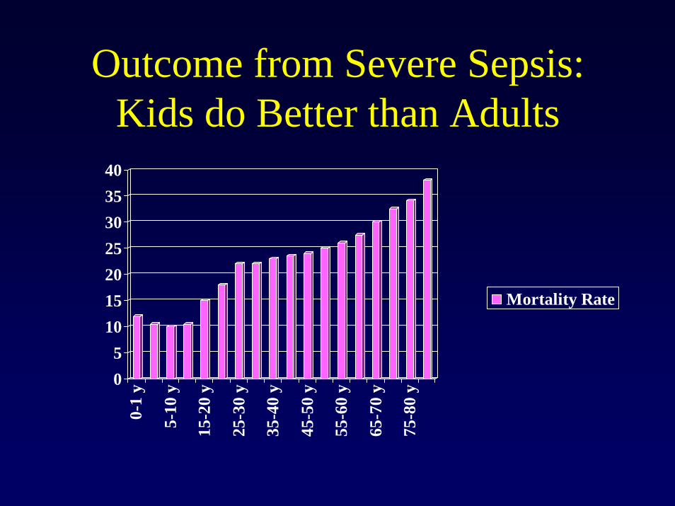
Outcome from Severe Sepsis: Kids do Better than Adults
05
10152025303540
0-1
y
5-10
y
15-2
0 y
25-3
0 y
35-4
0 y
45-5
0 y
55-6
0 y
65-7
0 y
75-8
0 y
Mortality Rate

Improving Outcomes from Pediatric Shock with Continuous Innovation
0%10%20%30%40%50%60%70%80%90%
100%
1968 UnivMinn
1985NCMC
1999 U.S. 2000 StMary's
U.K.
Mortality

Pediatric Mortality Absent If Normal MAP-CVP and Capillary Refill (< 3 sec) Attained in the First Hour
(Han et al, Pediatric Research, 108a, Volume 47(4)2000)
# Nonsurvivors/#Patients (Mortality%)74 min 116 min 166
minShock Present 17/57(30%) 13/44 (30%) 13/51 (25%)Shock Absent 0/27 (0%)* 4/40 (10%) 4/33 (12%)
Less than 25% (13/57) of patients in shock at the time of team arrival had received appropriate therapy as

Adult Mortality Reduced by 50% when SVC O2 sat maintained > 70% in the first 6 hours (Rivers et al. NEJM
2001)Fluids/pressors used to maintain normal MAP-CVP in treatment and Control GroupsIf SVC O2 sat < 70% then treatment group given pRBCs if Hgb < 10 gm/dL and /or Dobutamine if Hgb > 10 gm/dL
What CI does SVC O2 sat > 70% (Rivers et al NEJM, 2001) represent?
Oxygen consumption = CI (SaO2-MVO2) x 1.36 x %Hgb
135 = 3.3 (100-70 ) x 1.36 x 10to
244 = 6.0 (100-70) x 1.36 x 10

Hemodynamics Predict Pediatric Septic Shock Outcomes!
• Pollack reported 57% mortality (CCM 1985)

Fluid Resuscitation

Fluid Resuscitation Decreases Inflammation
• Mouse cecal ligation and puncture has attenuated TNFα and IL-1β mRNA expression with fluid bolus (crystalloid better than serum) (Wilson et al J Trauma 41,1996).
• Septic shock patients given HES infusions have reduced levels of soluble adhesion molecules compared to albumin orpentoxyfilline treated (Boldt et al Crit Car Med 24 1996)

Fluid Increases CO and Survival in Experimental Sepsis
• Endotoxin and bacteria shock models only achieve a hyperdynamic state after 60 cc/kg volume bolus (Carrol et al
Am J Physiol 243,1982)• Fluid resuscitation improves outcome
in rabbit TSS (Lee et al Infect Immun 59,1991) and I.P. E. coli septic shock (Ottosson et
al Circ Shock 35,1991; Hoban et al Circ Shock 34, 1991).

Fluid Resuscitation Decreases Inflammation
• Mouse cecal ligation and puncture has attenuated TNFα and IL-1β mRNA expression with fluid bolus (crystalloid better than serum) (Wilson et al J Trauma 41,1996).
• Septic shock patients given HES infusions have reduced levels of soluble adhesion molecules compared to albumin orpentoxyfilline treated (Boldt et al Crit Car Med 24 1996)

Fluid Increases O2 Consumption in Adult Septic Shock

Historical Perspective
• In the past, 20 cc/kg recommended as maximum bolus in pediatric septic shock due to two concerns with potential harm from vigorous volume resuscitation 1) Fear of pulmonary edema 2) Fear of SIADH in concomitant meningitis

Role of Early Fluid Resuscitation in Pediatric Septic Shock (Carcillo et al JAMA 266,1991)
• 34 patients in ED over 6 year period with septic shock and PAC at 6 hours.
• Evaluated amount of fluid resuscitation and relation to following outcome variables:cardiogenic pulmonary edema, non-cardiogenic pulmonary edema, persistenthypovolemia, and survival

Epidemiology
• Overall mortality rate was 47%• 82 % ventilator supported by 6 hrs• 100% inotrope/vasopressor by 6hrs• 33 ml/kg volume (9 ml/kg colloid) at 1 hr
95 ml/kg volume (37ml/kg colloid) at 6 hrs

Fluid Resuscitation

60 cc/kg Fluid Associated with 90% Survival!
• 0-20 cc/kg had increased hypovolemia and mortality of 68% (Similar to Pollack et al, 1987)
• > 40 cc/kg (mean 60 cc/kg) eliminated hypovolemia,improved survival and no increased incidence of pulmonary edema or herniation syndromes.

Outcomes

Outcomes

Conclusions
• Repeated 20 ml/kg fluid boluses may be administered in excess of 60 ml/kg in the first hour and 120 ml/kg in the first 6 hours if BP, pulses, mental status, and urine output suggest that perfusion is decreased.
• Intravascular monitoring may be necessary to diagnose hypovolemia, NCPE, and CPE.

Pediatric Fluid ResuscitationAfter the First 6 Hours
• Feltes et al performed echocardiography on 5 children with hemodynamically stable sepsis and 10 children with shock (Crit Care Med 22,1994).
• All 5 hemodynamically stable patients had normal performance, contractility, and preload. 3 of 5 had increased afterload.
Pediatric Fluid ResuscitationAfter the First 6 Hours
• Of the 10 patients with septic shock 4 had decreased contractility and increasedafterload, 1 had decreased afterload, and 1 had a severe preload deficit.
• Despite aggressive volume resuscitation 6 of 10 had diminished preload. Ventricular loading abnormalities persisted in 5 of 10 patients for 3 to 6 days.

Inotrope, vasopressor, and vasodilator support for fluid
refractory shock

Cardiovascular therapy improves outcome in experimental fluid
refractory shock • Inotropes and antibiotics improve outcome
in experimental sepsis (Natanson et al, 1990)• Inotropes and vasopressors increase IL-10
and reduce inflammation through cAMP effects.(Vincent et al, 1997)

Cardiovascular support improves experimental outcome

Fluid Refractory Pediatric Septic Shock
• Volume resuscitated sepsis patients a) required inotropes/vasopressors by 6 hours (Carcillo et al JAMA 266, 1991)
• b) showed decreased contractility (4 of 10) (Feltes et al Crit Care Med 22, 1994)
• Volume resuscitated severe pediatric burn patients show inotrope-dependent reduced LVSWI (Reynolds et al J Pediatr Surg 30,1995)

Fluid Refractory Pediatric Septic Shock
• Despite aggressive volume resuscitation, shock may persist secondary to cardiac and vascular dysfunction
• Inotropes, vasopressors, and /or vasodilators are required in fluid refractory pediatric septic shock.

Hemodynamic Support in Fluid Refractory Pediatric Shock
• 50 children with septic shock refractory to > 60 cc/kg volume with PCWP > 8 and functioning PAC at 6 hr to 48 hr (3 centers, 4 year recruitment, 11 deaths)(Ceneviva et al Pediatrics 102:1998)
• Evaluated need of inotrope, vasopressor, and or vasodilator therapy to reverse shock at presentation and over the first 48 hours.

Goal directed therapies
• Reversal of clinical shock and maintenance of MAP-CVP appropriate for age. If unsuccessful then:
• CI > 3.3 and < 6.0 L/min/m2
• SVRI > 800 dyne-sec/cm5/m2
• SVRI < 1600 and > 800 dyne -sec/cm5/m2 if CI < 3.3 L/min/m2

Initial Therapies Needed to Overcome Fluid Refractory
Shock at Presentation• 18 % required a change in regimen from
inotrope to vasopressor, or vasopressor toinotrope
• 24 % required catecholamines for dopamine or dobutamine refractory shock
• 16 % required the addition of vasodilator therapy
Hemodynamic Support in Fluid Refractory Pediatric Shock
• 50 children with septic shock refractory to > 60 cc/kg volume with PCWP > 8 and functioning PAC at 6 hr to 48 hr (3 centers, 4 year recruitment, 11 deaths)(Ceneviva et al Pediatrics 102:1998)
• Evaluated need of inotrope, vasopressor, and or vasodilator therapy to reverse shock at presentation and over the first 48 hours.

Goal directed therapies
• Reversal of clinical shock and maintenance of MAP-CVP appropriate for age. If unsuccessful then:
• CI > 3.3 and < 6.0 L/min/m2
• SVRI > 800 dyne-sec/cm5/m2
• SVRI < 1600 and > 800 dyne -sec/cm5/m2 if CI < 3.3 L/min/m2
Initial Therapies Needed to Overcome Fluid Refractory
Shock at Presentation• 18 % required a change in regimen from
inotrope to vasopressor, or vasopressor toinotrope
• 24 % required catecholamines for dopamine or dobutamine refractory shock
• 16 % required the addition of vasodilator therapy

Therapeutic changes guided by initial PAC variables

Cardiovascular Agents

Low Cardiac Output Syndrome and High SVR is predominant in
Fluid Refractory Shock • 58% had a low CI (< 3.3 L/ min/m2 ) and
high SVRI (> 1600 dyne-sec/cm5)• Only 20% had a high CI and low SVRI.• Kids are different from adults!

Effect of Therapy on Hemodynamics

Progressive Myocardial Dysfunction was the Predominant
Cause of Persistent Shock• Over 48 hours, the children with initial low
CI required the addition of vasodilators and the children with initial high CI requiredinotropes to maintain CI > 3.3.
• Over 90% of children with persistent shock (n = 18) required inotrope and vasodilator therapy

Progressive myocardial dysfunction
05
1015202530354045
Day 1 Day 3
InotropeVasodilatorVasopressor
Recurrent or Persistent Shock can also be caused by a complete change in hemodynamic state
• 10% of children showed a complete change in hemodynamic state from inotrope tovasopressor or vasopressor to inotropetherapy requirements.
• This observation is supported by experimental models (Hoban et al, 1991)

Changes in Cardiovascular Therapies Required over time
0
5
10
15
20
25G
roup
I D
ay 1
Day
3
Gro
up II
Day
1
Day
3
Gro
up II
I Day
1
Day
3
inotropeinotrope + vasodilatorvasopressorinotrope + vasopressor

Catecholamine Refractory shock
• 20 % of patients were catecholamine and nitrosovasodilator refractory.
• These patients responded to the addition of a) hydrocortisone therapy (n = 5) b) PDE type III inhibitors (n = 4) c) vasopressin (n = 1)
• ECMO was used in two refractory shock patients.

Catecholamine Refractory Shock and Adrenal Insufficiency
• Risk Factors include a) Purpura Fulminansand b) History of Prior Chronic Steroid Use.
• 52% of 33 children (n =16 meningococcus) with septic shock had AI (Hatherill et al CCM 26,1998)
• 27% of 97 children with sepsis showed adrenal infarction/hemorrhage at autopsy (Amoo-Lamptey,unpublished)

Catecholamine Refractory Shock and Adrenal Insufficiency
• Adrenal shock is indistinguishable from septic shock ie low CI/ high SVR, or high CI/ low SVR state (Bouachor et al, Intens Care Med
20,1994) so should be diagnosed by ACTH stimulation or risk factors
• Hydrocortisone 50 mg/kg (AHFS 1998, Sumarmo et
al Pediatr 69, 1982) or less?

Mechanism of Catecholamine Insensitivity
• LPS reduces β-receptor stimulated accumulation of cAMP. Forskolin bypasses this defect suggesting receptor, G-protein or PDE effects of LPS (Shepherd et al 1986, Campbell et al 1993)
• Increased catecholamine levels and need for doubling of catecholamine infusions in patients suggests receptor desensitization (Kovarik et al 1987, Reithman et al 1989)
Low CO syndrome reversible with PDE III inhibitors
• Children are less responsive to dopamine and dobutamine and may require epinephrine.(Perkin et al 1982)
• The inodilators milrinone (Barton et al 1996,Linsay et al 1998) and amrinone (Irazusta et al 2000) improve CI in catecholamine refractory, and catecholamine and nitrosovasodilator refractory shock (Ceneviva et al 1998)
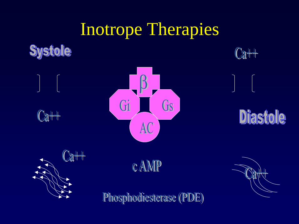
Inotrope Therapies
ECMO is effective rescue for Low CO syndrome
• ECMO reduces mortality in refractory neonatal septic shock to 20%.(Meyer et al 1995)
• ECMO reduces mortality in refractory pediatric septic shock to 50%.(Goldman et al 1997, Beca et al 1994)
• ECMO rescues adult Hantavirus victims (Low CO/ high SVR syndrome)(Hallin et al 1996, Crowley et al 1998)

Mechanism of Vascular Catecholamine Insensitivity
• LPS reduces vascular α receptor numbers and phosphoinositide second messenger production. (Carcillo et al 1988, Deaciuc et al1987)
• Norepinephrine reverses dopamine-resistant vascular failure
• Vasopressin reverses norepinephrineresistant vascular failure.

Vasoconstrictors and Dilators

Reduced CO syndrome not vasoplegia was associated with
mortality • The overall survival rate was 80%,
72% in children with low CI and 90% in children with high CI.
• This survival rate compared favorably to that reported by Pollack et al when 60 cc/kg fluid resuscitation and goal directed therapy was not practiced.

Outcome of Fluid Refractory Shock with Goal Directed
Hemodynamic Support
0102030405060708090
100
Group I Group II Group III Pollack etal 1985
Survival

Conclusions
• Unlike adults, children with fluid refractory shock are frequently hypodynamic and respond to inotrope and vasodilator therapy.
• Because hemodynamic states are heterogeneous and change with time, an incorrect cardiovascular therapeutic regimen should be suspected in any child with persistent shock.

Conclusions
• PAC indicated in a subgroup of children with fluid refractory septic shock (Zaritsky,
Current Concepts in Pediatric Emergency and Critical Care)• Outcomes can be improved when 60 cc/kg
fluid resuscitation and goal-directed cardiovascular therapy is applied.

Fluid responsive
Refractory shockPlace pulmonary artery catheter and direct fluid, inotrope,vasopressor,vasodilator, and hormonal therapies to attain normal
MAP-CVP and CI > 3.3 and < 6.0 L/min/m2
Place pulmonary artery catheter and direct fluid, inotrope,vasopressor,vasodilator, and hormonal therapies to attain normal
MAP-CVP and CI > 3.3 and < 6.0 L/min/m2
Figure 1 Stepwise management of hemodynamic support with goals of normal perfusion and perfusion pressure (MAP-CVPin infants and children with septic shock. Proceed to next step if shock persists.
Give hydrocortisoneGive hydrocortisone
At Risk of Adrenal Insufficiency? Catecholamine -resistant shock Not at Risk?
Titrate epinephrine for cold shock, norepinephrine for warm shock to normal MAP-CVP and SVC O2 saturation > 70%
Titrate epinephrine for cold shock, norepinephrine for warm shock to normal MAP-CVP and SVC O2 saturation > 70%
Fluid refractory-dopamine resistant shock
Establish central venous access, begin dopamine therapy and establish arterial monitoring .
Establish central venous access, begin dopamine therapy and establish arterial monitoring .
Fluid refractory shock
Push 20cc/kg isotonic saline or colloid boluses up to and over 60 cc/kgCorrect hypoglycemia and hypocalcemia
Push 20cc/kg isotonic saline or colloid boluses up to and over 60 cc/kgCorrect hypoglycemia and hypocalcemia
Recognize decreased mental status and perfusion.Maintain airway and establish access according to PALS guidelines.
Recognize decreased mental status and perfusion.Maintain airway and establish access according to PALS guidelines.
Observe in PICUObserve in PICU
Consider ECMOConsider ECMO
Persistent Catecholamine-resistant shock
15 min
Normal Blood Pressure Low Blood Pressure Low Blood Pressure Cold Shock Cold Shock Warm Shock
SVC O2 sat < 70% SVC O2 sat < 70%Add vasodilator or Type III PDE inhibitor Titrate Volume and Epinephrine Titrate Volume andNorepinephrine
with volume loading (?vasopressin or angiotensin)
0 min5 min
Do not give hydrocortisoneDo not give hydrocortisone60 min

Figure 2 Stepwise management of hemodynamic support with goals of normal perfusion and perfusion pressure (MAP-CVP)and pre and post-ductal oxygen saturation difference <5%, and central venous O2 sat > 70% in near term-newborns with septic sh
Push 10cc/kg isotonic crystalloid or colloid boluses to 60 cc/kg, correct hypoglycemia,and hypocalcemia. Begin prostaglandin infusion until echocardiogram shows no ductal-
dependent lesion.
Push 10cc/kg isotonic crystalloid or colloid boluses to 60 cc/kg, correct hypoglycemia,and hypocalcemia. Begin prostaglandin infusion until echocardiogram shows no ductal-
dependent lesion.
Recognize decreased perfusion, cyanosis, RDS.Maintain airway and establish access according to NRP guidelines.
Recognize decreased perfusion, cyanosis, RDS.Maintain airway and establish access according to NRP guidelines.0 min
5 min
Establish Central Venous and Arterial AccessTitrate dopamine and dobutamine
Establish Central Venous and Arterial AccessTitrate dopamine and dobutamine
Fluid-refractory shock
Fluid responsive
15 min
Observein NICU
Observein NICU
Fluid refractory-dopamine resistant shockTitrate epinephrine. Systemic alkalinization if PPHN is present
Inhaled nitric oxide Inhaled nitric oxide
Refractory Shock
Cold or Warm Shock Poor RV function PPHN, CVC O2 sat < 70%
Titrate vasodilator Type III PDE
inhibitor with volume loading
Titrate vasodilator Type III PDE
inhibitor with volume loading
Cold shockNormal blood pressure Poor LV function, CVC O2 sat < 70%
60 min
ECMOECMO
Warm shockLow blood pressure
Catecholamine-resistant shock
Direct therapies using echocardiogram and arterial and CVP monitoring
Titrate volume and epinephrine(? Vasopressin or angiotensin)

Figure 3 Recommendation for stepwise management of hemodynamic supportof shock in the first hour.
Recognize shock,maintain airwayestablish access.
Recognize shock,maintain airwayestablish access.0 min
5 min
Liver up Liver DownPush 10cc/kg isotonic crystalloid begin
prostaglandin infusion (neonate only) and or dobutamine until
echocardiogram r/o ductal-dependent lesion.
Push 10cc/kg isotonic crystalloid begin prostaglandin infusion (neonate only) and
or dobutamine untilechocardiogram r/o ductal-dependent
15 min
Push 60 cc/kg fluid, consider hemorrhagic shock
Push 60 cc/kg fluid, consider hemorrhagic shock
lesion.
Titrate epinephrine (if neonate with PPHN titrate NO)
Catecholamine-resistant shock
Consider adrenal insufficiency, hydrocortisone 50 mg/kg
60 min
Tirate volume and epinephrine
Tirate volume and epinephrine
Refractory Shock
Cold Shock with Low Blood Pressure , SVC O2 sat < 70%
Titrate vasodilator or Type III PDE
inhibitor with volume loading
Titrate vasodilator or Type III PDE
inhibitor with volume loading
Cold shockNormal blood pressure, SVC O2
sat < 70%
Warm shockLow blood pressure
Titrate volume andnorepinephrine(? Vasopressin or ?angiotensin)
ECMOECMO

What is the Pathophysiology of Sepsis Induced Multiple Organ Failure?
• No patients died from shock in the first 48hours,instead all deaths occurred with MOF usally after 7 days.
• We performed a clinico-pathologic correlate study in two parts to determine the pathophysiology of sepsis-induced MOF in children admitted to the Children’s Hospital of Pittsburgh intensive care unit

Methods Part I : ClinicopathologicCorrelates
• One hundred children admitted to the PICU with sepsis and organ failure were enrolled with IRB and parental approval
• Plasma samples were attained on day 1 and 3 of sepsis
• Autopsies were performed on 11 of 13 children who died.

Methods Part II: Pathologic Correlates (1988-1998)
• A ten year search of the CHP pathology autopsy bank revealed 56 autopsies with a pre-mortem diagnosis of sepsis, 12 autopsies with a pre-mortem diagnosis of pneumonia without sepsis, and 21 autopsies with a pre-op diagnosis of organ failure without infection (lung failure n =10, heart failure n = 4, liver failure n = 4, immune disease n = 2, metabolic disease n = 1)

Pro-inflammatory Cytokines and Nitric Oxide

White Cell Diapedesis

Thrombosis and Antifibrinolysis
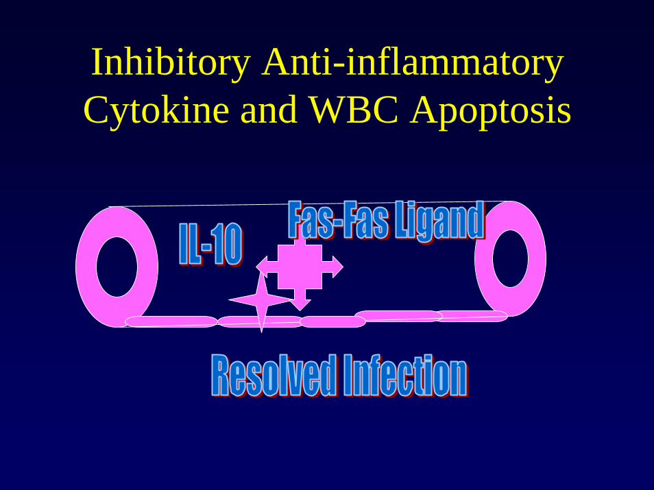
Inhibitory Anti-inflammatory Cytokine and WBC Apoptosis
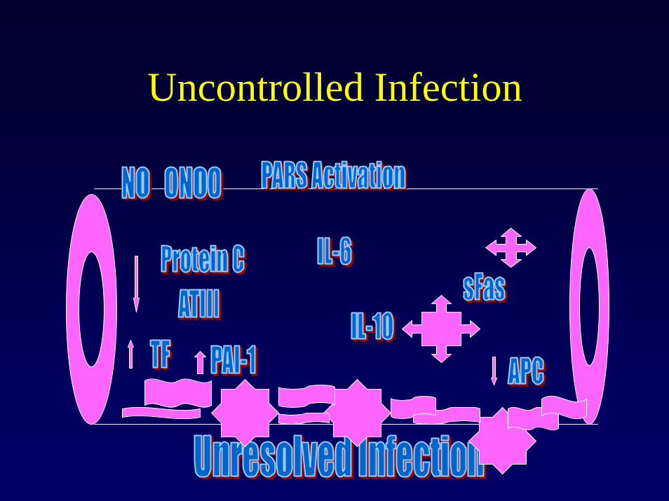
Uncontrolled Infection

Hypotheses
• Clinical correlates will show evidence of persistent inflammation and coagulation in children with persistent sepsis-induced MOF.
• Pathologic correlates will show increased evidence of persistent and unrecognized infection and thrombosis in children who die with sepsis-associated MOF.

Part I: Outcomes in sepsis-induced MOF at CHP-PICU
• < 3 organ failure - 96% survival• Resolved > 3 organ failure by day 3 - 88%
survival • Persistent > 3 organ failure at day 3- 65%
survival• Autopsy (11/13) showed that 10 children
died of uncontrolled infection and 1 child died of thrombosis.

0 60 120 1800
60
120
180
0601201800
60
120
180
0
30
6090
120
150
180
210
240270
300
330
0 60 1200
60
120
0601200
60
120
0
30
6090
120
150
180
210
240270
300
330
NMOF
RMOFPMOF
Controls
Nitrite
PCT
IL-6
IL-10E-SelICAM
VCAM
TM
TF
TFA
PAIPAIA Def
LacFas-R
Fas-L
Nitrite
PCT
IL-6
IL-10E-SelICAM
VCAM
TM
TF
TFA
PAIPAIA Def
LacFas-R
Fas-L
Nitrite
PCT
IL-6
IL-10E-SelICAM
VCAM
TM
TF
TFA
PAIPAIA Def
LacFas-R
Fas-L
Nitrite
PCT
IL-6
IL-10E-SelICAM
VCAMTM
TF
TFA
PAIPAIA Def
LacFas-R
Fas-L
Day 1 of Sepsis

0 60 120 1800
60
120
180
0601201800
60
120
180
0
30
60
90
120
150
180
210
240
270
300
330
0 60 120 1800
60
120
180
0601201800
60
120
180
0
30
60
90
120
150
180
210
240
270
300
330
0 60 120 1800
60
120
180
0601201800
60
120
180
0
30
60
90
120
150
180
210
240
270
300
330
NMOF
RMOF PMOF
0 60 120 1800
60
120
180
0601201800
60
120
180
0
30
60
90
120
150
180
210
240
270
300
330
Controls
Nitrite
PCT
IL-6
IL-10E-Sel
ICAM
VCAM
TM
TF
TFA
PAIPAIA
Def
Lac
Fas-R
Fas-L
Nitrite
PCT
IL-6
IL-10E-Sel
ICAM
VCAM
TM
TF
TFA
PAIPAIA
Def
Lac
Fas-R
Fas-L
Nitrite
PCT
IL-6
IL-10E-Sel
ICAM
VCAM
TM
TF
TFA
PAIPAIA
Def
Lac
Fas-R
Fas-L
Nitrite
PCT
IL-6
IL-10E-Sel
ICAM
VCAM
TM
TF
TFA
PAIPAIA
Def
LacFas-R
Fas-L
Day 3 of Sepsis

Uncontrolled Infection
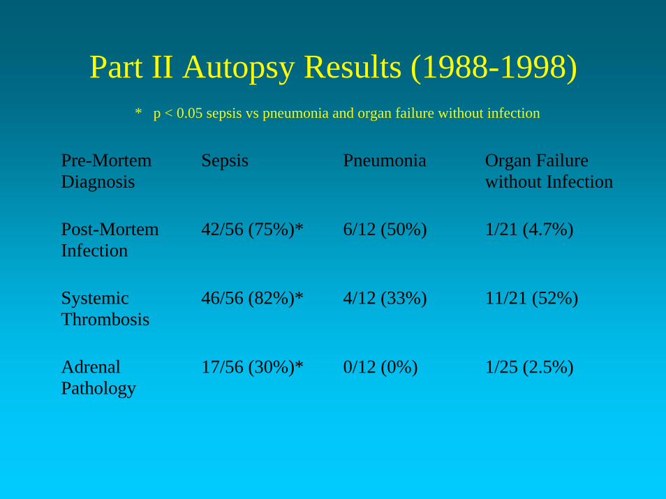
Part II Autopsy Results (1988-1998) * p < 0.05 sepsis vs pneumonia and organ failure without infection
Pre-MortemDiagnosis
Sepsis Pneumonia Organ Failurewithout Infection
Post-MortemInfection
42/56 (75%)* 6/12 (50%) 1/21 (4.7%)
SystemicThrombosis
46/56 (82%)* 4/12 (33%) 11/21 (52%)
AdrenalPathology
17/56 (30%)* 0/12 (0%) 1/25 (2.5%)
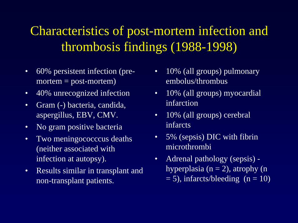
Characteristics of post-mortem infection and thrombosis findings (1988-1998)
• 60% persistent infection (pre-mortem = post-mortem)
• 40% unrecognized infection• Gram (-) bacteria, candida,
aspergillus, EBV, CMV.• No gram positive bacteria• Two meningococccus deaths
(neither associated with infection at autopsy).
• Results similar in transplant and non-transplant patients.
• 10% (all groups) pulmonary embolus/thrombus
• 10% (all groups) myocardial infarction
• 10% (all groups) cerebral infarcts
• 5% (sepsis) DIC with fibrin microthrombi
• Adrenal pathology (sepsis) -hyperplasia (n = 2), atrophy (n = 5), infarcts/bleeding (n = 10)

Conclusion
• Pathologic correlates support persistent/unrecognized infection as the leading cause of death in children with sepsis-induced MOF in our PICU.
• Systemic thrombosis can contribute to mortality associated with organ failure with and without sepsis.
• Adrenal pathology is common only with sepsis-induced MOF.
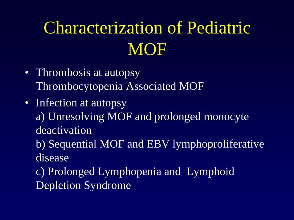
Characterization of Pediatric MOF
• Thrombosis at autopsy Thrombocytopenia Associated MOF
• Infection at autopsy a) Unresolving MOF and prolonged monocyte deactivation b) Sequential MOF and EBV lymphoproliferativedisease c) Prolonged Lymphopenia and Lymphoid Depletion Syndrome

Thrombocytopenia Associated MOF
• Platelet count less than 100,000 and 3 or greater organ failure
• Occurs with (20% of patients) and without (80% of patients) prolonged PT/PTT.-

Platelet
vWF
vWF-CP
tPA PGI
Endothelium
Platelet
vWF-CP
Platelet
vWF
vWF Platelet
Homeostasis
tPA

TFPI
Heparin ATIII
Prot C
APC
+
PGI
Thrombomodulin
PGI
TFPI
HomeostasisHomeostasistPA

Platelet
Platelet
Platelet
Platelet
Platelet
Platelet
Fibrin
vWFvWF:Platelet Thrombus:Platelet Thrombus
PAI-1PAI-1tPA
Platelet
vWF
vWF
Platelet
vWF

Fibrin
PAI-1Platelet
Platelet
PlateletFibrin
PlateletvWF
Platelet
Platelet
Platelet
PlateletFibrin
PlateletvWF
Platelet
EndotheliumPAI-1 tPA
Fibrin ThrombusFibrin Thrombus

vWF
Platelet
vWFShear stress

Endothelium
Platelet
Platelet
vWFX vWF-CP
vWF-CP Ab

Fibrin
Platelet
Platelet
PlateletPlatelet
Platelet
Platelet
PlateletvWF
Platelet
Platelet
Platelet
Platelet
Platelet
Platelet
Fibrin
vWFvWF

Endothelium
Endothelium PAI-1
vWF
TF
TF
vWF
vWFPAI-1
TF
PAI-1
VII

TF TF
vWFPAI-1
PAI-1
TFVII
vWFTF
Platelet
Platelet
Platelet
Platelet Platelet
Platelet
Platelet

Endothelium
Endothelium PAI-1 TF
PAI-1
PAI-1
PAI-1
PAI-1
vWF
vWF
TFPI
PlasminPlasminogen
PAI-1
X
PAI-1 PAI-1
PAI-1

TF
PAI-1
PAI-1
vWF
TFPI
Platelet
Platelet
Platelet
PlateletvWF
Platelet
Platelet
Platelet

THROMBOSIS
Prot C
Thrombomodulin
APC
Prothrombin Thrombin
HeparinAntithrombin I I I
Tissue Factor
TFPI
Endothelium
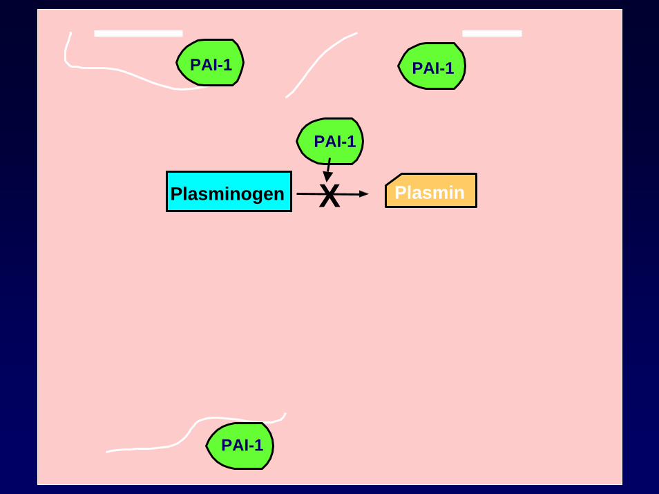
FIBRINOLYSIS
PAI-1 (-)
Plasminogen
(-)
Fibrinogen Fibrin
(-)
(-)
(-)
(+)
TPAStreptokinaseUrokinase(+)
Aminocaproic AcidPlasminTranexamine
Aprotinin

Non-specific therapies: Plasma infusion or exchange
• Plasma Exchange
• Removes Abs to vWF cleaving protease
• Removes vWF• Removes PAI-1• Removes tissue factor
• Plasma Infusion
• Restores clotting factors (VII, VIII, X etc)
• Restores vWF cleaving protease
• Restores prostacyclin• Restores protein C and
antithrombin III• Restores tPA

Recommended therapy for TTP
• Steroids for 24 hours.• Plasma exchange 11/2 volumes then 1
volume per day (median 18 days) requires calcium infusion during therapy.
• If recalcitrant consider cryopreserved supernatant or SD plasma.
• Vincristine
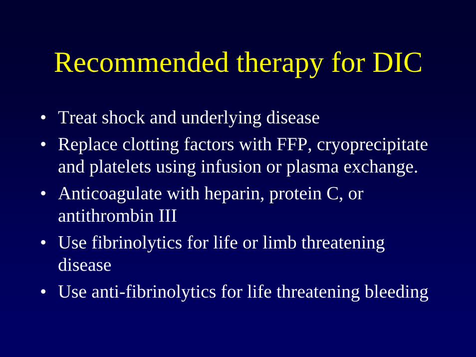
Recommended therapy for DIC
• Treat shock and underlying disease• Replace clotting factors with FFP, cryoprecipitate
and platelets using infusion or plasma exchange.• Anticoagulate with heparin, protein C, or
antithrombin III• Use fibrinolytics for life or limb threatening
disease• Use anti-fibrinolytics for life threatening bleeding

Our approach to Thrombotic Microangiopathy
• Remove source of thromboticmicroangiopathy.
• Consider antithrombotic agents.• If thromboctopenia associated MOF occurs
then we use the TTP-based therapeutic protocol.

Therapeutic Trial of Plasma exchange for Thrombocytopenia Associated MOF
• Patients with platelet count < 100,000 and three or greater organ failure for 24 hours.
• Randomized to TTP-based plasma exchange protocol or usual therapy until resolution of MOF.
• Twenty patients met criteria; Only ten agreed to consent to randomization.
• Mean therapy 12 days, ranged 4 to 28 days.

PELODPediatric Logistic Organ Dysfunction Score
DAY
0 5 10 15 20 25 30
PELO
D
0
20
40
60
80
100
Plasma ExchangeNo Plasma Exchange
Figure 3. Pediatric Logistic Organ Dysfunction Score, Mean with standarderror for patients who received plasma exchange therapy (N = 5) and who did not receive plasma exchange therapy (N = 5) for each day x 28 days.
17
PELOD decreased from 25.0 ± 2.0 to 0.8 ±0.6 with plasma exchangeat 28 d
PELOD increased from 25.4 ± 2.3 to73.6 ±18.4 without plasma exchange at 28 d
p < 0.001, power = 1.0, 2F-RM ANOVA
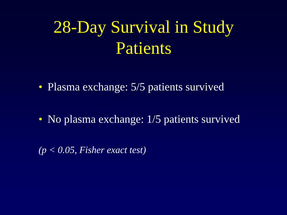
28-Day Survival in Study Patients
• Plasma exchange: 5/5 patients survived
• No plasma exchange: 1/5 patients survived
(p < 0.05, Fisher exact test)

Outcomes in children at the Children’s Hospital of Pittsburgh during the 2 years since study completion
• Plasma Exchange• 28 day mortality 10%• 1 year mortality 20%• n = 20 patients
• No Plasma Exchange• 28 day mortality 80%• 1 year mortality 85%• n = 10 patients

MOF Associated with Infection at Autopsy

Host Response to Inflammation
APC
Antigenpresentation;costimulatorysignals

The TH1 , TH2 Paradigm
IL-3GM-CSFTNF-∝Chemokines
IL-3GM-CSFTNF-∝Chemokines

Influence of Cytokines onCell Mediated Response
Effector Maturation

Strategies ofImmunesuppression

Provocative Hypothesisand Speculation
• For the most part, children with BMT or solid organ transplantation who die of ARDS or MOF in PICUs do so as a result of fatal immunoparalysis caused by reversible iatrogenic immunesuppression.
• It has become acceptable to die from ARDS, MOF, sepsis, or PTLD; but not from rejection or GVHD.
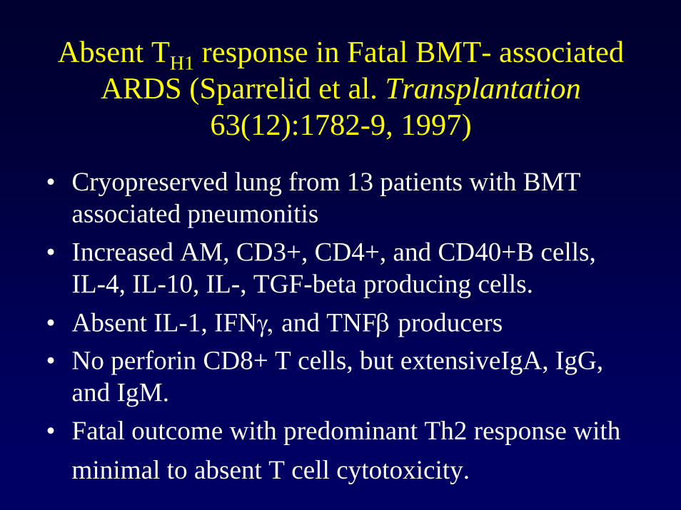
Absent TH1 response in Fatal BMT- associated ARDS (Sparrelid et al. Transplantation
63(12):1782-9, 1997)
• Cryopreserved lung from 13 patients with BMT associated pneumonitis
• Increased AM, CD3+, CD4+, and CD40+B cells, IL-4, IL-10, IL-, TGF-beta producing cells.
• Absent IL-1, IFNγ, and TNFβ producers• No perforin CD8+ T cells, but extensiveIgA, IgG,
and IgM.• Fatal outcome with predominant Th2 response with
minimal to absent T cell cytotoxicity.

Immunoparalysis in Adults with abdominal surgery associated sepsis (HD
Volk Intens Care Med, 1996)
• Defined as HLA-DR+ monocytes < 30%• Persists for at least 5 days associated with
19% (24/126) survival rate.• Not detectable or only briefly associated
with 80% (121/149) survival rate.

Outcome in adult transplant patients with sepsis (HD Volk et al Intens Care Med 1996)HLA-DR +monocytes
Rapid taper Survivingpatients
Survivinggrafts
< 30% No 1/12 (8%) None
< 30% Yes 30/33 (90%) 29/30
> 30% No 28/28 (100%) 28/28
> 30 % Yes 4/4 (100%) 0/4
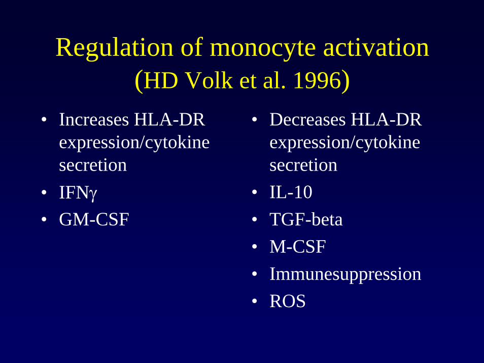
Regulation of monocyte activation(HD Volk et al. 1996)
• Decreases HLA-DR expression/cytokine secretion
• IL-10• TGF-beta• M-CSF• Immunesuppression• ROS
• Increases HLA-DR expression/cytokine secretion
• IFNγ• GM-CSF

Reversal of Immuneparalysis Using IFNγ
(HD Volk Intens Care Med 1996)Treatment(48 hour)
Patient group(n = 7)
% HLA-DR+monocytes
Control Healthy donor 87 +/- 5
IFNγ Healthy donor 93 +/- 11
Control Patients 28 +/- 8
IFNγ Patients 69+/- 6*

GM-CSF therapy in children after orthotopic liver transplantation
(Trinidade et al. J Hepatology 1998)
• 13 children (9 with neutropenia, 10 with infection) received a mean of 15 days (4-49 days) of GM-CSF therapy.
• No attributable episodes of rejection• Thought to have improved ability to fight
infection.

Unresolving MOF and Prolonged Monocyte Deactivation
• Septic Shock patients receiving exogenous immunesuppression with TH2 agents including tacrolimus, cyclosporine A who cannot clear infection.
• Children who recover from septic shock but then suffer secondary sepsis usually with gram negative bacteria or fungus, but also with Staphylococcus Aureus.

Immune Paralysis and HLA-DR
•HLA-DR is a class II MHC molecule which is expressed on the surfaces of antigen presenting cells, including monocytes.
•Its expression by monocytes is decreased in the anti-inflammatory state, as measured by flow cytometry.
•HLA-DR is strongly expressed on the surfaces of 90-100% of monocytes in healthy adults and neonates
•Expression of HLA-DR on less than 30% of circulating monocytes is considered to be a marker of Immune Paralysis in adults. Janeway, Immunobiology 1997

CD4 Count The T-lymphocyte Contribution
• The the importance of the CD4+ lymphocyte in the maintenance of immune function has been demonstrated in the HIV literature.
• CD4 counts of <750, 500, and 200 cells/mm in the first, second, and subsequent years of life have been associated with an increased risk for the development of infection and death. (CDC 1995) Janeway, Immunobiology 1997

CD4 CountThe T-lymphocyte Contribution
Janeway, Immunobiology 1997
• The CD4 antigen serves as a co-receptor on certain T-lymphocytes, facilitating interaction with MHC class II molecules on antigen presenting cells.
• Role in Immune Paralysis?
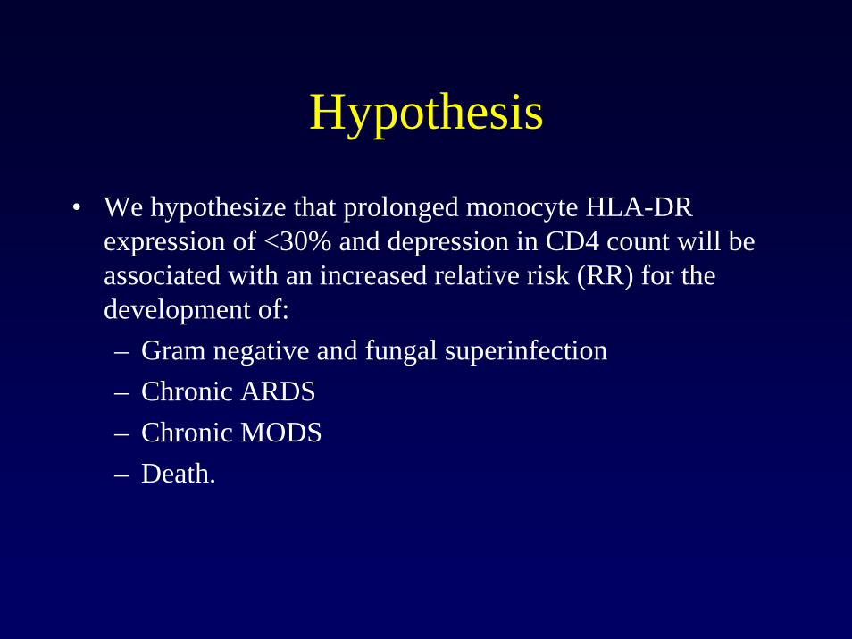
Hypothesis
• We hypothesize that prolonged monocyte HLA-DR expression of <30% and depression in CD4 count will be associated with an increased relative risk (RR) for the development of: – Gram negative and fungal superinfection– Chronic ARDS– Chronic MODS– Death.

Results to Date
• Total of 50 patients:– age 1 day to 22 years– 29 males– 21 females– 24 transplant, 26 non-transplant

EpidemiologyTransplant Patients
8 - Liver6 - Liver / Small bowel2 - Heart2 - Lung3 - Bone marrow2 - Heart / Lung1 - Kidney

EpidemiologyImmunosuppressive Medications
Patients Drug20 Tacrolimus12 Mycophenolate3 Cyclosporin1 Rapamycin1 ATGAM25 Methylprednisolone

Persistently Depressed Monocyte HLA-DR Expression Associated with Adverse
OutcomesRelative risks with 95%CI for low values after Day 3
Outcome HLA-DR CD4 Count HLA-DR and CD4Count
Superinfection 9.9 (3.3, 30) 1.6 (0.4, 3.2) 5.3 (2.3, 12)
Chronic ARDS 4.6 (2.2, 9.9) 1.6 (0.8, 3.3) 3.9 (2.0, 7.6)
Chronic MODS 12 (3.3, 50) 1.4 (0.5, 3.5) 5.6 (2.1, 14)
Death 8.5 (2.0, 35) 1.7 (0.5, 5.8) 5.5 (1.6, 18)

HLA-DR expression is decreased in the setting of superinfection.P=0.003; two way ANOVA
0102030405060708090
100
1 3 5 7 9 11 13
Day
HLA-DR (%+)
Superinfection n=17
No Superinfection n=33
Controls n=12
*p<0.05; SNK test
**

0102030405060708090
100
onset 1 week
HLA-DR (%+)
Persistent Infection n=5
Resolved Infection n=12
Controls n=12
*p<0.05; SNK test
*
HLA-DR Expression and Outcome of Superinfection p=0.01; two way
ANOVA

Relative risk of superinfection with prolonged depression of HLA-DR:
Monocyte HLA-DR<30% after Day 3: 14/16 (88%)
Monocyte HLA-DR>30% after Day 3: 3/34 (9%)
RR: 9.9 95%CI: 3.3, 30
0102030405060708090
100
1 3 5 7 9 11 13
Day
HLA-DR (%+)
Chronic ARDS n=19
No Chronic ARDSn=31
Controls n=12
*p<0.05; SNK test
**
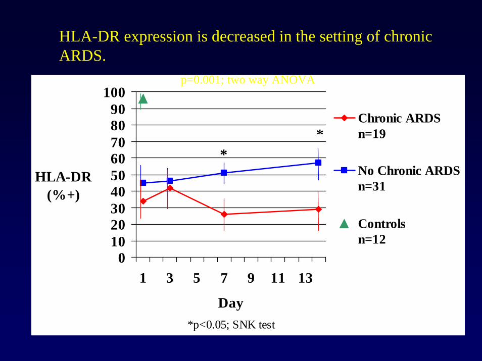
HLA-DR expression is decreased in the setting of chronic ARDS.
p=0.001; two way ANOVA
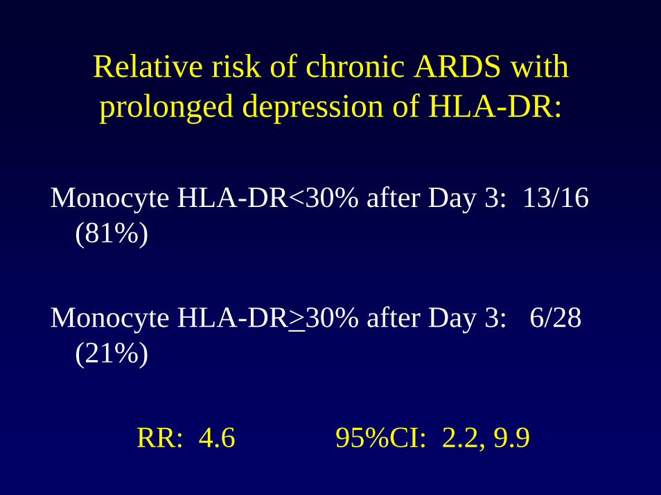
Relative risk of chronic ARDS with prolonged depression of HLA-DR:
Monocyte HLA-DR<30% after Day 3: 13/16 (81%)
Monocyte HLA-DR>30% after Day 3: 6/28 (21%)
RR: 4.6 95%CI: 2.2, 9.9

HLA-DR expression is decreased in the setting of chronic MODS.P=0.002; two way ANOVA
0102030405060708090
100
1 3 5 7 9 11 13
Day
HLA-DR (%+)
Chronic MODS n=14
No Chronic MODSn=36
Controls n=12
*p<0.05; SNK test
*
*

Relative risk of chronic MODS with prolonged depression of HLA-DR:
Monocyte HLA-DR<30% after Day 3: 12/16 (75%)
Monocyte HLA-DR>30% after Day 3: 2/34 (6%)
RR: 12 95%CI: 3.3, 50
0102030405060708090
100
1 3 5 7 9 11 13
Day
HLA-DR (%+)
Death n=10
Survival n=40
Controls n=12
*p<0.05; SNK test
*
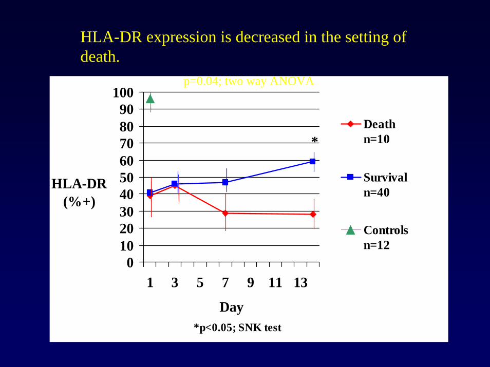
HLA-DR expression is decreased in the setting of death.
p=0.04; two way ANOVA

Relative risk of death with prolonged depression of HLA-DR:
Monocyte HLA-DR<30% after Day 3: 8/16 (50%)
Monocyte HLA-DR>30% after Day 3: 2/34 (6%)
RR: 8.5 95%CI: 2.0, 35

Relative risks for adverse outcomes with depressed HLA-DR in non-transplant patients:
RR 95%CISuperinfection 8.5 2.3, 31Chronic ARDS 3.2 0.98, 10Chronic MODS 12 1.5, 95Death 11 1.4, 88

0102030405060708090
100
Onset 1 weekfollow up
HLA-DR (%+)
0
5
10
15
20
Onset 1 weekfollow up
Tacr
olim
us le
vel (
ng/m
l)
Reduction in FK506 increased DR expression without rejection, with 100% survival (n=7). Failure to decrease FK506 resulted in prolonged
HLA-DR depression and 100% mortality (n=6). (p<0.002; two way ANOVA)
*p<0.05; SNK test
*
*p<0.05; SNK test
*
HLA-DR Expression
Tacrolimus level

0102030405060708090
100
Onset 1 week follow up
HLA-DR (%+)
Tacrolimus(ng/ml)
Control n=12
Tapering tacrolimus in patients with HLA-DR>30% resulted in 100% survival and no rejection. (n=6)
0102030405060708090
100
Initiation ofsteroids
1 week follow up
HLA-DR (%+)
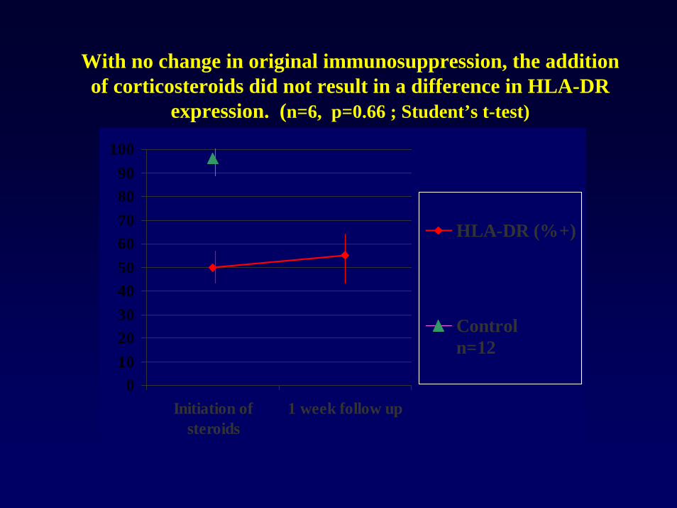
Control n=12
With no change in original immunosuppression, the addition of corticosteroids did not result in a difference in HLA-DR
expression. (n=6, p=0.66 ; Student’s t-test)

0102030405060708090
100
1 6 11 16 21 26 31 36 41 46 51 56
PICU Day
HLA-DR (%+)
Tacrolimus level(ng/ml)
black bars = periods of treatment with GM-CSF
HLA-DR expression increased in response to GM-CSF therapy in one patient.

0102030405060708090
100
1 7 13 19 25 31 37 43 49 55
PICU Day
HLA-DR (%+)
TNF-alpha(pg/ml/10)
black bars = periods of treatment with GM-CSF
Inducible TNF-alpha production increased along with HLA-DR expression.

Unresolving MOF with persistent infection and too many or too few lymphocytes
• Too Many Lymphocytes Sequential MOF ( liver and renal failure) a) Viral sepsis (Epstein Barr Virus) b) Lymphoproliferative disease (PTLD or x-linked)
• Too Few lymphocytes Unresolving MOF and prolonged lymphopenia(ALC < 1000 >7 days) Lymphoid depletion at autopsy

Fas, Fas ligand, and Liver Apoptosis/Necrosis
• Viral sepsis and lymphoproliferative disease associated with high circualting Fas Ligand and Fas and liver failure associated MOF.
• Autopsy shows lymphocyte infiltration of liver and hepatic destruction.
• Successful therapy B lymphocyte antibody now in compassionate clinical trial.

Lymphocyte Depletion Syndrome
• Prolactin prevents apoptosis of B and T lymphocytes.
• Prolonged lymphopenia (ALC < 1000) associated with secondary infection, unresolving MOF, and death.
• Lymphocyte depletion syndrome-absent lymphocytes in lymph nodes and spleen associated with prolonged lymphopenia and hypoprolactinemia (OR 10.8).

Planned Clinical Studies
• GM-CSF for prolonged monocyte deactivation.
• Prolactin for hypo-prolactinemic patients with prolonged lymphopenia.
• Protein C concentrate for patients at risk of bleeding (contraindication to activated protein C RCT)

InvestigatorsLesley Doughty Brown UniversityGary Ceneviva Penn State UniversityNeal Thomas Penn State UniversityJerril Green Arkansas Children’s Hospital Michael Whalen Massdachussetts General Hospital Sandra Kaplan University of PittsburghDanny Kofos Methodist Hospital (San Antonio, TX) Yong Han University of PittsburghMark Hall Ohio State UniversityTrung Nguyen University of PittsburghKate Felmet University of PittsburghScott Watson University of PittsburghRon Jaffe University of PittsburghShekhar Venkataraman University of PittsburghRobert Clark University of PitsburghDerek Angus University of Pittsburgh