sensory receptors. (a) receptor is afferent neuron.(b) receptor regulates afferent neuron. to cns...
TRANSCRIPT
Sensory Receptors
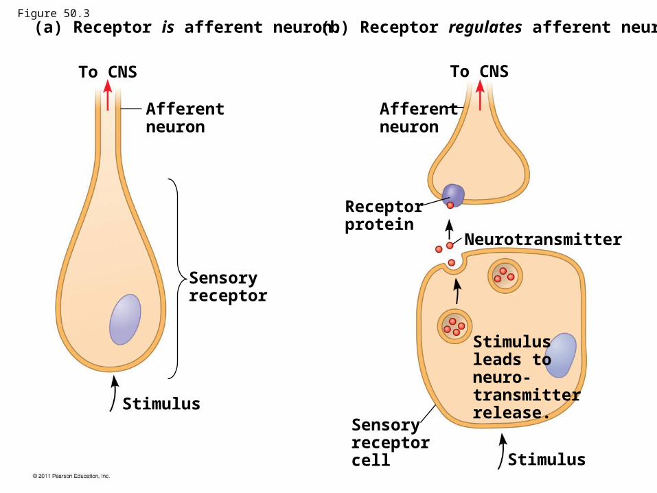
(a) Receptor is afferent neuron. (b) Receptor regulates afferent neuron.
To CNS
Afferentneuron
Afferentneuron
To CNS
Receptorprotein
Sensoryreceptor
Stimulus
Neurotransmitter
Sensoryreceptorcell Stimulus
Stimulusleads toneuro-transmitterrelease.
Figure 50.3
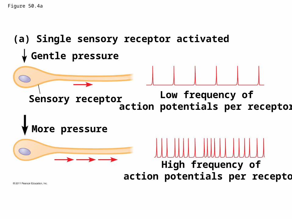
Figure 50.4a
(a) Single sensory receptor activated
Gentle pressure
Sensory receptor
More pressure
Low frequency ofaction potentials per receptor
High frequency ofaction potentials per receptor
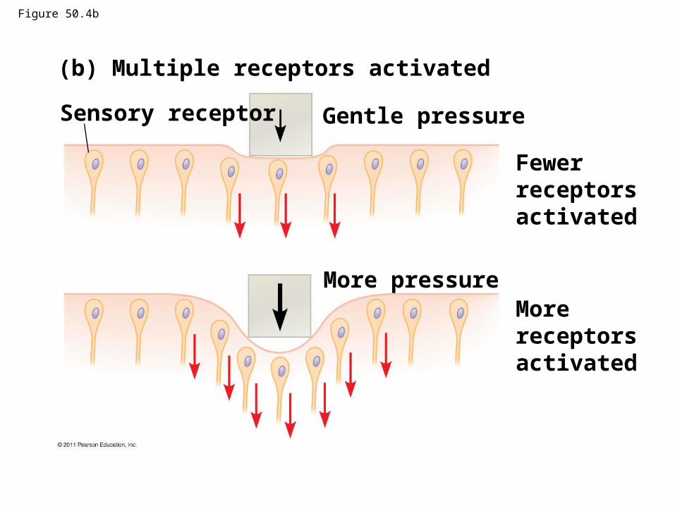
Figure 50.4b
(b) Multiple receptors activated
Sensory receptor Gentle pressure
More pressure
Fewerreceptorsactivated
Morereceptorsactivated
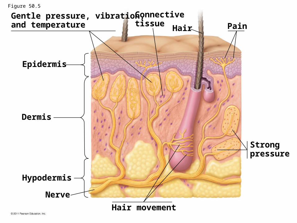
Gentle pressure, vibration,and temperature
Connectivetissue
Hair Pain
Epidermis
Dermis
Hypodermis
Nerve
Hair movement
Strongpressure
Figure 50.5
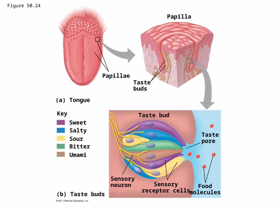
Figure 50.24
Papillae
Papilla
Tastebuds
(a) Tongue
Key
SweetSalty
SourBitter
Umami
Taste bud
Sensoryneuron Sensory
receptor cellsFood
molecules
Tastepore
(b) Taste buds
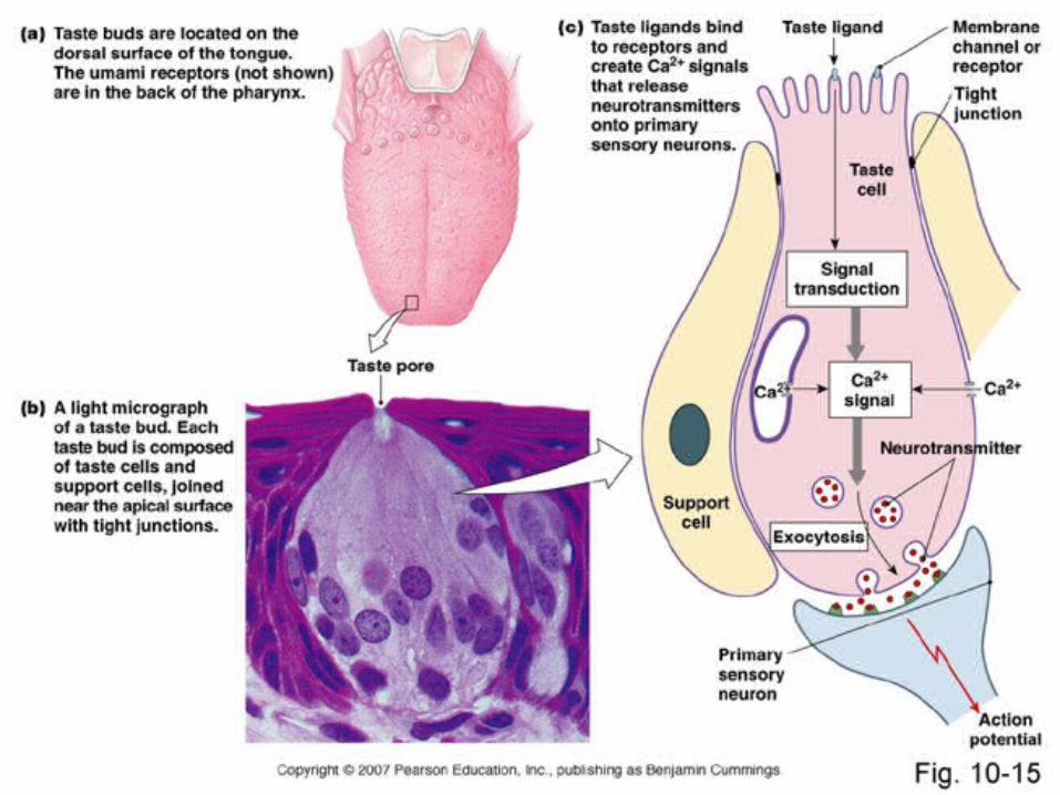
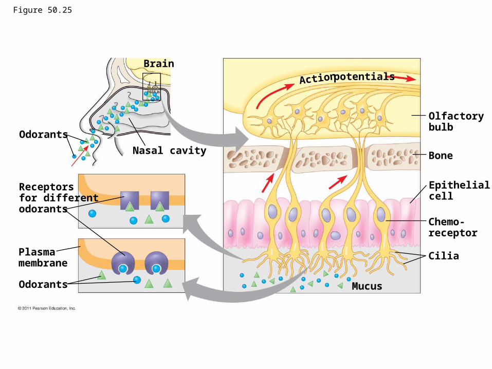
Figure 50.25
Odorants
Brain
Nasal cavity
Receptors for differentodorants
Plasmamembrane
Odorants
potentialsAction
Olfactorybulb
Bone
Epithelialcell
Chemo-receptor
Cilia
Mucus
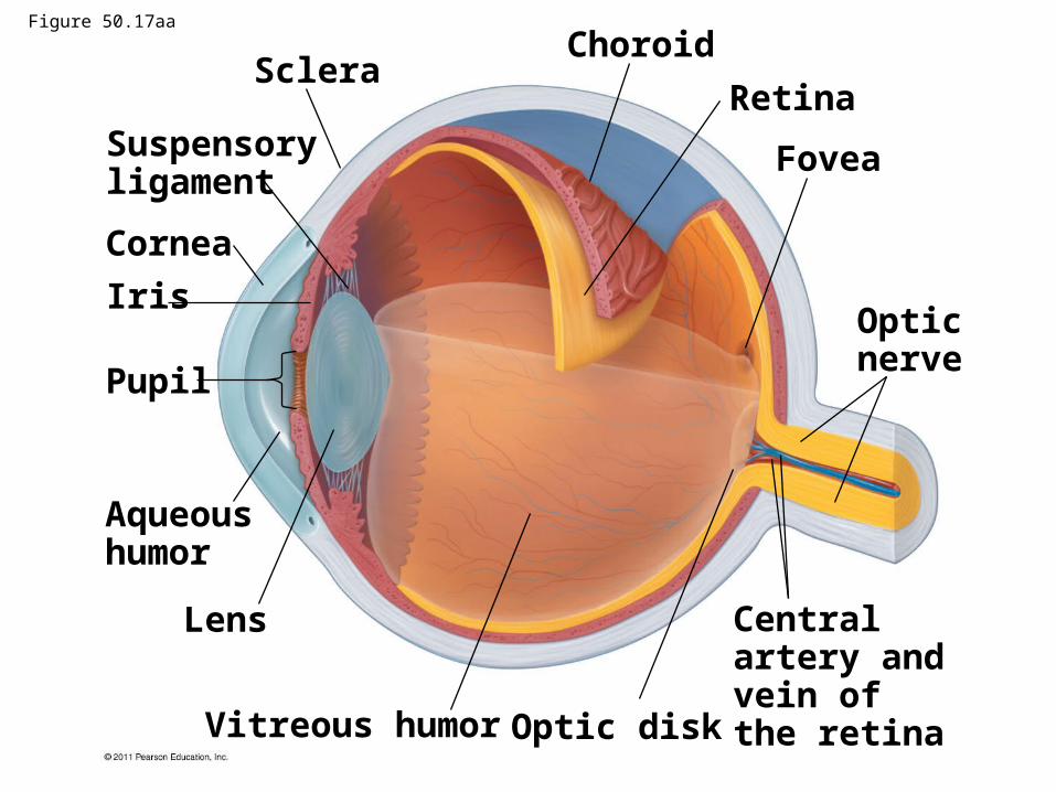
Sclera
Suspensoryligament
Cornea
Iris
Pupil
Aqueoushumor
Lens
Vitreous humor Optic disk
Centralartery andvein of the retina
Opticnerve
Fovea
Retina
ChoroidFigure 50.17aa
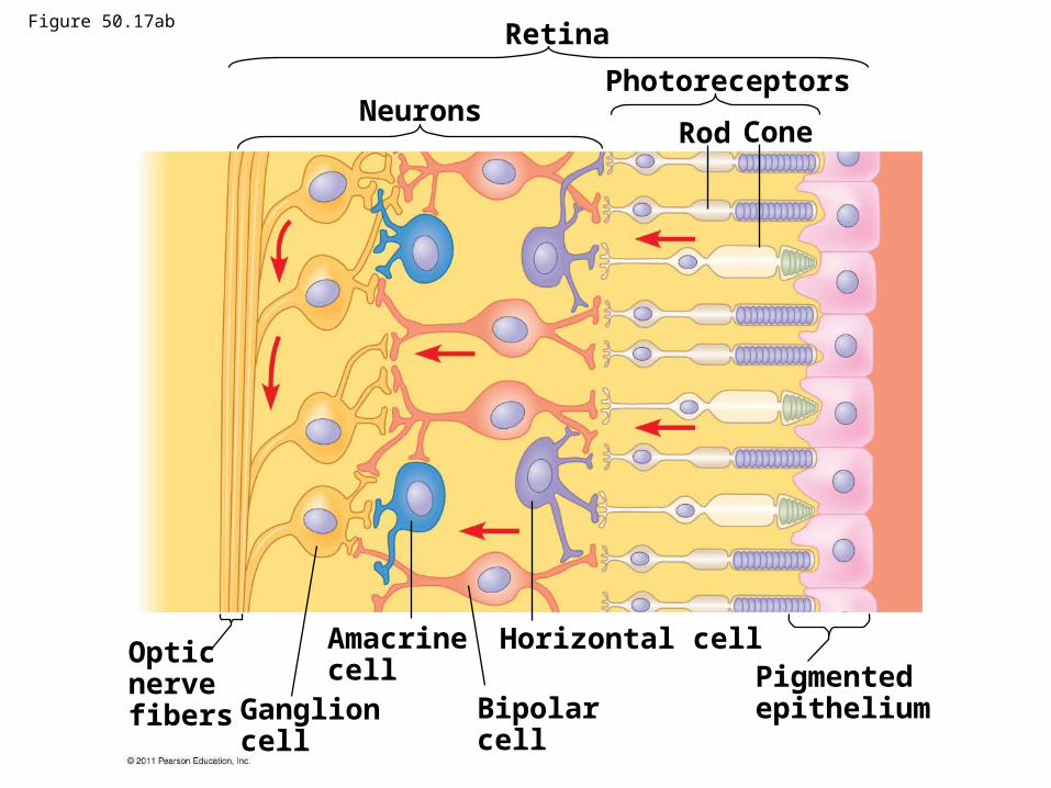
Retina
NeuronsPhotoreceptors
Rod Cone
Opticnervefibers Ganglion
cell
Amacrinecell
Bipolarcell
Horizontal cellPigmentedepithelium
Figure 50.17ab
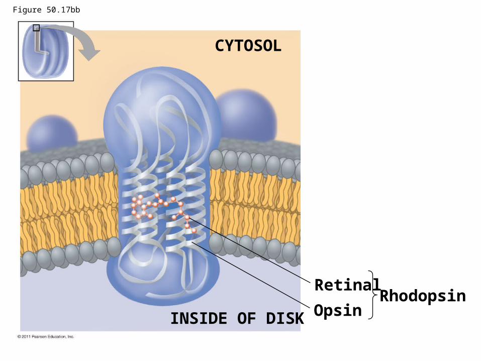
CYTOSOL
INSIDE OF DISK
Retinal
OpsinRhodopsin
Figure 50.17bb
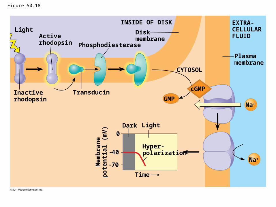
Figure 50.18
Light
Inactiverhodopsin
Activerhodopsin
Transducin
Phosphodiesterase
INSIDE OF DISK
Diskmembrane
CYTOSOL
GMP
cGMP
Na
Na
EXTRA-CELLULARFLUID
Plasmamembrane
Dark Light
Hyper-polarization
Time
0
40
70Mem
bra
ne
po
ten
tial
(m
V)
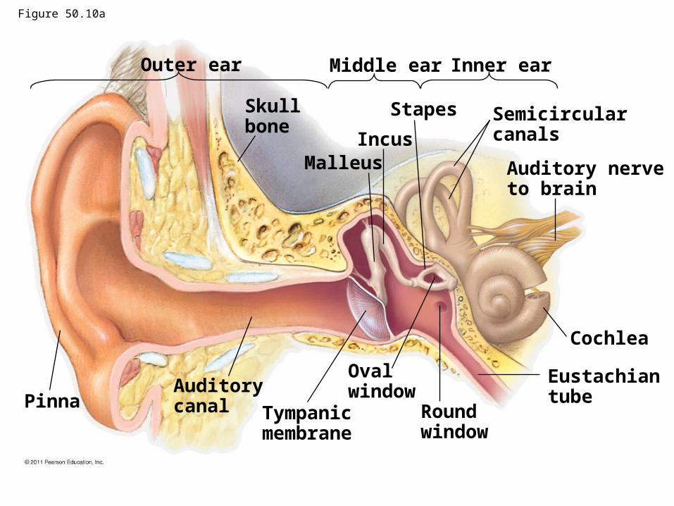
Outer ear Middle ear Inner ear
Skullbone
MalleusIncus
Stapes Semicircularcanals
Auditory nerveto brain
Cochlea
Eustachiantube
Roundwindow
Ovalwindow
Tympanicmembrane
AuditorycanalPinna
Figure 50.10a
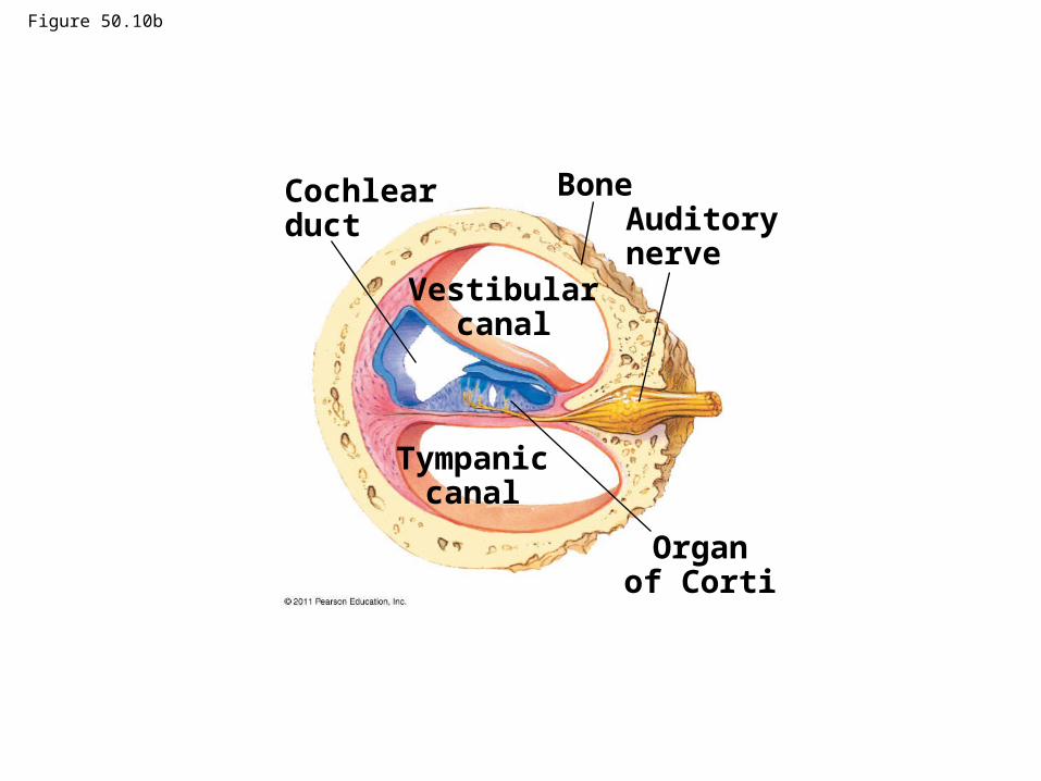
Figure 50.10b
Cochlearduct
BoneAuditorynerve
Vestibularcanal
Tympaniccanal
Organof Corti
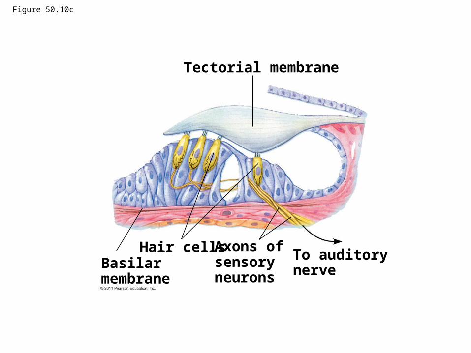
Figure 50.10c
Tectorial membrane
Basilarmembrane
Hair cells Axons ofsensoryneurons
To auditorynerve
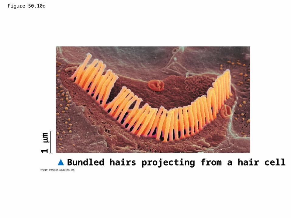
Figure 50.10d
1 m
Bundled hairs projecting from a hair cell
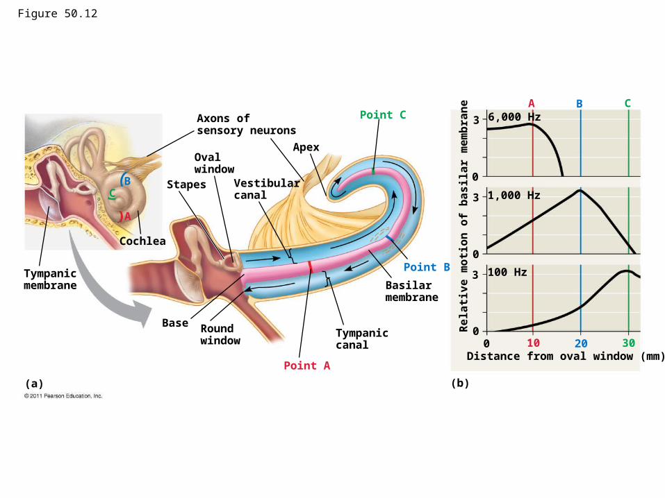
Figure 50.12
Tympanicmembrane
Cochlea
Stapes
Base
(a)
Roundwindow
Point A
A
A
10Tympaniccanal
Basilarmembrane
Point B
B
B
20
Point C
C
C
30
Apex
Vestibularcanal
Oval window
Axons ofsensory neurons
(b)
3
3
3
0
0
00
Distance from oval window (mm)
6,000 Hz
1,000 Hz
100 Hz
Re
lati
ve
mo
tio
n o
f b
as
ila
r m
em
bra
ne
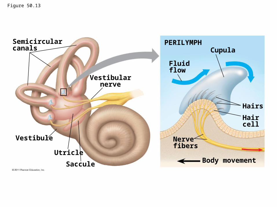
Semicircularcanals
Vestibularnerve
Vestibule
Utricle
Saccule
PERILYMPH
Fluidflow
Cupula
Hairs
Haircell
Nervefibers
Body movement
Figure 50.13