sensitivity and specificity of a urinary screening test used in an emergency setting to detect...
TRANSCRIPT

Accepted Manuscript
Sensitivity and specificity of a urinary screening test used in an emergency setting todetect abnormal first trimester pregnancies
João L.G. Teixeira, BSc Paola Rabaioli, BSc Ricardo F. Savaris, MD, PhD
PII: S0002-9378(14)00636-X
DOI: 10.1016/j.ajog.2014.06.056
Reference: YMOB 9916
To appear in: American Journal of Obstetrics and Gynecology
Received Date: 10 March 2014
Revised Date: 5 June 2014
Accepted Date: 23 June 2014
Please cite this article as: Teixeira JLG, Rabaioli P, Savaris RF, Sensitivity and specificity of a urinaryscreening test used in an emergency setting to detect abnormal first trimester pregnancies, AmericanJournal of Obstetrics and Gynecology (2014), doi: 10.1016/j.ajog.2014.06.056.
This is a PDF file of an unedited manuscript that has been accepted for publication. As a service toour customers we are providing this early version of the manuscript. The manuscript will undergocopyediting, typesetting, and review of the resulting proof before it is published in its final form. Pleasenote that during the production process errors may be discovered which could affect the content, and alllegal disclaimers that apply to the journal pertain.
MANUSCRIP
T
ACCEPTED
ACCEPTED MANUSCRIPT 1
Title page
Title
Sensitivity and specificity of a urinary screening test used in an emergency setting
to detect abnormal first trimester pregnancies
List of authors
João L. G. TEIXEIRA, BSc a
Paola RABAIOLI, BSc b
Ricardo F. SAVARIS, MD, PhD a,b
This study was conducted at Hospital de Clínicas de Porto Alegre, in Porto Alegre, Rio
Grande do Sul, Brazil.
a Programa de Pós-Graduação em Ciências Cirúrgicas, Universidade Federal do Rio
Grande do Sul Departamento e Serviço de Ginecologia e Obstetrícia, Universidade
Federal do Rio Grande do Sul, Porto Alegre, RS, Brazil, 90035-903
b Departamento de Ginecologia e Obstetrícia, Universidade Federal do Rio Grande do
Sul, Porto Alegre, RS, Brazil, 90035-903
Disclosure
The authors report no conflict of interest.
Sources of financial support for the research
This work was supported by Fundação de Incentivo a Pesquisa e Ensino (FIPE) –
Hospital de Clínicas de Porto Alegre – grant 11-0113
Reprint requests and corresponding author:
Ricardo F Savaris
Serviço de Ginecologia e Obstetrícia - Hospital de Clínicas de Porto Alegre

MANUSCRIP
T
ACCEPTED
ACCEPTED MANUSCRIPT 2
Rua Ramiro Barcelos, 2350/1124
Porto Alegre – RS – Brazil
90035-003
E-mail: [email protected]
Fax: 55 51 33598117
Abstract word count: 201
Word count: 2673

MANUSCRIP
T
ACCEPTED
ACCEPTED MANUSCRIPT 3
Condensation
A commercial urine screening test does not have a high specificity to rule out ectopic
pregnancy or miscarriage when used in an emergency room setting.
Short version of the article:
Urine screening test to detect abnormal first trimester pregnancy.
MANUSCRIP
T
ACCEPTED
ACCEPTED MANUSCRIPT 4
ABSTRACT
Objective: To evaluate the performance of a commercial urinary test to screen for
abnormal first trimester pregnancies in women presenting to an emergency room.
Study Design: In this prospective observational cohort, women with a confirmed first
trimester pregnancy (gestational age < 12 weeks) provided a urine sample for
diagnosing the viability of their gestation. Pregnancy viability and location testing were
confirmed by ultrasound and/or laparoscopy.
Results: From 815 eligible patients for the study, 12 were excluded for not having a
confirmed pregnancy (n=6) or for lost to follow up (n=6). A total of 803 patients
underwent testing and completed follow-up. The pre-test probability of an abnormal
pregnancy was 44% (9% for ectopic pregnancy and 35% for miscarriage). The test had
the following parameters to identify an abnormal first trimester pregnancy [%(95%
CI)]: sensitivity:13 (10 to 17); specificity:82 (78 to 86); positive predictive value:36 (28
to 46); negative predictive value: 54 (50 to 58); accuracy: 47%; positive likelihood
ratio=0.74 (0.53 to 1.03); negative likelihood ratio=1.06 (1 to 1.12). The reproducibility
of the test in our study was high (Kappa Index between readers 0.89 (95% confidence
interval 0.77 to 1).
Conclusion: In our emergency setting, we were not able to confirm that the commercial
test is adequate to detect or exclude an abnormal first trimester pregnancy.
KEY WORDS: Inexscreen®; sensitivity; specificity; pregnancy; first trimester;
screening test; abnormal pregnancy

MANUSCRIP
T
ACCEPTED
ACCEPTED MANUSCRIPT 5
Introduction
Vaginal bleeding and pelvic pain are common complaints in emergency facilities.1 Once
the diagnosis of pregnancy is established, the next step is to identify the location and
viability of this pregnancy.2 The main differential diagnoses are miscarriage and ectopic
pregnancy. The use of vaginal ultrasound (VUS) and quantitative β-hCG are the main
diagnostic tools.3 These diagnostic modalities can be both time consuming and
expensive. The search of a biomarker for miscarriage and ectopic pregnancy has been
pursued by many researches.4-6
The peculiarities of β-hCG and its different plasmatic levels in normal and abnormal
pregnancy led the medical industry to launch in the market Inexscreen®, a point-of-care
urinary test. Inexscreen® is a urinary lateral flow test with two windows, A and B.
Window “A” detects the intact hCG (i-hCG) and window “B” detects the β-core
fragments, the nicked β-hCG and the β-hCG isoforms. These isoforms are called human
chorionic gonadotropin related protein (hCGRP). The hCGRP:i-hCG ratio is
significantly decreased in ectopic pregnancies and miscarriages. As a rapid urinary test,
without the need of special equipment or specialized staff, Inexcreen® was developed
as a screening tool to rule out abnormal pregnancy. In 2011, Mazouz et al published the
initial results of Inexscreen® in the clinical context. According to their data,
Inexscreen® had a negative predictive value of 99.3% and 96.6% for ectopic pregnancy
and miscarriage, respectively.7 A negative predictive value >99% for ectopic pregnancy
is ideal in the clinical context, given the fact that time and costs can be reduced.
However, the authors recognized this first report should be confirmed with a larger
prospective study.7 The objective of this study was to estimate the diagnostic accuracy
of Inexscreen® test compared to ultrasonography and surgery in diagnosing abnormal
pregnancy in the first trimester.

MANUSCRIP
T
ACCEPTED
ACCEPTED MANUSCRIPT 6
Materials and methods
Participants
The study population consisted in a consecutive series of subjects who attended the
gynecological emergency department (GED) of Hospital de Clínicas de Porto Alegre
between April 14th, 2011 and October 31st, 2013.
Participant recruitment
Consecutive pregnant women between 14 and 49 year old who attended the GED for
any reason were invited to participate in the study.
Inclusion and exclusion criteria
Subjects were included if they had a pregnancy < 12 weeks according to the last
menstrual period or previous gestational ultrasonography. Subjects were excluded if
their pregnancy was not confirmed by urinary β-hCG, if they had a pregnancy of ≥12
weeks, or if they did not give written consent to participate in the study. Pregnancy and
gestational age were confirmed by urinary and/or serum β-hCG and ultrasound
respectively.
Data collection
After signing the written consent, a standard questionnaire was obtained. Next, a fresh
urine sample was collected from the patient for the index text (Inexscreen®). Subjects
were followed with serial plasma β-hCG and transvaginal ultrasound. In case of
pregnancy of unknown location, subjects were followed every 48 h or every week until
the outcome of pregnancy was identified.
Reference standard and its rationale
A transvaginal ultrasound was used as the reference standard to confirm the presence of
a viable, or a non-viable pregnancy. Vaginal ultrasound was performed within 4 hours
after the Inexscreen® test. Pregnancy viability was defined was defined as the presence
MANUSCRIP
T
ACCEPTED
ACCEPTED MANUSCRIPT 7
of intrauterine embryo/fetus with cardiac activity. Miscarriage was defined by the
absence of visible heartbeat in an embryo with crown-rump length ≥7 mm, or if the
mean gestational sac diameter was ≥25 mm and no structure was visualized inside. The
diagnostic algorithm published by Mol et al was used for diagnosis of ectopic
pregnancy.8 Ectopic pregnancy was confirmed by surgery and pathology report.
Technical specifications
Inexscreen® (Humasis Co., Ltd, Gyeonggi-do, 431-836, Korea) was the index test for
screening viable or non-viable first trimester pregnancy. Urine specimens were
collected in a clean, dry, plastic container and Inexscreen® was run within 10 minutes
of collection. With the disposable pipette provided with the Inexscreen® kit, 5 drops of
urine were loaded into the sample well of the Inexscreen® test device. Interpretation of
the test result was performed after 5 minutes, according to the manufacturer’s
instructions. Briefly, Inexscreen® test has 2 windows (A and B), and two lines in each
window (C and T). Line C is the internal control; if it is absent in one of the windows,
the test is discarded and a new test used. The intensity of the T line was defined by
visual comparison with standards given by the manufacturer. The intensity of the lines
was graduated in a grading system of whole numbers between 0 and 10. The cut-off
between normal and abnormal followed the manufacturer instructions. A ratio of the
intensity of lines A>B was consider abnormal; a ratio A≤B was consider normal.
Persons executing and reading Inexscreen ®
Two experts in Inexscreen® reading (JLG, RFS) trained and supervised the clinical
physicians (n=8), the residents and medical students that were in rotation every month
(total n= 37/30.5 months). The physicians, residents and medical students evaluated the
intensity of line T in windows A and B.
Persons executing ultrasound and pathological analysis
MANUSCRIP
T
ACCEPTED
ACCEPTED MANUSCRIPT 8
Board certified radiologists and pathologists were responsible for executing transvaginal
ultrasound and pathological analysis, respectively. Readers of the Inexscreen® test,
radiologists and pathologists were blind to the results of each other’s test.
Statistical methods and sample size
The performance of the Inexscreen® test was calculated using a 95% confidence
interval (95%CI) for sensitivity, specificity, positive and negative predictive values, and
positive and negative likelihood ratios. Kappa index was used to assess inter- and
intraobserver agreement. Reproducibility of the test, i.e., normal or abnormal, was
verified between two independent observers using 60 cases comprising the whole
spectrum of results, i.e., from 0 to 10. Digital pictures of lines A and B of these 60 cases
were taken and stored in a file. Results from a senior researcher (RFS) were compared
to the results of a naïve researcher for inter-observer agreement. The senior researcher
scored the same 60 cases in two different occasions for intra-observer agreement.
Analyses were performed using Prism 6.0 (GraphPad, San Diego, CA), online Kappa
calculator (http://vassarstats.net/kappa.html) and online diagnostic test calculator
(http://araw.mede.uic.edu/cgi-bin/testcalc.pl).
Sample size was calculated according to the nomogram described in the literature.9 The
following parameters were used: an estimated incidence of ectopic pregnancy of 10%
(±5%) and an estimated specificity of 95% (with a precision of ±5%). Sample size
calculation yielded a total number of 730 subjects. For cases of miscarriage, where the
estimated incidence of miscarriage was 40%, the minimal number of subjects was 180.
With these parameters we were able to verify with a 95% CI, that Inexscreen® has
specificity between 90 and 100% to diagnose ectopic pregnancy in a population where
the prevalence is between 5 to 15%.
Ethical issues

MANUSCRIP
T
ACCEPTED
ACCEPTED MANUSCRIPT 9
This study was submitted and approved by Comite de Ética em Pesquisa of Hospital de
Clínicas de Porto Alegre, the local Institutional Review Board (number 11-0113).
Inexscreen® was evaluated in the gynecological emergency department (GED) of the
Hospital de Clínicas de Porto Alegre, in Porto Alegre, RS, Brazil.
Results
Beginning and end dates of recruitment
Data collection was performed between April 14th, 2011 and October 28th, 2013.
Clinical and demographic characteristics of the study population
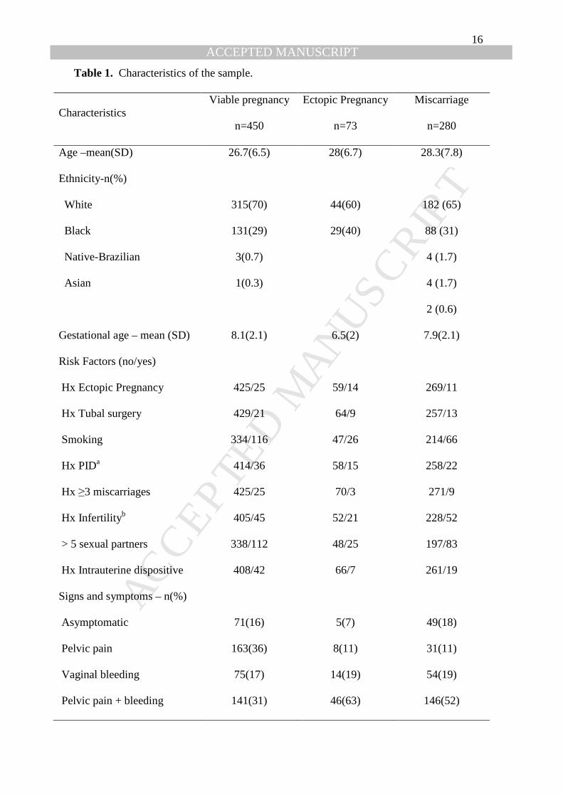
Most of the participants were white and the mean gestational age was 8.1, 6.5 and 7.9
weeks for intrauterine pregnancy, ectopic pregnancy and miscarriage, respectively
(Table 1).
Number of participants
A total of 815 Inexscreen® tests were used in the study and none of them were
discarded for being faulty, i.e., negative control line. Six subjects did not satisfy the
inclusion criteria and were excluded; six were lost to follow-up. A total of 803 subjects
satisfied inclusion criteria and had a vaginal ultrasound or were submitted to surgery
(Fig. 1). The median follow-up was 3 days (range 1 and 60 days).
Time interval between tests results
The time interval between the Inexscreen® test and the vaginal ultrasound was 4 hours
or less. No treatment was administered in between.
Distribution of the severity of the disease
A total of 125 subjects were asymptomatic with no or ≥ 1 risk factor. Asymptomatic
subjects without risk factor comprised 6.6% (53 out of 803) of the study population.
From these 53 subjects, 39.6% had an abnormal pregnancy (3 cases were ectopic

MANUSCRIP
T
ACCEPTED
ACCEPTED MANUSCRIPT 10
pregnancy; 18 cases of miscarriage). The majority of subjects (93.4%) had pelvic pain
and/or vaginal bleeding.
Cross tabulation of the Inexscreen ® and transvaginal ultrasound
All 803 subjects included in the study underwent transvaginal ultrasound and had a
urine sample analyzed by Inexscreen ®. Positive Inexscreen® (abnormal) in abnormal
pregnancy was seen in 47 cases; abnormal Inexscreen in normal pregnancy was seen in
81 cases. Normal Inexscreen® in abnormal pregnancy was seen in 306 cases, and
normal Inexscreen® in normal pregnancy was seen in 369 cases. There were no missing
or indeterminate results.
Adverse events from Inexscreen® and vaginal ultrasound
No adverse event occurred by performing the index test (Inexscreen®) or reference
standard (transvaginal ultrasound).
Estimates of the test
The test performance to identify an abnormal first trimester pregnancy was as
following: sensitivity:13% (95% CI, 10 to 17%); specificity:82% (95% CI, 78 to 85%);
positive predictive value:37% (95% CI, 28 to 46%); negative predictive value: 55%
(95% CI, 50 to 58%); accuracy: 52%; positive likelihood ratio: 0.74 (95% CI, 0.53 to
1.03); negative likelihood ratio: 1.06 (95% CI, 1 to 1.12). The pretest probability of an
abnormal pregnancy was 44%; the posttest probability after a positive and negative test
was 37 and 45%, respectively (Table 2). Similar results were observed in a subgroup of
symptomatic subjects with gestational age between 5 and 8 weeks of pregnancy (Table
3).
No indeterminate results or outliers were observed. The six cases that were lost to
follow-up were excluded from the analysis.

MANUSCRIP
T
ACCEPTED
ACCEPTED MANUSCRIPT 11
Kappa index for inter- and intraobserver agreement was 0.89 (95% CI, 0.77 to 1) and
0.93 (95% CI, 0.84 to 1), respectively.
Discussion
Our results revealed that Inexscreen® correctly identified 6 cases of women that did not
have a positive β-hCG (Figure 1). Similar to the results published by Mazous et al,7 the
specificity of the test was around 82%; in our setting, however, the test was neither
sensitive, nor specific to rule in or to rule out abnormal pregnancy with a precision
higher than 95% (Table 2). The lower performance of the test in our scenario can be
seen by the posttest probability of a positive and negative test: 37 and 45%, values that
did not differ much from the pretest probability, i.e., 44% (Table 2). The negative
predictive value (NPV) for ectopic pregnancy was 85% (95% CI, 81 to 88), compared to
the higher 96.6% (95% CI; 91 to 98%) reported by Mazous. However, the sensitivity of
the test found in our study differed substantially from that of Mazous. While we found a
low sensitivity, 13% (95% CI, 10 to 17), Mazous et al found a sensitivity of 92% (95%
CI, 84 to 97).7 Possible explanations for these discrepancies could be related to the fact
we used the test in asymptomatic pregnant women that attended the gynecological
emergency department for non-obstetric reasons, which alters the pre-test probability.
This subgroup represented 6% of the population, but 39.6% of the cases had an
abnormal pregnancy, and they would benefit the most for a screening test. However, if
only women with pelvic pain and/or vaginal bleeding were included, as it was in the
original work of Mazous et al, the negative predictive value for ectopic pregnancy
would remain low, i.e., 80% (95% CI, 74 to 85%), as shown in Table 3. Predictive
values of a test are influenced by the prevalence of a condition.10 Therefore, it is
important to conduct studies of screening tests in real-life scenarios, in populations that
reproduce the true prevalence of ectopic pregnancy and miscarriage. If these steps were

MANUSCRIP
T
ACCEPTED
ACCEPTED MANUSCRIPT 12
not taken, a selection bias would ensue. In our study, the prevalence of ectopic
pregnancy was 9% (95% CI, 7 to 11%), which is in accordance to the literature.7, 11 The
prevalence of miscarriage, 35% (95% CI, 31 to 38%), was higher than in the population
presented by Mazouz et al,7 but similar to a larger study published by Tude-Byass et
al.12
Other possibilities for the discrepancies could be related to the inclusion criteria. In our
study we included pregnant women with <12 weeks of gestational age. Mazous et al
included pregnancies between 5 and 8 weeks according to the last menstrual period. An
analysis of this subgroup, symptomatic women with gestational age between 5 and 8
weeks, revealed that the performance of the test did not change much (Table 3).
Reading the test could be an issue, since it compares the intensity of the lines in the
cassette to a standard card provided in each kit. The design of this point of care test is
simple, and it is easy to read. The end result lies into two categories: normal, when
intensity of line A≤B, or abnormal, when intensity of line A>B. These results are
straightforward when the intensity of the line is ≥3. The high Kappa index (κ=0.89)
found in our study reflects this agreement among readers. This result is in accordance
with Mazous et al.7
Of note, clear diagnosis of ectopic pregnancy, i.e., gestational sac or fetal heartbeat
outside the uterus, was made by ultrasound in 10 subjects and only 4 tests were
abnormal. Lastly, another possible explanation could be related to rationale of the test.
Inxescreen® is based in the hCGRP:i-hCG ratio; this ratio is significantly decreased in
ectopic pregnancies and miscarriages. Borrelli et al analyzed the serum and urine levels
of the intact hCG and the hCG fragments in first trimester pregnancy of women who
presented at an outpatient emergency department. They found that urine hCG variants
had less significant performance compared to those of serum in distinguishing ectopic

MANUSCRIP
T
ACCEPTED
ACCEPTED MANUSCRIPT 13
pregnancy from viable pregnancy.13 Indeed, we note that Inexscreen® could
alternatively be used to estimate hCG levels at urine, as the test provides different levels
of hCG in urine:< 200 mUI/ml, 200 to 1000, 1000 to 5000, 5000 to 10000 and >10000
mUI/mL. Physicians could benefit from this information to plan the timing of
ultrasound, or to evaluate the rising or fall of hCG compared to previous plasma values.
The strengths of the study were the sample size, the low rate of dropout and the real-
world scenario with a prospective follow-up. One of the limitations of this study was
that we did not correlate the hCG levels of all cases of ectopic pregnancy to the levels
found by Inexscreen®. Indeed, this could be further tests as a clinical applicability of
the test: to identify the discriminatory zone and the use of vaginal ultrasound.3
In conclusion, in this setting, Inexscreen® has a specificity of 82%, a value in range of
what is published in the literature, but it has 13% of sensitivity. The test is not adequate
to rule in or to rule out an abnormal pregnancy in the first trimester in our setting.
Further prospective studies are necessary to confirm our results.
References
1. Matteson KA, Weitzen SH, Lafontaine D, Phipps MG. Accessing care: use
of a specialized women's emergency care facility for nonemergent
problems. J Womens Health (Larchmt) 2008;17:269-77.
2. Lee AS, Cohen SL, Anderson JR, Chanmugam A, Bienstock JL. The effect
of gynecologic algorithm pathways on emergency department visit times. J
Emerg Med 2013;44:217-24.
3. Barnhart KT, Simhan H, Kamelle SA. Diagnostic accuracy of ultrasound
above and below the beta-hCG discriminatory zone. Obstet Gynecol
1999;94:583-7.
MANUSCRIP
T
ACCEPTED
ACCEPTED MANUSCRIPT 14
4. Daponte A, Deligeoroglou E, Garas A, Pournaras S, Hadjichristodoulou C,
Messinis IE. Activin a and follistatin as biomarkers for ectopic pregnancy
and missed abortion. Dis Markers 2013;35:497-503.
5. Senapati S, Barnhart KT. Biomarkers for ectopic pregnancy and pregnancy
of unknown location. Fertil Steril 2013;99:1107-16.
6. Rausch ME, Barnhart KT. Serum biomarkers for detecting ectopic
pregnancy. Clin Obstet Gynecol 2012;55:418-23.
7. Mazouz S, Lee JK, Fernandez H. Evaluation of a urinary test as a diagnostic
tool of a nonprogressive pregnancy. Fertil Steril 2011;95:783-6.
8. Mol BW, van Der Veen F, Bossuyt PM. Implementation of probabilistic
decision rules improves the predictive values of algorithms in the diagnostic
management of ectopic pregnancy. Hum Reprod 1999;14:2855-62.
9. Malhotra RK, Indrayan A. A simple nomogram for sample size for
estimating sensitivity and specificity of medical tests. Indian J Ophthalmol
2010;58:519-22.
10. Grimes DA, Schulz KF. Uses and abuses of screening tests. Lancet
2002;359:881-4.
11. Alkatout I, Honemeyer U, Strauss A et al. Clinical diagnosis and treatment
of ectopic pregnancy. Obstet Gynecol Surv 2013;68:571-81.
12. Tunde-Byass M, Cheung VY. The value of the early pregnancy assessment
clinic in the management of early pregnancy complications. J Obstet
Gynaecol Can 2009;31:841-4.

MANUSCRIP
T
ACCEPTED
ACCEPTED MANUSCRIPT 15
13. Borrelli PT, Butler SA, Docherty SM, Staite EM, Borrelli AL, Iles RK.
Human chorionic gonadotropin isoforms in the diagnosis of ectopic
pregnancy. Clin Chem 2003;49:2045-9.
MANUSCRIP
T
ACCEPTED
ACCEPTED MANUSCRIPT 16
Table 1. Characteristics of the sample.
Characteristics Viable pregnancy
n=450
Ectopic Pregnancy
n=73
Miscarriage
n=280
Age –mean(SD) 26.7(6.5) 28(6.7) 28.3(7.8)
Ethnicity-n(%)
White 315(70) 44(60) 182 (65)
Black 131(29) 29(40) 88 (31)
Native-Brazilian 3(0.7) 4 (1.7)
Asian 1(0.3) 4 (1.7)
2 (0.6)
Gestational age – mean (SD) 8.1(2.1) 6.5(2) 7.9(2.1)
Risk Factors (no/yes)
Hx Ectopic Pregnancy 425/25 59/14 269/11
Hx Tubal surgery 429/21 64/9 257/13
Smoking 334/116 47/26 214/66
Hx PIDa 414/36 58/15 258/22
Hx ≥3 miscarriages 425/25 70/3 271/9
Hx Infertilityb 405/45 52/21 228/52
> 5 sexual partners 338/112 48/25 197/83
Hx Intrauterine dispositive 408/42 66/7 261/19
Signs and symptoms – n(%)
Asymptomatic 71(16) 5(7) 49(18)
Pelvic pain 163(36) 8(11) 31(11)
Vaginal bleeding 75(17) 14(19) 54(19)
Pelvic pain + bleeding 141(31) 46(63) 146(52)

MANUSCRIP
T
ACCEPTED
ACCEPTED MANUSCRIPT 17
a Hx PID= History of Pelvic Inflammatory Disease.
b Hx Infertility: History of infertility, defined as failure to achieve a clinical pregnancy
after 12 months or more of regular unprotected sexual intercourse.

MANUSCRIP
T
ACCEPTED
ACCEPTED MANUSCRIPT 18
Table 2. Performance of Inexscreen® test compared to Vaginal Ultrasound, or
surgery/pathology in 803 women. Numbers are % (95% confidence interval), except for
likelihood ratios.
Parameter Total Ectopic pregnancy Miscarriage
Pre-test probability 44 9 35
Sensitivity 13 (10 to 17) 14 (6 to 23) 13 (9 to 18)
Specificity 82 (78 to 85) 82 (78 to 85) 82 (78 to 85)
PPV 37 (28 to 46) 11 (5 to 19) 31 (23 to 40)
NPV 55 (50 to 58) 85 (81 to 88) 60 (56 to 64)
Accuracy 52 72 56
LR+ 0.74 (0.53 to 1.03) 0.76 (0.41 to 1.4) 0.73 (0.51 to 1.05)
LR - 1.06 (1 to 1.12) 1.05 (0.95 to 1.16) 1.06 (0.99 to 1.13)
PPT + 37 11 31
PPT - 45 15 40
PPV: positive predictive value
NPV: negative predictive value
LR+: positive likelihood ratio
LR-: negative likelihood ratio
PPT+: post test probability if test is positive
PPT-: post test probability if test is negative

MANUSCRIP
T
ACCEPTED
ACCEPTED MANUSCRIPT 19
Table 3. Performance of Inexscreen® test compared to Vaginal Ultrasound, or
surgery/pathology in 403 symptomatic women with gestational age between 5 and 8
weeks. Numbers are % (95% confidence interval), except for likelihood ratios.
Parameter Total Ectopic pregnancy Miscarriage
Pre-test probability 45 13 33
Sensitivity 16 (11 to 22) 13 (6 to 25) 17 (9 to 18)
Specificity 81 (75 to 86) 81 (75 to 86) 81 (75 to 86)
PPV 42 (30 to 54) 15 (6 to 27) 36 (23 to 40)
NPV 53 (47 to 59) 80 (74 to 85) 62 (56 to 64)
Accuracy 51 68 57
LR+ 0.86 (0.6 to 1.3) 0.72 (0.3 to 1.5) 0.92 (0.6 to 1.4)
LR - 1.03 (0.9 to 1.1) 1.07 (0.94 to 1.2) 1.02 (0.9 to 1.1)
PPT + 42 10 31.2
PPT - 47 14 33.4
PPV: positive predictive value
NPV: negative predictive value
LR+: positive likelihood ratio
LR-: negative likelihood ratio
PPT+: post test probability if test is positive
PPT-: post test probability if test is negative

MANUSCRIP
T
ACCEPTED
ACCEPTED MANUSCRIPT 20
Figure legends
Figure 1. Flow chart of the study. Ect Preg= ectopic pregnancy

MANUSCRIP
T
ACCEPTED
ACCEPTED MANUSCRIPT