selenium in health and disease
TRANSCRIPT

1
1
COMPREHENSIVE INVITED REVIEW
Selenium in Human Health and Disease
Susan J Fairweather-Tait,1 Yongping Bao,1 Martin R Broadley,2 Rachel Collings,1
Dianne Ford,3 John E Hesketh,3 and Rachel Hurst1
1School of Medicine, Health Policy & Practice, University of East Anglia, Norwich,
Norfolk NR4 7TJ, UK.
2School of Biosciences, University of Nottingham, Sutton Bonington Campus,
Loughborough, Leicestershire LE12 5RD, UK.
3Institute for Cell & Molecular Biosciences, Medical School, Newcastle University,
Newcastle NE2 4HH, UK.
Running head: selenium and human health
Proofs to: Professor Susan Fairweather-Tait, School of Medicine, Health Policy &
Practice, University of East Anglia, Norwich, Norfolk NR4 7TJ, UK.
Tel: +44 1603 591304
Fax: +44 1603 593752
E-mail: [email protected]
Word count (excluding references and figure legends): 25,624
References: 435
Reviewing Editors: Carla Boitani, Marcus Conrad, Arthur Cooper, Vadim
Gladyshev, Kum Kum Khanna, William Manzanares, Jakob Moskovitz, Laura Papp,
K. Sandeep Prabhu, Lutz Schomburg, Gerhard N Schrauzer, Alan Shenkin and
Fulvio Ursini
Page 1 of 154 A
ntio
xida
nts
& R
edox
Sig
nalin
gSe
leni
um in
Hum
an H
ealth
and
Dis
ease
(do
i: 10
.108
9/ar
s.20
10.3
275)
Thi
s ar
ticle
has
bee
n pe
er-r
evie
wed
and
acc
epte
d fo
r pu
blic
atio
n, b
ut h
as y
et to
und
ergo
cop
yedi
ting
and
proo
f co
rrec
tion.
The
fin
al p
ublis
hed
vers
ion
may
dif
fer
from
this
pro
of.

2
2
I. Introduction
II. Selenium in the environment
a. Soil selenium
b. Food sources and selenium species
i. Bread and cereals
ii. Meat, fish and eggs
iii. Milk, dairy products and beverages
iv. Fruit and vegetables
v. Selenium-enriched foods
c. Selenium intake
i. Dietary surveys
ii. Global variation in intake
iii. Selenium intake from dietary supplements
III. Selenium absorption and metabolism
a. Absorption of dietary selenium
b. The biochemical interconversion of selenium species
c. Systemic transport of selenium
IV. Selenium status
a. Measurement of status
b. Global variation in status
c. Changes in selenium status in relation to environmental
factors
V. Functions of selenium in the human body
a. Thyroid hormone metabolism
i. Thyroid hormone synthesis and the role of
selenoproteins in thyroid gland function and
protection
ii. Prioritization of the selenium supply to the thyroid
gland and to iodothyronine deiodinases (DIOs)
Page 2 of 154A
ntio
xida
nts
& R
edox
Sig
nalin
gSe
leni
um in
Hum
an H
ealth
and
Dis
ease
(do
i: 10
.108
9/ar
s.20
10.3
275)
Thi
s ar
ticle
has
bee
n pe
er-r
evie
wed
and
acc
epte
d fo
r pu
blic
atio
n, b
ut h
as y
et to
und
ergo
cop
yedi
ting
and
proo
f co
rrec
tion.
The
fin
al p
ublis
hed
vers
ion
may
dif
fer
from
this
pro
of.
3
3
iii. Functions of the DIOs and their potential role in
health and disease
b. Antioxidant defense systems and oxidative metabolism
i. Glutathione peroxidases (GPxs)
ii. Thioredoxin reductases (TXNRDs)
iii. Other seleoproteins involved in the antioxidant
defense system
c. Immune system
VI. Clinical disorders
a. Deficiency
i. Keshan disease
ii. Role of selenium in Kashin-Beck disease
b. Toxicity
VII. Effects on health
a. Cardiovascular disease
b. Cancer
i. Total cancer incidence and mortality
ii. Gastrointestinal cancers
iii. Prostate cancer
iv. Other cancers
v. Summary of selenium and cancer research, and
ranges that may offer protection
vi. Selenium supplementation as an adjuvant therapy in
radiation or chemotherapy treatment
vii. Effect of genotype and polymorphisms relating to
selenium and cancer risk
c. Diabetes
d. Inflammation and inflammatory disorders
e. Fertility
Page 3 of 154 A
ntio
xida
nts
& R
edox
Sig
nalin
gSe
leni
um in
Hum
an H
ealth
and
Dis
ease
(do
i: 10
.108
9/ar
s.20
10.3
275)
Thi
s ar
ticle
has
bee
n pe
er-r
evie
wed
and
acc
epte
d fo
r pu
blic
atio
n, b
ut h
as y
et to
und
ergo
cop
yedi
ting
and
proo
f co
rrec
tion.
The
fin
al p
ublis
hed
vers
ion
may
dif
fer
from
this
pro
of.
4
4
f. Genetics of selenoproteins
VIII. Selenium in critical illness
IX. Dietary reference intakes
X. Conclusions and future perspectives
Page 4 of 154A
ntio
xida
nts
& R
edox
Sig
nalin
gSe
leni
um in
Hum
an H
ealth
and
Dis
ease
(do
i: 10
.108
9/ar
s.20
10.3
275)
Thi
s ar
ticle
has
bee
n pe
er-r
evie
wed
and
acc
epte
d fo
r pu
blic
atio
n, b
ut h
as y
et to
und
ergo
cop
yedi
ting
and
proo
f co
rrec
tion.
The
fin
al p
ublis
hed
vers
ion
may
dif
fer
from
this
pro
of.

5
5
Abstract
This review covers current knowledge of selenium in the environment, dietary
intakes, metabolism and status, functions in the body, thyroid hormone
metabolism, antioxidant defense systems and oxidative metabolism, and the
immune system. Selenium toxicity and links between deficiency and Keshan
disease and Kashin-Beck disease are described. The relationship between
selenium intake/status and various health outcomes, in particular gastrointestinal
and prostate cancer, cardiovascular disease, diabetes, and male fertility, are
reviewed, and recent developments in genetics of selenoproteins are outlined.
The rationale behind current dietary reference intakes of selenium is explained
and examples of differences between countries and/or expert bodies are given.
Throughout the review, gaps in knowledge and research requirements are
identified. More research is needed to improve our understanding of selenium
metabolism and requirements for optimal health. Functions of the majority of the
selenoproteins await characterization, the mechanism of absorption has yet to be
identified, measures of status need to be developed, and effects of genotype on
metabolism require further investigation. The relationships between selenium
intake/status and health, or risk of disease, are complex but require elucidation in
order to inform clinical practice and to refine dietary recommendations and
develop effective public health policies.
Page 5 of 154 A
ntio
xida
nts
& R
edox
Sig
nalin
gSe
leni
um in
Hum
an H
ealth
and
Dis
ease
(do
i: 10
.108
9/ar
s.20
10.3
275)
Thi
s ar
ticle
has
bee
n pe
er-r
evie
wed
and
acc
epte
d fo
r pu
blic
atio
n, b
ut h
as y
et to
und
ergo
cop
yedi
ting
and
proo
f co
rrec
tion.
The
fin
al p
ublis
hed
vers
ion
may
dif
fer
from
this
pro
of.
6
6
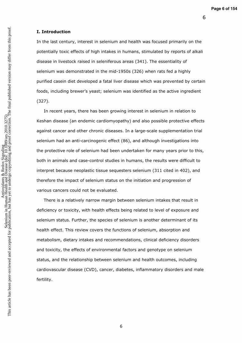
I. Introduction
In the last century, interest in selenium and health was focused primarily on the
potentially toxic effects of high intakes in humans, stimulated by reports of alkali
disease in livestock raised in seleniferous areas (341). The essentiality of
selenium was demonstrated in the mid-1950s (326) when rats fed a highly
purified casein diet developed a fatal liver disease which was prevented by certain
foods, including brewer’s yeast; selenium was identified as the active ingredient
(327).
In recent years, there has been growing interest in selenium in relation to
Keshan disease (an endemic cardiomyopathy) and also possible protective effects
against cancer and other chronic diseases. In a large-scale supplementation trial
selenium had an anti-carcinogenic effect (86), and although investigations into
the protective role of selenium had been undertaken for many years prior to this,
both in animals and case-control studies in humans, the results were difficult to
interpret because neoplastic tissue sequesters selenium (311 cited in 402), and
therefore the impact of selenium status on the initiation and progression of
various cancers could not be evaluated.
There is a relatively narrow margin between selenium intakes that result in
deficiency or toxicity, with health effects being related to level of exposure and
selenium status. Further, the species of selenium is another determinant of its
health effect. This review covers the functions of selenium, absorption and
metabolism, dietary intakes and recommendations, clinical deficiency disorders
and toxicity, the effects of environmental factors and genotype on selenium
status, and the relationship between selenium and health outcomes, including
cardiovascular disease (CVD), cancer, diabetes, inflammatory disorders and male
fertility.
Page 6 of 154A
ntio
xida
nts
& R
edox
Sig
nalin
gSe
leni
um in
Hum
an H
ealth
and
Dis
ease
(do
i: 10
.108
9/ar
s.20
10.3
275)
Thi
s ar
ticle
has
bee
n pe
er-r
evie
wed
and
acc
epte
d fo
r pu
blic
atio
n, b
ut h
as y
et to
und
ergo
cop
yedi
ting
and
proo
f co
rrec
tion.
The
fin
al p
ublis
hed
vers
ion
may
dif
fer
from
this
pro
of.
7
7
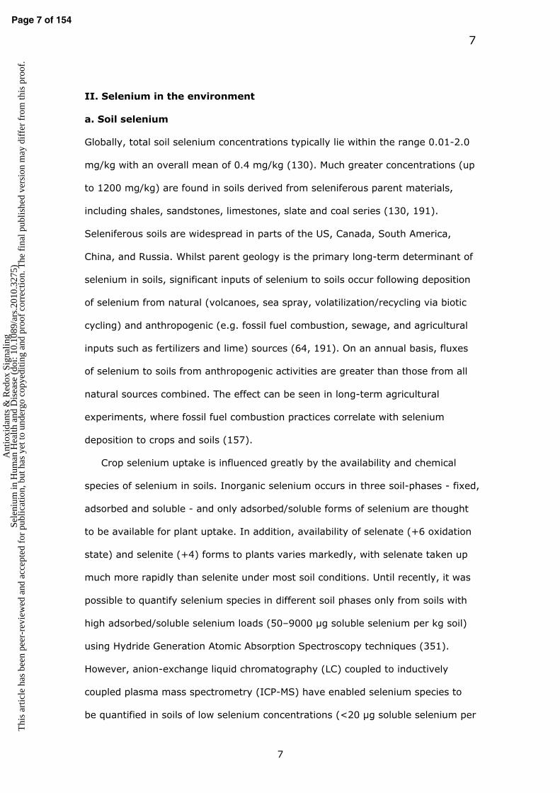
II. Selenium in the environment
a. Soil selenium
Globally, total soil selenium concentrations typically lie within the range 0.01-2.0
mg/kg with an overall mean of 0.4 mg/kg (130). Much greater concentrations (up
to 1200 mg/kg) are found in soils derived from seleniferous parent materials,
including shales, sandstones, limestones, slate and coal series (130, 191).
Seleniferous soils are widespread in parts of the US, Canada, South America,
China, and Russia. Whilst parent geology is the primary long-term determinant of
selenium in soils, significant inputs of selenium to soils occur following deposition
of selenium from natural (volcanoes, sea spray, volatilization/recycling via biotic
cycling) and anthropogenic (e.g. fossil fuel combustion, sewage, and agricultural
inputs such as fertilizers and lime) sources (64, 191). On an annual basis, fluxes
of selenium to soils from anthropogenic activities are greater than those from all
natural sources combined. The effect can be seen in long-term agricultural
experiments, where fossil fuel combustion practices correlate with selenium
deposition to crops and soils (157).
Crop selenium uptake is influenced greatly by the availability and chemical
species of selenium in soils. Inorganic selenium occurs in three soil-phases - fixed,
adsorbed and soluble - and only adsorbed/soluble forms of selenium are thought
to be available for plant uptake. In addition, availability of selenate (+6 oxidation
state) and selenite (+4) forms to plants varies markedly, with selenate taken up
much more rapidly than selenite under most soil conditions. Until recently, it was
possible to quantify selenium species in different soil phases only from soils with
high adsorbed/soluble selenium loads (50–9000 µg soluble selenium per kg soil)
using Hydride Generation Atomic Absorption Spectroscopy techniques (351).
However, anion-exchange liquid chromatography (LC) coupled to inductively
coupled plasma mass spectrometry (ICP-MS) have enabled selenium species to
be quantified in soils of low selenium concentrations (<20 µg soluble selenium per
Page 7 of 154 A
ntio
xida
nts
& R
edox
Sig
nalin
gSe
leni
um in
Hum
an H
ealth
and
Dis
ease
(do
i: 10
.108
9/ar
s.20
10.3
275)
Thi
s ar
ticle
has
bee
n pe
er-r
evie
wed
and
acc
epte
d fo
r pu
blic
atio
n, b
ut h
as y
et to
und
ergo
cop
yedi
ting
and
proo
f co
rrec
tion.
The
fin
al p
ublis
hed
vers
ion
may
dif
fer
from
this
pro
of.

8
8
kg soil) (351). In UK soils of low selenium status, adsorbed/soluble selenium
concentrations are generally two orders of magnitude lower than total soil
selenium, and consist primarily of selenite and KH2PO4-extractable organic
selenium forms (351).
b. Food sources and selenium species
The amount of selenium in the diet largely depends on where crops are grown
and cultivated, the soil/fodder to which animals are exposed, and the actual foods
consumed. The effect of selenium species on bioavailability has been reviewed
recently (117) and data on the selenium content of foods are available (117, 135,
302, 372). The main food groups providing selenium in the diet are bread and
cereals, meat, fish, eggs and milk/dairy products (Fig. 1). Some Brazil nuts are a
particularly rich source, with selenium concentrations ranging from ~0.03-512
mg/kg fresh weight (302).
i. Bread and cereals
The selenium content of bread and cereals can vary widely from ~0.01-30 mg/kg
(302). On average, bread and cereals provide a quarter of the selenium intake in
the UK (Fig. 1). The predominant species of selenium in wheat and bread are
selenomethionine (usually ~55-85%), selenocysteine (~4-12%) and selenate/ite
(~12-19%) (400, 405).
ii. Meat, fish and eggs
The selenium content of meat depends on many factors. Offal contains relatively
high levels of selenium, in particular liver and kidneys; the selenium
concentrations of kidney, liver and heart tissue from beef were 4.5, 0.93 and 0.55
mg/kg respectively, whereas muscle was in the region of 0.2 mg/kg (193).
Supplementation of cattle with selenium-enriched yeast increased muscle
selenium concentration to ~0.6 mg/kg (193). In the US, the average selenium
Page 8 of 154A
ntio
xida
nts
& R
edox
Sig
nalin
gSe
leni
um in
Hum
an H
ealth
and
Dis
ease
(do
i: 10
.108
9/ar
s.20
10.3
275)
Thi
s ar
ticle
has
bee
n pe
er-r
evie
wed
and
acc
epte
d fo
r pu
blic
atio
n, b
ut h
as y
et to
und
ergo
cop
yedi
ting
and
proo
f co
rrec
tion.
The
fin
al p
ublis
hed
vers
ion
may
dif
fer
from
this
pro
of.
9
9
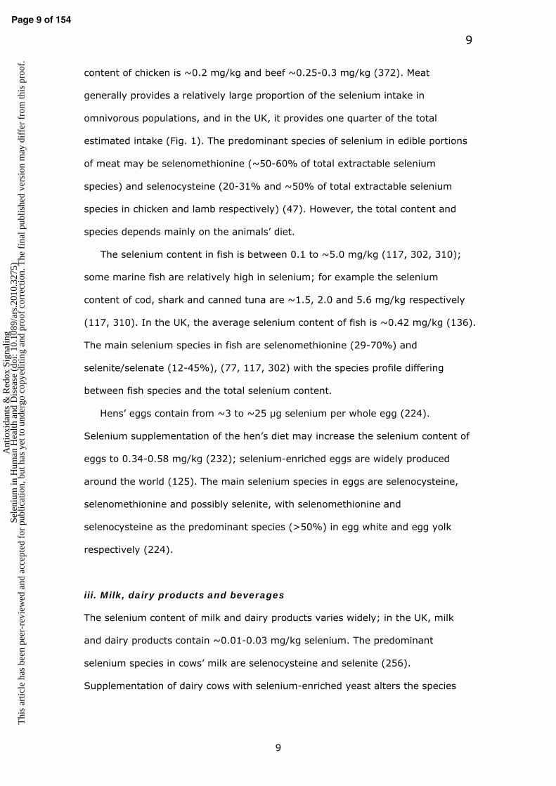
content of chicken is ~0.2 mg/kg and beef ~0.25-0.3 mg/kg (372). Meat
generally provides a relatively large proportion of the selenium intake in
omnivorous populations, and in the UK, it provides one quarter of the total
estimated intake (Fig. 1). The predominant species of selenium in edible portions
of meat may be selenomethionine (~50-60% of total extractable selenium
species) and selenocysteine (20-31% and ~50% of total extractable selenium
species in chicken and lamb respectively) (47). However, the total content and
species depends mainly on the animals’ diet.
The selenium content in fish is between 0.1 to ~5.0 mg/kg (117, 302, 310);
some marine fish are relatively high in selenium; for example the selenium
content of cod, shark and canned tuna are ~1.5, 2.0 and 5.6 mg/kg respectively
(117, 310). In the UK, the average selenium content of fish is ~0.42 mg/kg (136).
The main selenium species in fish are selenomethionine (29-70%) and
selenite/selenate (12-45%), (77, 117, 302) with the species profile differing
between fish species and the total selenium content.
Hens’ eggs contain from ~3 to ~25 µg selenium per whole egg (224).
Selenium supplementation of the hen’s diet may increase the selenium content of
eggs to 0.34-0.58 mg/kg (232); selenium-enriched eggs are widely produced
around the world (125). The main selenium species in eggs are selenocysteine,
selenomethionine and possibly selenite, with selenomethionine and
selenocysteine as the predominant species (>50%) in egg white and egg yolk
respectively (224).
iii. Milk, dairy products and beverages
The selenium content of milk and dairy products varies widely; in the UK, milk
and dairy products contain ~0.01-0.03 mg/kg selenium. The predominant
selenium species in cows’ milk are selenocysteine and selenite (256).
Supplementation of dairy cows with selenium-enriched yeast alters the species
Page 9 of 154 A
ntio
xida
nts
& R
edox
Sig
nalin
gSe
leni
um in
Hum
an H
ealth
and
Dis
ease
(do
i: 10
.108
9/ar
s.20
10.3
275)
Thi
s ar
ticle
has
bee
n pe
er-r
evie
wed
and
acc
epte
d fo
r pu
blic
atio
n, b
ut h
as y
et to
und
ergo
cop
yedi
ting
and
proo
f co
rrec
tion.
The
fin
al p
ublis
hed
vers
ion
may
dif
fer
from
this
pro
of.
10
10
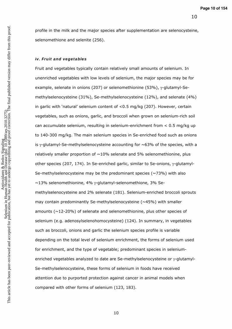
profile in the milk and the major species after supplementation are selenocysteine,
selenomethione and selenite (256).
iv. Fruit and vegetables
Fruit and vegetables typically contain relatively small amounts of selenium. In
unenriched vegetables with low levels of selenium, the major species may be for
example, selenate in onions (207) or selenomethionine (53%), γ-glutamyl-Se-
methylselenocysteine (31%), Se-methylselenocysteine (12%), and selenate (4%)
in garlic with ‘natural’ selenium content of <0.5 mg/kg (207). However, certain
vegetables, such as onions, garlic, and broccoli when grown on selenium-rich soil
can accumulate selenium, resulting in selenium-enrichment from < 0.5 mg/kg up
to 140-300 mg/kg. The main selenium species in Se-enriched food such as onions
is γ-glutamyl-Se-methylselenocysteine accounting for ~63% of the species, with a
relatively smaller proportion of ~10% selenate and 5% selenomethionine, plus
other species (207, 174). In Se-enriched garlic, similar to Se-onions, γ-glutamyl-
Se-methylselenocysteine may be the predominant species (~73%) with also
~13% selenomethionine, 4% γ-glutamyl-selenomethione, 3% Se-
methylselenocysteine and 2% selenate (181). Selenium-enriched broccoli sprouts
may contain predominantly Se-methylselenocysteine (~45%) with smaller
amounts (~12-20%) of selenate and selenomethionine, plus other species of
selenium (e.g. adenosylselenohomocysteine) (124). In summary, in vegetables
such as broccoli, onions and garlic the selenium species profile is variable
depending on the total level of selenium enrichment, the forms of selenium used
for enrichment, and the type of vegetable; predominant species in selenium-
enriched vegetables analyzed to date are Se-methylselenocysteine or γ-glutamyl-
Se-methylselenocysteine, these forms of selenium in foods have received
attention due to purported protection against cancer in animal models when
compared with other forms of selenium (123, 183).
Page 10 of 154A
ntio
xida
nts
& R
edox
Sig
nalin
gSe
leni
um in
Hum
an H
ealth
and
Dis
ease
(do
i: 10
.108
9/ar
s.20
10.3
275)
Thi
s ar
ticle
has
bee
n pe
er-r
evie
wed
and
acc
epte
d fo
r pu
blic
atio
n, b
ut h
as y
et to
und
ergo
cop
yedi
ting
and
proo
f co
rrec
tion.
The
fin
al p
ublis
hed
vers
ion
may
dif
fer
from
this
pro
of.

11
11
v. Selenium-enriched foods
The only permitted species of selenium added to foods for particular nutritional
use in Europe, including baby formula milk and total parental nutrition foods, are
sodium selenite, sodium selenate and sodium hydrogen selenite (127), whereas
the predominant selenium species in most ‘natural’ and unenriched foods is
selenomethionine (Fig. 2). Selenium-enrichment through fertilization or feeding
supplements to animals changes the selenium species profile in some foods e.g.
eggs, onions, garlic and broccoli (124, 183, 207, 224), but wheat and meat tend
to retain the predominant selenium species as selenomethionine (47, 405). The
selenium speciation of foods and the effect of processing and cooking on the
species profile is a priority for future research.
c. Selenium intake
i. Dietary surveys
The contribution to selenium intake from drinking water (130) and air is
insignificant, except for individuals working in industries with an occupational
exposure risk (e.g. metal recovery and paint production). Dietary selenium
intakes can be estimated from dietary surveys, food composition tables, market
basket type surveys, and/or composite dietary analyses. There are inherent
uncertainties in using all of these data, since dietary surveys are prone to
misreporting, and robust primary data on the selenium concentration of different
foodstuffs are often lacking or below technical limits of detection and/or
quantification. For example, in the UK, the most recent published dietary survey
reporting mineral intakes is the National Diet and Nutrition Survey (NDNS) (158,
165). However, under-reporting is ~25% (307), and there can be over-reporting
of foods perceived to be healthy, for example cereals and vegetables (275, 385).
Food composition tables may lack robust selenium concentration data for many
food groups. For example, the Sixth Summary Edition of McCance and
Widdowson’s The Composition of Foods contains mineral concentration data for
Page 11 of 154 A
ntio
xida
nts
& R
edox
Sig
nalin
gSe
leni
um in
Hum
an H
ealth
and
Dis
ease
(do
i: 10
.108
9/ar
s.20
10.3
275)
Thi
s ar
ticle
has
bee
n pe
er-r
evie
wed
and
acc
epte
d fo
r pu
blic
atio
n, b
ut h
as y
et to
und
ergo
cop
yedi
ting
and
proo
f co
rrec
tion.
The
fin
al p
ublis
hed
vers
ion
may
dif
fer
from
this
pro
of.

12
12
up to 3423 types of food and drink products (135). Of these, selenium data are
not reported for 1161 products, “trace” or zero selenium is reported for 467
products, and the selenium content of a further 470 products are estimates.
Among the 1325 products for which selenium concentration data are reported,
241 products have the lowest reported selenium concentration of 1 µg/100 g,
which may introduce rounding errors. In the UK’s 2006 Total Diet Survey, a
market basket survey of 24 UK towns reported selenium concentration data
above the limit of quantification for only 7 out of 20 food groups (136). However,
subject to these caveats, it is still possible to provide estimates of selenium intake,
and even target specific food groups, by integrating dietary survey data with food
composition data (62).
ii. Global variation in selenium intake
Individual dietary selenium intakes across the world are estimated to range from
3-7000 µg/day (130, 297, 300, 410). The highest levels of intake have been
recorded in seleniferous regions of China (>4990 µg/day (422)) and Venezuela
(130, 298, 299). For European countries where estimates are available, mean
intakes are typically <50 µg/day per person (298, 299) which are close to or
below the recommended nutrient intake level (127, 300). In regions of relatively
high selenium intake in India, intakes were estimated to be 475 µg/day for
women and 632 µg/day for men, with >80% of the selenium intake provided by
consumption of cereals grown in high selenium soil in the local region (164). For
large areas of the world (e.g. Africa and many parts of Asia and Latin America),
dietary selenium intake estimates are unavailable. Examples of selenium intakes
across the world are shown in Fig. 3.
With respect to selenium intake from habitual diet, the UK is one of the
countries with the lowest estimated selenium intake (Fig. 3). The mean selenium
intake from food sources for men and women (aged 19-64) were 55 and 43
µg/day respectively. In comparison, intakes in the US are much higher: the mean
Page 12 of 154A
ntio
xida
nts
& R
edox
Sig
nalin
gSe
leni
um in
Hum
an H
ealth
and
Dis
ease
(do
i: 10
.108
9/ar
s.20
10.3
275)
Thi
s ar
ticle
has
bee
n pe
er-r
evie
wed
and
acc
epte
d fo
r pu
blic
atio
n, b
ut h
as y
et to
und
ergo
cop
yedi
ting
and
proo
f co
rrec
tion.
The
fin
al p
ublis
hed
vers
ion
may
dif
fer
from
this
pro
of.

13
13
intake from food in the US was 133.5 ± 2.42 µg/day for men and 92.6 ± 1.57
µg/day for women (370). There is a very wide variation in selenium intake around
the world, and when undertaking risk-benefit analysis, an important task due to
the narrow ‘safe’ range of selenium, it is also important to take the use of dietary
supplements into account.
iii. Selenium intake from dietary supplements
There are many different formulations of supplements available worldwide with
varying doses and species of selenium, and selenium is often included in
multivitamin/mineral supplements. The selenium content of supplements in the
US indicate that they provide between 10-200 µg/day (371). Dietary supplement
use in the US and Europe is common, with over 50% of the population surveyed
in the US regularly consuming dietary supplements (289). In the UK, 35% of
adults (27% of men and 41% of women) reported taking supplements,
predominantly fish oils and multivitamins/minerals (137), some containing
selenium. Denmark and Finland report 32-60% use of supplements, and in Poland
and Spain 8-11% of men and 10-18% of women consume dietary supplements.
(127). A recent evaluation and comparison of national intake survey data from
several countries in Europe, comparing intake from supplements and habitual diet,
estimated that the contribution from dietary supplements was between 6-45% of
the total estimated selenium intake for adult men and women, with country
specific differences (127). In Finland, where selenium fertilizers are mandatory,
the mean estimated selenium intake from the habitual diet for men and women is
79.5 and 56.1 µg/day respectively. Consumption of dietary supplements by 32%
of men and 58% of women was calculated to provide an extra 5.3 µg/day for men
and 10.5 µg/day for women (127).
The contribution of selenium-containing supplements is likely to provide an
average additional intake of 5-30 µg/day, but obviously this will vary widely
depending on habitual diet and the content/formulation of the supplement. Also
Page 13 of 154 A
ntio
xida
nts
& R
edox
Sig
nalin
gSe
leni
um in
Hum
an H
ealth
and
Dis
ease
(do
i: 10
.108
9/ar
s.20
10.3
275)
Thi
s ar
ticle
has
bee
n pe
er-r
evie
wed
and
acc
epte
d fo
r pu
blic
atio
n, b
ut h
as y
et to
und
ergo
cop
yedi
ting
and
proo
f co
rrec
tion.
The
fin
al p
ublis
hed
vers
ion
may
dif
fer
from
this
pro
of.

14
14
the absorption and metabolism of selenium depends on the selenium species in
the supplement as well as the dose/amount consumed and selenium status of the
individual. A substantial proportion of supplements available contain one species
of selenium (mainly in multivitamin/mineral supplements), as selenomethionine,
Se-methylselenocysteine, selenite or selenate. However selenium-enriched yeast
is a complex mixture of several different species of selenium and usually contains
more than four different species, including ∼23-84% selenomethionine, 3-21%
selenocysteine, 1-20% Se-methylselenocysteine/γ-glutamyl Se-
methylselenocysteine, 0.5-5% Se-adenosyl-selenohomocysteine, ∼4% selenate
plus other selenium species which may vary according to the media and growth
conditions of the selenium-enriched yeast (302, 400).
III. Selenium absorption and metabolism
There is limited knowledge about the biochemical interconversions involved in the
metabolism of the different selenium species in mammals, and information
concerning tissue specificity of pathways remains scant. The absorption of
selenium for assimilation and excretion through these pathways potentially
involves multiple membrane transport mechanisms, but it is a topic that has
received little attention to date.
a. Absorption of dietary selenium
The identity of the transporter proteins responsible for the absorption of dietary
selenium remains uncertain. Membrane transport proteins with the capacity to
mediate uptake of organic forms of selenium have been identified on the basis of
quantification of the total selenium content of Xenopus laevis oocytes expressing
individual transporters (from injected in-vitro-transcribed mRNA) and provided
with different test substrates. These studies revealed that the selenoamino acids
selenomethionine, methylselenocysteine and selenocysteine, but not the
seleonoderivatives selenobetaine or selenocystamine, are transported effectively
Page 14 of 154A
ntio
xida
nts
& R
edox
Sig
nalin
gSe
leni
um in
Hum
an H
ealth
and
Dis
ease
(do
i: 10
.108
9/ar
s.20
10.3
275)
Thi
s ar
ticle
has
bee
n pe
er-r
evie
wed
and
acc
epte
d fo
r pu
blic
atio
n, b
ut h
as y
et to
und
ergo
cop
yedi
ting
and
proo
f co
rrec
tion.
The
fin
al p
ublis
hed
vers
ion
may
dif
fer
from
this
pro
of.

15
15
by the suite of intestinal (and renal) amino acids transporters, in particular by the
B0 and b0+rBAT systems (263). The SLC26 multifunctional anion exchanger family
are good candidates for intestinal selenate transport, based on published
observations concerning inhibition of selenate transport in various experimental
epithelial systems by the SLC26 inhibitor, 4,4’-diisothiocyano-2,2’-disulfonic acid
stilbene (DIDS), and by substrates including sulfate and oxalate (336, 406). This
view is substantiated by unpublished data demonstrating expression of several
SLC26 family members in human intestine and in the intestinal cell line Caco-2
and inhibition by selenate of sulfate uptake by Caco-2 cells (Dianne Ford,
personal communication).
b. The biochemical interconversion of selenium species
Assimilation of dietary selenium into selenoproteins occurs through a series of
interconversions about which many details are still lacking. An overview of the
metabolic pathways is given in Fig. 4. For clarity, selenide (H2Se) is considered as
a central point in the metabolic interconversions of both organic and inorganic
selenium compounds. Dietary selenomethionine is converted to selenocysteine
(also obtained directly from the diet, as is Se-methylselenocysteine) via the
intermediate selenocystathionine through the action of cystathionine β-synthase
and then cystathionine γ-lyase (97, 267). Selenomethionine released through
protein catabolic processes enters the process of metabolic interconversion in the
same way and, unlike selenocysteine, is incorporated non-specifically in place of
methionine into proteins, depending on availability. Selenocysteine β-lyase
releases selenide (H2Se) from selenocysteine (97, 267); an alternative route for
the release of selenide from selenomethionine may be through the action of gut
bacterial methionase (97). Dietary Se-methylselenocysteine can be converted to
methylselenol (CH3SeH) in a cystathione γ-lyase-catalysed reaction (287), which
can in turn be demethylated to produce selenide (356). Selenite can be reduced
to selenide directly through the action of thioredoxin reductase, TXNRD, (itself a
Page 15 of 154 A
ntio
xida
nts
& R
edox
Sig
nalin
gSe
leni
um in
Hum
an H
ealth
and
Dis
ease
(do
i: 10
.108
9/ar
s.20
10.3
275)
Thi
s ar
ticle
has
bee
n pe
er-r
evie
wed
and
acc
epte
d fo
r pu
blic
atio
n, b
ut h
as y
et to
und
ergo
cop
yedi
ting
and
proo
f co
rrec
tion.
The
fin
al p
ublis
hed
vers
ion
may
dif
fer
from
this
pro
of.
16
16
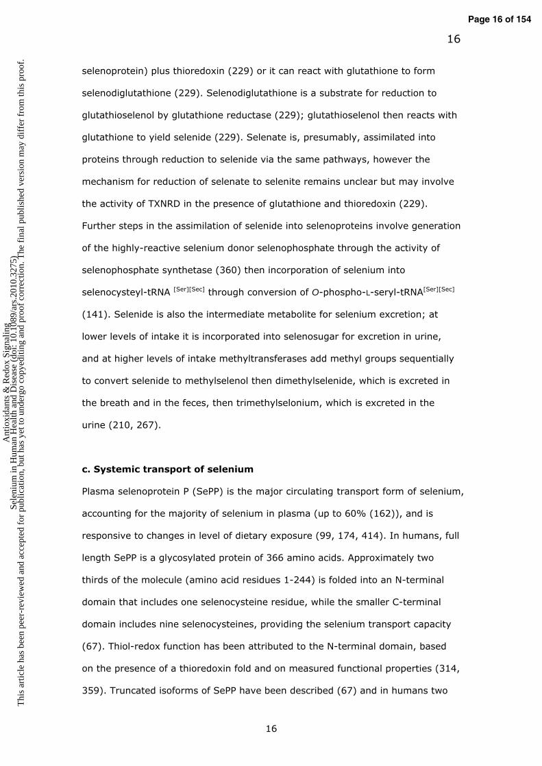
selenoprotein) plus thioredoxin (229) or it can react with glutathione to form
selenodiglutathione (229). Selenodiglutathione is a substrate for reduction to
glutathioselenol by glutathione reductase (229); glutathioselenol then reacts with
glutathione to yield selenide (229). Selenate is, presumably, assimilated into
proteins through reduction to selenide via the same pathways, however the
mechanism for reduction of selenate to selenite remains unclear but may involve
the activity of TXNRD in the presence of glutathione and thioredoxin (229).
Further steps in the assimilation of selenide into selenoproteins involve generation
of the highly-reactive selenium donor selenophosphate through the activity of
selenophosphate synthetase (360) then incorporation of selenium into
selenocysteyl-tRNA [Ser][Sec] through conversion of O-phospho-L-seryl-tRNA[Ser][Sec]
(141). Selenide is also the intermediate metabolite for selenium excretion; at
lower levels of intake it is incorporated into selenosugar for excretion in urine,
and at higher levels of intake methyltransferases add methyl groups sequentially
to convert selenide to methylselenol then dimethylselenide, which is excreted in
the breath and in the feces, then trimethylselonium, which is excreted in the
urine (210, 267).
c. Systemic transport of selenium
Plasma selenoprotein P (SePP) is the major circulating transport form of selenium,
accounting for the majority of selenium in plasma (up to 60% (162)), and is
responsive to changes in level of dietary exposure (99, 174, 414). In humans, full
length SePP is a glycosylated protein of 366 amino acids. Approximately two
thirds of the molecule (amino acid residues 1-244) is folded into an N-terminal
domain that includes one selenocysteine residue, while the smaller C-terminal
domain includes nine selenocysteines, providing the selenium transport capacity
(67). Thiol-redox function has been attributed to the N-terminal domain, based
on the presence of a thioredoxin fold and on measured functional properties (314,
359). Truncated isoforms of SePP have been described (67) and in humans two
Page 16 of 154A
ntio
xida
nts
& R
edox
Sig
nalin
gSe
leni
um in
Hum
an H
ealth
and
Dis
ease
(do
i: 10
.108
9/ar
s.20
10.3
275)
Thi
s ar
ticle
has
bee
n pe
er-r
evie
wed
and
acc
epte
d fo
r pu
blic
atio
n, b
ut h
as y
et to
und
ergo
cop
yedi
ting
and
proo
f co
rrec
tion.
The
fin
al p
ublis
hed
vers
ion
may
dif
fer
from
this
pro
of.
17
17
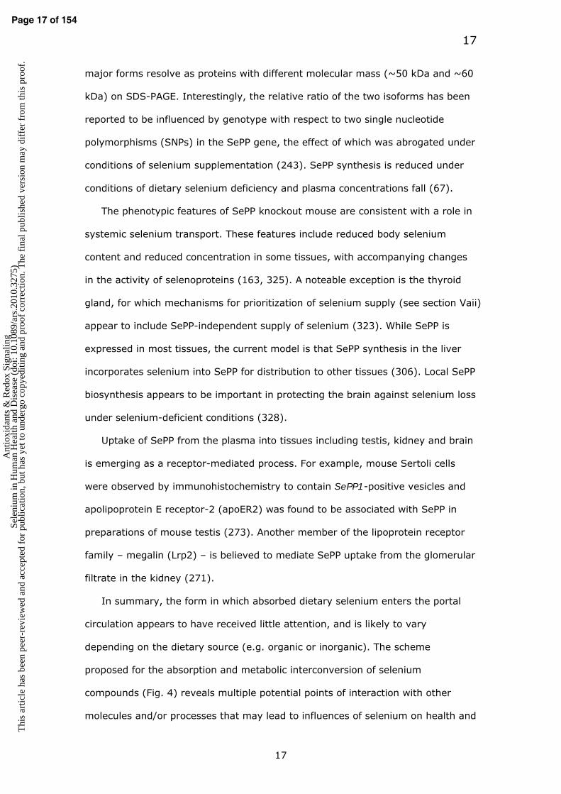
major forms resolve as proteins with different molecular mass (~50 kDa and ~60
kDa) on SDS-PAGE. Interestingly, the relative ratio of the two isoforms has been
reported to be influenced by genotype with respect to two single nucleotide
polymorphisms (SNPs) in the SePP gene, the effect of which was abrogated under
conditions of selenium supplementation (243). SePP synthesis is reduced under
conditions of dietary selenium deficiency and plasma concentrations fall (67).
The phenotypic features of SePP knockout mouse are consistent with a role in
systemic selenium transport. These features include reduced body selenium
content and reduced concentration in some tissues, with accompanying changes
in the activity of selenoproteins (163, 325). A noteable exception is the thyroid
gland, for which mechanisms for prioritization of selenium supply (see section Vaii)
appear to include SePP-independent supply of selenium (323). While SePP is
expressed in most tissues, the current model is that SePP synthesis in the liver
incorporates selenium into SePP for distribution to other tissues (306). Local SePP
biosynthesis appears to be important in protecting the brain against selenium loss
under selenium-deficient conditions (328).
Uptake of SePP from the plasma into tissues including testis, kidney and brain
is emerging as a receptor-mediated process. For example, mouse Sertoli cells
were observed by immunohistochemistry to contain SePP1-positive vesicles and
apolipoprotein E receptor-2 (apoER2) was found to be associated with SePP in
preparations of mouse testis (273). Another member of the lipoprotein receptor
family – megalin (Lrp2) – is believed to mediate SePP uptake from the glomerular
filtrate in the kidney (271).
In summary, the form in which absorbed dietary selenium enters the portal
circulation appears to have received little attention, and is likely to vary
depending on the dietary source (e.g. organic or inorganic). The scheme
proposed for the absorption and metabolic interconversion of selenium
compounds (Fig. 4) reveals multiple potential points of interaction with other
molecules and/or processes that may lead to influences of selenium on health and
Page 17 of 154 A
ntio
xida
nts
& R
edox
Sig
nalin
gSe
leni
um in
Hum
an H
ealth
and
Dis
ease
(do
i: 10
.108
9/ar
s.20
10.3
275)
Thi
s ar
ticle
has
bee
n pe
er-r
evie
wed
and
acc
epte
d fo
r pu
blic
atio
n, b
ut h
as y
et to
und
ergo
cop
yedi
ting
and
proo
f co
rrec
tion.
The
fin
al p
ublis
hed
vers
ion
may
dif
fer
from
this
pro
of.
18
18
disease and so provide important targets for future research. Membrane transport
proteins that are involved in the absorption of dietary selenium should be
identified, and potential interactions with dietary components and oral
pharmaceuticals investigated to aid the development of dietary recommendations
and health policy. Establishing the extent to which specific metabolic
interconversions of selenium compounds are tissue- or organ-specific, or
ubiquitous, will provide a more detailed model for selenium handling on a whole-
body level, including identification of the molecular forms in which selenium is
transported between the tissues; the view that SePP produced in the liver is the
major circulating from of selenium is substantiated by robust evidence, but it is
likely that other selenoproteins, or other forms of selenium, are also important
transported forms, perhaps with some element of tissue-specificity with respect
to production and uptake. Notable in this context is the form in which selenium
leaves the intestinal enterocyte and in which it is presented to the liver,
ultimately for incorporation into SePP. While receptor-mediated endocytosis,
involving specific receptor molecules, is emerging as the mechanism for uptake of
SePP in specific tissues, the mechanisms involved in delivery of selenium to the
tissues in general remains unknown. A good understanding of selenium
metabolism and transport on a whole-body level is essential if research aimed to
establish the relationships between selenium, health and disease is to be
optimally targeted, so these gaps in knowledge should be addressed as a matter
of priority.
IV. Selenium status
a. Measurement of status
Methods to assess the selenium status of populations have been reviewed
extensively (23, 130, 191). In some situations, the overall selenium status of
large populations can be predicted by examining the chemical composition of the
terrestrial environment, in particular soil selenium content, and by analyzing the
Page 18 of 154A
ntio
xida
nts
& R
edox
Sig
nalin
gSe
leni
um in
Hum
an H
ealth
and
Dis
ease
(do
i: 10
.108
9/ar
s.20
10.3
275)
Thi
s ar
ticle
has
bee
n pe
er-r
evie
wed
and
acc
epte
d fo
r pu
blic
atio
n, b
ut h
as y
et to
und
ergo
cop
yedi
ting
and
proo
f co
rrec
tion.
The
fin
al p
ublis
hed
vers
ion
may
dif
fer
from
this
pro
of.
19
19
selenium composition of generic foodstuffs and dietary habits. At an individual
level, selenium status can be assessed from hair, toenail and urinary analysis, or
more directly by assaying the selenium concentration of blood (whole blood,
plasma, serum or erythrocyte/platelet fractions), and bioassays of selenoproteins
in different blood fractions may provide more accurate estimates of
functional/physiological selenium status.
The analysis of selenium in hair and toenails is useful as a long term
biomarker (23). As both tissues are easy to access and are non-invasive, they are
also suitable for fieldwork but samples must be prepared with care to avoid
contamination. For example, hair samples can be affected by selenium-containing
shampoo residues. Wide variation in hair selenium concentration has been
reported in populations of contrasting selenium status in China (<0.1 to >100
mg/kg hair) (130), but positive correlations have been found between selenium
status and toenail selenium concentrations (316). Urinary selenium excretion can
also be used to determine absorption in bioavailability studies, or to assess
compliance in intervention studies and is a useful responsive biomarker (23).
Plasma or serum selenium is one of the most commonly used biomarkers of
selenium status, and in a meta-analysis of fourteen studies plasma selenium
responded to selenium-supplementation or selenium-depletion across all
subgroups (23). Plasma selenium is relatively easy to obtain provided trace
element-free collection tubes are available. Whole blood selenium also responds
significantly to supplementation and is therefore also a useful biomarker.
However, there are only a limited number of studies reporting whole blood
selenium and so the inter-individual heterogeneity is unclear and the length of
time needed to incorporate selenium into red blood cells renders it less
responsive than plasma to changes in selenium intake. It is possible to assess the
selenium concentration of erythrocytes and platelets although samples require
almost immediate processing. Since much of the selenium in red blood cells is
Page 19 of 154 A
ntio
xida
nts
& R
edox
Sig
nalin
gSe
leni
um in
Hum
an H
ealth
and
Dis
ease
(do
i: 10
.108
9/ar
s.20
10.3
275)
Thi
s ar
ticle
has
bee
n pe
er-r
evie
wed
and
acc
epte
d fo
r pu
blic
atio
n, b
ut h
as y
et to
und
ergo
cop
yedi
ting
and
proo
f co
rrec
tion.
The
fin
al p
ublis
hed
vers
ion
may
dif
fer
from
this
pro
of.
20
20
associated with hemoglobin (144), erythrocyte selenium is again less responsive
than plasma selenium.
Although serum/plasma selenium is a useful measure of selenium status and
short-term responses to changes in intake, it is not ideal for assessing selenium
status in populations due to the high level of inter-individual heterogeneity (23).
Serum/plasma selenium is also affected by confounding factors including smoking,
alcoholism and some disease states. For example HIV/AIDS appears to lower
plasma selenium levels (9, 105). There is also an apparent decline in plasma
selenium in the elderly in certain populations (92, 269, 380) which may occur
independently of intake and one study suggested that the bioavailability of
selenium is influenced by aging (269). In addition, there is a large effect of
dietary selenium species on plasma selenium concentrations. For example,
organic species of selenium are readily incorporated into plasma albumin unlike
inorganic species (68, 262).
The expression of individual selenoproteins may be useful measures of
selenium status (23, 174, 281). There are a total of 25 human genes which
express selenoproteins (211); quantification of a combination of the
selenoproteins may be needed to measure selenium status (111). The most
commonly used group of selenoproteins are the glutathione peroxidases, GPx1,
GPx3 and GPx4. GPx activities in plasma, erythrocytes and platelets generally
respond to supplementation only in selenium-deficient populations since plasma
GPx3 activity normally plateaus with intakes ≥ 65 µg/day. The selenium intake
required to achieve maximal activity of plasma GPx activity has been used to set
Dietary Reference Intake (DRI) values in the US (274) (section XI). Plasma SePP
responds in a dose-dependent way to selenium supplementation (23, 99, 162,
174, 253, 414) and may be a more sensitive selenium status biomarker over a
wider range of intake/status than some other markers, e.g. platelet GPx1 or GPx3
(174, 414); SePP concentration may reach a maximum at a plasma selenium
Page 20 of 154A
ntio
xida
nts
& R
edox
Sig
nalin
gSe
leni
um in
Hum
an H
ealth
and
Dis
ease
(do
i: 10
.108
9/ar
s.20
10.3
275)
Thi
s ar
ticle
has
bee
n pe
er-r
evie
wed
and
acc
epte
d fo
r pu
blic
atio
n, b
ut h
as y
et to
und
ergo
cop
yedi
ting
and
proo
f co
rrec
tion.
The
fin
al p
ublis
hed
vers
ion
may
dif
fer
from
this
pro
of.

21
21
concentration in the region of 125 ng/ml (to convert to µmol/L divide by 78.96)
and an intake ~100 µg/day (174).
b. Global variation in status
As with global estimates of selenium intake, there is very wide geographical
variation in plasma and serum selenium concentrations (87, 130, 297, 377, 410).
In areas of China in which Keshan Disease and Kashin-Beck Disease (an endemic,
chronic, degenerative osteoarthropathy – see Section VIa) are prevalent, serum
selenium concentrations as low as ~12-20 ng/ml have been reported, whereas in
seleniferous regions of the US, serum selenium can rise to 200 ng/ml (410). For
healthy adults, the proposed reference range is 39.5-197.4 ng/ml (410). However,
this range carries considerable uncertainties with regard to sufficiency since
maximal plasma GPx activity occurs at plasma selenium concentrations ~70-90
ng/ml (262, 365), and maximal SePP activity occurs at a plasma selenium
concentration ~120 ng/ml (174). Fordyce (2005) reports a more conservative
‘normal’ range of serum selenium of 60-105 ng/ml (130). In a review of 65
studies published between 1995 and 2003, serum or plasma selenium
concentrations in European healthy adults ranged from 50.22 to 145.29 ng/ml
but with the skewed range, most fell below the mean selenium concentration of
78.96 ng/ml (1.00 µmol/L) (377).
c. Changes in selenium status in relation to environmental factors
Case studies from New Zealand, China, Finland and the UK clearly demonstrate a
link between supply of selenium in the soil-to-crop pathway and changes in
selenium intake and status among populations. In New Zealand, increased
selenium intakes correlated with imports of Australian wheat which contain higher
levels of selenium (366, 368, 395). In China, a detailed geochemical analysis of
areas with high incidence of Keshan disease in the late 1990s showed that total
soil selenium was not inversely correlated with Keshan disease incidence, as
Page 21 of 154 A
ntio
xida
nts
& R
edox
Sig
nalin
gSe
leni
um in
Hum
an H
ealth
and
Dis
ease
(do
i: 10
.108
9/ar
s.20
10.3
275)
Thi
s ar
ticle
has
bee
n pe
er-r
evie
wed
and
acc
epte
d fo
r pu
blic
atio
n, b
ut h
as y
et to
und
ergo
cop
yedi
ting
and
proo
f co
rrec
tion.
The
fin
al p
ublis
hed
vers
ion
may
dif
fer
from
this
pro
of.
22
22
expected, but Keshan disease incidence was, however, inversely correlated with
water soluble soil selenium (reviewed by Johnson et al. 2010 (191)).
In Finland, the link between the supply of selenium in the soil-to-crop pathway
and changes in selenium intake and status among populations has been
demonstrated in a spectacular manner (reviewed by Broadley et al. 2006 (64)).
In 1983, legislation was introduced to incorporate selenium into all multi-nutrient
fertilizers (20, 114-116, 297, 378, 379). The policy aimed to produce a 10-fold
selenium-enrichment of cereal grains (379). Multi-nutrient fertilizer formulations
were altered to include 16 mg selenium/kg fertilizer for arable and horticultural
crops and 6 mg selenium/kg fertilizer for fodder crop and hay production. Initially,
crop selenium concentrations increased by more than 10-fold and a single level of
supplementation of 6 mg selenium/kg fertilizer commenced in June 1990 (379).
In 1998, selenium supplementation was increased to 10 mg selenium/kg fertilizer
for all crops (64).
Selenium fertilizers in Finland increased crop selenium content, dietary intakes
and the selenium status of the Finnish population (64, 116, 297). The selenium
concentration of wheat bran increased ten-fold and fruit and vegetable crops 10-
to-100-fold (114); pig muscle meat and liver increased from 0.08 and 0.49
mg/kg respectively in 1985 to a peak of 0.30 and 0.73 mg/kg in 1989 (379).
Average selenium intakes rose from 25 µg/day in 1975/1976 to 124 µg/day in
1989 (116). Between 1975/6 and 1989, intake of selenium from cereals increased
from 9 to 30 µg/day, from fruit and vegetables from 0.4 to 4 µg/day, and more
modest (<10-fold) increases were reported in meat, fish, dairy products and eggs
(116). Increases in blood (378) and serum (392) selenium concentrations were
also reported, including a study of Finnish children (<15 years), whose serum
selenium increased from a mean of 69 ng/ml (range: 43-114 ng/ml) in 1985 to
100 ng/ml (range: 76-124 ng/ml) in 1986 (392). In adults, serum selenium
increased from 82 ng/ml (range: 49-107 ng/ml) in 1985 to 103 (range: 69-136
ng/ml) in 1986.
Page 22 of 154A
ntio
xida
nts
& R
edox
Sig
nalin
gSe
leni
um in
Hum
an H
ealth
and
Dis
ease
(do
i: 10
.108
9/ar
s.20
10.3
275)
Thi
s ar
ticle
has
bee
n pe
er-r
evie
wed
and
acc
epte
d fo
r pu
blic
atio
n, b
ut h
as y
et to
und
ergo
cop
yedi
ting
and
proo
f co
rrec
tion.
The
fin
al p
ublis
hed
vers
ion
may
dif
fer
from
this
pro
of.

23
23
In the UK, there is also evidence to support an environmental link between
selenium supply and selenium status, albeit more subtle than in Finland.
Selenium intake is relatively low, ~50-60 µg per day, and some populations may
not be consuming sufficient selenium for optimal plasma GPx3 activity or optimal
SePP concentration (~100 µg/day (174)). The evidence for a decline in selenium
intake in the UK since the 1970-80s shows that in 1974 and 1985 estimates of
selenium intake were ~60 µg/day but by 1991, estimated intakes of 24-31 to 60
µg/day per person were reported (136, 248). In studies from 1994, 1995, 1997
and 2000, selenium intakes ranged from 29-43 µg/day, but in 2006 an increase
to 48-58 µg/day per person was reported, with the caveat that there are
analytical uncertainties for 13 out of 20 food groups analyzed for selenium (136).
There are several reasons for the apparent decline in selenium intake and
status in the UK over the last 30 years (2, 49, 163, 252). Cereals, and especially
wheat, comprise ~30% of the energy intake of the average UK diet (137) and the
single largest dietary change affecting selenium intake and status is likely to be a
reduction in consumption of imported milling wheat containing higher levels of
selenium in the grain, and increased consumption of UK-grown wheat containing
low levels of grain selenium. In 1970 >5 Mt of wheat was imported into the UK,
mostly used for milling and human consumption (Fig. 5a) (120). Of this, 2.2 Mt
came from the US and Canada (235). Since 1990, wheat imports to UK have
averaged 1.2 Mt/year (range 0.73-1.67 Mt). In 2005, of the 1.35 Mt/year
imported to UK, 0.37 Mt was imported from Canada and 0.10 Mt from the US (Fig.
5b), and by the 2007/08 season, 82% of wheat for human consumption was
reportedly being grown in the UK (64). The primary reason for the decline in
imports was the widespread adoption of the Chorleywood Bread Process in the
early 1960s which enabled UK (and other European Union, EU)-grown milling
wheat to replace higher protein content North American-grown wheat in bread
production. Subsequently, the UK became a member of European Economic
Page 23 of 154 A
ntio
xida
nts
& R
edox
Sig
nalin
gSe
leni
um in
Hum
an H
ealth
and
Dis
ease
(do
i: 10
.108
9/ar
s.20
10.3
275)
Thi
s ar
ticle
has
bee
n pe
er-r
evie
wed
and
acc
epte
d fo
r pu
blic
atio
n, b
ut h
as y
et to
und
ergo
cop
yedi
ting
and
proo
f co
rrec
tion.
The
fin
al p
ublis
hed
vers
ion
may
dif
fer
from
this
pro
of.
24
24
Community (EEC) in 1973, which introduced EU import tariffs and led to a rapid
decline in non-EU sourced wheat.
Recently, it has been shown that UK wheat grain selenium concentration can
be increased by 16–26 ng/g fresh grain, for each gram of selenium applied per
hectare (applied as sodium selenate (Na2SeO4)) (63). The concentration of
selenium in UK-sourced wheat grain is usually 28 ng/g, with a narrow
interquartile range of ≤19 ng/g, n=452). There has been little apparent change
since the early 1980s (2), although increased sulfur usage may cause a decrease
in selenium uptake by wheat (352). In contrast, the selenium concentration of
US-sourced wheat grain is 457 (n=190; (148)) and 370 (n=290; (407)) ng/g and
Canadian-sourced wheat grain is reportedly 760 ng/g (58). The higher
concentration of selenium in US and Canadian wheat grain is primarily due to the
higher plant-available selenium concentrations found naturally in soils of their
wheat-growing regions and lower-yielding (i.e. typically more mineral dense)
grain. Most soils in the UK are naturally low in selenium (64, 130) and there is no
evidence for soil selenium depletion due to more intensive cropping (119).
However, since coal is a rich source of selenium, general reductions in coal usage
and desulfurization technologies may also have reduced selenium inputs to crops
(2, 157). Changes in dietary patterns, such as decreased consumption of offal, is
a further contributing factor (191).
Rice is another staple food that affects selenium status in certain populations.
Williams et al. (2009) recently conducted a spatially resolved analysis of variation
in selenium concentration in polished rice grains (403). Over 1000 samples of rice
were purchased from major rice producing/exporting countries and there is wide
variation between and within countries. For example, rice sourced from the US
and India had average levels of selenium >30 times greater than rice sourced
from Egypt (Fig. 6). Within-countries, rice selenium concentration varied by up to
10-fold. Therefore, for public health policy spatial links between geochemistry,
Page 24 of 154A
ntio
xida
nts
& R
edox
Sig
nalin
gSe
leni
um in
Hum
an H
ealth
and
Dis
ease
(do
i: 10
.108
9/ar
s.20
10.3
275)
Thi
s ar
ticle
has
bee
n pe
er-r
evie
wed
and
acc
epte
d fo
r pu
blic
atio
n, b
ut h
as y
et to
und
ergo
cop
yedi
ting
and
proo
f co
rrec
tion.
The
fin
al p
ublis
hed
vers
ion
may
dif
fer
from
this
pro
of.
25
25
crop uptake, dietary intake and plasma/serum selenium concentration need to be
resolved.
V. Functions of selenium in the human body
a. Thyroid hormone metabolism
The redox-protective effects of selenoproteins may be of particular importance in
the thyroid gland, whose long-lived cells generate H2O2 (and so also reactive
oxygen species (ROS)) required for the synthesis of thyroid hormones. This likely
role is reflected in the abundance of selenium in the thyroid gland (206) and
perhaps by the high priority given to maintaining selenium supply to the thyroid
gland under conditions where availability is restricted. Also of particular relevance
is the direct involvement of selenoenzymes, the iodothyronine deiodinases (DIOs),
in thyroid hormone metabolism.
i. Thyroid hormone synthesis and the role of selenoproteins in thyroid
gland function and protection
The pathway for synthesis of the thyroid hormones tetra-iodothyronine (T4) and
3,3’,5’ tri-iodothyronine (T3) is shown schematically as Fig. 7, based on evidence
from a variety of sources (48, 134, 145, 322, 343), and highlighting the roles of
selenoproteins. The expression in the thyrocyte of several selenoproteins,
including glutathione peroxidases (GPx1, GPx3 (secreted) and GPx4), thioredoxin
reductases 1 and 2 (TXNRD1 and TXNRD2), deiodinases 1 and 2 (DIO1, DIO2),
Sep15, SePP and selenoproteins M and S (319) may contribute to its high
selenium content, and the ability of some of these (GPx1, GPx3, GPx4, TXNRD1,
TXNRD2), along with intracellular catalase and peroxiredoxins, to degrade excess
H2O2, may be important in antioxidant defense and redox control (205). An
additional protective role for selenium in the thyroid gland is indicated by the
positive outcomes of clinical trials involving selenium supplementation to reduce
antibody load in cases of autoimmune thyroiditis (257).
Page 25 of 154 A
ntio
xida
nts
& R
edox
Sig
nalin
gSe
leni
um in
Hum
an H
ealth
and
Dis
ease
(do
i: 10
.108
9/ar
s.20
10.3
275)
Thi
s ar
ticle
has
bee
n pe
er-r
evie
wed
and
acc
epte
d fo
r pu
blic
atio
n, b
ut h
as y
et to
und
ergo
cop
yedi
ting
and
proo
f co
rrec
tion.
The
fin
al p
ublis
hed
vers
ion
may
dif
fer
from
this
pro
of.

26
26
ii. Prioritization of the selenium supply to the thyroid gland and to DIOs
Observations made in rats fed severely selenium-deficient diets provided early
evidence that selenium supply to the thyroid gland is prioritized. Whereas levels
of GPx activity and GPx1 mRNA became virtually undetectable in liver and heart,
mRNA levels were maintained in the thyroid gland and activity was reduced by
only 50%. Similarly, DIO activity and DIO1 mRNA were maintained in thyroid but
reduced in liver (43). There is also evidence that the DIOs, both in the thyroid
gland and in non-thyroid tissues, are preferentially supplied above other
selenoproteins with selenium to retain activity. For example, 5’-deiodinase
activity and DIO1 mRNA were retained under conditions of selenium depletion
sufficient to reduce GPx activity and GPx1 mRNA in an epithelial cell line (147).
The clinical phenotype of an abnormality in thyroid hormone metabolism resulting
from a mutation in SEBP2 (selenocysteine insertion sequence (SECIS) binding
protein 2, which interacts with the 3’UTR SECIS of selenoproteins to re-code the
UGA (stop) codon to facilitate selenocysteine incorporation) appeared to be
dominated by effects on DIO activity (103), perhaps reflecting an important role
for SECISBP2 in the prioritization of selenium supply to the DIOs. Further
evidence that functionality of the SECIS is important in selenium prioritization
more generally includes the identification of a wide range (up to 1000-fold) in the
UGA-recoding activity of different human SECIS sequences (213).
iii. Functions of the DIOs and their potential role in health and disease
Thyroid hormones are important signaling molecules with essential roles in cell
function and in tissue development and physiology; thus, perturbations in their
levels, potentially including through effects of selenium status on their synthesis,
have potential consequences for health. While some T4 deiodination occurs in the
thyroid gland, it has been estimated that around 80% of circulating T3 is
generated through DIO activity in the peripheral tissues (345). DIO2 is now
Page 26 of 154A
ntio
xida
nts
& R
edox
Sig
nalin
gSe
leni
um in
Hum
an H
ealth
and
Dis
ease
(do
i: 10
.108
9/ar
s.20
10.3
275)
Thi
s ar
ticle
has
bee
n pe
er-r
evie
wed
and
acc
epte
d fo
r pu
blic
atio
n, b
ut h
as y
et to
und
ergo
cop
yedi
ting
and
proo
f co
rrec
tion.
The
fin
al p
ublis
hed
vers
ion
may
dif
fer
from
this
pro
of.

27
27
recognized to be the deiodinase primarily responsible for deiodination of the pro-
hormone T4 at the 5’ position to generate active T3 (345). The roles of the three
DIOs in interconversion of the thyroid hormones, including inactivation by 5-
deionidation, are shown in Fig. 7.
A wealth of evidence supports the view that the relative levels of expression of
the different DIOs in specific tissues and at specific developmental stages or in
response to challenges such as tissue injury, illness and nutritional deficiency is
balanced to promote appropriate control of cell proliferation and/or differentiation
through control of thyroid hormone activation and inactivation, as reviewed
recently (345). For example, compensatory increases in tissue DIO2 activity
observed in iodine deficiency or hypothyroidism increased local T3 production
(112, 282). Adequate selenium nutrition may thus be particularly important in
cases of hypothyroidism to facilitate increased DIO activity in tissues for which
the selenium supply is a lower priority than for the thyroid gland.
Variation in DIO genes appears to influence thyroid hormone metabolism and
activity (283). Two SNPs in the DIO1 gene that have been associated with
alterations in the ratio of active T3 to the inactive metabolite 3,3’,5’
triiodothyronine (rT3) are of particular interest because they are located in the
3’UTR of the mRNA so may (in a manner similar to SNPs in the 3’UTR regions of
GPX4 and SePP (241, 243)) mediate effects through selenocysteine incorporation
into DIO1 and so, speculatively, may show an interaction with selenium status.
In conclusion, given the evidence that the non-thyroid tissues have lower
priority for supply with selenium under conditions of restriction and that DIO
induction may be an important adaptive response of tissues to particular
challenges, more information on the interactions and processes is required to
understand links between selenium, health and disease.
b. Antioxidant defense system and oxidative metabolism
i. Glutathione Peroxidases (GPxs)
Page 27 of 154 A
ntio
xida
nts
& R
edox
Sig
nalin
gSe
leni
um in
Hum
an H
ealth
and
Dis
ease
(do
i: 10
.108
9/ar
s.20
10.3
275)
Thi
s ar
ticle
has
bee
n pe
er-r
evie
wed
and
acc
epte
d fo
r pu
blic
atio
n, b
ut h
as y
et to
und
ergo
cop
yedi
ting
and
proo
f co
rrec
tion.
The
fin
al p
ublis
hed
vers
ion
may
dif
fer
from
this
pro
of.
28
28
Amongst the selenoproteins are 5 GPxs: cytosolic GPx (GPx1), gastrointestinal-
specific GPx (GPx2), plasma GPx (GPx3), and phospholipid hydroperoxide GPx
(GPx4) are well characterized major selenoenzymes of the human antioxidant
defense systems (230). GPx1-3 catalyze the reduction of hydrogen peroxide and
organic hydroperoxides, whereas GPx4 can directly reduce phospholipid
hydroperoxides and cholesterol hydroperoxides. GPx6 is an olfactory epithelium
and embryonic tissue-specific GPx (211). GPx1 and GPx2 have well characterized
antioxidant functions, as indicated by the greater susceptibility of mice lacking
both GPx1 and 2 to an oxidative challenge (85). Responses of transgenic mice
lacking or over expressing GPx1 suggest novel roles for GPx1 in relation to both
reactive oxygen species and reactive nitrogen species, and a link to insulin
secretion and insulin resistance (217, 394). GPx3 is a key antioxidant enzyme in
the plasma and acts as a functional parameter for selenium status assessment,
and GPx3 deficiency has been associated with CVD and stroke (46, 399).
GPx activity and expression have been used in many human studies as
biomarkers for selenium status (355). It has been shown in GPx1(-/-) mice that
GPx1 deficiency plays a major role in cardiac dysfunction in angiotensin II-
dependent hypertension (18). Furthermore, there have been attempts to
correlate genetic polymorphism of selenoenzymes with risk of disease, including
cancer and heart disease. A recent study provided some evidence that SNPs in
GPx1 and GPx4, and their interaction with variants in other selenoprotein genes,
may influence colorectal cancer risk (242). In another study, with a population
who had advanced distal colorectal carcinoma, SNPs in SEPP1 and TXNRD1 were
identified to be associated with adenoma risk but not the GPX1-4 variants
investigated (286).
ii. Thioredoxin Reductases (TXNRDs)
TXNRDs are involved in the control of cellular proliferation, cell survival and
apoptosis through the control of thioredoxin (Trx) activity and redox state, and
Page 28 of 154A
ntio
xida
nts
& R
edox
Sig
nalin
gSe
leni
um in
Hum
an H
ealth
and
Dis
ease
(do
i: 10
.108
9/ar
s.20
10.3
275)
Thi
s ar
ticle
has
bee
n pe
er-r
evie
wed
and
acc
epte
d fo
r pu
blic
atio
n, b
ut h
as y
et to
und
ergo
cop
yedi
ting
and
proo
f co
rrec
tion.
The
fin
al p
ublis
hed
vers
ion
may
dif
fer
from
this
pro
of.

29
29
play a crucial role in biological response to oxidative stress. Three TXNRDs have
been identified in mammals: TXNRD1 in the cytosol/nucleus, TXNRD2 in
mitochondria, and thioredoxin glutathione reductase in the testis, with the last
also possessing glutathione and glutaredoxin reductase activity (279). TXNRD,
Trx, and NADPH constitute the thioredoxin system, a major cellular redox system
present in all living organisms (19). TXNRDs have a wide range of substrates
including small molecules such as hydrogen peroxide, lipid hydroperoxides, and
ascorbate, lipoic acid, ubiquinone and Trx (413). ROS are a major contributing
factor to the pathogenesis of CVD. The thioredoxin system plays an important
role in scavenging ROS. Trx, glutaredoxin, peroxiredoxin and their isoforms, are
involved in interaction with signaling pathways thus making them attractive
targets for clinical intervention (3). TXNRD is the only known enzyme able to
reduce oxidized Trx which regulates a plethora of redox signaling events (153).
Reduced Trx provides electrons to ribonucleotide reductase, essential for DNA
synthesis, by converting ribonucleotide to deoxyribonucleotides. Moreover, the
Trx system participates in many cellular signaling pathways by controlling the
activity of transcription factors containing critical cysteines in their DNA-binding
domains, such as nuclear factor kappa B (NFκB), activator protein-1 (AP-1), p53,
and the glucocorticoid receptor (223).
There is growing evidence that redox regulation by the TXNRD systems plays
a crucial role in the biological response against oxidative stress and in cell
proliferation, apoptosis and the modulation of inflammation (70, 342, 362). Trx
can bind to apoptosis signal regulating kinase 1 (ASK1) and modulates apoptosis
(223, 315, 258). TXNRD is also over-expressed in many tumor cells and
contributes to drug resistance, therefore TXNRD was considered a new target for
anticancer drugs (140). Interestingly, in addition to selenium, dietary
phytochemicals such as isothiocyanates and polyphenols can also up-regulate
TXNRD and GPx2 expression in both tumor and normal cell lines via the Keap1-
Nrf2-ARE pathway (203, 220, 429).
Page 29 of 154 A
ntio
xida
nts
& R
edox
Sig
nalin
gSe
leni
um in
Hum
an H
ealth
and
Dis
ease
(do
i: 10
.108
9/ar
s.20
10.3
275)
Thi
s ar
ticle
has
bee
n pe
er-r
evie
wed
and
acc
epte
d fo
r pu
blic
atio
n, b
ut h
as y
et to
und
ergo
cop
yedi
ting
and
proo
f co
rrec
tion.
The
fin
al p
ublis
hed
vers
ion
may
dif
fer
from
this
pro
of.

30
30
iii. Other selenoproteins involved in the antioxidant defense system
Recently, it has been suggested that selenoproteins, including SelW, SelM, SelT
and the 15kDa selenoprotein, are members of a novel redox protein family that
share the common feature of containing a thioredoxin-like fold with a CxxSec
redox fold (1, 39). Furthermore, over-expression of SelW in cultured cells led to
lower sensitivity to challenge from hydrogen peroxide (190), suggesting that
SelW has an antioxidant function. Selenoprotein H (SelH) is a nucleolar
thioredoxin-like protein with a unique expression pattern and structural studies
suggest that SelH is a redox-sensing DNA binding protein (266, 278). A recent
study described a knock-out mouse deficient in selenoprotein MsrB1 (SelR) with
increased levels of malondialdehyde, protein carbonyls, protein methionine
sulfoxide, and glutathione disulfide as well as reduced levels of free and protein
thiols indicating that MsrB1 plays an important role in redox regulation (128).
SePP has a purported thiol-redox function as a member of the thioredoxin
superfamily (67) and may protect against oxidative damage (347). In addition
selenoproteins N and K may have antioxidant roles (17, 228).
In summary, several of the selenoproteins may be involved in the antioxidant
defense system as highlighted in section Vb above, there are many other
important functions of the human selenoproteins such as the role of SelW and
SelN in muscle function reviewed recently in Lescure et al. 2009 (218) and other
functions discussed throughout this review, for example, throughout chapter VI:
clinical disorders section below. For recent reviews focused in detail on
selenoproteins refer to references 39, 230, 279, 301, 332 and 433.
c. Immune system
Studies in experimental and farm animals indicate that selenium deficiency affects
both cell-mediated and humoral components of the immune response (21, 167,
344). In humans, limited data suggest that when intakes of selenium are sub-
Page 30 of 154A
ntio
xida
nts
& R
edox
Sig
nalin
gSe
leni
um in
Hum
an H
ealth
and
Dis
ease
(do
i: 10
.108
9/ar
s.20
10.3
275)
Thi
s ar
ticle
has
bee
n pe
er-r
evie
wed
and
acc
epte
d fo
r pu
blic
atio
n, b
ut h
as y
et to
und
ergo
cop
yedi
ting
and
proo
f co
rrec
tion.
The
fin
al p
ublis
hed
vers
ion
may
dif
fer
from
this
pro
of.
31
31
optimal selenium supplements can enhance immune responses (167). Low serum
selenium in humans is associated with low levels of natural killer cells (304) and
selenium supplementation (200 μg/day) increased T-lymphocyte-driven tumor
lysis and lymphocyte proliferation (200). In rats, selenium deficiency lowers levels
of IgA, M and G; selenium-deficient lymphocytes show lower mitogen-stimulated
proliferation, and in cell culture, selenium promotes human neutrophil function
(21). Despite these observations the details of how selenium intake influences the
immune system remains poorly understood, with the most information being
available on the effects of severe selenium deficiency and selenoprotein knock-
out in response to viral infection.
A landmark discovery in selenium biology during the past 30 years was the
observation by Chinese scientists that Keshan disease, a myocarditis found in
selenium-deficient areas of China, is associated with a combination of low
selenium intake and Coxsackie virus B (CVB) infection (see Section VI a i).
Molecular hybridization and immunohistochemistry analysis of post-mortem
material has shown CVB3 to be present in the majority of Keshan diseases cases.
Furthermore, feeding mice a selenium-deficient diet in combination with CVB
infection leads to pathological changes in the myocardium similar to those found
in Keshan disease (multiple necrosis lesions of the myocardium and inflammatory
cell infiltration). Overall, these data demonstrate that enteroviruses, especially
CVB, are closely associated with the viral myocarditis of Keshan disease (107,
431).
Selenium intake has subsequently been found to affect the progression of
other viral infections in animals (34). For example, selenium deficiency results in
greater lung pathology in mice infected with influenza virus compared with
selenium-adequate mice; selenium-deficient mice showed an altered immune
response to an infection with a virulent strain of influenza virus and the viral
genome changed to a more virulent genotype. Selenium deficiency also has a
significant impact on the morphology and influenza-induced host defense
Page 31 of 154 A
ntio
xida
nts
& R
edox
Sig
nalin
gSe
leni
um in
Hum
an H
ealth
and
Dis
ease
(do
i: 10
.108
9/ar
s.20
10.3
275)
Thi
s ar
ticle
has
bee
n pe
er-r
evie
wed
and
acc
epte
d fo
r pu
blic
atio
n, b
ut h
as y
et to
und
ergo
cop
yedi
ting
and
proo
f co
rrec
tion.
The
fin
al p
ublis
hed
vers
ion
may
dif
fer
from
this
pro
of.
32
32
responses in human airway epithelial cells (35). Porcine circovirus type 2
replication in PK-15 cells is inhibited by DL-selenomethionine in a concentration-
dependent manner (277). Except for Keshan disease no causal links between
selenium intake and viral response have been demonstrated in humans, but
selenium status and viral responses are associated. For example, selenium
supplementation was shown to modulate the response of healthy volunteers of
marginally low plasma selenium status to a disabled polio virus (65), confirming
that selenium status determines the ability to respond to a viral infection. In
addition, epidemiological studies suggest a correlation between severity of AIDS
and selenium deficiency, but to date it is not clear whether this reflects a
protective role for selenium during HIV infection or an altered nutritional selenium
status as a result of the disease (31, 167, 227). Similar observations have been
made with the influenza virus. Selenium status not only modulates responses to
influenza virus (261) but GPx1 activity is important in determining the response
(33, 337). However, the extent to which other selenoproteins are involved in
determining the effects of selenium status on responses to viral infection is poorly
understood, although selenium depletion was shown in a cell culture model to
affect influenza virus-induced cytokine production in bronchial epithelial cells
(189).
Immune responses are intimately linked to inflammatory processes and these
in turn are inter-related to production of ROS and redox control processes. For
example, ROS production can increase expression of inflammatory cytokines
through increased NF-κB activity (369). It is possible that selenium modulates
inflammatory and immune processes through redox functions. To some extent
this is illustrated by the observations that transgenic mice with a mutant Secys-
tRNA that causes depletion of all selenoproteins, but with variations in extent
between selenoprotein and tissues, showed changes in the pattern of certain
cytokines (e.g. interferon–γ) (337). In addition, the potential importance of redox
signaling via selenoproteins is also shown by the effects of GPx1 knockout on viral
Page 32 of 154A
ntio
xida
nts
& R
edox
Sig
nalin
gSe
leni
um in
Hum
an H
ealth
and
Dis
ease
(do
i: 10
.108
9/ar
s.20
10.3
275)
Thi
s ar
ticle
has
bee
n pe
er-r
evie
wed
and
acc
epte
d fo
r pu
blic
atio
n, b
ut h
as y
et to
und
ergo
cop
yedi
ting
and
proo
f co
rrec
tion.
The
fin
al p
ublis
hed
vers
ion
may
dif
fer
from
this
pro
of.

33
33
infection. However, mutant Secys-tRNA mice failed to show differences in lung
pathology following influenza virus infection, suggesting that a basal threshold
level of GPx1 is sufficient to maintain response to the virus. Interestingly, it was
recently reported that the selenoprotein thioredoxin reductase 1, also a key factor
in redox control, can negatively regulate the activity of the HIV-1 encoded
transcriptional activator, Tat, in human macrophages (194).
White blood cells such as lymphocytes, macrophages and neutrophils require
ROS and pro-inflammatory molecules for their activation, differentiation and
phagocytosis (132). Thus, since selenoproteins may influence these signaling
pathways they may in turn be expected to be crucial for these cell functions. For
example, neutrophils require oxidative radical production to achieve microbial
killing and selenium deficiency lowers the ability of neutrophils to kill ingested
microbes, probably partly due to lower GPx1 activity and thus impaired radical
metabolism (21). Macrophages are key cells in the signaling and activation of
inflammatory responses, but this action also produces ROS therefore it must be
carefully controlled and counteracted. Studies in which selenium supplemented
macrophages were stimulated with LPS (a bacterial endotoxin) found that
supplementation with selenium suppressed TNF-α and COX-2 (cyclooxygenase-2)
expression (386). However, Carlson et al. 2009 (79) found that macrophages
without any selenoproteins still exhibited normal inflammatory responses,
although higher levels of ROS were seen. In a similar experiment, mice with
selenoprotein-less T-cells also exhibited increased ROS levels, reduced numbers
of mature T-cells and defective antibody responses (340).
Low selenium status has been associated with reduced serum IL6 in elderly
people (388), an observation that is consistent with links between selenium,
selenoproteins and inflammatory signaling. In addition to potential metabolic links
between GPx1, ROS and inflammatory cytokines such as the interleukins, results
from a series of studies suggest that selenium levels affect eicosanoid metabolism.
Studies of both severely selenium-deficient animals and selenium-deficient cells in
Page 33 of 154 A
ntio
xida
nts
& R
edox
Sig
nalin
gSe
leni
um in
Hum
an H
ealth
and
Dis
ease
(do
i: 10
.108
9/ar
s.20
10.3
275)
Thi
s ar
ticle
has
bee
n pe
er-r
evie
wed
and
acc
epte
d fo
r pu
blic
atio
n, b
ut h
as y
et to
und
ergo
cop
yedi
ting
and
proo
f co
rrec
tion.
The
fin
al p
ublis
hed
vers
ion
may
dif
fer
from
this
pro
of.
34
34
culture suggest that selenium supply, through its influence on GPxs, has an
inhibitory regulatory effect on 5’ lipoxygenase activity in lymphocytes (179, 397)
and thus on generation of pro-inflammatory leukotrienes. In addition, over-
expression of GPx4 in transfected basophils has also been reported to suppress 5’
lipoxygenase activity. Further evidence for a metabolic link between GPx4 and
leukotrienes has come from the finding that individuals with different allelic
variants of a common SNP in the GPX4 gene (rs713041) have been found to have
different levels of 5’ lipoxygenase product levels in their lymphocytes (382),
suggesting that lymphocyte GPx4 activity influences pro-inflammatory leukotriene
activity in lymphocytes.
The observation that a combination of severe selenium and iodine deficiency
causes a thyroid atrophy that does not respond to iodine supplementation due to
inflammatory damage to the thyroid, has led to studies of selenium in thyroiditis
(205). Indeed, several clinical trials have reported that selenium supplementation
reduces inflammatory markers in patients with autoimmune thyroiditis, and this
has led to the hypothesis that even a moderate selenium deficiency can be a
causative factor in autoimmune thyroid disease in patients who have genetic
susceptibility to autoimmune disorders.
In summary, there is a growing body of evidence that selenium status affects
immune function, in particular the ability to respond to viral infection. The
mechanisms underlying these effects are poorly understood but may involve
modulation of ROS and inflammatory signaling pathways through the antioxidant
and redox functions of selenoproteins.
VI. Clinical disorders
a. Deficiency
i. Keshan Disease
Keshan disease is an endemic cardiomyopathy observed in selenium-deficient
areas of China. Its name originates from a severe outbreak in Keshan County,
Page 34 of 154A
ntio
xida
nts
& R
edox
Sig
nalin
gSe
leni
um in
Hum
an H
ealth
and
Dis
ease
(do
i: 10
.108
9/ar
s.20
10.3
275)
Thi
s ar
ticle
has
bee
n pe
er-r
evie
wed
and
acc
epte
d fo
r pu
blic
atio
n, b
ut h
as y
et to
und
ergo
cop
yedi
ting
and
proo
f co
rrec
tion.
The
fin
al p
ublis
hed
vers
ion
may
dif
fer
from
this
pro
of.
35
35
Heilongjiang Province, China in 1935. The main clinical features of Keshan disease
are acute or chronic episodes of a heart disorder characterized by cardiogenic
shock and/or congestive heart failure. Keshan disease can be clinically classified
into four types: acute, sub-acute, chronic, and latent. Dilatation of the heart is
commonly observed (143).
The etiology of Keshan disease is not yet fully understood. Originally, the
proposed risk factors included Coxsackie virus (CVB), type A streptococci, and
organic toxicants produced by parasitic fungi on cereals or putrid organic
substances in water and/or soil environments. In the 1970s, the low selenium
status of the inhabitants in Keshan disease-endemic areas became evident, and
subsequently, the preventive effect of selenium supplementation on Keshan
disease was identified. Further improvements in selenium status were associated
with a decline in Keshan disease in the endemic areas and confirmed the link
between selenium and Keshan disease.
In China, the selenium-deficient zone from the Northeast to the Southwest
involves 16 provinces, and historical data indicated that the population at risk was
>100 million. Keshan disease surveillance has been a national program since
1990, an important component of which is the determination of selenium levels in
Keshan disease-endemic areas (425).
The geographical distribution of topsoil selenium in China and its relationship
to Keshan disease (and Kashin-Beck disease) indicates that the selenium
concentrations in topsoil of affected areas are typically below 0.125 mg/kg. In
contrast, the concentration in areas without disease is 0.224 mg/kg, and the
excessive level is more than 3 mg/kg (361). Nutritional studies showed that the
mean hair selenium contents were <0.122 mg/kg in endemic areas and >0.200
mg/kg in non-Keshan disease-endemic zones, whereas the average blood
selenium concentration of people in Keshan disease endemic areas was no more
than 253 nmol/L (20 ng/ml). The selenium content of muscle, heart, liver, and
Page 35 of 154 A
ntio
xida
nts
& R
edox
Sig
nalin
gSe
leni
um in
Hum
an H
ealth
and
Dis
ease
(do
i: 10
.108
9/ar
s.20
10.3
275)
Thi
s ar
ticle
has
bee
n pe
er-r
evie
wed
and
acc
epte
d fo
r pu
blic
atio
n, b
ut h
as y
et to
und
ergo
cop
yedi
ting
and
proo
f co
rrec
tion.
The
fin
al p
ublis
hed
vers
ion
may
dif
fer
from
this
pro
of.
36
36
kidney in Keshan disease patients are up to 10-fold lower than healthy subjects
(143).
Recent studies suggest that genetic polymorphisms in selenoproteins may be
associated with susceptibility to Keshan disease. Lei et al. (2009) measured the
concentration of blood selenium and the activity and polymorphisms of cellular
GPx1 in 71 KD patients and 290 controls (216). Results suggested that selenium
deficiency in carriers with the GPx1 leucine-containing allele is associated with low
GPx1 enzyme activity, which may, in turn, increase the incidence of Keshan
disease.
The link between selenium and Keshan disease was further strengthened with
results of a selenium supplementation trial. Between 1974 and 1977, sodium
selenite or placebo tablets were given to children at high risk of Keshan disease.
Concurrent with an increase in blood selenium concentrations in the treated group
(n = 6767), 17 acute and sub-acute Keshan disease cases were reported
compared with 106 in the placebo group (n = 5445). After 4 y, there had been 53
deaths in the controls whereas only one selenium-treated subject had died (421).
Supplementation of individuals with selenium tablets (as sodium selenite) has
been effective in preventing the development of Keshan disease (418). However,
as not all people living in the low selenium areas suffered from Keshan disease
other causal factors such as virus infections were proposed.
In animal studies, Bai et al. (1980) demonstrated that mice fed grains from
Keshan disease areas developed a deficiency in selenium (28). When these mice
were infected with a strain of Coxsackie virus B4 that was isolated from a Keshan
disease victim, the mice developed severe heart pathology; whereas, mice that
were fed grains from non-Keshan disease endemic areas developed only mild
heart pathology when infected with the virus. This study suggested that together
with the deficiency in selenium, an infection of CVB was required for the
development of Keshan disease (28). A further study demonstrated that selenium
deficiency was responsible for driving changes in the viral genome and changing a
Page 36 of 154A
ntio
xida
nts
& R
edox
Sig
nalin
gSe
leni
um in
Hum
an H
ealth
and
Dis
ease
(do
i: 10
.108
9/ar
s.20
10.3
275)
Thi
s ar
ticle
has
bee
n pe
er-r
evie
wed
and
acc
epte
d fo
r pu
blic
atio
n, b
ut h
as y
et to
und
ergo
cop
yedi
ting
and
proo
f co
rrec
tion.
The
fin
al p
ublis
hed
vers
ion
may
dif
fer
from
this
pro
of.
37
37
normally avirulent pathogen into a virulent one (36). Moreover, influenza virus
exhibits increased virulence towards the selenium-deficient mice (34).
Selenium deficiency may affect the expression of selenoenzymes such as
GPX1. One study showed that 50% of GPx1 knockout mice (GPx-/-) infected with
CVB3/0 developed myocarditis, whereas infected wild-type mice (Gpx1+/+) were
resistant (no mice developed myocarditis) (33). This study suggests that
antioxidant protection is important for protection against CVB3-induced
myocarditis.
Following the isolation of enteroviruses from patients with KD during
outbreaks of Keshan disease in selenium-deficient rural areas of southwestern
China, an association of enterovirus infection with Keshan disease and its
outbreaks in selenium-deficient areas has been established (284). To date, many
studies strongly suggest a dual etiology that involves both a nutritional deficiency
of selenium as well as an infection with an enterovirus (34, 173).
There is an interest in identifying potential protective dietary compounds, for
example sulforaphane (SFN), a hydrolysis product of glucosinolate from
cruciferous vegetables which is a potent inducer for a battery of antioxidant
enzymes including quinone oxidoreductase, glutathione transferases, UDP-
glucuronyltransferase, γ-glutamylcysteine synthetase, heme oxygenase, aldo-
keto reductase, thioredoxin reductase, and GPxs. The mechanism of interactions
between Se and SFN in antioxidant enzyme expression was mainly via
Nrf2/Keap1 system (60). Sun et al. (2004) conducted a study to investigate
whether SFN can protect the myocardium of selenium-deficient mice against viral
infection and demonstrated that GPx activity in groups given SFN was
significantly higher than in the control groups without SFN (353). Further, both
the incidence and extent of myocardium injury in viral groups with SFN were
significantly lower than those in the viral groups without SFN. It was therefore
concluded that SFN affords a degree of protection against Coxsackie virus B3m-
induced mouse cardiomyopathy.
Page 37 of 154 A
ntio
xida
nts
& R
edox
Sig
nalin
gSe
leni
um in
Hum
an H
ealth
and
Dis
ease
(do
i: 10
.108
9/ar
s.20
10.3
275)
Thi
s ar
ticle
has
bee
n pe
er-r
evie
wed
and
acc
epte
d fo
r pu
blic
atio
n, b
ut h
as y
et to
und
ergo
cop
yedi
ting
and
proo
f co
rrec
tion.
The
fin
al p
ublis
hed
vers
ion
may
dif
fer
from
this
pro
of.
38
38
ii. Role of selenium in Kashin-Beck disease
Kashin-Beck disease (KBD) is an endemic, chronic, degenerative
osteoarthropathy that is present in selenium-deficient areas in the world, and is
mainly found in a diagonal belt from northeast to southwest China, and also in
Mongolia, Siberia and North Korea. The disease was first described in 1848 by
Nickolay Kashin in the Bajkal area of Russia (196) and later in 1906 by Eugene
Beck (32). The etiology of KBD is largely unknown. The risk factors seem to
include mycotoxins such as Trichothecene mycotoxin (T-2) from contaminated
storage grains, and organic substances such as humic acid and fulvic acid in
drinking water, Coxsackie B3 virus infection, and deficiency in trace elements,
mainly selenium and iodine (348). The original theory proposed by Russian
researchers, was that KBD was caused by a mycotoxin. However, the focus on
the disease gradually shifted to China where the causal theory was based on the
effects of selenium deficiency and interactions with mycotoxins (8). Amongst all
the risk factors selenium was the most studied, and disturbances of selenoprotein
expression and/or function are associated with both Keshan and Kashin-Beck
diseases (204).
In the KBD endemic areas the levels of selenium in both soil and human
biological samples are much lower than that in areas without KBD. Ge & Yang
(1993) reported that average hair selenium concentrations in residents of KBD
endemic areas were 1.19 ± 0.34 nmol/g compared with 4.81 ± 2.27 nmol/g in
non-endemic areas (143). Blood GPx activities were 74.0 ± 12.8 kU/L in endemic
groups of the population and 95.6 ± 8.9 kU/L in non-endemic groups. Oral
supplementation of the endemic group with sodium selenite (1-2 mg/wk) for 2
month increased GPx activity to 94.0 ± I 1 .5 kU/L, indicating that the population
was selenium deficient (389). Many epidemiological studies conclude that KBD is
mainly common in low selenium areas where patients are in a selenium-deficient
condition, and selenium supplementation is effective at preventing a worsening of
Page 38 of 154A
ntio
xida
nts
& R
edox
Sig
nalin
gSe
leni
um in
Hum
an H
ealth
and
Dis
ease
(do
i: 10
.108
9/ar
s.20
10.3
275)
Thi
s ar
ticle
has
bee
n pe
er-r
evie
wed
and
acc
epte
d fo
r pu
blic
atio
n, b
ut h
as y
et to
und
ergo
cop
yedi
ting
and
proo
f co
rrec
tion.
The
fin
al p
ublis
hed
vers
ion
may
dif
fer
from
this
pro
of.

39
39
metaphysis change and in promoting repair (75, 83). For example, in the KBD
region in China, serum selenium concentration was on average 36-37 ±17 ng/ml,
compared to a similar region with no evidence of KBD where serum selenium was
~63 ±15 ng/ml. A majority (>50%) of participants in the KBD area had serum
selenium <37 ng/ml, compared to only 16% in the non-disease area (data from
2006 – reported in Shi et al. 2010 (338)). There was 13% prevalence of Kashin-
Beck in certain regions of China (338).
In areas where severe selenium deficiency is endemic, iodine deficiency is also
a risk factor for KBD (252) and correction of iodine deficiency should be
undertaken before selenium supplementation in order to avoid hypothyroidism.
However, in another study, Zhang et al. (2001) studied selenium, iodine and
fungal contamination in the Yulin District, including 3 villages where KBD was
endemic, whereas there were no cases of KBD in the 4th village. Results showed
that low hair selenium concentration and presence of fungal cereal contamination
were significantly associated with an increased risk of KBD, but low urine iodine
was not (430).
T-2 is a naturally-occurring mould byproduct of Fusarium sp. fungus which is
toxic to humans and animals and was found at high levels (2.0~1549.9 ng/g) in
contaminated grain (353). T-2 toxin-containing food can lead to some pathologic
changes in the cartilage of guinea pigs that are similar to the changes observed in
KBD patients (415). Therefore, the T-2 toxin contamination was proposed as a
possible cause of KBD (424). In support of this hypothesis, cell culture studies
demonstrated that T-2 toxin can inhibit aggrecan synthesis in human
chondrocytes, promote aggrecanases and pro-inflammatory cytokines expression
and aggrecan degradation; selenium can inhibit the effects of T-2 toxin (222). A
previous study by the same group showed that selenium can partly block T-2
toxin induced apoptosis in chondrocytes (82).
In human studies, Cao et al. (2008) demonstrated there was significant
aggrecanase-mediated proteoglycan degradation in both adult and child KBD
Page 39 of 154 A
ntio
xida
nts
& R
edox
Sig
nalin
gSe
leni
um in
Hum
an H
ealth
and
Dis
ease
(do
i: 10
.108
9/ar
s.20
10.3
275)
Thi
s ar
ticle
has
bee
n pe
er-r
evie
wed
and
acc
epte
d fo
r pu
blic
atio
n, b
ut h
as y
et to
und
ergo
cop
yedi
ting
and
proo
f co
rrec
tion.
The
fin
al p
ublis
hed
vers
ion
may
dif
fer
from
this
pro
of.

40
40
patients and altered CD44 metabolism was also involved in KBD pathogenesis
(76). Again in a cell culture study, Wojewoda et al. (2010) found that selenium
decreased ROS generation and increased the level and activity of antioxidant
enzymes such as GPx and TXNRD (404).
So far, the contribution of any particular genotype towards the risk of
developing KDB is unknown. A recent paper studied the associations between
genetic variation in selenoprotein genes and susceptibility to KBD. The genotypic
and allelic frequency of GPx1 Pro198Leu were significantly different between KBD
patients and the control group (P=0.013, P=0.037, respectively). A significant
increased KBD risk was observed in individuals with Pro/Leu or Leu/Leu (odds
ratio, OR =1.78; 95% confidence interval: 1.13~2.81) compared with Pro/Pro.
Moreover, the GPx activity in whole blood decreased significantly in a subgroup of
individuals representing Pro/Leu and Leu/Leu compared to Pro/Pro (P<0.01)
(416). In contrast, in this study, no associations were found between KBD risk
and other gene polymorphisms such as TXNRD2, SEPP1 and DIO2. Moreover, Shi
et al. (2010) suggest that variants of the chromosomal short tandem repeats
D11S4094, D11S4149, D2S338, and D2S305 might be associated with KBD (338).
Downey et al. (2009) studied the effects of skeletal selenoprotein deficiency using
a transgenic mouse line to trigger Trsp gene deletions in osteochondroprogenitors
(96). Trsp encodes selenocysteine tRNA[Ser]Sec, which is required for the
incorporation of selenocysteine residues into selenoproteins. The mutant mice
exhibited growth retardation, epiphyseal growth plate abnormalities, and delayed
skeletal ossification, as well as marked chondronecrosis of articular, auricular, and
tracheal cartilages. Phenotypically, the mice replicated a number of the
pathological features of KBD (96), supporting the notion that Se deficiency is
important to the development of KBD.
Moreno-Reyes et al. (2003) reported that selenium supplementation in Tibet
had no effect on established KBD, growth, or thyroid function once iodine
deficiency was corrected (251). Therefore it was suggested that iodine, but not
Page 40 of 154A
ntio
xida
nts
& R
edox
Sig
nalin
gSe
leni
um in
Hum
an H
ealth
and
Dis
ease
(do
i: 10
.108
9/ar
s.20
10.3
275)
Thi
s ar
ticle
has
bee
n pe
er-r
evie
wed
and
acc
epte
d fo
r pu
blic
atio
n, b
ut h
as y
et to
und
ergo
cop
yedi
ting
and
proo
f co
rrec
tion.
The
fin
al p
ublis
hed
vers
ion
may
dif
fer
from
this
pro
of.

41
41
selenium, deficiency should be corrected in Tibetan children with KBD. The results
of a systematic review demonstrated no convincing evidence that selenium,
vitamin A, vitamin C or the combination product of selenium, vitamin A, C, and E
are effective in the treatment of any type of arthritis (75). A meta-analysis of 5
randomized controlled trials (RCTs) and 10 non-RCTs assessed the efficacy of
selenium supplementation for prevention of KBD osteoarthropathy in children
(435). Significant effects were demonstrated and the results indicated that the
current evidence supports the benefits of selenium supplementation for
prevention of KBD in children although the evidence was limited by potential
biases and confounders. Results from a 3-year trial in which 1064 children ages 3
to 10 have been given iodine and selenium and either a cocktail of micronutrients
(copper, manganese, zinc, and vitamins A, C, and E), or a placebo are expected
to be published soon by Mathieu and colleagues which may help resolve the
controversy.
Recent comparative microarray analysis of gene expression profiles between
primary knee osteoarthritis (OA) and KBD demonstrated a clear difference in the
gene expression profile in cartilage from patients with KBD compared with that
from patients with OA (98), implying there are different mechanisms responsible
for the development and progression of these two diseases (393). The genes with
a lower expression in KBD than normal cartilage included chondrocyte metabolism,
ECM, DNA modification, and transcription factors, and genes with a higher
expression in KBD than normal cartilage included signaling transduction, cell cycle,
and apoptosis. The differential gene expression specific for KBD may indicate a
specific mechanism that is responsible for the destruction of the articular cartilage
in KBD. The data also suggest that cartilage degeneration, apoptosis induction
pathways, and matrix metabolism might be more important in KBD cartilage.
Finally, the development of protein chip technology has enabled the
application of high throughput proteomics to identify potential biomarkers for a
variety of diseases including KBD. Wang et al. (2008) reported marked serum
Page 41 of 154 A
ntio
xida
nts
& R
edox
Sig
nalin
gSe
leni
um in
Hum
an H
ealth
and
Dis
ease
(do
i: 10
.108
9/ar
s.20
10.3
275)
Thi
s ar
ticle
has
bee
n pe
er-r
evie
wed
and
acc
epte
d fo
r pu
blic
atio
n, b
ut h
as y
et to
und
ergo
cop
yedi
ting
and
proo
f co
rrec
tion.
The
fin
al p
ublis
hed
vers
ion
may
dif
fer
from
this
pro
of.

42
42
proteomic changes in KBD using surface-enhanced laser desorption/ionization
time-of-flight mass spectrometry (SELDI-TOF MS) (391), and it would be
plausible to use this technique in human intervention trials to examine the effect
of selenium supplementation.
b. Toxicity
Although much less common than selenium deficiency, selenium toxicity can
affect individuals as a result of over-supplementation (234), accidental or
deliberate (suicidal) ingestion of very high doses (215), or through high levels in
the food supply. Characteristic features of selenosis occur in population groups
exposed to unusually high levels of dietary selenium, and include brittle hair and
brittle, thickened, stratified nails, leading to loss in some cases, along with an
odour of garlic on the breath and skin (234, 300). Additional symptoms, including
vomiting and pulmonary oedema, are a feature of more acute selenium poisoning
(215).
In Enshi, in the Chinese province of Hubei, an outbreak of illness in which the
most notable and prevalent symptoms were loss of nails and hair reached a peak
between 1961 and 1964 when it affected almost half of the population. This
condition was later diagnosed as severe selenosis, attributed to soil with very
high selenium content (422). It was identified that the period of peak prevalence
of selenosis was due to drought causing failure of the (lower selenium) rice crop,
leading to consumption of alternative crops, with higher selenium content.
Analysis of vegetables and cereals grown in the area after the period of peak
prevalence confirmed high levels of selenium, which were up to 1500-fold greater
than levels measured in the same foods taken from a nearby selenium-deficient
area, where Keshan disease was endemic. Average daily intake of selenium was
estimated to be 4990 µg. Analysis of samples of human hair, blood and urine
from residents of the affected area revealed that selenium concentrations were
Page 42 of 154A
ntio
xida
nts
& R
edox
Sig
nalin
gSe
leni
um in
Hum
an H
ealth
and
Dis
ease
(do
i: 10
.108
9/ar
s.20
10.3
275)
Thi
s ar
ticle
has
bee
n pe
er-r
evie
wed
and
acc
epte
d fo
r pu
blic
atio
n, b
ut h
as y
et to
und
ergo
cop
yedi
ting
and
proo
f co
rrec
tion.
The
fin
al p
ublis
hed
vers
ion
may
dif
fer
from
this
pro
of.

43
43
far in excess of concentrations measured in samples from individuals resident in a
selenium-adequate area.
In parts of the Punjab State in the North-Western region of India, crops and
fodder contain very high selenium levels and selenosis is observed in cattle. Signs
of selenium toxicity are observed in people consuming locally grown food (Fig. 8)
(164). Daily intakes of selenium were estimated to be 632 and 475 µg/day in
men and women respectively, and corresponding values from non-seleniferous
areas were 65 and 52 µg/day (94). Policies to manage the situation include non-
consumption of crop produce by the farmers, dilution with produce from non-
endemic regions and application of manures to reduce selenium accumulation in
crops (95).
Levels of dietary exposure at which selenium becomes toxic and selenosis
develops are difficult to establish, because toxicity is affected by the form in
which selenium is available in the food supply, and probably also by combination
with other dietary components and, possibly, interactions with genotype. High
levels of selenium in diets based predominantly on meat sources appear to be
particularly well-tolerated, as exemplified by the high daily selenium intake of the
Inuit of North Greenland, estimated as 193-5885 µg (150). This intake results in
blood selenium concentrations in the order of 1000 µg/l (300), but is not
associated with symptoms of toxicity. Comparison of levels of selenium in blood
and urine in the Enshi study population and also in samples from populations in
South Dakota (169) revealed toxicity associated with lower concentrations than
those that result in symptoms of selenosis in Venezuela (422), probably reflecting
exposure to dietary selenium in different forms. Suicide by exposure to very high
levels of selenium has been associated generally with ingestion of selenite or
selenate (215), an observation that probably reflects the form of selenium in
substances used in suicide attempts, rather than necessarily being indicative of
greater toxicity of inorganic compared with organic selenium compounds.
Page 43 of 154 A
ntio
xida
nts
& R
edox
Sig
nalin
gSe
leni
um in
Hum
an H
ealth
and
Dis
ease
(do
i: 10
.108
9/ar
s.20
10.3
275)
Thi
s ar
ticle
has
bee
n pe
er-r
evie
wed
and
acc
epte
d fo
r pu
blic
atio
n, b
ut h
as y
et to
und
ergo
cop
yedi
ting
and
proo
f co
rrec
tion.
The
fin
al p
ublis
hed
vers
ion
may
dif
fer
from
this
pro
of.
44
44
The mechanisms underlying selenium toxicity remain unknown; suggested
mechanisms include induction of oxidative stress (375) and the replacement of
sulfur with selenium in hair and keratin, leading to structural defects (376).
Research on this topic, based on various in vitro and in vivo models (reviewed in
Valdiglesias et al. 2009 (375)) is complicated by likely effects on measured
outcomes of the form and dose of selenium administered and limitations with
respect to extrapolating results from in vitro assays to human exposure. The
eventual evolution of systems-biology-based approaches to describing selenium
metabolism and interactions with cells and tissues will guide the most relevant
experimental systems for understanding selenium toxicity at a molecular and
whole body level.
VII. Effects on health
a. Cardiovascular disease
Selenium is essential for selenium-dependent antioxidant enzymes such as GPxs,
TXNRD, SePP, and other selenoproteins. Because of the antioxidant properties of
selenium and/or selenoenzymes, it has been hypothesized that selenium may
prevent CVD. Many observational studies investigating the association of low
selenium concentrations with cardiovascular outcomes and randomized trials
investigating whether selenium supplements prevent coronary heart disease (CHD)
have been inconclusive.
A meta-analysis (126) synthesized results from observational studies of the
association of selenium biomarkers with CHD endpoints and from results of
clinical trials of the efficacy of selenium supplements in preventing CHD. The
conclusions were that observational studies showed an inverse association
between selenium concentrations and CHD incidence, although the data need
further validation, whereas randomized trials were inconclusive with respect to
the effect of selenium supplementation. Another meta-analysis of 13 prospective
cohort studies found a moderate inverse relationship between plasma/serum
Page 44 of 154A
ntio
xida
nts
& R
edox
Sig
nalin
gSe
leni
um in
Hum
an H
ealth
and
Dis
ease
(do
i: 10
.108
9/ar
s.20
10.3
275)
Thi
s ar
ticle
has
bee
n pe
er-r
evie
wed
and
acc
epte
d fo
r pu
blic
atio
n, b
ut h
as y
et to
und
ergo
cop
yedi
ting
and
proo
f co
rrec
tion.
The
fin
al p
ublis
hed
vers
ion
may
dif
fer
from
this
pro
of.

45
45
selenium and CHD although the interpretation of these data is complicated by
potential residual confounding and publication bias (260). Finally, a recent report
by Xun et al. (2010), examined the longitudinal association between toenail
selenium levels and subclinical atherosclerosis over an 18-year period, and no
associations were observed between toenail selenium and measures of subclinical
atherosclerosis among young American adults (417).
Selenium supplementation does not appear to reduce the risk of CVD in
healthy individuals. Supplements of 200 µg/day in individuals free of CVD at
baseline were not significantly associated with any CVD endpoints during 7.6
years follow-up of the entire blinded phase of the Nutritional Prevention of Cancer
Trial (1983–1996) (350), and a randomized, placebo-controlled trial in healthy
men given 300 µg/day of selenium as high-selenium yeast for 48 wk suggested
that selenium supplements were not likely to improve endothelial function or
peripheral arterial responsiveness in healthy North American men receiving
adequate selenium from their diets (155). Moreover, increased consumption of
wheat biofortified with selenium does not modify biomarkers of cancer risk,
oxidative stress, or immune function in healthy Australian males (412).
Although it appears that selenium supplementation has no effect on CVD risk
in healthy individuals, in a very recent AtheroGene study, low selenium
concentration was associated with future cardiovascular death in patients with
acute coronary syndrome, although no effect on stable angina pectoris (232).
Furthermore, in the selenium Therapy in Coronary Artery disease Patients
(SETCAP) Study, supplementation of sodium selenite increased GPx1 activity in
endothelial cells and in coronary artery disease (CAD) patients. Therefore, long
term studies are needed to demonstrate whether CAD outcome can be improved
by selenium (320). A cross-sectional study on the association of serum selenium
with the prevalence of peripheral arterial disease among 2,062 US men and
women 40 years of age or older participating in the National Health and Nutrition
Examination Survey (NHANES), 2003–2004, suggested that the effects of
Page 45 of 154 A
ntio
xida
nts
& R
edox
Sig
nalin
gSe
leni
um in
Hum
an H
ealth
and
Dis
ease
(do
i: 10
.108
9/ar
s.20
10.3
275)
Thi
s ar
ticle
has
bee
n pe
er-r
evie
wed
and
acc
epte
d fo
r pu
blic
atio
n, b
ut h
as y
et to
und
ergo
cop
yedi
ting
and
proo
f co
rrec
tion.
The
fin
al p
ublis
hed
vers
ion
may
dif
fer
from
this
pro
of.
46
46
selenium on athlerosclerosis are non-linear and may follow a U-shaped
relationship (54).
In summary, the observational evidence that low selenium concentrations are
associated with cardiovascular risk should be treated as suggestive but not
definitive. There is uncertainty about cause and effect, therefore time-resolved
and prospective studies are needed in different pathological settings. Furthermore,
when investigating the relationship between selenium and disease risk future
studies need to determine not only selenium status but also genotype in relation
to selenoproteins and related pathways (301).
b. Cancer
There are a multitude of studies investigating the effect of selenium on cancer;
several recent reviews focus on the potential mechanisms of action using
evidence from in vitro cell culture studies and in vivo, mainly animal model,
studies (187, 231, 332, 433). Proposed mechanisms of the effects of selenium on
cancer are summarized in Fig. 9. They include regulation of cell cycle and
apoptosis, antioxidant effect through the action of selenoproteins, in particular,
GPx1, GPx4, Sep15, SEPP1, TXNRD1 (301, 433), modulation of angiogenesis (231)
and the extracellular matrix (79, 175), histone deacetylase inhibition (287),
carcinogen detoxification, induction of GSTs, alteration of DNA damage and repair
mechanisms, and also immune system modulation (187, 231, 332, 433).
However, the effects of selenium on cancer are species-specific, dose-specific,
cancer type-specific and may be affected by genotype and the bioavailability of
selenium (Fig. 9), discussed in more detail below. In this review we discuss data
from human studies, including RCTs and epidemiological data, with a particular
focus on the species and dose of selenium in interventions and plasma/serum
selenium status.
i. Total cancer incidence and mortality
Page 46 of 154A
ntio
xida
nts
& R
edox
Sig
nalin
gSe
leni
um in
Hum
an H
ealth
and
Dis
ease
(do
i: 10
.108
9/ar
s.20
10.3
275)
Thi
s ar
ticle
has
bee
n pe
er-r
evie
wed
and
acc
epte
d fo
r pu
blic
atio
n, b
ut h
as y
et to
und
ergo
cop
yedi
ting
and
proo
f co
rrec
tion.
The
fin
al p
ublis
hed
vers
ion
may
dif
fer
from
this
pro
of.
47
47
Willett et al. 1983 (401) reported an association between selenium and cancer in
which the relative risk (RR) of cancer was higher in individuals with low plasma
selenium concentrations (<115 ng/ml compared with 128 to >154 ng/ml) (Fig.
10A). The Nutritional Prevention of Cancer (NPC) trial showed a protective effect
of selenium-enriched yeast supplements (200 µg/day) on total cancer mortality
(86), and also total cancer incidence, but only for men with low plasma selenium
concentrations (<121.6 ng/ml at baseline) (101). Participants with baseline
plasma selenium >121.6ng/ml who consumed selenium-enriched yeast 200
µg/day had a trend towards elevated cancer incidence (101).
In one of the large nutrition intervention trials in Linxian, China, with over
20,000 participants, supplementation for ~5 years with 15 mg β-carotene, 30 mg
α-tocopherol and 50 µg/day selenium resulted in a small significant decrease in
total cancer mortality (55, 56, 390). In the SU.VI.MAX trial (138) the antioxidant
status of participants at baseline was related to the risk of cancer in men but not
women, and a large 2 year intervention study with selenium and allitridum (a
synthetic compound similar to bioactive forms in garlic) with follow-up of cancer
incidence for 5 years showed a reduced RR of all tumors in men but not women
(221). A meta-analysis investigating the effect of antioxidant supplements on
primary cancer incidence and mortality by Bardia et al. (2008) concluded that
selenium supplementation was associated with reduced cancer incidence in men
but not women (29).
ii. Gastrointestinal cancers
A meta-analysis of 5 studies published up to 2007 on the effect of selenium on
gastrointestinal cancers, showed that selenium supplementation was associated
with a ~25-60% reduction in gastrointestinal cancers (overall RR: 0.59, 95 % CI:
0.46-0.75) (49); gastrointestinal cancer included esophageal, gastric, small
intestine, colorectal, pancreatic, liver and biliary tract (49). The RR for esophageal
Page 47 of 154 A
ntio
xida
nts
& R
edox
Sig
nalin
gSe
leni
um in
Hum
an H
ealth
and
Dis
ease
(do
i: 10
.108
9/ar
s.20
10.3
275)
Thi
s ar
ticle
has
bee
n pe
er-r
evie
wed
and
acc
epte
d fo
r pu
blic
atio
n, b
ut h
as y
et to
und
ergo
cop
yedi
ting
and
proo
f co
rrec
tion.
The
fin
al p
ublis
hed
vers
ion
may
dif
fer
from
this
pro
of.

48
48
cancer was 0.40, and for gastric, colorectal and hepatocellular carcinoma the RR’s
were 0.76, 0.48 and 0.56 respectively.
A dietary intervention trial in China showed a significant decrease in
esophageal cancer prevalence (364), and also a significant decrease in total
mortality and gastric cancer mortality (56, 288, 390). The reduction in
esophageal cancer mortality was observed after 10 years post-supplementation in
the population group <55 years of age (288). Overall, whilst the results are very
striking, the anti-cancer effects cannot be attributed to selenium alone since the
population was also supplemented with vitamin E and β-carotene.
Another large intervention trial in China, with over 5000 participants,
investigated the effect of a combined dose of selenium (50 µg/day as sodium
selenite) and garlic compound, allitridum (200 mg/day) compared with placebo
group on gastric cancer and total cancer incidence (221). The concentration of
plasma/serum selenium concentration was not reported in this study and it is not
known whether the effects were due to selenium alone, allitridium alone or the
combination. However, the intervention with selenium and allitridium for 2 years
and the follow-up of cancer incidence for 5 years following the intervention period,
showed that the RR of all tumors and gastric cancer were reduced, but in men
only (221).
As part of a large prospective study conducted in the Netherlands (The
Netherlands Cohort Study), the selenium status (toenail selenium) of a subgroup
of cases with esophageal cancer and controls (total n=2750) were compared. An
inverse association between toenail selenium content and esophageal squamous
cell carcinoma and gastric cardia carcinoma was observed (346). The
multivariable adjusted RR for esophageal squamous cell carcinoma and gastric
cancer were 0.37 (95% CI: 0.16-0.86) and 0.52 (95% CI: 0.27-1.02)
respectively, for the highest quartile selenium status (toenail selenium > 0.613
µg/g) compared with the lowest quartile (toenail selenium ≤ 0.498 µg/g) (346).
The blood selenium concentrations and selenium intakes were not reported, but
Page 48 of 154A
ntio
xida
nts
& R
edox
Sig
nalin
gSe
leni
um in
Hum
an H
ealth
and
Dis
ease
(do
i: 10
.108
9/ar
s.20
10.3
275)
Thi
s ar
ticle
has
bee
n pe
er-r
evie
wed
and
acc
epte
d fo
r pu
blic
atio
n, b
ut h
as y
et to
und
ergo
cop
yedi
ting
and
proo
f co
rrec
tion.
The
fin
al p
ublis
hed
vers
ion
may
dif
fer
from
this
pro
of.
49
49
the mean intake of selenium in the Netherlands is estimated to be relatively low,
~43 and 57 µg/day for women and men respectively (127).
Selenium-enriched yeast supplements (200 µg/day) in the NPC trial
significantly reduced the incidence of colorectal cancer and adenomas in
participants with low baseline selenium status who were current smokers (304);
the mean baseline plasma selenium concentration for all volunteers on the study
was 114 ng/ml, and only the group with baseline plasma selenium <105.5 ng/ml
had a significantly reduced risk of colon cancer after selenium supplementation.
Colorectal adenoma cancer patients in Norway were given a daily supplement
containing 101 µg selenium, 15 mg β-carotene, 150 mg vitamin C, 75 mg vitamin
E, and 1.6 g calcium for 3 years but there was no overall effect of the supplement
on the incidence of colon polyps or the growth of adenomas. However, patients
<60 years and those with lower incidence of adenomas at inclusion potentially
benefited more from the supplement compared with the placebo (168).
Patients with colon adenomas had significantly reduced serum selenium
concentration (57±3.97 ng/ml) and other markers of selenium status compared
with a control group (serum selenium of 71 ng/ml) (10). Supplementation of
these patients with 500 µg/day selenite increased serum selenium concentration
to ~87 ng/ml, and in the colon tissue there were specific effects on different
biomarkers, for example GPx increased slightly in response to supplementation.
However, markers of cancer growth and progression and biomarkers of effect
were not quantified in this study (10). Comparing colorectal cancer tissue with
normal mucosa showed a significant reduction or even a loss of SePP mRNA in
colon cancer tissue compared with normal tissue. This result was specific for SePP
since gastrointestinal GPx differed between tumor and normal tissues, but overall
was not significantly different (12). In colon cancer there is likely to be a
differential regulation of various selenoproteins and initial evidence points to the
relative importance of SePP.
Page 49 of 154 A
ntio
xida
nts
& R
edox
Sig
nalin
gSe
leni
um in
Hum
an H
ealth
and
Dis
ease
(do
i: 10
.108
9/ar
s.20
10.3
275)
Thi
s ar
ticle
has
bee
n pe
er-r
evie
wed
and
acc
epte
d fo
r pu
blic
atio
n, b
ut h
as y
et to
und
ergo
cop
yedi
ting
and
proo
f co
rrec
tion.
The
fin
al p
ublis
hed
vers
ion
may
dif
fer
from
this
pro
of.
50
50
Pooled analysis of the effects of blood selenium concentration on colorectal
adenoma risk, from 3 randomized trials, the polyp prevention trial (317), the
wheat bran fiber trial (239) and the polyp prevention study (146), as described
by Jacobs et al. 2004 (188) demonstrated that those who had plasma selenium
concentrations in the highest quartile (median 150 ng/ml) had statistically
significant lower odds of developing new colorectal adenomas than those with
selenium levels in the lowest quartile (median 113 ng/ml), refer to Fig. 10B (188).
The baseline blood selenium concentration in the three studies was ~130ng/ml.
Generally, the relationship between selenium and risk of esophageal or gastric
cancer is unclear since the human studies to date have investigated low dose
selenium (50 µg/day) with a combination of other supplements, or have looked at
the association with selenium over a relatively narrow range of selenium status.
There is, however, an indication of a protective effect of selenium against
colorectal cancer, although the dose-response has not yet been elucidated.
iii. Prostate cancer
A case-control study in the US showed that serum selenium concentrations >151
ng/ml were associated with reduced risk of prostate cancer compared with serum
selenium below 119 ng/ml (Figure 10C). The effect was also associated with
serum α-tocopherol concentration: men with low serum α-tocopherol and high
serum selenium concentration were at decreased risk of prostate cancer (384). A
meta-analysis of epidemiological studies, including case-control and nested case-
control studies reporting selenium status (in toenail, serum or plasma) and
prostate cancer incidence (n=20 studies included up to 2005) shows an inverse
association between serum levels and risk of cancer (61). Another meta-analysis
of epidemiological data of selenium intake and prostate cancer risk (113)
indicated that the level of selenium intake and the stage of prostate cancer were
important factors. In a comparison of studies reporting prostate cancer risk
relative to selenium intake and disease status Etminan et al. (2005) reported that
Page 50 of 154A
ntio
xida
nts
& R
edox
Sig
nalin
gSe
leni
um in
Hum
an H
ealth
and
Dis
ease
(do
i: 10
.108
9/ar
s.20
10.3
275)
Thi
s ar
ticle
has
bee
n pe
er-r
evie
wed
and
acc
epte
d fo
r pu
blic
atio
n, b
ut h
as y
et to
und
ergo
cop
yedi
ting
and
proo
f co
rrec
tion.
The
fin
al p
ublis
hed
vers
ion
may
dif
fer
from
this
pro
of.

51
51
men with late stage prostate cancer and high level of selenium intake had a
pooled RR of 0.69 (95% CI: 0.48-1.01), suggesting that in this population a
relatively high selenium intake may reduce cancer risk by ~ 30% (113). West et
al. (1991) found that for men (aged 68-74) with aggressive prostate cancer and
with estimated high intake of selenium (in the two highest quartiles, 139-183
µg/day and >183 µg/day) the RR was increased up to 1.8 (95% CIs were 0.7-
4.3 and 0.8-4.4 respectively), compared to lower intakes of selenium (<106
µg/day) (398). This indicates that there is likely to be a relatively narrow range of
benefit in terms of selenium intake and chemopreventive effect, and the optimal
intake requires further elucidation in different populations.
Selenium supplementation, together with other antioxidants, decreased the
risk of prostate cancer (244) but the effects were related to prostate specific
antigen (PSA) score; in men with normal PSA concentration <3 µg/l, the
reduction in prostate cancer risk was significant, whereas with PSA >3 µg/l the
supplementation was associated with slightly increased risk. In the Nutritional
Prevention of Cancer (NPC) trial, baseline PSA also appeared to be linked to the
effect of selenium, the protective effect of the selenium-yeast supplement being
more effective for men who had baseline PSA ≤ 4 ng/ml (100). In a group of men
with high-grade prostatic intraepithelial neoplasia (PIN), daily supplementation
with 200 µg selenium (as selenomethionine) plus 60 mg vitamin E and 100 mg
isoflavanoids (42 mg genistein, 22.8 mg glycitin and 35.2 mg daidzin) for 6
months decreased PSA concentration and was associated with a trend in
decreased prostate cancer diagnosis in repeat biopsy prostate tissue (192).
Unfortunately, the study design did not include a placebo or control group (192)
and further research is required to investigate the potential reduction in prostate
cancer with the supplementation regimen and to investigate which supplement
component(s) may be most effective.
More recently, the largest randomized placebo-controlled trial to date
investigating the effect of selenium (as selenomethionine) on prostate cancer risk,
Page 51 of 154 A
ntio
xida
nts
& R
edox
Sig
nalin
gSe
leni
um in
Hum
an H
ealth
and
Dis
ease
(do
i: 10
.108
9/ar
s.20
10.3
275)
Thi
s ar
ticle
has
bee
n pe
er-r
evie
wed
and
acc
epte
d fo
r pu
blic
atio
n, b
ut h
as y
et to
und
ergo
cop
yedi
ting
and
proo
f co
rrec
tion.
The
fin
al p
ublis
hed
vers
ion
may
dif
fer
from
this
pro
of.

52
52
the SELECT trial, (226) did not show a reduction in prostate cancer risk for the
population of relatively healthy men studied who consumed 200 µg/day
selenomethionine supplement (n=8752) compared with the placebo group
(n=8696) over 5 years. Supplementation with L-selenomethionine (200 µg/day)
plus vitamin E (400 IU/day all rac-α-tocopheryl acetate) as part of the SELECT
study, in a healthy selenium replete group, did not reduce prostate cancer risk
either (226). This can probably be attributed to the relatively high selenium
status (median baseline plasma selenium concentration ~135 ng/ml) of the
population studied, and because selenomethionine may not be the most effective
anti-carcinogenic form of selenium (182, 183, 290). The NPC trial showed that
selenium-enriched yeast may be associated with a reduction in prostate cancer
risk, and this protection was confined to those men who had a low baseline
plasma selenium concentration <123.2 ng/ml (86, 100). All participants on the
SELECT trial were also allowed to consume a multivitamin tablet (containing 400
IU/day of vitamin D3, plus other multivitamins) and were freely supplied with this
multivitamin (225). Other studies have shown that multivitamin use may not
protect against cancer, and may increase the risk of certain types of cancer (214).
Finally, the World Cancer Research Fund (WCRF) meta-analyses of the studies to
date published on selenium and prostate cancer suggest that selenium may be
more effective in protecting against aggressive prostate cancer and its
progression (408), so selenium may be beneficial for patients who already have
prostate cancer.
The effects of selenium are clearly specific to cancer type and stage (61, 113,
408) and the relative risks and benefits of low/replete/high selenium status
should be considered carefully. For example, the NPC trial (86) demonstrated that
200 µg/day selenium-enriched yeast reduced prostate, lung and colon cancer risk
but slightly increased the risk of skin cancer in the cohort who had previously had
skin cancer (86). The dose is critical as illustrated by the fact that a relatively
high dose of selenium-yeast, 400 µg/day, did not reduce total cancer incidence
Page 52 of 154A
ntio
xida
nts
& R
edox
Sig
nalin
gSe
leni
um in
Hum
an H
ealth
and
Dis
ease
(do
i: 10
.108
9/ar
s.20
10.3
275)
Thi
s ar
ticle
has
bee
n pe
er-r
evie
wed
and
acc
epte
d fo
r pu
blic
atio
n, b
ut h
as y
et to
und
ergo
cop
yedi
ting
and
proo
f co
rrec
tion.
The
fin
al p
ublis
hed
vers
ion
may
dif
fer
from
this
pro
of.
53
53
(303), whereas 200 µg/day selenium-yeast did (86). For selenium and prostate
cancer the dose, species, status of the population and cancer type/grade are all
important factors linked to outcome and cancer prevention. From a review of the
literature, it seems probable that plasma/serum selenium between >120 and
<160 ng/ml may be associated with a protective effect, this level of plasma
selenium is normally achieved through consumption of approximately 100-150 µg
selenium/day. Improving our understanding of the relationship between selenium
and risk and progression of prostate cancer and the underlying mechanisms is a
current research priority. One such interesting future research area may surround
the action of selenium on viral infection in cancer, for example, some patient
cohorts in the US with malignant prostate cancer have recently been found to
have xenotrophic murine leukemia virus-related (XMRV) virus present (318). The
relative importance of SePP in prostate may also be a key interesting target, since
SePP concentration has been found to be significantly reduced in serum (245) and
SEPP1 gene expression down-regulated in some types of malignant prostate
cancer compared with normal prostate tissue (74). Studies in healthy humans
show that SePP reaches a maximum at approximately 120-125ng/ml plasma
selenium (174), and optimal selenium intake for certain populations with prostate
cancer require further determination.
iv. Other cancers
Many cell culture and animal model experiments have focused on the
mechanisms of action of selenium on breast cancer, reviewed by El-Bayoumy and
Sinha 2004 (109). But there are only a few human studies investigating selenium
status and breast cancer. An association between GPx1 polymorphism and breast
cancer risk was reported (295). Further, a large study investigating the
association between several polymorphisms in 10 key genes associated with
oxidative damage repair in more than 4,000 women with breast cancer linked two
polymorphisms in GPx4 with increased risk of mortality (373). In a population
Page 53 of 154 A
ntio
xida
nts
& R
edox
Sig
nalin
gSe
leni
um in
Hum
an H
ealth
and
Dis
ease
(do
i: 10
.108
9/ar
s.20
10.3
275)
Thi
s ar
ticle
has
bee
n pe
er-r
evie
wed
and
acc
epte
d fo
r pu
blic
atio
n, b
ut h
as y
et to
und
ergo
cop
yedi
ting
and
proo
f co
rrec
tion.
The
fin
al p
ublis
hed
vers
ion
may
dif
fer
from
this
pro
of.

54
54
based case-control study, using data from the Shanghai breast cancer study with
over 1,000 participants, breast cancer risk was increased in women with the
manganese superoxide dismutase (SOD2) Ala/Ala genotype compared to the
Val/Val genotype, especially in those women who were premenopausal and had a
higher body mass index (73). In a recent study, toenail selenium content was
inversely associated with levels of chromosome damage in BRCA1 carriers with
estimated selenium intake of 90 µg/day and toenail selenium concentration of
1.00 µg/g (208). The authors suggested that ‘selenium supplementation may be
beneficial for BRCA1 carriers’. However, any potential effect of selenium
supplementation on the reduction in chromosome damage is likely to be limited
to low intake/status populations.
A meta-analysis of epidemiological studies published on selenium and lung
cancer concluded that selenium may protect against lung cancer in low selenium
regions, intake <55 µg/day and serum selenium <100 ng/ml (432).
In relation to skin cancer, a combined supplement containing selenium-
enriched yeast (providing a daily dose of 120 mg vitamin C, 30 mg vitamin E, 6
mg β-carotene, 100 µg selenium and 20 mg zinc) was associated with increased
incidence, in particular melanoma skin cancer, in women when compared with the
placebo group over a follow-up period of ~7.5 years in the SU.VI.MAX trial (159).
In patients who had a prior history of skin cancer (non-melanoma), consumption
of 200 µg/day selenium-enriched yeast increased the risk of skin cancer
(squamous cell carcinoma and total melanoma skin cancer) compared with the
placebo group (86, 102). It seems unlikely that ‘optimal selenium status’ or
selenium supplementation regimens can offer protection against skin cancer from
the human study data to date and higher selenium status and intakes may be
associated with increased risk of skin cancer.
A long-term intervention trial in China with selenized table salt fortified with
15 ppm sodium selenite for over 8 years in over 20,000 individuals showed that
the incidence of primary liver cancer decreased by 35% in the selenium
Page 54 of 154A
ntio
xida
nts
& R
edox
Sig
nalin
gSe
leni
um in
Hum
an H
ealth
and
Dis
ease
(do
i: 10
.108
9/ar
s.20
10.3
275)
Thi
s ar
ticle
has
bee
n pe
er-r
evie
wed
and
acc
epte
d fo
r pu
blic
atio
n, b
ut h
as y
et to
und
ergo
cop
yedi
ting
and
proo
f co
rrec
tion.
The
fin
al p
ublis
hed
vers
ion
may
dif
fer
from
this
pro
of.

55
55
supplemented group compared with the control non-supplemented group (427).
Supplementation with selenium-enriched yeast (200 µg/day) reduced the
incidence of primary liver cancer in hepatitis B surface antigen positive individuals
compared to the placebo group (427). Hepatitis B viral infection was prevalent in
~15% of the population in the Qidong region of China, where this intervention
study was completed; those who had hepatitis B had a 200-fold increased risk of
primary liver cancer. Selenium reduced the incidence in this population but the
exact mechanisms for this protection against liver cancer are not known.
In a study carried out in Taiwan, men who were hepatitis B positive and had
hepatocellular carcinoma had a significantly lower mean plasma selenium level of
131.6 ng/ml compared with the hepatitis B negative control group (150.2 ng/ml)
(426). Among the men who had hepatitis virus infection, the plasma selenium
concentration was associated with a difference in odds ratio for hepatocellular
carcinoma (Fig. 10D). There was a decrease in hepatocellular carcinoma with an
increase in plasma selenium concentration, but the effect in terms of cancer
protection was non-linear; >162.3 ng/ml the number of hepatocellular carcinoma
cases started to increase and the odds ratio was on a trend upwards, indicating
perhaps >160 ng/ml was above the associated ‘protective’ range of plasma
selenium. Overall, men who had hepatitis virus infection and plasma selenium
between 150-160 ng/ml had significantly less risk of developing hepatocellular
carcinoma compared with men who had plasma selenium <124.9 ng/ml.
v. Summary of selenium and cancer research, and ranges that may offer
protection
Although direct comparisons of odds ratios, hazard ratios (HR), and relative risks
for many studies are not possible because the results are study-specific, there is
a consistent trend throughout several of the human studies demonstrating
potential protective effects with plasma /serum selenium between ~120-
160ng/ml and reduced risk of some types of cancer when compared with the low
Page 55 of 154 A
ntio
xida
nts
& R
edox
Sig
nalin
gSe
leni
um in
Hum
an H
ealth
and
Dis
ease
(do
i: 10
.108
9/ar
s.20
10.3
275)
Thi
s ar
ticle
has
bee
n pe
er-r
evie
wed
and
acc
epte
d fo
r pu
blic
atio
n, b
ut h
as y
et to
und
ergo
cop
yedi
ting
and
proo
f co
rrec
tion.
The
fin
al p
ublis
hed
vers
ion
may
dif
fer
from
this
pro
of.

56
56
plasma selenium status, namely <120 ng/ml. Above 160 ng/ml the cancer
protective effect is likely to diminish and the risk perhaps increases for some
types of cancer. Literature from the 1950’s and 1960’s showed that an
inappropriately high dose range of selenium may actually increase the incidence
of certain types of cancer in animal models and selenium used to be classed as a
carcinogen in animals when used at high exposure (84, 334). Therefore a careful
balance, ensuring selenium intakes and selenium status fall in the relatively
narrow base of the U-shaped risk-response curve, is critical for potential
modulation of certain cancer-type specific risk profiles.
vi. Selenium supplementation as an adjuvant therapy in radiation or
chemotherapy treatment
Initial evidence for the role of optimal selenium status in protecting against
toxicity and unwanted effects of chemotherapy and radiotherapy in cancer
patients seems to be promising, especially considering the often observed
‘depleted’ selenium status in many cases of cancer. Several studies have shown
the potentially protective and beneficial effects of selenium (mainly selenite)
supplementation, to protect against toxicity and side-effects of radiotherapy and
chemotherapy treatments, in particular cisplatin therapy (22, 66, 170, 434, 255).
However, the effect of selenium and other antioxidants may only be beneficial for
reducing the chemotherapy side effects for certain types of cancer in certain
combinations. For example, Weijl et al. (2004) found no reduction in organ
toxicity or other markers of toxicity with a supplementation regimen of selenium
plus vitamins C and E (110, 396). The potential beneficial effects of selenium
supplementation and therapy treatment for cancer patients are also likely to be
selenium dose and species specific, and are also treatment and cancer specific.
For example, large super-doses (>2000 µg/day) of selenomethionine to increase
plasma selenium to >15-20 µM in cancer patients (various types of cancer
including colorectal, small and non-small lung cancer, sarcoma and urachal)
Page 56 of 154A
ntio
xida
nts
& R
edox
Sig
nalin
gSe
leni
um in
Hum
an H
ealth
and
Dis
ease
(do
i: 10
.108
9/ar
s.20
10.3
275)
Thi
s ar
ticle
has
bee
n pe
er-r
evie
wed
and
acc
epte
d fo
r pu
blic
atio
n, b
ut h
as y
et to
und
ergo
cop
yedi
ting
and
proo
f co
rrec
tion.
The
fin
al p
ublis
hed
vers
ion
may
dif
fer
from
this
pro
of.

57
57
undergoing treatment with irinotecan did not seem to provide any overall
additional benefit to the patient and did not decrease the toxicity of the treatment
(118).
In certain at-risk populations, selenium supplementation and optimization of
selenium status should be tested further as an adjuvant to radio- and
chemotherapy. The potential mechanisms of action of selenium and radiotherapy
treatment of prostate cancer may include effects on DNA damage pathway, cell
cycle control plus antioxidant effects (357).
vii. Effect of genotype and polymorphisms relating to selenium and
cancer risk
Various SNPs in selenoprotein genes SEPP1, GPX1, GPX4 and SEP15 have been
associated with cancer risk in humans (including lung, colorectal, head and neck,
prostate, breast, bladder, lymphoma and liver cancers) (section VIIf below, 160,
301, 433).
Other SNPs have also been linked to the effect of selenium on cancer. In a
case-control study investigating selenium intake and esophageal squamous cell
carcinoma risk plus the involvement of two polymorphisms (in aldehyde
dehydrogenase-2, ALDH2 and x-ray repair cross-complementing 1, XRCC1), the
cohort with ALDH2 Lys/Lys and XRCC1 399Gln/Gln or Gln/Arg genotypes, plus low
selenium intake from the diet (<14.6 µg/day) and exposure to tobacco and
alcohol, had significantly increased risk of esophageal squamous cell carcinoma
(72). The population cohort studied (n=633 in total) was from a selenium
deficient region in China (median selenium intake estimated to be 25.9 µg/day)
and where esophageal cancer has the highest worldwide incidence rate (72).
Nearly 60% of the patients investigated with esophageal cancer had a selenium
intake <14.6-22.1 µg/d and there was a lower risk of esophageal cancer in the
highest selenium intake group (≥43.2 µg/day) (72).
Page 57 of 154 A
ntio
xida
nts
& R
edox
Sig
nalin
gSe
leni
um in
Hum
an H
ealth
and
Dis
ease
(do
i: 10
.108
9/ar
s.20
10.3
275)
Thi
s ar
ticle
has
bee
n pe
er-r
evie
wed
and
acc
epte
d fo
r pu
blic
atio
n, b
ut h
as y
et to
und
ergo
cop
yedi
ting
and
proo
f co
rrec
tion.
The
fin
al p
ublis
hed
vers
ion
may
dif
fer
from
this
pro
of.

58
58
Genetic variants of the gene encoding manganese superoxide dismutase
(SOD2) were linked with the association of selenium and prostate cancer risk;
men who had the AA genotype and higher plasma selenium concentration
(>139.8 ng/ml) had a lower risk of aggressive prostate cancer (RR: 0.60, 95% CI:
0.32-1.12), whereas men with the VV or VA genotype and higher selenium
concentration (>139.8 ng/ml) had an increased risk (RR: 1.82, 95% CI: 1.27-
2.61). The overall suggestion from this cohort study with prostate cancer patients
(n=489) from the US indicated that the association between plasma selenium
concentration and prostate cancer risk may be modified by different SOD2
genotype (81). Cooper et al. 2008 (89) showed that there was an interaction
between SOD2 and SEPP SNPs and prostate cancer risk in a large (n=4871 in
total) population case-control study in Sweden. Men who had the SOD2Ala16+
and were homozygous for SEPP1Ala234, had a significantly higher risk of prostate
cancer and aggressive prostate cancer (OR: 1.43, 95% CI: 1.17-1.76; OR: 1.60,
95% CI: 1.22-2.09) compared with SOD2val16 homozygotes. This relationship
between the SOD2 and SEPP1 polymorphisms and prostate cancer risk was more
pronounced in smokers (89).
The SEPP1Ala234Thr and rs7579 polymorphisms may also be linked to the
relative abundance of the two isoforms (50 and 60 kDa forms) of selenoprotein P
in plasma; the 60kDa form of selenoprotein P was significantly reduced in the
plasma of colorectal cancer patients compared to controls and this was linked to
SEPP1 genotype (243). In a relatively small case-control cohort study (n=80
participants in total) investigating the effect of GPX1Pro198Leu and Sep15
1125G/A polymorphisms on selenium status and gene expression in bladder
cancer patients compared with healthy controls, the GPX1 and SEP15
polymorphisms were not associated with the gene expression levels of GPX1,
GPX3, SEP15 or SEPP or with plasma selenium concentration, but in patients with
bladder cancer SEP15 and GPX3 were correlated with grade of cancer. The
expression level of genes, GPX1, GPX3, SEP15 and SEPP1 were significantly
Page 58 of 154A
ntio
xida
nts
& R
edox
Sig
nalin
gSe
leni
um in
Hum
an H
ealth
and
Dis
ease
(do
i: 10
.108
9/ar
s.20
10.3
275)
Thi
s ar
ticle
has
bee
n pe
er-r
evie
wed
and
acc
epte
d fo
r pu
blic
atio
n, b
ut h
as y
et to
und
ergo
cop
yedi
ting
and
proo
f co
rrec
tion.
The
fin
al p
ublis
hed
vers
ion
may
dif
fer
from
this
pro
of.
59
59
decreased (1.3-2.0-fold) in leukocytes from bladder cancer patients compared
with the healthy controls (309). In a selenium supplementation trial, BRCA1
mutation carriers had increased levels of DNA damage marker, (8-oxodG) in
leukocyte DNA compared with the control group without the BRCA1 mutation, and
after supplementation with 300 µg/day sodium selenite, the DNA damage marker
level significantly decreased (106).
Genetic variation in selenoprotein genes and other genes may impact both the
response to selenium and cancer risk/outcome, which adds complexity to the
relationship between selenium and cancer and requires further investigation to
pinpoint the key genetic associations and mechanisms of effect.
c. Diabetes
The evidence supporting an effect of selenium on the risk of diabetes is variable,
occasionally conflicting, and limited to very few human studies. Following a trial
investigating the effect of selenium supplementation (200 μg/day) on skin cancer,
subsequent analysis showed that there was an increased risk of developing type 2
diabetes in the supplemented group (349). The participants in the trial were
North American and generally selenium replete. Supplementation over a period of
seven years increased plasma selenium concentrations to ~180-190 ng/ml (HR:
1.55, 95% CI: 1.03-2.33), and it was observed that the risk for developing
diabetes was greatest in participants with the highest tertile of baseline selenium
status, >121.6 ng/ml (HR: 2.70, 95% CI: 1.30-5.61). These findings suggest that
the Upper Tolerable Intake Level for selenium, currently set at 400 μg/day by the
US DRI committee (274), may need to be revised, and that there could be
adverse effects associated with higher dietary intakes that are not as overt as the
typical toxicity signs associated with selenosis (51).
Evidence from analysis of NHANES III (52) supports these findings; the
adjusted mean serum selenium concentrations were slightly, but significantly,
higher in diabetics compared to those without the disease; and comparison of the
Page 59 of 154 A
ntio
xida
nts
& R
edox
Sig
nalin
gSe
leni
um in
Hum
an H
ealth
and
Dis
ease
(do
i: 10
.108
9/ar
s.20
10.3
275)
Thi
s ar
ticle
has
bee
n pe
er-r
evie
wed
and
acc
epte
d fo
r pu
blic
atio
n, b
ut h
as y
et to
und
ergo
cop
yedi
ting
and
proo
f co
rrec
tion.
The
fin
al p
ublis
hed
vers
ion
may
dif
fer
from
this
pro
of.
60
60
highest (>137.66 ng/ml) with the lowest (<111.62 ng/ml) quintile gave an odds
ratio of 1.57 (95% CI: 1.16-2.13). Conversely, recent evidence from a European
population suggests that the incidence of diabetes was greater in men with lower
median plasma selenium concentrations compared to higher concentrations
(71.06 ng/ml versus 101.86 ng/ml) (4). This study, conducted in an elderly
French population, found a sex-specific protective effect of higher selenium status
at baseline on later occurrence of dysglycemia, i.e. risk of dysglycemia was
significantly lower in men with plasma selenium in the highest tertile (1.19-1.97
ng/ml) compared to those in the lowest tertile (0.18-1.00 ng/ml) (HR: 0.48, 95%
CI: 0.25-0.92), but no significant relationship was observed in women.
Cross-sectional case-control analyses have also given mixed results. A number
of studies have found a lower selenium status in diabetic patients compared to
controls (202, 259), which is in contrast to the findings from the NHANES III
analysis (52, 212). Analysis of the Health Professionals Follow-up Study found the
prevalence of diabetes to be greater in men with the lowest tertile of toenail
selenium (OR: 0.43, 95% CI: 0.28-0.64) compared to the highest tertile (291).
The study also analyzed men with both diabetes and CVD compared to controls
but no association was found. Due to the global variations in selenium status
these studies may not be directly comparable, and the results may in fact indicate
a U-shaped risk curve, which could be further complicated by other diseases
associated with diabetes.
The mechanisms behind this potential U-shaped risk association have not yet
been clearly defined. In its role as an antioxidant, particularly within the GPxs,
selenium is likely to be important in reducing oxidative stress, an important risk
factor for developing diabetes. There are also plausible suggestions that selenium
can influence glucose metabolism. However, at high intakes it is also conceivable
that reactive oxygen species could be generated or selenium may accumulate in
the organs associated with glucose metabolism (51). In patients with diabetes,
selenium supplementation (960 µg/day) reduced NF-κB levels to those
Page 60 of 154A
ntio
xida
nts
& R
edox
Sig
nalin
gSe
leni
um in
Hum
an H
ealth
and
Dis
ease
(do
i: 10
.108
9/ar
s.20
10.3
275)
Thi
s ar
ticle
has
bee
n pe
er-r
evie
wed
and
acc
epte
d fo
r pu
blic
atio
n, b
ut h
as y
et to
und
ergo
cop
yedi
ting
and
proo
f co
rrec
tion.
The
fin
al p
ublis
hed
vers
ion
may
dif
fer
from
this
pro
of.
61
61
comparable with non-diabetic controls (121). Animal model work has also
suggested a role for selenium supplementation in reducing some biochemical
effects of diabetes. Hwang et al. 2007 (176) found that selenium supplementation
of NOD (non-obese diabetic mice) counteracted ER stress through stimulation of
PERK-eIF2 and IRE1-JNK pathways and also activating the insulin-signalling
pathway. The study also suggested that treatment with selenium may influence
aspects of other chronic diseases associated with diabetes, e.g. by decreasing the
levels of serum markers of liver damage. SelS was first isolated (at the time
called Tanis) during work on diabetic rats and is thought to be an important link
in the relationship between diabetes, inflammation and CVD (387). Expression of
SelS was found to be significantly lower in diabetic rats in a fed state compared to
normal control animals and appears to be regulated by glucose. The importance
of SelS in inflammatory responses is described further in section VII d. Whatever
the mechanisms responsible, current evidence implies that both low and high
selenium intakes could influence the risk of diabetes, and this relationship
requires further investigation through good quality human studies.
d. Inflammation and inflammatory disorders
In addition to being an important antioxidant, selenium has anti-inflammatory
properties. The underlying mechanisms have recently been reviewed elsewhere
(104). In summary, there are a number of ways in which selenium can influence
inflammatory responses, including the inhibition of the NF-κB cascade which
induces the production of interleukins and tumor necrosis factor-α (TNF-α) (209).
Evidence also suggests that SelS has a key role in inflammatory responses, first
identified in diabetic rats (387). Serum amyloid A (SAA) is an acute phase
response protein produced in the liver and SelS has been identified as a potential
receptor for the protein (387), thus also establishing a link between selenium and
CVD (as SAA is incorporated into HDL cholesterol). Polymorphisms in the SELS
gene have been linked to variations in markers of inflammation; with one
Page 61 of 154 A
ntio
xida
nts
& R
edox
Sig
nalin
gSe
leni
um in
Hum
an H
ealth
and
Dis
ease
(do
i: 10
.108
9/ar
s.20
10.3
275)
Thi
s ar
ticle
has
bee
n pe
er-r
evie
wed
and
acc
epte
d fo
r pu
blic
atio
n, b
ut h
as y
et to
und
ergo
cop
yedi
ting
and
proo
f co
rrec
tion.
The
fin
al p
ublis
hed
vers
ion
may
dif
fer
from
this
pro
of.
62
62
particular variant -105G A showing significantly impaired expression of SelS (91).
However, a recent case-control study indicated that there is no association
between 6 polymorphisms in the SELS gene and autoimmune inflammatory
disorders including arthritis and diabetes (238). There is evidence to suggest a
sex-specific effect of selenium on inflammatory responses (324) which may
explain some of the variation in findings related to inflammatory disorders. There
is a degree of sexual dimorphism in the distribution of selenium throughout the
organs of the body, which could impact on selenium status and prioritization of
certain systems or tissues in times of deficiency. However, presently little
consideration is given to the effect of sex in either animal or human
experimentation (324).
Chronic inflammatory disorders are normally associated with a decrease in
selenium status, and cross-sectional case-control studies have suggested that
patients with inflammatory disorders such as cystic fibrosis (247), acne (246),
and inflammatory bowel disease (268) may have a lower selenium status than
healthy controls. Therefore supplementation with selenium could possibly
alleviate some of the symptoms of such disorders through increasing antioxidant
activity and suppressing inflammatory conditions. Unlike the potential
preventative benefits of selenium seen for other health issues, most of the
research surrounding inflammatory disorders has been focused on
supplementation as an alternative therapy, or treatment, for patients.
A systematic review (6) of selenium supplementation for chronic asthma
patients found only one RCT of sufficient quality to assess the efficacy of
intervention. In this study (151) significant improvements were seen in the
overall clinical evaluation of patients in the supplemented group, however, these
differences were not seen in the individual parameters measured. All other
assessed studies were either not randomized or before-after trials, and therefore
did not meet the inclusion criteria for the review. The authors therefore concluded
that although there was some indication of a positive effect of selenium
Page 62 of 154A
ntio
xida
nts
& R
edox
Sig
nalin
gSe
leni
um in
Hum
an H
ealth
and
Dis
ease
(do
i: 10
.108
9/ar
s.20
10.3
275)
Thi
s ar
ticle
has
bee
n pe
er-r
evie
wed
and
acc
epte
d fo
r pu
blic
atio
n, b
ut h
as y
et to
und
ergo
cop
yedi
ting
and
proo
f co
rrec
tion.
The
fin
al p
ublis
hed
vers
ion
may
dif
fer
from
this
pro
of.

63
63
supplementation, high quality evidence is currently lacking. A more recent larger
RCT (333) concluded that selenium supplementation did not result in any
significant improvements in either lung function or asthma-related quality of life
scores.
Other studies have indicated a varied response to supplementation for asthma
patients. One hypothesis for the variation in intervention results is that while
selenium may have important antioxidant properties, it can also enhance the
immune reactions responsible for the allergic responses of the disorder (167).
Mouse model work has suggested that low and high selenium status may produce
smaller allergic responses than a moderate selenium status (166). This may
explain some of the variation in human trial results, assuming that patients
supplemented in the various studies had diverse selenium status at baseline.
However, a large European-wide case-control analysis of selenium levels in
asthma patients and healthy matched controls revealed no association between
status and risk of the disease (71). There are, of course, other variables to
consider, such as the specific type of asthma, population characteristics and
concurrent use of asthma drugs which are likely to further complicate an already
intricate relationship.
Selenium has also been postulated as a potential therapy for rheumatoid
arthritis (RA) patients. Like many other inflammatory disorders, selenium status
appears to be lower in RA patients than in controls (summarized in Tarp 1995
(363)). A recent systematic review of the use of antioxidants for treatment of
arthritis concluded that the five trials identified (for selenium and RA) were
generally of poor quality and that few conclusions could be drawn on the efficacy
of supplementation. No meta-analysis was performed because in some instances
data reporting was incomplete, but most of the included studies did not report
any significant effect of selenium supplementation on clinical outcomes (75).
Although there appears to be good evidence from case-control studies
suggesting lower selenium status in patients with inflammatory conditions
Page 63 of 154 A
ntio
xida
nts
& R
edox
Sig
nalin
gSe
leni
um in
Hum
an H
ealth
and
Dis
ease
(do
i: 10
.108
9/ar
s.20
10.3
275)
Thi
s ar
ticle
has
bee
n pe
er-r
evie
wed
and
acc
epte
d fo
r pu
blic
atio
n, b
ut h
as y
et to
und
ergo
cop
yedi
ting
and
proo
f co
rrec
tion.
The
fin
al p
ublis
hed
vers
ion
may
dif
fer
from
this
pro
of.
64
64
compared to healthy controls, there is little supporting evidence from high quality
RCTs for a therapeutic effect of selenium supplementation. This could, in part, be
explained by the dual functionality of selenium, influencing both antioxidant and
immune responses. Further high quality interventions are required to establish
these relationships.
e. Fertility
The use of selenium supplements for fertility problems in some domestic animal
species prompted an investigation into the relationship between selenium and
impaired fertility in both men and women, and reproductive outcomes. Much of
the current evidence has been focused on the role of selenium in male
spermatogenesis and semen quality (e.g. sperm count, semen volume, motility
and morphology), but links have also been made to female reproductive issues
such as pre-eclampsia and miscarriage (296). The evidence supporting a role for
selenium in female fertility is limited, although there are data to suggest that
women with unexplained infertility may have lower selenium levels in the
follicular fluid than those with explained infertility (280). A study in which couples
were assessed over a period of five years found that the pregnancy rate was
greatest in the mid-range of selenium status (50); however, status was only
measured in the semen of the men and therefore these findings require cautious
interpretation as the exposure of both partners would not necessarily be similar.
There is a larger body of evidence supporting a potential role for selenium,
and antioxidants in general, in post-conception physiology and complicated
pregnancies. Infants born to mothers with the lowest selenium status in the early
stages of pregnancy have significantly lower birthweights than those born to
mothers with higher selenium status (57). Cross-sectional analysis suggests that
women with pre-eclampsia have both a significantly lower selenium status in the
latter stages of pregnancy and lower levels of placental GPx at delivery than
healthy pregnant women (24). Pre-eclampsia is characterized by an increase in
Page 64 of 154A
ntio
xida
nts
& R
edox
Sig
nalin
gSe
leni
um in
Hum
an H
ealth
and
Dis
ease
(do
i: 10
.108
9/ar
s.20
10.3
275)
Thi
s ar
ticle
has
bee
n pe
er-r
evie
wed
and
acc
epte
d fo
r pu
blic
atio
n, b
ut h
as y
et to
und
ergo
cop
yedi
ting
and
proo
f co
rrec
tion.
The
fin
al p
ublis
hed
vers
ion
may
dif
fer
from
this
pro
of.
65
65
the usual inflammatory responses that occur during pregnancy. Subsequently,
following the discovery that SelS is associated with inflammatory responses (91),
a retrospective genetic analysis of a case-control study in Norway identified an
increased risk of pre-eclampsia in women carrying the allele associated with
impaired SelS expression (254). Miscarriage has also been linked with selenium
status; Barrington et al. 1996 (30) found that women recently suffering a
miscarriage in the first trimester of pregnancy had significantly lower selenium
status than pregnant women at the same gestational age. A decrease in
antioxidant enzyme activity (particularly the GPxs) is attributed to the effect
(428).
The relationship between selenium and male fertility has been widely studied
using animal models and cross-sectional analyses of semen samples. However,
the effect of dietary supplementation on fertility measures has not been widely
studied through human interventions, and has thus far given inconsistent results.
Behne et al. (1982) (38) showed that the testis are a primary target for selenium
within the body (Fig. 4), and during times of deficiency the supply of the
micronutrient to the male gonads appears to be prioritized. The selenium content
of the testis is high, and increases during puberty. SePP is required to transport
selenium, particularly to the testis where apoER2 is known to act as a receptor
(273). In Sepp1 knock-out mice the semen quality is severely compromised, and
interestingly, wildtype mice fed low selenium diets show almost identical
problems, but these are reversed upon feeding a high selenium diet (270, 272).
The majority of selenium found within the testis is incorporated into the
selenoprotein GPx4, which is expressed in particularly large amounts and is now
thought to have multiple roles within spermatogenesis. The selenium-containing
GPx enzymes are considered to have key antioxidant activities, scavenging and
protecting cells from reactive oxygen species. GPx4 fulfils this role within the
testis, and is highly expressed and active during the process of sperm maturation.
GPx4 also has a structural role within the mature spermatozoa, most of the
Page 65 of 154 A
ntio
xida
nts
& R
edox
Sig
nalin
gSe
leni
um in
Hum
an H
ealth
and
Dis
ease
(do
i: 10
.108
9/ar
s.20
10.3
275)
Thi
s ar
ticle
has
bee
n pe
er-r
evie
wed
and
acc
epte
d fo
r pu
blic
atio
n, b
ut h
as y
et to
und
ergo
cop
yedi
ting
and
proo
f co
rrec
tion.
The
fin
al p
ublis
hed
vers
ion
may
dif
fer
from
this
pro
of.

66
66
selenium content of mature spermatozoa is still present as GPx4, however, the
activity of the enzyme is negligible (374). During the final phases of sperm
maturation GPx4 forms interlinking structures and comprises >50% of the
mitochondrial containing capsule of mature spermatozoa; a unique example of a
GPx enzyme forming a keratin-like structure and subsequently losing its activity
(374). The position of the capsule, in the mid-piece of the spermatozoa, is likely
to explain the structural defects commonly seen in selenium deficient animals,
particularly the brittle and weak connection between the head and tail regions
(411). Two recent animal studies which used spermatocyte-specific GPx4
knockouts or mice lacking expression of mitochondrial GPx4 both found that these
mice were infertile; characterized by a reduced number of spermatozoa plus
increased abnormalities (178, 321). Other selenoproteins present in the testis
include selenoproteins V, W, K, 15ka and S, but the specific function of these
within the testis remains unknown (59).
Three different measures of selenium content in semen can be made: the
selenium concentration in the semen as a whole, the concentration in the seminal
plasma, and the concentration in the sperm. The choice of compartment is critical
in assessment of selenium concentration. Sperm selenium content is well
regulated and does not appear to be heavily influenced by dietary intake. Seminal
plasma, however, is largely composed of secretions from other glands (notably
the prostate) and therefore may not accurately reflect the selenium present
within the testis. Semen selenium takes both measures into account but it is, to a
certain extent, dependent upon sperm density (37).
Semen selenium values are typically about a third of the value of blood
plasma selenium (312) and extremes of semen concentration have been
associated with reduced semen quality, particularly motility (50). Many cross-
sectional analyses have been conducted to attempt to establish a relationship
between infertility and selenium content of semen. Takasaki et al. (1987) (358)
found no significant difference between the selenium concentration in whole
Page 66 of 154A
ntio
xida
nts
& R
edox
Sig
nalin
gSe
leni
um in
Hum
an H
ealth
and
Dis
ease
(do
i: 10
.108
9/ar
s.20
10.3
275)
Thi
s ar
ticle
has
bee
n pe
er-r
evie
wed
and
acc
epte
d fo
r pu
blic
atio
n, b
ut h
as y
et to
und
ergo
cop
yedi
ting
and
proo
f co
rrec
tion.
The
fin
al p
ublis
hed
vers
ion
may
dif
fer
from
this
pro
of.
67
67
semen or seminal plasma of fertile and infertile men, although the sperm
selenium content was significantly higher in the infertile group. The exact
proportion of semen selenium that is contributed by sperm appears to vary, and
not only according to the sperm count. Behne et al. (1988) (37) found a
correlation between the sperm count of men seeking treatment for infertility and
the contribution of sperm selenium to whole semen concentrations, but the
proportion ranged from 0 to 41% and was not in agreement with previous studies
which suggested a value of around 15% regardless of sperm count (50).
Since the discovery of the importance of the GPxs to male fertility, particularly
GPx4, a number of cross-sectional analyses of their relevance to measures of
male fertility have been conducted. Alkan et al. (1997) (5) reported that levels of
GPx in the seminal plasma of infertile men were lower than those of fertile men,
which in turn led to higher levels of reactive oxygen species. GPx4 expression is
significantly lower in the spermatozoa of some men with reduced fertility, but this
only appears to account for about a quarter of infertile men (180). A comparison
of the rescued GPx4 activity of specimens from fertile and infertile men found the
range of activity to be significantly lower in the latter (131).
Relatively few intervention studies have been conducted and these have
yielded mixed results. Scott et al. (1998) (330) showed that supplementation
with selenium (100 μg/day, as L-selenomethionine) improved motility after three
months. However, the patients recruited for the trial had a low initial selenium
status and had low motility levels at onset of treatment. An earlier study
administering 200 μg/day as either selenium-enriched yeast or sodium selenite
showed no significant effect on semen quality measurements in either group
(184). A controlled feeding trial, in which men were given either high (297 μg/day)
or low (13 μg/day) selenium containing diets for 99 days, found no changes in
sperm selenium or androgen levels throughout the trial (156). However, there
was a significant decrease in the fraction of motile sperm in the high selenium
group, and an overall decrease in sperm count in both groups. It was
Page 67 of 154 A
ntio
xida
nts
& R
edox
Sig
nalin
gSe
leni
um in
Hum
an H
ealth
and
Dis
ease
(do
i: 10
.108
9/ar
s.20
10.3
275)
Thi
s ar
ticle
has
bee
n pe
er-r
evie
wed
and
acc
epte
d fo
r pu
blic
atio
n, b
ut h
as y
et to
und
ergo
cop
yedi
ting
and
proo
f co
rrec
tion.
The
fin
al p
ublis
hed
vers
ion
may
dif
fer
from
this
pro
of.

68
68
hypothesized that the latter could be a result of seasonal fluctuations in sperm
production. Selenium supplementation with selenium-enriched yeast (247 μg/day)
also resulted in no significant changes in testosterone levels or ratios in a small
group of healthy adult males (108).
In one of the largest intervention trials to date, Safarinejad & Safarinejad
(2009) (313) conducted a 2x2 factorial trial to study the effects of selenium and
N-acetyl-cysteine (NAC) on a group of 468 infertile men. 100 μg of selenium/day
was administered to one group for 26 weeks which resulted in an increase in
testosterone and all semen quality parameters. Similar patterns were seen in the
NAC group and the size of effect was increased in the group receiving both types
of supplement. The authors suggest that the positive effects seen in this trial may
have been a result of the larger study groups, the population setting (with
selenium status likely to be influential), the form of selenium administered and
the specific targeting of their study to a group of infertile men with conditions
most likely to respond to supplementation. A further intervention trial by Hawkes
et al. (2009) (154) supplemented healthy men with 300 μg/day selenium-
enriched yeast for 48 weeks and found no effect on testosterone levels or semen
quality measures. However, semen volume and sperm selenium decreased, and
velocity and normal morphology increased in both the supplemented and placebo
groups. The study also confirmed that sperm selenium levels are almost entirely
unaffected by recent dietary intakes, as the concentration in the supplemented
group did not alter despite large significant increases in blood selenium levels.
In addition to selenium supplementation trials, a number of studies have
assessed combinations of micronutrients as a therapy for infertility in male
patients. Keske-Ammar et al. (2003) (197) compared a combination of selenium
(as “Bio-selenium”, 225 μg/day) and vitamin E with a ‘control’ of B vitamins (a
standard therapy) in infertile men. Although a significant improvement was seen
in mobility after 3 months in the vitamin E-selenium group, less than half of the
originally recruited patients completed the study. A small study (n=9)
Page 68 of 154A
ntio
xida
nts
& R
edox
Sig
nalin
gSe
leni
um in
Hum
an H
ealth
and
Dis
ease
(do
i: 10
.108
9/ar
s.20
10.3
275)
Thi
s ar
ticle
has
bee
n pe
er-r
evie
wed
and
acc
epte
d fo
r pu
blic
atio
n, b
ut h
as y
et to
und
ergo
cop
yedi
ting
and
proo
f co
rrec
tion.
The
fin
al p
ublis
hed
vers
ion
may
dif
fer
from
this
pro
of.

69
69
supplementing infertile men with a combination of selenium (100 μg/day organic
selenium) and vitamin E reported significant improvements in motility, normal
morphology and percentage of live spermatozoa following a treatment period
compared to a baseline control period (381).
The disparity in results from intervention and observational studies makes it
difficult to disentangle selenium-semen quality relationships. The considerable
natural variation in semen parameters, even within defined fertility categories,
makes large sample sizes imperative. Furthermore, the selenium status of the
population, form of selenium administered and duration of intervention all varied
in the few intervention trials conducted. Duration could be particularly important
if seasonal variations can account for large differences in semen measures, as
suggested by Hawkes et al. 2001 (156). Few studies have actually looked at
reproductive outcomes associated with semen parameters and selenium intake.
One intervention trial reported a paternity rate of 11% in the supplemented group
(330), and a cohort followed up reproductive outcomes for up to 5 years,
reporting that pregnancy rate was highest in the mid-ranges of semen selenium
(50). It is clear, however, that much is still unanswered regarding the influence of
selenium on male fertility and, in particular, actual reproductive outcomes.
Further high quality interventions are required to establish whether selenium has
any discernable therapeutic effects for male infertility, and if so in which
populations and circumstances. Evidence to date suggests that high dietary
intakes (although below the upper safety limits) may be as detrimental as
deficiency to male fertility, and therefore determining the optimal range for health
is all the more pertinent.
f. Genetics of selenoproteins
Theoretically, there are three broad routes by which selenoprotein function can
differ between individuals: firstly, different dietary intakes affect selenoprotein
synthesis and activity; secondly, genetic variants in a selenoprotein gene
Page 69 of 154 A
ntio
xida
nts
& R
edox
Sig
nalin
gSe
leni
um in
Hum
an H
ealth
and
Dis
ease
(do
i: 10
.108
9/ar
s.20
10.3
275)
Thi
s ar
ticle
has
bee
n pe
er-r
evie
wed
and
acc
epte
d fo
r pu
blic
atio
n, b
ut h
as y
et to
und
ergo
cop
yedi
ting
and
proo
f co
rrec
tion.
The
fin
al p
ublis
hed
vers
ion
may
dif
fer
from
this
pro
of.

70
70
(mutations or SNPs) lead to altered protein function or regulation; and thirdly, a
combination of dietary intake and genetic variants affect selenoprotein function.
It is well established that selenoprotein synthesis varies with dietary intake and
that there is a hierarchy in sensitivity to selenium intake (39, 42). In addition, a
small number of mutations have been identified in selenium-related genes that
lead to clinical disease. For example, a mutation in the gene encoding SECIS
Binding Protein 2 SBP2 causes an amino acid change which results in altered
selenocysteine incorporation into selenoproteins, and as a result impaired thyroid
hormone action due to low deiodinase expression (103). Recently, newly
identified nonsense mutations in this gene have been shown to lead to a complex
syndrome that includes myopathic, thyroid and neurological features (122). In
addition, a congenital muscular dystrophy has been associated with a rare
mutation in the region of the Selenoprotein N gene predicted to correspond to the
SECIS within the 3’UTR (7). In these cases the effect of the rare mutation is
independent of selenium intake – they give rise to genetic diseases. In contrast,
links between common SNPs, alone or in combination with sub-optimal dietary
selenium, and risk of multi-factorial diseases such as cancer and heart disease
remain to be established. However, over the past few years a number of SNPs in
selenoprotein or selenium-related genes have been shown to have functional
consequences and thus to be worthy of study in relation to disease risk.
Selenocysteine incorporation occurs during translation by a mechanism that
requires a specific RNA stem-loop structure (SECIS) in the 3’ untranslated region
(3’UTR) of the mRNAs (39, 160) and therefore it is important to consider SNPs
selenoprotein gene regions corresponding to 3’UTR sequences and not only those
in promoter or coding regions. Indeed in selenoprotein genes SNPs identified as
being functional have been found in coding, promoter and 3’UTR regions.
The first coding region SNP identified in a selenoprotein gene was rs1050450
which causes a Pro to Leu amino acid change in GPx1 and which alters enzyme
thermal stability and enzyme activity (133). The variant has been found to alter
Page 70 of 154A
ntio
xida
nts
& R
edox
Sig
nalin
gSe
leni
um in
Hum
an H
ealth
and
Dis
ease
(do
i: 10
.108
9/ar
s.20
10.3
275)
Thi
s ar
ticle
has
bee
n pe
er-r
evie
wed
and
acc
epte
d fo
r pu
blic
atio
n, b
ut h
as y
et to
und
ergo
cop
yedi
ting
and
proo
f co
rrec
tion.
The
fin
al p
ublis
hed
vers
ion
may
dif
fer
from
this
pro
of.

71
71
the relationship between plasma selenium and blood cell GPx1 activity (185).
More recently, a coding region SNP in the SEPP1 gene (rs3877899) was identified
(240) and this is predicted to cause a Thr to Ala change in the amino-acid
sequence. The SNP apparently alters SEPP function since it leads to altered
responses of blood cell GPx1, GPx4 and TR1 to selenium supplementation and
also affects the proportion of SePP isoforms in plasma (240, 243).
Reporter genes have proved useful in assessing the functionality of promoter
region polymorphisms in the selenoprotein genes. For example, the promoter
region of the SePP1 gene contains a TC repeat sequence, and a SNP in this
sequence has been found to cause lower promoter activity when linked to a
reporter gene and expressed in a liver cell line (11). More recently, eight linked
variants have been identified in the promoter region of the GPx3 gene; reporter
studies have suggested differences in promoter activity between the two
haplotypes suggesting that there are functional variants within these groups
(383). In addition, a SNP has been found in the promoter region of the
Selenoprotein S gene at position -105 and this variant has been found to alter
both the levels of markers of inflammation such as TNF-α and interleukin 1β and
the response to endoplasmic reticulum-related stress (91).
Reporter genes have also proved useful in exploring the functionality of SNPs
in gene regions corresponding to the 3’UTR of selenoprotein mRNAs.
Approximately 10 years ago two SNPs, a C/T substitution at position 811 (rs5845)
and a G/A at position 1125 (rs5859) were found in the region of the Sep15 gene
that corresponds to the 3’UTR of the mRNA and expression studies using the
sequences linked to a reporter gene showed that the combination of the variants
influenced read-through at a UGA codon (172). In addition, a variant in the 3’UTR
region of the GPX4 gene (rs713041) has also been found to determine
selenoprotein deiodinase reporter gene activity in transfected Caco-2 cells; the
two allelic variants of this rs713041 SNP in GPx4 promote reporter activity to
differing extents in selenium-deficient and selenium-supplemented conditions
Page 71 of 154 A
ntio
xida
nts
& R
edox
Sig
nalin
gSe
leni
um in
Hum
an H
ealth
and
Dis
ease
(do
i: 10
.108
9/ar
s.20
10.3
275)
Thi
s ar
ticle
has
bee
n pe
er-r
evie
wed
and
acc
epte
d fo
r pu
blic
atio
n, b
ut h
as y
et to
und
ergo
cop
yedi
ting
and
proo
f co
rrec
tion.
The
fin
al p
ublis
hed
vers
ion
may
dif
fer
from
this
pro
of.

72
72
(46). The C variant promotes reporter gene activity to a greater extent and this
would be expected to result from greater selenocysteine incorporation into the
deiodinase reporter. In addition, in vitro RNA-protein binding assays show that
transcripts corresponding to the T and C variants differ in their ability to form
RNA-protein complexes (241) with the C variant having the stronger binding
properties. Furthermore data from a selenium supplementation trial showed that
this SNP affected responses of GPx4, GPx1 and GPx3 protein expression or
activity in response to selenium supplementation or withdrawal (241). In addition,
a G/A variant has been found at position 25191 in the 3’UTR region of the SEPP1
gene (rs7579) and on the basis of results from a human selenium
supplementation trial the SNP appears to be functional. Rs7579 was found to
modulate both plasma and lymphocyte GPx activities, plasma concentrations of
SePP post-supplementation and the proportion of SEPP isoforms found in plasma
(240).
The allele frequencies of a limited number of these SNPs have been assessed
in disease association studies (reviewed in Hesketh 2008 (160) and Rayman 2009
(301)) and although the cohorts analyzed have been relatively small the analyses
have led to suggestions that some of the variants may be associated with disease
risk. Variants in the family of GPx gene family have been linked to cancer risk.
The Leu variant of GPx1 (rs 1050450) has been reported to increase susceptibility
to lung, breast and bladder cancer, possibly when combined with the influence of
either a second SNP in the gene encoding the antioxidant defense protein
manganese superoxide dismutase or environmental factors such as alcohol
consumption and smoking (90, 171, 177, 293, 295). These studies suggest that
this allele, in combination with increased cell stress, affects disease susceptibility.
To date studies of the association of rs713041 (a T/C SNP in the 3’UTR of GPX4)
have produced contradictory results with one small UK study indicating that the T
variant is associated with a lower risk of colon cancer (44), but a recent larger
study in a Czech population showing that the T variant is associated with a higher
Page 72 of 154A
ntio
xida
nts
& R
edox
Sig
nalin
gSe
leni
um in
Hum
an H
ealth
and
Dis
ease
(do
i: 10
.108
9/ar
s.20
10.3
275)
Thi
s ar
ticle
has
bee
n pe
er-r
evie
wed
and
acc
epte
d fo
r pu
blic
atio
n, b
ut h
as y
et to
und
ergo
cop
yedi
ting
and
proo
f co
rrec
tion.
The
fin
al p
ublis
hed
vers
ion
may
dif
fer
from
this
pro
of.

73
73
risk of colorectal cancer (CRC) (242). In addition, results from a large association
study suggest a link between genotype at this SNP and susceptibility to breast
cancer (373). Other variants have been reported in the GPx4 gene but there was
no clear relationship between any of these variants and sperm viability or fertility.
SNPs in the promoter region of the GPx3 gene fall into two haplotype groups and
the group that showed a lower activity in reporter gene assays was also present
at higher frequency in children and young adults with arterial ischemic stroke
(383).
Since SePP has a key role in selenium transport it might be expected that
variants in this gene would influence risk of diseases in which selenium intake has
been implicated as a determining factor. However, the evidence for such
associations is limited. Initial studies suggested that neither the TC promoter
polymorphism nor the Ala-Thr SNP in SEPP1 (rs3877899) show altered allele
frequencies in colorectal cancer patients (11, 12). However, a more recent study
has studied different SNPs in the SEPP1 gene and reported that a combination of
several SNPs in SEPP1 promoter modify risk of colorectal adenoma (286).
Interestingly, recent studies have suggested that rs7579 is associated with
altered risk of CRC and rs3877899. In combination with the rare allele for the
manganese superoxide dismutase SNP rs4880 affects risk of prostate cancer in
smokers (89).
An association between the combined rs5845 and rs5859 variants in Sep15
and breast cancer risk has been reported and, in addition, the genotype for
rs5859 has been observed to influence lung cancer risk in smokers (186). Two
small association studies have showed no evidence that the -105G>A SNP in the
promoter of SelS affects risk of ulcerative colitis or other autoimmune
inflammatory diseases (331). However, a recent study in a Japanese population
has suggested the variant affects the risk of gastric cancer (338). In addition, a
recent association study has linked another variant in SelS (rs 34713741) to CRC
risk (242).
Page 73 of 154 A
ntio
xida
nts
& R
edox
Sig
nalin
gSe
leni
um in
Hum
an H
ealth
and
Dis
ease
(do
i: 10
.108
9/ar
s.20
10.3
275)
Thi
s ar
ticle
has
bee
n pe
er-r
evie
wed
and
acc
epte
d fo
r pu
blic
atio
n, b
ut h
as y
et to
und
ergo
cop
yedi
ting
and
proo
f co
rrec
tion.
The
fin
al p
ublis
hed
vers
ion
may
dif
fer
from
this
pro
of.

74
74
In summary, genetic variants in regions of selenoprotein genes corresponding
to promoter, coding region or 3’UTR have been identified and shown to cause
functional changes. To date, disease-association studies of these SNPs have been
inconclusive and there is a need to carry out more extensive studies of larger
cohorts so as to incorporate analysis of an appropriate range of SNPs to assess
variation across the selenoprotein pathway as a whole, and in combination with
selenium status/intake. Results of the small association studies carried out to
date suggest that such future extensive studies will be important, especially when
they consider interactions between different variants and also take environmental
and dietary factors into account.
VIII Selenium in critical illness
Selenium is generally accepted as an essential component of total parenteral
nutrition since in its absence deficiency symptoms are observed (198, 292), and
it has been shown to have a positive effect on immune function in patients on
home parenteral nutrition for short-bowel syndrome (285).
Critically ill patients, including those with burns (40), have reduced plasma GPx
activity and selenium concentrations (236, 237), in particular selenoprotein P
(129). The magnitude of the decrease in plasma selenium appears to reflect the
severity of the disease (15) and the concentration continues to fall over time for
patients in intensive care (152). There is an accompanying increase in urinary
selenium excretion (201) although these losses are not enough to account for the
reduction in plasma selenium concentrations which must reflect the redistribution
of body selenium. In the light of these observations it has been suggested that
there is a higher demand for selenium in critical illness, and recommendations
made for selenium to be included in parenteral nutrition and/or for selenium to be
administered intravenously (25, 335).
Page 74 of 154A
ntio
xida
nts
& R
edox
Sig
nalin
gSe
leni
um in
Hum
an H
ealth
and
Dis
ease
(do
i: 10
.108
9/ar
s.20
10.3
275)
Thi
s ar
ticle
has
bee
n pe
er-r
evie
wed
and
acc
epte
d fo
r pu
blic
atio
n, b
ut h
as y
et to
und
ergo
cop
yedi
ting
and
proo
f co
rrec
tion.
The
fin
al p
ublis
hed
vers
ion
may
dif
fer
from
this
pro
of.
75
75
The reason for the transfer of selenium from plasma into other body
compartments is not known, and the underlying mechanisms have yet to be
elucidated. In critical illness TSH, thyroxine and thyroxintriodothyronine (T3) are
low and reverse T3 is elevated (195). Although the etiology and consequences of
changes in thyroid hormones are unclear, it is likely to be a direct effect of
cytokines rather than selenium insufficiency per se (142). The hierarchy in
synthesis of selenoproteins when selenium supply is inadequate gives preference
to the three iodothyronine deiodinases involved in thyroid metabolism (45).
Furthermore, selenium supplementation has no effect on thyroid hormone levels
in critically ill patients (16).
Excessive oxidative stress plays a key role in the development of complications of
critical illness, such as systemic inflammatory response syndrome (SIRS). Several
selenoproteins are enzymes involved in antioxidant defences and redox regulation,
such as the GPxs, thioredoxin reductases and methionine sulfoxide reductase.
Sepsis is an important cause of mortality in intensive care unit patients; infection
and endotoxemia provoke a cascade of localised and systemic responses,
including increased free radical production, cytokines and lipid peroxidation (139).
It has been proposed that low GPx activity in critically ill patients and low total,
but increased glutathione disulfide levels, plus increased free radicals in body
compartments may contribute to multi-organ failure (149). Selenium
supplementation (158-454 μg/d) was found to increase plasma selenium and GPx
activity in severely septic patients in intensive care, but thyroid function tests, C-
reactive protein and F2 isoprostanes were unaffected (250). An intriguing
hypothesis to explain the observed redistribution of selenium in septic shock and
SIRS is that selenoprotein P binds strongly to the endothelium, hence the fall in
plasma concentration, which is particularly notable just before death (129).
High-dose selenium supplementation has been reported to decrease mortality in
septic shock, especially when using a bolus administration, whereas studies using
Page 75 of 154 A
ntio
xida
nts
& R
edox
Sig
nalin
gSe
leni
um in
Hum
an H
ealth
and
Dis
ease
(do
i: 10
.108
9/ar
s.20
10.3
275)
Thi
s ar
ticle
has
bee
n pe
er-r
evie
wed
and
acc
epte
d fo
r pu
blic
atio
n, b
ut h
as y
et to
und
ergo
cop
yedi
ting
and
proo
f co
rrec
tion.
The
fin
al p
ublis
hed
vers
ion
may
dif
fer
from
this
pro
of.
76
76
a continuous administration fail to find any benefit. In septic shock patients given
high-dose selenium administration by continuous infusion (selenium as sodium
selenite (4000 μg on the first day, 1000 μg/day for the 9 following days) or
placebo), there was no difference in mortality rates or adverse events rates.
Conversely, when patients with severe SIRS, sepsis, and septic shock were given
1000 μg of selenium as sodium-selenite as a 30-min bolus injection, followed by
14 daily continuous infusions of 1000 μg intravenously, or placebo, mortality rate
was reduced (14).
Heyland et al. (2005) (161) undertook a systematic review to investigate whether
antioxidant supplementation (including selenium) improved the survival of
critically ill patients. Sub-group analysis of 7 studies showed a trend [RR: 0.59,
95% CI: 0.32, 1.08 p=0.09] towards lower mortality with high dose (500-1000
μg/d) selenium supplements, either alone or in combination with other
antioxidants, but not with lower doses (<500 μg/d); there was no effect of
selenium on infectious complications. A Cochrane Review on the impact of
selenium supplementation in critically ill adults was published in 2004 and
updated in 2007 (27). Ten randomized trials involving 1172 patients were
included but they were reported to be generally poor quality (inadequate size
and/or methodology). The administration of intravenous sodium selenite was not
demonstrated to result in a significant reduction in mortality of intensive care
patients. There was also no effect on the development of infection, or number of
days on a ventilator, length of intensive care unit stay, length of hospital stay or
quality of life. Since these systematic reviews were published, the results of a trial
examining the influence of early antioxidant supplementation (selenium, zinc,
vitamin C and thiamine) on clinical outcome of critically ill patients in intensive
care with conditions characterised by oxidative stress, namely cardiac valve or
coronary bypass surgery with postoperative complications, major trauma and
severe subarachnoid hemorrhage have been published (41). Plasma selenium
Page 76 of 154A
ntio
xida
nts
& R
edox
Sig
nalin
gSe
leni
um in
Hum
an H
ealth
and
Dis
ease
(do
i: 10
.108
9/ar
s.20
10.3
275)
Thi
s ar
ticle
has
bee
n pe
er-r
evie
wed
and
acc
epte
d fo
r pu
blic
atio
n, b
ut h
as y
et to
und
ergo
cop
yedi
ting
and
proo
f co
rrec
tion.
The
fin
al p
ublis
hed
vers
ion
may
dif
fer
from
this
pro
of.
77
77
increased and C-reactive protein decreased faster in the test versus the placebo
group, but infectious complications did not differ and length of stay in hospital
was only reduced in the trauma group.
In conclusion, despite the well documented fall in circulating selenium in critical
illness, there is limited evidence for any meaningful improvement in the outcome
that can be attributed to selenium supplementation. A large trial (SIGNET) in
critical care patients has recently been completed to determine whether selenium
and glutamine offer any potential to enhance host defence and thereby reduce
infections and mortality (13). Another trial (CRISIS) is currently underway,
investigating the prophylactic effect of enteral supplementation with zinc,
selenium and glutamine on nosocomial infections and sepsis in critically ill
children (78). When the results of these trials are published, it will hopefully be
possible to develop a transparent and robust policy for selenium administration in
critical illness.
IX Dietary reference intakes
The range of intake between which selenium deficiency and toxicity occurs is
relatively narrow, with current estimates suggesting that intakes below 30 μg/day
are inadequate and those exceeding 900 μg/day are potentially harmful (409,
420). Globally, dietary intakes traverse both of these guidelines due to the
influence of geochemistry on the quantity of selenium in foods. The best known
examples of the influence of local geochemistry on selenium intakes are within
China, where there are areas with extremely low and high selenium consumption.
Much of the data pertaining to the risks of excessive and deficient intakes have
been derived from these areas.
The methodology and terminology of Dietary Reference Intakes (DRIs) can be
confusing. Essentially, DRIs encapsulate a range of different values recommended
by expert bodies, although even the term DRI is not exclusive, with Dietary
Page 77 of 154 A
ntio
xida
nts
& R
edox
Sig
nalin
gSe
leni
um in
Hum
an H
ealth
and
Dis
ease
(do
i: 10
.108
9/ar
s.20
10.3
275)
Thi
s ar
ticle
has
bee
n pe
er-r
evie
wed
and
acc
epte
d fo
r pu
blic
atio
n, b
ut h
as y
et to
und
ergo
cop
yedi
ting
and
proo
f co
rrec
tion.
The
fin
al p
ublis
hed
vers
ion
may
dif
fer
from
this
pro
of.

78
78
Reference Values (DRVs) and Nutrient Intake Values (NIVs) having similar
definitions. Although there have been attempts to align the terminology (199), at
present it is as diverse as the values themselves. Each set of DRIs typically
includes values equivalent to an Estimated Average Requirement (EAR), the mean
intake level that would meet the needs of half the population; and a
Recommended Dietary Allowance (RDA), the intake level at which the needs of
97.5% of the population are met (2 SDs above the EAR, Fig. 11). In addition,
many bodies have issued advice on the Tolerable Upper Intake Level (UL), the
highest daily intake level that poses no adverse effects to health and normally
includes the application of an uncertainty factor; and some have also designated
a Lower Level of Intake (LI), normally defined as the minimum intake required to
maintain proper function or prevent deficiency symptoms.
Current dietary recommendations aim not only to prevent overt deficiency,
but in most cases the DRIs have been set to achieve an optimum status. For
selenium, the most commonly used marker for deriving current recommendations
is the optimization of plasma GPx3 activity. Despite this, DRIs for selenium are
diverse (Fig. 12). This variation is largely due to the selection of papers from
which to derive an evidence base, the use of varying average body weights, and
the interpolation techniques applied. The DRIs for adults published by a selection
of countries and expert bodies are shown in Table 2. Some of the
recommendations, notably the UK (93) and the European Food Safety Authority
(EFSA) (329) DRIs, are almost twenty years old, and since then further data from
studies have become available. However, when the Nordic Nutrition Council and
the World Health Organization (WHO) updated their recommendations in 2004
(265, 410), they concluded that there was no significant new evidence to
incorporate into their decisions and the reference values were left unchanged,
despite nearly a decade elapsing since the previous reports. Two of the most
recently published recommendations, WHO (410) and Australia and New Zealand
(AU/NZ) (26), are quite different, with the Recommended Dietary Intake (RDI,
Page 78 of 154A
ntio
xida
nts
& R
edox
Sig
nalin
gSe
leni
um in
Hum
an H
ealth
and
Dis
ease
(do
i: 10
.108
9/ar
s.20
10.3
275)
Thi
s ar
ticle
has
bee
n pe
er-r
evie
wed
and
acc
epte
d fo
r pu
blic
atio
n, b
ut h
as y
et to
und
ergo
cop
yedi
ting
and
proo
f co
rrec
tion.
The
fin
al p
ublis
hed
vers
ion
may
dif
fer
from
this
pro
of.
79
79
RDA equivalent) of AU/NZ more than twice the value of the equivalent
Recommended Nutrient Intake (RNI) set by WHO. The AU/NZ recommendations
are in fact more in line with those published by the UK back in 1991 (Table 2).
The choice of data for determining recommendations, and its interpretation, is
often specific to the country or the committee setting the DRIs. The UK panel
chose to set recommendations based on assessments of intake and status data
from dietary surveys within its own country. From these data they concluded that,
at the time of review, the whole blood selenium concentrations within the
population were just over the 100 μg/l value suggested for optimization of GPx
activity (367), and therefore the average diet consumed by UK residents was
adequate to meet requirements. The Reference Nutrient Intake (RNI, RDA
equivalent) value was calculated using the current intakes and set at an
equivalent to 1 μg/kg per day (93), but there were insufficient data to derive an
EAR. In comparison, WHO recommendations were set on the basis of two-thirds
maximal plasma GPx3 activity rather than full saturation (the end point used by
most expert bodies) because they saw no obvious health benefit of maximal
saturation. Furthermore, the average body weight used by the committee was 65
kg for men, significantly less than in many Western countries, in order to better
reflect the needs of populations in developing countries (410). WHO set their
recommendations using data from a trial in which severely depleted individuals
were supplemented with varying amounts of selenium over time (423).
Several other DRI committees have also used data from the trials conducted
in China by Yang et al. 1987 (423) including EFSA (329), the Nordic Nutrition
Council (265) and the Institute of Medicine (IOM) (274). The latter, however,
averaged the suggested requirement of 41 μg/day from the Yang trial (423)
(interpolated to 52 μg/day for American males) with a study from New Zealand
(99) that suggested a figure of 38 μg/day to arrive at their EAR of 45 μg/day for
adults. The panel reviewing the literature for the AU/NZ DRIs in 2005 (26) also
used evidence from the Duffield et al. 1999 (99) study in which New Zealanders
Page 79 of 154 A
ntio
xida
nts
& R
edox
Sig
nalin
gSe
leni
um in
Hum
an H
ealth
and
Dis
ease
(do
i: 10
.108
9/ar
s.20
10.3
275)
Thi
s ar
ticle
has
bee
n pe
er-r
evie
wed
and
acc
epte
d fo
r pu
blic
atio
n, b
ut h
as y
et to
und
ergo
cop
yedi
ting
and
proo
f co
rrec
tion.
The
fin
al p
ublis
hed
vers
ion
may
dif
fer
from
this
pro
of.
80
80
with low blood selenium concentrations were selected for a supplementation trial
where various doses of selenomethionine (up to 40 μg/day) were provided, on
top of a habitual dietary intake just under 30 µg/day, for 20 weeks. Plasma GPx3
activity only reached a plateau in the 40 μg/day supplement group, suggesting an
intake of around 70 μg/day is necessary for deficient populations. However, the
panel also used evidence from a more recent trial that was conducted in China
and involved groups supplemented with either selenomethionine or selenite up to
doses of 66 μg/day, plus a habitual average intake estimated as 10 μg/day, also
for 20 weeks (414). GPx3 activity reached a plateau in the selenomethionine
group at a supplemental intake of 37 μg/day and in the selenite group at 66
μg/day.
Many countries and bodies have chosen not to distinguish recommended
intakes for men and women, selecting instead just one value for adults. The IOM
concludes that although Keshan disease has been recorded in women of
reproductive age this was only an issue in extremely deficient populations and not
currently of concern, even in China, given recent improved intakes. The DRIs,
however, are calculated on a basis of average male body weight and therefore
are more than adequate to meet female needs (274). The majority of differences
in the gender specific recommendations are as a result of calculations made on
average body weights. The Japanese recommendations have assigned different
values for the Tolerable Upper Intake Level for each gender (Table 1), which is
not the practice of the majority of panels as the value already incorporates an
uncertainty factor (249). None of the recommendations discriminate between
different forms of selenium, although this might be of relevance for upper limits.
DRI panels tend to set requirements for children and adolescents by
extrapolating data from the adult values on the basis of metabolic body weight.
Although far from ideal, there is currently very limited evidence for setting EARs
for these population groups. Infants under the age of 1 year are not covered by
this method of estimation. Instead, an Adequate Intake (AI) is set which is based
Page 80 of 154A
ntio
xida
nts
& R
edox
Sig
nalin
gSe
leni
um in
Hum
an H
ealth
and
Dis
ease
(do
i: 10
.108
9/ar
s.20
10.3
275)
Thi
s ar
ticle
has
bee
n pe
er-r
evie
wed
and
acc
epte
d fo
r pu
blic
atio
n, b
ut h
as y
et to
und
ergo
cop
yedi
ting
and
proo
f co
rrec
tion.
The
fin
al p
ublis
hed
vers
ion
may
dif
fer
from
this
pro
of.

81
81
on observations and evidence from the literature of intakes that appear to be
adequate to meet the population needs. These estimations are usually based on
average measurements from human milk, which are assumed to be adequate
during the first year of life.
The process of setting DRIs using data from trials conducted in populations
exposed to very different selenium intakes could potentially be misleading. There
is evidence to suggest that there is a significant adaptation to usual intakes within
a population, and therefore historical intakes will affect the selenium balance of
an individual. This theory is supported by trials in American men (219) in which
an intake of 80 μg/day was necessary to balance losses. Conversely, Luo et al.
1985 (233) conducted a similar study in Chinese men and estimated the figure to
be 7.4 μg/day. The wide variation in these figures can partly be attributed to
differences in body weight of the subjects, but also the difference in selenium
body pool size.
The choice of plasma GPx3 as a biomarker for use in setting dietary reference
values is subject to debate. The two trials that served as the basis for the AU/NZ
recommendations (99, 414) both reported that plasma selenoprotein P did not
plateau in any of their intervention groups, suggesting that it reaches a maximum
at intakes in excess of 70 μg/day. The Nordic Nutrition Council conceded that
although intakes of 30-40 μg/day appear adequate to optimize plasma GPx3,
other GPxs may require considerably higher intakes to reach a plateau (265). The
choice of biomarker has a significant impact on the reference value that is derived,
as does the cut-off point chosen. A recent systematic review of selenium
biomarkers concluded that for most measures of selenium status there are
insufficient data to determine the conditions for which different biomarkers are
useful (23). There is also currently some debate as to whether recommendations
should be set to prevent overt deficiency symptoms or to maximize optimal
health. This discussion is further fuelled by evidence that suggests intakes higher
than current recommendations may reduce the risk of certain chronic diseases,
Page 81 of 154 A
ntio
xida
nts
& R
edox
Sig
nalin
gSe
leni
um in
Hum
an H
ealth
and
Dis
ease
(do
i: 10
.108
9/ar
s.20
10.3
275)
Thi
s ar
ticle
has
bee
n pe
er-r
evie
wed
and
acc
epte
d fo
r pu
blic
atio
n, b
ut h
as y
et to
und
ergo
cop
yedi
ting
and
proo
f co
rrec
tion.
The
fin
al p
ublis
hed
vers
ion
may
dif
fer
from
this
pro
of.
82
82
such as cancer and CVD (see Section VII a&b). Currently, although many DRI
expert bodies acknowledge these relationships, the general conclusion is that the
evidence is not sufficient to use for deriving DRI values.
In a recent risk modeling exercise, Renwick et al. 2008 (308) compared
the data used for setting the current DRIs with other potential, but less
substantiated, health effects. In the model, the range of intake which provided a
very low (<0.001%) risk of deficiency or toxicity, according to data used by DRI
committees, was 90 to 500 μg/day. The potential complications (Fig. 13) of using
alternative health end-points was illustrated by the addition of lines modeled for
an anti-cancer effect (using data from Clark et al. 1996 (86)) and an increased
prothrombin time effect (using data from Yang et al. 1989 (419)). This eliminates
the ‘safe’ range, and results in an overlap for beneficial and adverse effects,
thereby highlighting the complications of including risk-benefit assessment as
part of the DRI process.
A further consideration that is pertinent to DRIs is selenium bioavailability.
Although the bioavailability of selenium compounds is generally high (compared
to iron, for example), there are marked differences in the absorption of the
organic and inorganic forms (117). The bioavailability of different species of
selenium from varying food sources is not well defined and requires further
research. In addition, the quantity of selenium within specific foods, even those
grown in the same region, can vary considerably and therefore a larger error
factor is sometimes applied to selenium requirements to counteract
inconsistencies in intake.
The EURRECA Network of Excellence is a European Commission Framework
Programme 6 (FP6) funded project that aims to address some of the
discrepancies currently evident in dietary recommendations throughout Europe.
During the first stage of the research activities current data on the
recommendations set by panels was collated and transformed into an online
database (80), which will be a resource to the wider community interested in
Page 82 of 154A
ntio
xida
nts
& R
edox
Sig
nalin
gSe
leni
um in
Hum
an H
ealth
and
Dis
ease
(do
i: 10
.108
9/ar
s.20
10.3
275)
Thi
s ar
ticle
has
bee
n pe
er-r
evie
wed
and
acc
epte
d fo
r pu
blic
atio
n, b
ut h
as y
et to
und
ergo
cop
yedi
ting
and
proo
f co
rrec
tion.
The
fin
al p
ublis
hed
vers
ion
may
dif
fer
from
this
pro
of.

83
83
DRIs. Further work is planned to develop a series of instruments designed to
revise and standardize the way micronutrient reference values are derived. In
addition, it is hoped that the Network will identify areas of research that are
needed in order to progress DRI development, and also address the concept of
personalized nutrition. The influence of genotype on nutrient requirements is still
an emerging field but evidence suggests that certain polymorphisms may alter
the metabolism of dietary selenium (240, 241), see section VII f. One particular
remit of the Network is to pay special attention to vulnerable groups within
populations. There is evidence to suggest that those on parenteral nutrition, HIV
positive patients, alcoholics, and the chronically ill may all be at risk of poor
selenium status. No DRI panel has yet issued advice for specific population
subgroups, but it may be the first step towards a more personalized approach,
and it is increasingly evident that the identification of ‘at risk’ groups is extremely
important for the development of public health policies.
X Conclusions and Perspectives
As outlined throughout this review a great deal of research is needed to improve
our understanding of selenium metabolism, which is currently rather limited
compared with many other nutrients. The mechanisms of absorption have not yet
been identified, and various roles of selenium within the body are awaiting
characterization. Robust measures of status in relation to short- and long-term
exposure and biochemical, functional and health outcomes need to be developed.
Existing possible relationships between blood selenium concentration and
selenium function or health effects are summarized (Fig. 14). However, the
relationship between selenium intake/status and risk of disease is complex, as
exemplified by the observation that the effects of selenium supplementation trials
are cancer type specific (location of tumor and grade/severity of disease) and
specific to populations or individuals, being dependent on baseline selenium
status, intake, metabolism, and genotype. The potential for some selenium
Page 83 of 154 A
ntio
xida
nts
& R
edox
Sig
nalin
gSe
leni
um in
Hum
an H
ealth
and
Dis
ease
(do
i: 10
.108
9/ar
s.20
10.3
275)
Thi
s ar
ticle
has
bee
n pe
er-r
evie
wed
and
acc
epte
d fo
r pu
blic
atio
n, b
ut h
as y
et to
und
ergo
cop
yedi
ting
and
proo
f co
rrec
tion.
The
fin
al p
ublis
hed
vers
ion
may
dif
fer
from
this
pro
of.
84
84
species to inhibit certain types of cancer and their possible role as an adjuvant in
cancer therapy requires further investigation. Greater understanding of the
relationship between selenium and health will be assisted by a more complete
knowledge about the functions of selenoproteins and interactions with other
metabolites, which can be achieved using a systems biology approach. The
impact of genotype, in particular polymorphisms, is a key component of
personalized nutrition/medicine which will aid preventive medicine and
therapeutic clinical practice. Gender is another important parameter which is all
too often ignored during the design of trials and during the analyses. Finally, and
most urgently, in view of on-going activities in the US and Europe to update
current dietary reference intakes, biomarkers that can be used to derive selenium
requirements are needed to refine current dietary recommendations and to
develop public health policies.
Acknowledgments This review was carried out with partial financial support from the Commission of
the European Communities, specific RTD Programme "Quality of Life and
Management of Living Resources", within the 6th Framework Programme
(Contract No. FP6-036196-2 EURRECA: EURopean micronutrient
RECommendations Aligned) (RC, RH). This review does not necessarily reflect the
views of the Commission and in no way anticipates the future policy in this area.
Other financial support was provided from the University of East Anglia (SJF-T),
the BBSRC (Agri-Food Committee Industry Partnering Award, BB-G013969-1) and
by Yara (UK) Ltd (MRB). We thank Dr N.Tejo Prakash from Thapar University,
India for the photographs provided in this review.
Author Disclosure Statement
No competing financial interests exist.
Page 84 of 154A
ntio
xida
nts
& R
edox
Sig
nalin
gSe
leni
um in
Hum
an H
ealth
and
Dis
ease
(do
i: 10
.108
9/ar
s.20
10.3
275)
Thi
s ar
ticle
has
bee
n pe
er-r
evie
wed
and
acc
epte
d fo
r pu
blic
atio
n, b
ut h
as y
et to
und
ergo
cop
yedi
ting
and
proo
f co
rrec
tion.
The
fin
al p
ublis
hed
vers
ion
may
dif
fer
from
this
pro
of.
85
85
List of Abbreviations
AI, adequate intake; AIDS, acquired immune deficiency syndrome; AKR, aldo-
keto reductase; ALDH2, aldehyde dehydrogenase 2; AP-1, activator protein-1;
ApoER2, apolipoprotein E receptor 2; ASK1, apoptosis signal-regulating kinase 1;
CAD, coronary artery disease; CHD, coronary heart disease; CNS, central nervous
system; CRC, colorectal cancer; CVB, Coxsackie virus B; CVD, cardiovascular
disease; DIDS, diisothiocyano-2,2’-disulphonic acid stilbene; DIO, iodothyronine
deiodinase; DIO, iodothyronine deiodinase gene; DRI, dietary reference intake;
DRV, dietary reference value; EAR, estimated average requirement; EEC,
European Economic Community; EFSA, European Food Safety Authority; EU,
European Union; EURRECA, European Micronutrient Recommendations Aligned;
GCS, γ-glutamylcysteine synthetase; GI, gastrointestinal; GST, glutathione
transferase; GPx, glutathione peroxidase; GPX, glutathione peroxidase gene; HIV,
human immunodeficiency virus; HO, heme oxygenase; HR, hazard ratio; ICP-MS,
inductively coupled plasma mass spectrometry; IOM, Institute of Medicine; KBD,
Kashin-Beck disease; LC, liquid chromatography; LI, lower level of intake;
mnSOD, manganese superoxidase dismutase; NAC, N-acetyl cysteine; NDNS,
National Diet and Nutrition Survey; NF-κB, nuclear factor kappa B; NHANES,
National Health and Nutrition Examination Survey; NIV, nutrient intake value;
NPC, Nutritional Prevention of Cancer trial; OA, osteoarthritis; OR, odds ratio; PIN,
prostatic intraepithelial neoplasia; PSA, prostate-specific antigen; QR, quinone
oxidoreductase; RA, rheumatoid arthritis; RCT, randomized controlled trial; RDA,
recommended dietary allowance; RNI, recommended nutrient intake; ROS,
reactive oxygen species; RR, relative risk; rT3, 3,3’,5’ triiodothyronine; Se,
selenium; SBP2, SECIS binding protein 2; SECIS, selenocysteine insertion
sequence; SELDI-TOF MS, surface-enhanced laser desorption/ionization time-of-
flight masss spectrometry; SelH, selenoprotein H; SelM, selenoprotein M; SelR,
selenoprotein R; SelS, selenoprotein S; SelT, selenoprotein T; SelW,
selenoprotein W; SEP15, 15kDa selenoprotein gene; SePP, selenoprotein P; SEPP,
Page 85 of 154 A
ntio
xida
nts
& R
edox
Sig
nalin
gSe
leni
um in
Hum
an H
ealth
and
Dis
ease
(do
i: 10
.108
9/ar
s.20
10.3
275)
Thi
s ar
ticle
has
bee
n pe
er-r
evie
wed
and
acc
epte
d fo
r pu
blic
atio
n, b
ut h
as y
et to
und
ergo
cop
yedi
ting
and
proo
f co
rrec
tion.
The
fin
al p
ublis
hed
vers
ion
may
dif
fer
from
this
pro
of.

86
86
selenoprotein P gene; SEPS, selenoprotein S gene; SEPW, selenoprotein W gene;
SFN, sulforaphane; SNP, single nucleotide polymorphism; SOD, superoxide
dismutase; SOD2, manganese superoxide dismutase gene; T-2, trichothecene
mycotoxin; T3, 3,3,5’ tri-iodothyronine; T4, tetra-iodothyronine or thyroxine;
TNF-α, tumor necrosis factor-α; Trx, thioredoxin; TXNRD, thioredoxin reductase;
TXNRD, thiredoxin reductase gene; UGT, UDP-glucuronyltransferase; UL,
tolerable upper intake level; WCRF, World Cancer Research Fund; WHO, World
Health Organization; XMRV, Xenotropic murine leukemia virus-related virus;
XRCC1, x-ray repair cross-complementing 1.
Page 86 of 154A
ntio
xida
nts
& R
edox
Sig
nalin
gSe
leni
um in
Hum
an H
ealth
and
Dis
ease
(do
i: 10
.108
9/ar
s.20
10.3
275)
Thi
s ar
ticle
has
bee
n pe
er-r
evie
wed
and
acc
epte
d fo
r pu
blic
atio
n, b
ut h
as y
et to
und
ergo
cop
yedi
ting
and
proo
f co
rrec
tion.
The
fin
al p
ublis
hed
vers
ion
may
dif
fer
from
this
pro
of.

87
87
References
1. Aachmann FL, Fomenko DE, Soragni A, Gladyshev VN, Dikiy A. Solution
structure of selenoprotein W and NMR analysis of its interaction with 14-3-
3 proteins. J Biol Chem 282: 37036-44, 2007.
2. Adams ML, Lombi E, Zhao FJ, McGrath SP. Evidence of low selenium
concentrations in UK bread-making wheat grain. Journal of the Science of
Food and Agriculture 82: 1160-1165, 2002.
3. Ahsan MK, Lekli I, Ray D, Yodoi J, Das DK. Redox regulation of cell
survival by the thioredoxin superfamily: an implication of redox gene
therapy in the heart. Antioxid Redox Signal 11: 2741-58, 2009.
4. Akbaraly TN, Arnaud J, Rayman MP, Hininger-Favier I, Roussel AM, Berr C,
Fontbonne A. Plasma selenium and risk of dysglycemia in an elderly
French population: results from the prospective Epidemiology of Vascular
Ageing Study. Nutr Metab (Lond) 7: 21, 2010.
5. Alkan I, Simsek F, Haklar G, Kervancioglu E, Ozveri H, Yalcin S, Akdas A.
Reactive oxygen species production by the spermatozoa of patients with
idiopathic infertility: relationship to seminal plasma antioxidants. J Urol
157: 140-3, 1997.
6. Allam MF, Lucane RA. Selenium supplementation for asthma. Cochrane
Database Syst Rev: CD003538, 2004.
7. Allamand V, Richard P, Lescure A, Ledeuil C, Desjardin D, Petit N, Gartioux
C, Ferreiro A, Krol A, Pellegrini N, Urtizberea JA, Guicheney P. A single
homozygous point mutation in a 3'untranslated region motif of
selenoprotein N mRNA causes SEPN1-related myopathy. EMBO Rep 7:
450-4, 2006.
8. Allander E. Kashin-Beck disease. An analysis of research and public health
activities based on a bibliography 1849-1992. Scand J Rheumatol Suppl 99:
1-36, 1994.
Page 87 of 154 A
ntio
xida
nts
& R
edox
Sig
nalin
gSe
leni
um in
Hum
an H
ealth
and
Dis
ease
(do
i: 10
.108
9/ar
s.20
10.3
275)
Thi
s ar
ticle
has
bee
n pe
er-r
evie
wed
and
acc
epte
d fo
r pu
blic
atio
n, b
ut h
as y
et to
und
ergo
cop
yedi
ting
and
proo
f co
rrec
tion.
The
fin
al p
ublis
hed
vers
ion
may
dif
fer
from
this
pro
of.
88
88
9. Allard JP, Aghdassi E, Chau J, Salit I, Walmsley S. Oxidative stress and
plasma antioxidant micronutrients in humans with HIV infection. Am J Clin
Nutr 67: 143-7, 1998.
10. Al-Taie OH, Seufert J, Karvar S, Adolph C, Mork H, Scheurlen M, Kohrle J,
Jakob F. Selenium supplementation enhances low selenium levels and
stimulates glutathione peroxidase activity in peripheral blood and distal
colon mucosa in past and present carriers of colon adenomas. Nutr Cancer
46: 125-30, 2003.
11. Al-Taie OH, Seufert J, Mork H, Treis H, Mentrup B, Thalheimer A, Starostik
P, Abel J, Scheurlen M, Kohrle J, Jakob F. A complex DNA-repeat structure
within the Selenoprotein P promoter contains a functionally relevant
polymorphism and is genetically unstable under conditions of mismatch
repair deficiency. Eur J Hum Genet 10: 499-504, 2002.
12. Al-Taie OH, Uceyler N, Eubner U, Jakob F, Mork H, Scheurlen M, Brigelius-
Flohe R, Schottker K, Abel J, Thalheimer A, Katzenberger T, Illert B,
Melcher R, Kohrle J. Expression profiling and genetic alterations of the
selenoproteins GI-GPx and SePP in colorectal carcinogenesis. Nutr Cancer
48: 6-14, 2004.
13. Andrews PJ. Selenium and glutamine supplements: where are we heading?
A critical care perspective. Curr Opin Clin Nutr Metab Care 13: 192-7,
2010.
14. Angstwurm MW, Engelmann L, Zimmermann T, Lehmann C, Spes CH, Abel
P, Strauss R, Meier-Hellmann A, Insel R, Radke J, Schuttler J, Gartner R.
Selenium in Intensive Care (SIC): results of a prospective randomized,
placebo-controlled, multiple-center study in patients with severe systemic
inflammatory response syndrome, sepsis, and septic shock. Crit Care Med
35: 118-26, 2007.
15. Angstwurm MW, Gaertner R. Practicalities of selenium supplementation in
critically ill patients. Curr Opin Clin Nutr Metab Care 9: 233-8, 2006.
Page 88 of 154A
ntio
xida
nts
& R
edox
Sig
nalin
gSe
leni
um in
Hum
an H
ealth
and
Dis
ease
(do
i: 10
.108
9/ar
s.20
10.3
275)
Thi
s ar
ticle
has
bee
n pe
er-r
evie
wed
and
acc
epte
d fo
r pu
blic
atio
n, b
ut h
as y
et to
und
ergo
cop
yedi
ting
and
proo
f co
rrec
tion.
The
fin
al p
ublis
hed
vers
ion
may
dif
fer
from
this
pro
of.
89
89
16. Angstwurm MW, Schopohl J, Gaertner R. Selenium substitution has no
direct effect on thyroid hormone metabolism in critically ill patients. Eur J
Endocrinol 151: 47-54, 2004.
17. Arbogast S, Ferreiro A. Selenoproteins and protection against oxidative
stress: selenoprotein N as a novel player at the crossroads of redox
signaling and calcium homeostasis. Antioxid Redox Signal 12: 893-904,
2010.
18. Ardanaz N, Yang XP, Cifuentes ME, Haurani MJ, Jackson KW, Liao TD,
Carretero OA, Pagano PJ. Lack of glutathione peroxidase 1 accelerates
cardiac-specific hypertrophy and dysfunction in angiotensin II
hypertension. Hypertension 55: 116-23, 2010.
19. Arner ES, Holmgren A. Physiological functions of thioredoxin and
thioredoxin reductase. Eur J Biochem 267: 6102-9, 2000.
20. Aro A, Alfthan G, Varo P. Effects of supplementation of fertilizers on
human selenium status in Finland. Analyst 120: 841-3, 1995.
21. Arthur JR, McKenzie RC, Beckett GJ. Selenium in the immune system. J
Nutr 133: 1457S-9S, 2003.
22. Asfour IA, El-Tehewi MM, Ahmed MH, Abdel-Sattar MA, Moustafa NN,
Hegab HM, Fathey OM. High-dose sodium selenite can induce apoptosis of
lymphoma cells in adult patients with non-Hodgkin's lymphoma. Biol Trace
Elem Res 127: 200-10, 2009.
23. Ashton K, Hooper L, Harvey LJ, Hurst R, Casgrain A, Fairweather-Tait SJ.
Methods of assessment of selenium status in humans: a systematic review.
Am J Clin Nutr 89: 2025S-2039S, 2009.
24. Atamer Y, Kocyigit Y, Yokus B, Atamer A, Erden AC. Lipid peroxidation,
antioxidant defense, status of trace metals and leptin levels in
preeclampsia. Eur J Obstet Gynecol Reprod Biol 119: 60-6, 2005.
25. Atkinson M, Worthley LI. Nutrition in the critically ill patient: part II.
Parenteral nutrition. Crit Care Resusc 5: 121-36, 2003.
Page 89 of 154 A
ntio
xida
nts
& R
edox
Sig
nalin
gSe
leni
um in
Hum
an H
ealth
and
Dis
ease
(do
i: 10
.108
9/ar
s.20
10.3
275)
Thi
s ar
ticle
has
bee
n pe
er-r
evie
wed
and
acc
epte
d fo
r pu
blic
atio
n, b
ut h
as y
et to
und
ergo
cop
yedi
ting
and
proo
f co
rrec
tion.
The
fin
al p
ublis
hed
vers
ion
may
dif
fer
from
this
pro
of.
90
90
26. Australian Government Department of Health and Aging, New Zealand
Ministry of Health and National Health and Medical Research Council.
Nutrient reference values for Australia and New Zealand. Commonwealth
of Australia; 2005. pp. 221-227.
27. Avenell A, Noble DW, Barr J, Engelhardt T. Selenium supplementation for
critically ill adults. Cochrane Database Syst Rev: CD003703, 2004.
28. Bai J. [The combined effect of selenium deficiency and viral infection on
the myocardium of mice (preliminary study) (author's transl)]. Zhongguo
Yi Xue Ke Xue Yuan Xue Bao 2: 29-31, 1980.
29. Bardia A, Tleyjeh IM, Cerhan JR, Sood AK, Limburg PJ, Erwin PJ, Montori
VM. Efficacy of antioxidant supplementation in reducing primary cancer
incidence and mortality: systematic review and meta-analysis. Mayo Clin
Proc 83: 23-34, 2008.
30. Barrington JW, Lindsay P, James D, Smith S, Roberts A. Selenium
deficiency and miscarriage: a possible link? Br J Obstet Gynaecol 103:
130-2, 1996.
31. Baum MK, Shor-Posner G, Lai S, Zhang G, Lai H, Fletcher MA, Sauberlich H,
Page JB. High risk of HIV-related mortality is associated with selenium
deficiency. J Acquir Immune Defic Syndr Hum Retrovirol 15: 370-4, 1997.
32. Beck EB. To the problem of disforming endemic osteoarthritis in the Baikal
area. Russk Vrach. Russian Physician 3: 74-5, 1906.
33. Beck MA, Esworthy RS, Ho YS, Chu FF. Glutathione peroxidase protects
mice from viral-induced myocarditis. FASEB J 12: 1143-9, 1998.
34. Beck MA, Levander OA, Handy J. Selenium deficiency and viral infection. J
Nutr 133: 1463S-7S, 2003.
35. Beck MA, Nelson HK, Shi Q, Van Dael P, Schiffrin EJ, Blum S, Barclay D,
Levander OA. Selenium deficiency increases the pathology of an influenza
virus infection. FASEB J 15: 1481-3, 2001.
Page 90 of 154A
ntio
xida
nts
& R
edox
Sig
nalin
gSe
leni
um in
Hum
an H
ealth
and
Dis
ease
(do
i: 10
.108
9/ar
s.20
10.3
275)
Thi
s ar
ticle
has
bee
n pe
er-r
evie
wed
and
acc
epte
d fo
r pu
blic
atio
n, b
ut h
as y
et to
und
ergo
cop
yedi
ting
and
proo
f co
rrec
tion.
The
fin
al p
ublis
hed
vers
ion
may
dif
fer
from
this
pro
of.

91
91
36. Beck MA, Shi Q, Morris VC, Levander OA. Rapid genomic evolution of a
non-virulent coxsackievirus B3 in selenium-deficient mice results in
selection of identical virulent isolates. Nat Med 1: 433-6, 1995.
37. Behne D, Gessner H, Wolters G, Brotherton J. Selenium, rubidium and zinc
in human semen and semen fractions. Int J Androl 11: 415-23, 1988.
38. Behne D, Hofer T, von Berswordt-Wallrabe R, Elger W. Selenium in the
testis of the rat: studies on its regulation and its importance for the
organism. J Nutr 112: 1682-7, 1982.
39. Bellinger FP, Raman AV, Reeves MA, Berry MJ. Regulation and function of
selenoproteins in human disease. Biochem J 422: 11-22, 2009.
40. Berger MM, Cavadini C, Bart A, Blondel A, Bartholdi I, Vandervale A, Krupp
S, Chiolero R, Freeman J, Dirren H. Selenium losses in 10 burned patients.
Clin Nutr 11: 75-82, 1992.
41. Berger MM, Soguel L, Shenkin A, Revelly JP, Pinget C, Baines M, Chiolero
RL. Influence of early antioxidant supplements on clinical evolution and
organ function in critically ill cardiac surgery, major trauma, and
subarachnoid hemorrhage patients. Crit Care 12: R101, 2008.
42. Bermano G, Arthur JR, Hesketh JE. Role of the 3' untranslated region in
the regulation of cytosolic glutathione peroxidase and phospholipid-
hydroperoxide glutathione peroxidase gene expression by selenium supply.
Biochem J 320 ( Pt 3): 891-5, 1996.
43. Bermano G, Nicol F, Dyer JA, Sunde RA, Beckett GJ, Arthur JR, Hesketh JE.
Tissue-specific regulation of selenoenzyme gene expression during
selenium deficiency in rats. Biochem J 311 ( Pt 2): 425-30, 1995.
44. Bermano G, Pagmantidis V, Holloway N, Kadri S, Mowat NA, Shiel RS,
Arthur JR, Mathers JC, Daly AK, Broom J, Hesketh JE. Evidence that a
polymorphism within the 3'UTR of glutathione peroxidase 4 is functional
and is associated with susceptibility to colorectal cancer. Genes Nutr 2:
225-32, 2007.
Page 91 of 154 A
ntio
xida
nts
& R
edox
Sig
nalin
gSe
leni
um in
Hum
an H
ealth
and
Dis
ease
(do
i: 10
.108
9/ar
s.20
10.3
275)
Thi
s ar
ticle
has
bee
n pe
er-r
evie
wed
and
acc
epte
d fo
r pu
blic
atio
n, b
ut h
as y
et to
und
ergo
cop
yedi
ting
and
proo
f co
rrec
tion.
The
fin
al p
ublis
hed
vers
ion
may
dif
fer
from
this
pro
of.

92
92
45. Berry MJ. Insights into the hierarchy of selenium incorporation. Nat Genet
37: 1162-3, 2005.
46. Bierl C, Voetsch B, Jin RC, Handy DE, Loscalzo J. Determinants of human
plasma glutathione peroxidase (GPx-3) expression. J Biol Chem 279:
26839-45, 2004.
47. Bierla K, Dernovics M, Vacchina V, Szpunar J, Bertin G, Lobinski R.
Determination of selenocysteine and selenomethionine in edible animal
tissues by 2D size-exclusion reversed-phase HPLC-ICP MS following
carbamidomethylation and proteolytic extraction. Anal Bioanal Chem 390:
1789-98, 2008.
48. Bizhanova A, Kopp P. Minireview: The sodium-iodide symporter NIS and
pendrin in iodide homeostasis of the thyroid. Endocrinology 150: 1084-90,
2009.
49. Bjelakovic G, Nikolova D, Simonetti RG, Gluud C. Antioxidant supplements
for preventing gastrointestinal cancers. Cochrane Database Syst Rev:
CD004183, 2008.
50. Bleau G, Lemarbre J, Faucher G, Roberts KD, Chapdelaine A. Semen
selenium and human fertility. Fertil Steril 42: 890-4, 1984.
51. Bleys J, Navas-Acien A, Guallar E. Selenium and diabetes: more bad news
for supplements. Ann Intern Med 147: 271-2, 2007.
52. Bleys J, Navas-Acien A, Guallar E. Serum selenium and diabetes in U.S.
adults. Diabetes Care 30: 829-34, 2007.
53. Bleys J, Navas-Acien A, Guallar E. Serum selenium levels and all-cause,
cancer, and cardiovascular mortality among US adults. Arch Intern Med
168: 404-10, 2008.
54. Bleys J, Navas-Acien A, Laclaustra M, Pastor-Barriuso R, Menke A, Ordovas
J, Stranges S, Guallar E. Serum selenium and peripheral arterial disease:
results from the national health and nutrition examination survey, 2003-
2004. Am J Epidemiol 169: 996-1003, 2009.
Page 92 of 154A
ntio
xida
nts
& R
edox
Sig
nalin
gSe
leni
um in
Hum
an H
ealth
and
Dis
ease
(do
i: 10
.108
9/ar
s.20
10.3
275)
Thi
s ar
ticle
has
bee
n pe
er-r
evie
wed
and
acc
epte
d fo
r pu
blic
atio
n, b
ut h
as y
et to
und
ergo
cop
yedi
ting
and
proo
f co
rrec
tion.
The
fin
al p
ublis
hed
vers
ion
may
dif
fer
from
this
pro
of.
93
93
55. Blot WJ, Li JY, Taylor PR, Guo W, Dawsey S, Wang GQ, Yang CS, Zheng SF,
Gail M, Li GY, et al. Nutrition intervention trials in Linxian, China:
supplementation with specific vitamin/mineral combinations, cancer
incidence, and disease-specific mortality in the general population. J Natl
Cancer Inst 85: 1483-92, 1993.
56. Blot WJ, Li JY, Taylor PR, Guo W, Dawsey SM, Li B. The Linxian trials:
mortality rates by vitamin-mineral intervention group. Am J Clin Nutr 62:
1424S-1426S, 1995.
57. Bogden JD, Kemp FW, Chen X, Stagnaro-Green A, Stein TP, Scholl TO.
Low-normal serum selenium early in human pregnancy predicts lower birth
weight. Nutrition Research 26: 497-502, 2006.
58. Boila RJ, Stothers SC, Campbell LD. The concentration of selenium in the
grain from wheat, barley and oats grown at selected locations throughout
Manitoba Canadian journal of animal science 73: 217-221, 1993.
59. Boitani C, Puglisi R. Selenium, a key element in spermatogenesis and male
fertility. Adv Exp Med Biol 636: 65-73, 2008.
60. Brigelius-Flohe R, Banning A. Part of the series: from dietary antioxidants
to regulators in cellular signaling and gene regulation. Sulforaphane and
selenium, partners in adaptive response and prevention of cancer. Free
Radic Res 40: 775-87, 2006.
61. Brinkman M, Reulen RC, Kellen E, Buntinx F, Zeegers MP. Are men with
low selenium levels at increased risk of prostate cancer? Eur J Cancer 42:
2463-71, 2006.
62. Broadley MR, Alcock J, Alford J, Cartwright P, Foot I, Fairweather-Tait SJ,
Hart DJ, Hurst R, Knott P, McGrath SP, Meacham MC, Norman K, Mowat H,
Scott P, Stroud JL, Tovey M, Tucker M, White PJ, Young SD, Zhao F-J.
Selenium biofortification of high-yielding winter wheat (Triticum aestivum
L.) by liquid or granular Se fertilisation. Plant Soil 332: 5-18, 2010.
Page 93 of 154 A
ntio
xida
nts
& R
edox
Sig
nalin
gSe
leni
um in
Hum
an H
ealth
and
Dis
ease
(do
i: 10
.108
9/ar
s.20
10.3
275)
Thi
s ar
ticle
has
bee
n pe
er-r
evie
wed
and
acc
epte
d fo
r pu
blic
atio
n, b
ut h
as y
et to
und
ergo
cop
yedi
ting
and
proo
f co
rrec
tion.
The
fin
al p
ublis
hed
vers
ion
may
dif
fer
from
this
pro
of.
94
94
63. Broadley MR, White PJ. Eats roots and leaves. Can edible horticultural
crops address dietary calcium, magnesium and potassium deficiencies?
Proceedings of the Nutrition Society doi:10.1017/S0029665110001588,
2010.
64. Broadley MR, White PJ, Bryson RJ, Meacham MC, Bowen HC, Johnson SE,
Hawkesford MJ, McGrath SP, Zhao F-J, Breward N, Harriman M, Tucker M.
Biofortification of UK food crops with selenium. Proceedings of the
Nutrition Society 65: 169-181, 2006.
65. Broome CS, McArdle F, Kyle JA, Andrews F, Lowe NM, Hart CA, Arthur JR,
Jackson MJ. An increase in selenium intake improves immune function and
poliovirus handling in adults with marginal selenium status. Am J Clin Nutr
80: 154-62, 2004.
66. Bruns F, Buntzel J, Mucke R, Schonekaes K, Kisters K, Micke O. Selenium
in the treatment of head and neck lymphedema. Med Princ Pract 13: 185-
90, 2004.
67. Burk RF, Hill KE. Selenoprotein P-expression, functions, and roles in
mammals. Biochim Biophys Acta 1790: 1441-7, 2009.
68. Burk RF, Hill KE, Motley AK. Plasma selenium in specific and non-specific
forms. Biofactors 14: 107-14, 2001.
69. Burk RF, Norsworthy BK, Hill KE, Motley AK, Byrne DW. Effects of chemical
form of selenium on plasma biomarkers in a high-dose human
supplementation trial. Cancer Epidemiol Biomarkers Prev 15: 804-10,
2006.
70. Burke-Gaffney A, Callister ME, Nakamura H. Thioredoxin: friend or foe in
human disease? Trends Pharmacol Sci 26: 398-404, 2005.
71. Burney P, Potts J, Makowska J, Kowalski M, Phillips J, Gnatiuc L, Shaheen
S, Joos G, Van Cauwenberge P, Van Zele T, Verbruggen K, Van Durme Y,
Derudder I, Wohrl S, Godnic-Cvar J, Salameh B, Skadhauge L, Thomsen G,
Zuberbier T, Bergmann KC, Heinzerling L, Renz H, Al-Fakhri N, Kosche B,
Page 94 of 154A
ntio
xida
nts
& R
edox
Sig
nalin
gSe
leni
um in
Hum
an H
ealth
and
Dis
ease
(do
i: 10
.108
9/ar
s.20
10.3
275)
Thi
s ar
ticle
has
bee
n pe
er-r
evie
wed
and
acc
epte
d fo
r pu
blic
atio
n, b
ut h
as y
et to
und
ergo
cop
yedi
ting
and
proo
f co
rrec
tion.
The
fin
al p
ublis
hed
vers
ion
may
dif
fer
from
this
pro
of.

95
95
Hildenberg A, Papadopoulos NG, Xepapadaki P, Zannikos K, Gjomarkaj M,
Bruno A, Pace E, Bonini S, Bresciani M, Gramiccioni C, Fokkens W,
Weersink EJM, Carlsen KH, Bakkeheim E, Loureiro C, Villanueva CM,
Sanjuas C, Zock JP, Lundback B, Janson C. A case-control study of the
relation between plasma selenium and asthma in European populations: A
GA2LEN project. Allergy: European Journal of Allergy and Clinical
Immunology 63: 865-871, 2008.
72. Cai L, You NC, Lu H, Mu LN, Lu QY, Yu SZ, Le AD, Marshall J, Heber D,
Zhang ZF. Dietary selenium intake, aldehyde dehydrogenase-2 and X-ray
repair cross-complementing 1 genetic polymorphisms, and the risk of
esophageal squamous cell carcinoma. Cancer 106: 2345-54, 2006.
73. Cai Q, Shu XO, Wen W, Cheng JR, Dai Q, Gao YT, Zheng W. Genetic
polymorphism in the manganese superoxide dismutase gene, antioxidant
intake, and breast cancer risk: results from the Shanghai Breast Cancer
Study. Breast Cancer Res 6: R647-55, 2004.
74. Calvo A, Xiao N, Kang J, Best CJ, Leiva I, Emmert-Buck MR, Jorcyk C,
Green JE. Alterations in gene expression profiles during prostate cancer
progression: functional correlations to tumorigenicity and down-regulation
of selenoprotein-P in mouse and human tumors. Cancer Res 62: 5325-35,
2002.
75. Canter PH, Wider B, Ernst E. The antioxidant vitamins A, C, E and
selenium in the treatment of arthritis: A systematic review of randomized
clinical trials. Rheumatology 46: 1223-1233, 2007.
76. Cao J, Li S, Shi Z, Yue Y, Sun J, Chen J, Fu Q, Hughes CE, Caterson B.
Articular cartilage metabolism in patients with Kashin-Beck Disease: an
endemic osteoarthropathy in China. Osteoarthritis Cartilage 16: 680-8,
2008.
77. Cappon CJ, Smith JC. Chemical form and distribution of mercury and
selenium in edible seafood. J Anal Toxicol 6: 10-21, 1982.
Page 95 of 154 A
ntio
xida
nts
& R
edox
Sig
nalin
gSe
leni
um in
Hum
an H
ealth
and
Dis
ease
(do
i: 10
.108
9/ar
s.20
10.3
275)
Thi
s ar
ticle
has
bee
n pe
er-r
evie
wed
and
acc
epte
d fo
r pu
blic
atio
n, b
ut h
as y
et to
und
ergo
cop
yedi
ting
and
proo
f co
rrec
tion.
The
fin
al p
ublis
hed
vers
ion
may
dif
fer
from
this
pro
of.

96
96
78. Carcillo J, Holubkov R, Dean JM, Berger J, Meert KL, Anand KJ,
Zimmerman J, Newth CJ, Harrison R, Willson DF, Nicholson C. Rationale
and design of the pediatric critical illness stress-induced immune
suppression (CRISIS) prevention trial. JPEN J Parenter Enteral Nutr 33:
368-74, 2009.
79. Carlson BA, Yoo MH, Sano Y, Sengupta A, Kim JY, Irons R, Gladyshev VN,
Hatfield DL, Park JM. Selenoproteins regulate macrophage invasiveness
and extracellular matrix-related gene expression. BMC Immunol 10: 57,
2009.
80. Cavelaars AE, Kadvan A, Doets EL, Tepsic J, Novakovic R, Dhonukshe-
Rutten R, Renkema M, Glibetic M, Bucchini L, Matthys C, Smith R, van't
Veer P, de Groot CP, Gurinovic M. Nutri-RecQuest: a web-based search
engine on current micronutrient recommendations. Eur J Clin Nutr 64
Suppl 2: S43-7, 2010.
81. Chan JM, Oh WK, Xie W, Regan MM, Stampfer MJ, King IB, Abe M, Kantoff
PW. Plasma selenium, manganese superoxide dismutase, and
intermediate- or high-risk prostate cancer. J Clin Oncol 27: 3577-83, 2009.
82. Chen J, Chu Y, Cao J, Yang Z, Guo X, Wang Z. T-2 toxin induces apoptosis,
and selenium partly blocks, T-2 toxin induced apoptosis in chondrocytes
through modulation of the Bax/Bcl-2 ratio. Food Chem Toxicol 44: 567-73,
2006.
83. Chen Y, Zhai, JM, Wang, ZL, Tan, XW, Xue, L, Geng, D. A comparative
research on the treatment effect of Se supplement, Vit C supplement and
cereals dryness on Kaschin-Beck disease. Chin J Ctrl Endem Dis 18: 343-6,
2003.
84. Cherkes LA, Aptekar SG, Volgarev MN. Tumors of the liver produced by
selenium. Biull Eksp Biol Med 3: 78-83, 1962.
Page 96 of 154A
ntio
xida
nts
& R
edox
Sig
nalin
gSe
leni
um in
Hum
an H
ealth
and
Dis
ease
(do
i: 10
.108
9/ar
s.20
10.3
275)
Thi
s ar
ticle
has
bee
n pe
er-r
evie
wed
and
acc
epte
d fo
r pu
blic
atio
n, b
ut h
as y
et to
und
ergo
cop
yedi
ting
and
proo
f co
rrec
tion.
The
fin
al p
ublis
hed
vers
ion
may
dif
fer
from
this
pro
of.
97
97
85. Chu FF, Esworthy RS, Chu PG, Longmate JA, Huycke MM, Wilczynski S,
Doroshow JH. Bacteria-induced intestinal cancer in mice with disrupted
Gpx1 and Gpx2 genes. Cancer Res 64: 962-8, 2004.
86. Clark LC, Combs GF, Jr., Turnbull BW, Slate EH, Chalker DK, Chow J, Davis
LS, Glover RA, Graham GF, Gross EG, Krongrad A, Lesher JL, Jr., Park HK,
Sanders BB, Jr., Smith CL, Taylor JR. Effects of selenium supplementation
for cancer prevention in patients with carcinoma of the skin. A randomized
controlled trial. Nutritional Prevention of Cancer Study Group. JAMA 276:
1957-63, 1996.
87. Combs GF, Jr. Selenium in global food systems. Br J Nutr 85: 517-47,
2001.
88. Combs GF, Jr., Clark LC, Turnbull BW. An analysis of cancer prevention by
selenium. Biofactors 14: 153-9, 2001.
89. Cooper ML, Adami HO, Gronberg H, Wiklund F, Green FR, Rayman MP.
Interaction between single nucleotide polymorphisms in selenoprotein P
and mitochondrial superoxide dismutase determines prostate cancer risk.
Cancer Res 68: 10171-7, 2008.
90. Cox DG, Tamimi RM, Hunter DJ. Gene x Gene interaction between MnSOD
and GPX-1 and breast cancer risk: a nested case-control study. BMC
Cancer 6: 217, 2006.
91. Curran JE, Jowett JB, Elliott KS, Gao Y, Gluschenko K, Wang J, Abel Azim
DM, Cai G, Mahaney MC, Comuzzie AG, Dyer TD, Walder KR, Zimmet P,
MacCluer JW, Collier GR, Kissebah AH, Blangero J. Genetic variation in
selenoprotein S influences inflammatory response. Nat Genet 37: 1234-41,
2005.
92. de Jong N, Gibson RS, Thomson CD, Ferguson EL, McKenzie JE, Green TJ,
Horwath CC. Selenium and zinc status are suboptimal in a sample of older
New Zealand women in a community-based study. J Nutr 131: 2677-84,
2001.
Page 97 of 154 A
ntio
xida
nts
& R
edox
Sig
nalin
gSe
leni
um in
Hum
an H
ealth
and
Dis
ease
(do
i: 10
.108
9/ar
s.20
10.3
275)
Thi
s ar
ticle
has
bee
n pe
er-r
evie
wed
and
acc
epte
d fo
r pu
blic
atio
n, b
ut h
as y
et to
und
ergo
cop
yedi
ting
and
proo
f co
rrec
tion.
The
fin
al p
ublis
hed
vers
ion
may
dif
fer
from
this
pro
of.

98
98
93. Department of Health. Dietary Reference Values for Food Energy and
Nutrients for the United Kingdom. In: Report on Health and Social
Subjects 41. London: Panel on Dietary Reference Values for Food Energy
and Nutrients for the United Kingdom; pp. 174-177, 1991.
94. Dhillon KS, Dhillon SK. Distribution and management of seleniferous soils.
Advances in Agronomy 79: 119-184, 2003.
95. Dhillon KS, Dhillon SK, Dogra R. Selenium accumulation by forage and
grain crops and volatilization from seleniferous soils amended with
different organic materials. Chemosphere 78: 548-56, 2010.
96. Downey CM, Horton CR, Carlson BA, Parsons TE, Hatfield DL, Hallgrimsson
B, Jirik FR. Osteo-chondroprogenitor-specific deletion of the selenocysteine
tRNA gene, Trsp, leads to chondronecrosis and abnormal skeletal
development: a putative model for Kashin-Beck disease. PLoS Genet 5:
e1000616, 2009.
97. Drake EN. Cancer chemoprevention: selenium as a prooxidant, not an
antioxidant. Med Hypotheses 67: 318-22, 2006.
98. Duan C, Guo X, Zhang XD, Yu HJ, Yan H, Gao Y, Ma WJ, Gao ZQ, Xu P,
Lammi M. Comparative analysis of gene expression profiles between
primary knee osteoarthritis and an osteoarthritis endemic to Northwestern
China, Kashin-Beck disease. Arthritis Rheum 62: 771-80, 2010.
99. Duffield AJ, Thomson CD, Hill KE, Williams S. An estimation of selenium
requirements for New Zealanders. Am J Clin Nutr 70: 896-903, 1999.
100. Duffield-Lillico AJ, Dalkin BL, Reid ME, Turnbull BW, Slate EH, Jacobs ET,
Marshall JR, Clark LC. Selenium supplementation, baseline plasma
selenium status and incidence of prostate cancer: an analysis of the
complete treatment period of the Nutritional Prevention of Cancer Trial.
BJU Int 91: 608-12, 2003.
101. Duffield-Lillico AJ, Reid ME, Turnbull BW, Combs GF, Jr., Slate EH,
Fischbach LA, Marshall JR, Clark LC. Baseline characteristics and the effect
Page 98 of 154A
ntio
xida
nts
& R
edox
Sig
nalin
gSe
leni
um in
Hum
an H
ealth
and
Dis
ease
(do
i: 10
.108
9/ar
s.20
10.3
275)
Thi
s ar
ticle
has
bee
n pe
er-r
evie
wed
and
acc
epte
d fo
r pu
blic
atio
n, b
ut h
as y
et to
und
ergo
cop
yedi
ting
and
proo
f co
rrec
tion.
The
fin
al p
ublis
hed
vers
ion
may
dif
fer
from
this
pro
of.
99
99
of selenium supplementation on cancer incidence in a randomized clinical
trial: a summary report of the Nutritional Prevention of Cancer Trial.
Cancer Epidemiol Biomarkers Prev 11: 630-9, 2002.
102. Duffield-Lillico AJ, Slate EH, Reid ME, Turnbull BW, Wilkins PA, Combs GF,
Jr., Park HK, Gross EG, Graham GF, Stratton MS, Marshall JR, Clark LC.
Selenium supplementation and secondary prevention of nonmelanoma skin
cancer in a randomized trial. J Natl Cancer Inst 95: 1477-81, 2003.
103. Dumitrescu AM, Liao XH, Abdullah MS, Lado-Abeal J, Majed FA, Moeller LC,
Boran G, Schomburg L, Weiss RE, Refetoff S. Mutations in SECISBP2 result
in abnormal thyroid hormone metabolism. Nat Genet 37: 1247-52, 2005.
104. Duntas LH. Selenium and inflammation: underlying anti-inflammatory
mechanisms. Horm Metab Res 41: 443-7, 2009.
105. Dworkin BM, Rosenthal WS, Wormser GP, Weiss L, Nunez M, Joline C, Herp
A. Abnormalities of blood selenium and glutathione peroxidase activity in
patients with acquired immunodeficiency syndrome and aids-related
complex. Biol Trace Elem Res 15: 167-77, 1988.
106. Dziaman T, Huzarski T, Gackowski D, Rozalski R, Siomek A, Szpila A, Guz
J, Lubinski J, Wasowicz W, Roszkowski K, Olinski R. Selenium
supplementation reduced oxidative DNA damage in adnexectomized
BRCA1 mutations carriers. Cancer Epidemiol Biomarkers Prev 18: 2923-8,
2009.
107. Eggers H, Mertens, T. Viruses and myocardium: notes of a virologist. Eur
Heart J 8: 129-133, 1987.
108. El-Bayoumy K, Richie JP, Jr., Boyiri T, Komninou D, Prokopczyk B, Trushin
N, Kleinman W, Cox J, Pittman B, Colosimo S. Influence of selenium-
enriched yeast supplementation on biomarkers of oxidative damage and
hormone status in healthy adult males: a clinical pilot study. Cancer
Epidemiol Biomarkers Prev 11: 1459-65, 2002.
Page 99 of 154 A
ntio
xida
nts
& R
edox
Sig
nalin
gSe
leni
um in
Hum
an H
ealth
and
Dis
ease
(do
i: 10
.108
9/ar
s.20
10.3
275)
Thi
s ar
ticle
has
bee
n pe
er-r
evie
wed
and
acc
epte
d fo
r pu
blic
atio
n, b
ut h
as y
et to
und
ergo
cop
yedi
ting
and
proo
f co
rrec
tion.
The
fin
al p
ublis
hed
vers
ion
may
dif
fer
from
this
pro
of.

100
100
109. El-Bayoumy K, Sinha R. Mechanisms of mammary cancer
chemoprevention by organoselenium compounds. Mutat Res 551: 181-97,
2004.
110. Elsendoorn TJ, Weijl NI, Mithoe S, Zwinderman AH, Van Dam F, De Zwart
FA, Tates AD, Osanto S. Chemotherapy-induced chromosomal damage in
peripheral blood lymphocytes of cancer patients supplemented with
antioxidants or placebo. Mutat Res 498: 145-58, 2001.
111. Elsom R, Sanderson P, Hesketh JE, Jackson MJ, Fairweather-Tait SJ,
Akesson B, Handy J, Arthur JR. Functional markers of selenium status: UK
Food Standards Agency workshop report. Br J Nutr 96: 980-4, 2006.
112. Escobar-Morreale HF, Obregon MJ, Hernandez A, Escobar del Rey F,
Morreale de Escobar G. Regulation of iodothyronine deiodinase activity as
studied in thyroidectomized rats infused with thyroxine or triiodothyronine.
Endocrinology 138: 2559-68, 1997.
113. Etminan M, FitzGerald JM, Gleave M, Chambers K. Intake of selenium in
the prevention of prostate cancer: a systematic review and meta-analysis.
Cancer Causes Control 16: 1125-31, 2005.
114. Eurola M, Ekholm Pi, Ylinen M, Koivistoinen P, Varo P. Effects of Selenium
Fertilization on the Selenium Content of Selected Finnish Fruits and
Vegetables. Acta Agriculturae Scandinavica 39: 345-350, 1989.
115. Eurola M, Hietaniemi V, Kontturi M, Tuuri H, Kangas A, Niskanen M,
Saastamoinen M. Selenium content of Finnish oats in 1997-1999: effect of
cultivars and cultivation techniques. Agricultural and Food Science 13: 46-
53, 2004.
116. Eurola MH EP, Ylinen ME, Koivistoinen PE, Varo PT. Selenium in Finnish
foods after beginning the use of selenate supplemented fertilizers. Journal
of the Science of Food and Agriculture 56: 57-70, 1991.
Page 100 of 154A
ntio
xida
nts
& R
edox
Sig
nalin
gSe
leni
um in
Hum
an H
ealth
and
Dis
ease
(do
i: 10
.108
9/ar
s.20
10.3
275)
Thi
s ar
ticle
has
bee
n pe
er-r
evie
wed
and
acc
epte
d fo
r pu
blic
atio
n, b
ut h
as y
et to
und
ergo
cop
yedi
ting
and
proo
f co
rrec
tion.
The
fin
al p
ublis
hed
vers
ion
may
dif
fer
from
this
pro
of.

101
101
117. Fairweather-Tait SJ, Collings R, Hurst R. Selenium bioavailability: current
knowledge and future research requirements. Am J Clin Nutr 91: 1484S-
1491S, 2010.
118. Fakih MG, Pendyala L, Brady W, Smith PF, Ross ME, Creaven PJ, Badmaev
V, Prey JD, Rustum YM. A Phase I and pharmacokinetic study of
selenomethionine in combination with a fixed dose of irinotecan in solid
tumors. Cancer Chemother Pharmacol 62: 499-508, 2008.
119. Fan M-S, Zhao F-J, Poulton PR, McGrath SP. Historical changes in the
concentrations of selenium in soil and wheat grain from the Broadbalk
experiment over the last 160 years. Science of The Total Environment 389:
532-538, 2008.
120. FAO. Food and Agriculture Organization of the United Nations. FAOSTAT
(Online). http://faostat.fao.org/site/342/default.aspx. March 2010.
121. Faure P, Ramon O, Favier A, Halimi S. Selenium supplementation
decreases nuclear factor-kappa B activity in peripheral blood mononuclear
cells from type 2 diabetic patients. Eur J Clin Invest 34: 475-81, 2004.
122. Ferreira Azevedo M, Barra GB, Naves LA, Ribeiro Velasco LF, Godoy Garcia
Castro P, de Castro LC, Amato AA, Miniard A, Driscoll D, Schomburg L, de
Assis Rocha Neves F. Selenoprotein-Related Disease in a Young Girl
Caused by Nonsense Mutations in the SBP2 Gene. J Clin Endocrinol Metab,
doi: 10.1210/j.c.2009-2611, 2010.
123. Finley JW. Selenium accumulation in plant foods. Nutr Rev 63: 196-202,
2005.
124. Finley JW, Ip C, Lisk DJ, Davis CD, Hintze KJ, Whanger PD. Cancer-
protective properties of high-selenium broccoli. J Agric Food Chem 49:
2679-83, 2001.
125. Fisinin VI, Papazyan TT, Surai PF. Producing selenium-enriched eggs and
meat to improve the selenium status of the general population. Crit Rev
Biotechnol 29: 18-28, 2009.
Page 101 of 154 A
ntio
xida
nts
& R
edox
Sig
nalin
gSe
leni
um in
Hum
an H
ealth
and
Dis
ease
(do
i: 10
.108
9/ar
s.20
10.3
275)
Thi
s ar
ticle
has
bee
n pe
er-r
evie
wed
and
acc
epte
d fo
r pu
blic
atio
n, b
ut h
as y
et to
und
ergo
cop
yedi
ting
and
proo
f co
rrec
tion.
The
fin
al p
ublis
hed
vers
ion
may
dif
fer
from
this
pro
of.
102
102
126. Flores-Mateo G, Navas-Acien A, Pastor-Barriuso R, Guallar E. Selenium
and coronary heart disease: a meta-analysis. Am J Clin Nutr 84: 762-73,
2006.
127. Flynn A, Hirvonen T, Mensink GB, Ocke MC, Serra-Majem L, Stos K,
Szponar L, Tetens I, Turrini A, Fletcher R, Wildemann T. Intake of selected
nutrients from foods, from fortification and from supplements in various
European countries. Food Nutr Res 53, 2009.
128. Fomenko DE, Novoselov SV, Natarajan SK, Lee BC, Koc A, Carlson BA, Lee
TH, Kim HY, Hatfield DL, Gladyshev VN. MsrB1 (methionine-R-sulfoxide
reductase 1) knock-out mice: roles of MsrB1 in redox regulation and
identification of a novel selenoprotein form. J Biol Chem 284: 5986-93,
2009.
129. Forceville X, Mostert V, Pierantoni A, Vitoux D, Le Toumelin P, Plouvier E,
Dehoux M, Thuillier F, Combes A. Selenoprotein P, rather than glutathione
peroxidase, as a potential marker of septic shock and related syndromes.
Eur Surg Res 43: 338-47, 2009.
130. Fordyce F. Selenium deficiency and toxicity in the environment. In:
Essentials of medical geology. Edited by Selinus O, Alloway B, Centeno J,
Finkelman R, Fuge R, Lindh U, Smedley P. London: Elsevier; pp. 373-415,
2005.
131. Foresta C, Flohe L, Garolla A, Roveri A, Ursini F, Maiorino M. Male fertility
is linked to the selenoprotein phospholipid hydroperoxide glutathione
peroxidase. Biol Reprod 67: 967-71, 2002.
132. Forman HJ, Torres M. Reactive oxygen species and cell signaling:
respiratory burst in macrophage signaling. Am J Respir Crit Care Med 166:
S4-8, 2002.
133. Forsberg L, de Faire U, Marklund SL, Andersson PM, Stegmayr B,
Morgenstern R. Phenotype determination of a common Pro-Leu
Page 102 of 154A
ntio
xida
nts
& R
edox
Sig
nalin
gSe
leni
um in
Hum
an H
ealth
and
Dis
ease
(do
i: 10
.108
9/ar
s.20
10.3
275)
Thi
s ar
ticle
has
bee
n pe
er-r
evie
wed
and
acc
epte
d fo
r pu
blic
atio
n, b
ut h
as y
et to
und
ergo
cop
yedi
ting
and
proo
f co
rrec
tion.
The
fin
al p
ublis
hed
vers
ion
may
dif
fer
from
this
pro
of.

103
103
polymorphism in human glutathione peroxidase 1. Blood Cells Mol Dis 26:
423-6, 2000.
134. Friedrichs B, Tepel C, Reinheckel T, Deussing J, von Figura K, Herzog V,
Peters C, Saftig P, Brix K. Thyroid functions of mouse cathepsins B, K, and
L. J Clin Invest 111: 1733-45, 2003.
135. Food Standards Agency (2002) McCance and Widdowson's. The
Composition of Foods: Sixth Summary Edition. Cambridge. Royal Society
of Chemistry.
136. Food Standards Agency (2009) ‘Survey on measurement of the
concentrations of metals and other elements from the 2006 UK total diet
study’. Food Survey Information Sheet 01/09, 2009: pp.16-17,33, 37-45.
Food Standards Agency, London, UK. Date of publication: 28/01/2009. The
URL for the document download:
http://www.food.gov.uk/multimedia/pdfs/fsis0109metals.pdf.
137. Food Standards Agency, Department of Health. National Diet and Nutrition
Survey, Headline Results of the Rolling Programme (2008/2009).
Published online: 09/02/2010. Section 5.5.2, 5.7, Tables: 5.18-5.21.
Available at:
http://www.food.gov.uk/science/dietarysurveys/ndnsdocuments/ndns0809
year1.
138. Galan P, Briancon S, Favier A, Bertrais S, Preziosi P, Faure H, Arnaud J,
Arnault N, Czernichow S, Mennen L, Hercberg S. Antioxidant status and
risk of cancer in the SU.VI.MAX study: is the effect of supplementation
dependent on baseline levels? Br J Nutr 94: 125-32, 2005.
139. Galley HF, Davies MJ, Webster NR. Xanthine oxidase activity and free
radical generation in patients with sepsis syndrome. Crit Care Med 24:
1649-53, 1996.
140. Gandin V, Fernandes AP, Rigobello MP, Dani B, Sorrentino F, Tisato F,
Bjornstedt M, Bindoli A, Sturaro A, Rella R, Marzano C. Cancer cell death
Page 103 of 154 A
ntio
xida
nts
& R
edox
Sig
nalin
gSe
leni
um in
Hum
an H
ealth
and
Dis
ease
(do
i: 10
.108
9/ar
s.20
10.3
275)
Thi
s ar
ticle
has
bee
n pe
er-r
evie
wed
and
acc
epte
d fo
r pu
blic
atio
n, b
ut h
as y
et to
und
ergo
cop
yedi
ting
and
proo
f co
rrec
tion.
The
fin
al p
ublis
hed
vers
ion
may
dif
fer
from
this
pro
of.
104
104
induced by phosphine gold(I) compounds targeting thioredoxin reductase.
Biochem Pharmacol 79: 90-101, 2010.
141. Ganichkin OM, Xu XM, Carlson BA, Mix H, Hatfield DL, Gladyshev VN, Wahl
MC. Structure and catalytic mechanism of eukaryotic selenocysteine
synthase. J Biol Chem 283: 5849-65, 2008.
142. Gartner R. Selenium and thyroid hormone axis in critical ill states: an
overview of conflicting view points. J Trace Elem Med Biol 23: 71-4, 2009.
143. Ge K, Yang G. The epidemiology of selenium deficiency in the etiological
study of endemic diseases in China. Am J Clin Nutr 57: 259S-263S, 1993.
144. Gibson RS. Principles of Nutritional Assessment New York: Second Edition.
Oxford University Press pp. 766-796, 2005.
145. Gnidehou S, Caillou B, Talbot M, Ohayon R, Kaniewski J, Noel-Hudson MS,
Morand S, Agnangji D, Sezan A, Courtin F, Virion A, Dupuy C. Iodotyrosine
dehalogenase 1 (DEHAL1) is a transmembrane protein involved in the
recycling of iodide close to the thyroglobulin iodination site. FASEB J 18:
1574-6, 2004.
146. Greenberg ER, Baron JA, Tosteson TD, Freeman DH, Jr., Beck GJ, Bond JH,
Colacchio TA, Coller JA, Frankl HD, Haile RW, et al. A clinical trial of
antioxidant vitamins to prevent colorectal adenoma. Polyp Prevention
Study Group. N Engl J Med 331: 141-7, 1994.
147. Gross M, Oertel M, Kohrle J. Differential selenium-dependent expression of
type I 5'-deiodinase and glutathione peroxidase in the porcine epithelial
kidney cell line LLC-PK1. Biochem J 306 (Pt 3): 851-6, 1995.
148. Hahn MH, Kuennen RW, Caruso JA, Fricke FL. Determination of trace
amounts of selenium in corn, lettuce, potatoes, soybeans, and wheat by
hydride generation/condensation and flame atomic absorption
spectrometry. J Agric Food Chem 29: 792-6, 1981.
Page 104 of 154A
ntio
xida
nts
& R
edox
Sig
nalin
gSe
leni
um in
Hum
an H
ealth
and
Dis
ease
(do
i: 10
.108
9/ar
s.20
10.3
275)
Thi
s ar
ticle
has
bee
n pe
er-r
evie
wed
and
acc
epte
d fo
r pu
blic
atio
n, b
ut h
as y
et to
und
ergo
cop
yedi
ting
and
proo
f co
rrec
tion.
The
fin
al p
ublis
hed
vers
ion
may
dif
fer
from
this
pro
of.

105
105
149. Hammarqvist F, Luo JL, Cotgreave IA, Andersson K, Wernerman J. Skeletal
muscle glutathione is depleted in critically ill patients. Crit Care Med 25:
78-84, 1997.
150. Hansen JC, Pedersen HS. Environmental exposure to heavy metals in
North Greenland. Arctic Med Res 41: 21-34, 1986.
151. Hasselmark L, Malmgren R, Zetterstrom O, Unge G. Selenium
supplementation in intrinsic asthma. Allergy 48: 30-6, 1993.
152. Hawker FH, Stewart PM, Snitch PJ. Effects of acute illness on selenium
homeostasis. Crit Care Med 18: 442-6, 1990.
153. Hawkes WC, Alkan Z. Regulation of redox signaling by selenoproteins. Biol
Trace Elem Res 134: 235-51, 2010.
154. Hawkes WC, Alkan Z, Wong K. Selenium supplementation does not affect
testicular selenium status or semen quality in North American men. J
Androl 30: 525-33, 2009.
155. Hawkes WC, Laslett LJ. Selenium supplementation does not improve
vascular responsiveness in healthy North American men. Am J Physiol
Heart Circ Physiol 296: H256-62, 2009.
156. Hawkes WC, Turek PJ. Effects of dietary selenium on sperm motility in
healthy men. J Androl 22: 764-72, 2001.
157. Haygarth PM, Cooke AI, Jones KC, Harrison AF, Johnston AE. Long-Term
Change in the Biogeochemical Cycling of Atmospheric Selenium:
Deposition to Plants and Soil. J. Geophys. Res. 98: 16769-16776, 1993.
158. Henderson L, Irving, K, Gregory, J et al. The National Diet and Nutrition
Survey: adults aged 19-64 years. Volume 3: Vitamin and mineral intake
and urinary analysis. London: Her Majesty’s Stationery Office (HMSO);
2003.
159. Hercberg S, Ezzedine K, Guinot C, Preziosi P, Galan P, Bertrais S, Estaquio
C, Briancon S, Favier A, Latreille J, Malvy D. Antioxidant supplementation
Page 105 of 154 A
ntio
xida
nts
& R
edox
Sig
nalin
gSe
leni
um in
Hum
an H
ealth
and
Dis
ease
(do
i: 10
.108
9/ar
s.20
10.3
275)
Thi
s ar
ticle
has
bee
n pe
er-r
evie
wed
and
acc
epte
d fo
r pu
blic
atio
n, b
ut h
as y
et to
und
ergo
cop
yedi
ting
and
proo
f co
rrec
tion.
The
fin
al p
ublis
hed
vers
ion
may
dif
fer
from
this
pro
of.
106
106
increases the risk of skin cancers in women but not in men. J Nutr 137:
2098-105, 2007.
160. Hesketh J. Nutrigenomics and selenium: gene expression patterns,
physiological targets, and genetics. Annu Rev Nutr 28: 157-77, 2008.
161. Heyland DK, Dhaliwal R, Suchner U, Berger MM. Antioxidant nutrients: a
systematic review of trace elements and vitamins in the critically ill patient.
Intensive Care Med 31: 327-37, 2005.
162. Hill KE, Xia Y, Akesson B, Boeglin ME, Burk RF. Selenoprotein P
concentration in plasma is an index of selenium status in selenium-
deficient and selenium-supplemented Chinese subjects. J Nutr 126: 138-
45, 1996.
163. Hill KE, Zhou J, McMahan WJ, Motley AK, Atkins JF, Gesteland RF, Burk RF.
Deletion of selenoprotein P alters distribution of selenium in the mouse. J
Biol Chem 278: 13640-6, 2003.
164. Hira CK, Partal K, Dhillon KS. Dietary selenium intake by men and women
in high and low selenium areas of Punjab. Public Health Nutr 7: 39-43,
2004.
165. Hoare J, Henderson, L, Bates, CJ et al. The National Diet & Nutrition
Survey: adults aged 19 to 64 years. Summary report London: Her
Majesty’s Stationery Office (HMSO); 2004.
166. Hoffmann PR. Selenium and asthma: a complex relationship. Allergy 63:
854-856, 2008.
167. Hoffmann PR, Berry MJ. The influence of selenium on immune responses.
Molecular Nutrition & Food Research 52: 1273-1280, 2008.
168. Hofstad B, Almendingen K, Vatn M, Andersen SN, Owen RW, Larsen S,
Osnes M. Growth and recurrence of colorectal polyps: a double-blind 3-
year intervention with calcium and antioxidants. Digestion 59: 148-56,
1998.
Page 106 of 154A
ntio
xida
nts
& R
edox
Sig
nalin
gSe
leni
um in
Hum
an H
ealth
and
Dis
ease
(do
i: 10
.108
9/ar
s.20
10.3
275)
Thi
s ar
ticle
has
bee
n pe
er-r
evie
wed
and
acc
epte
d fo
r pu
blic
atio
n, b
ut h
as y
et to
und
ergo
cop
yedi
ting
and
proo
f co
rrec
tion.
The
fin
al p
ublis
hed
vers
ion
may
dif
fer
from
this
pro
of.
107
107
169. Howe M. Selenium in the blood of South Dakotans. Arch Environ Health 34:
444-8, 1979.
170. Hu YJ, Chen Y, Zhang YQ, Zhou MZ, Song XM, Zhang BZ, Luo L, Xu PM,
Zhao YN, Zhao YB, Cheng G. The protective role of selenium on the
toxicity of cisplatin-contained chemotherapy regimen in cancer patients.
Biol Trace Elem Res 56: 331-41, 1997.
171. Hu YJ, Diamond AM. Role of glutathione peroxidase 1 in breast cancer:
loss of heterozygosity and allelic differences in the response to selenium.
Cancer Res 63: 3347-51, 2003.
172. Hu YJ, Korotkov KV, Mehta R, Hatfield DL, Rotimi CN, Luke A, Prewitt TE,
Cooper RS, Stock W, Vokes EE, Dolan ME, Gladyshev VN, Diamond AM.
Distribution and functional consequences of nucleotide polymorphisms in
the 3'-untranslated region of the human Sep15 gene. Cancer Res 61:
2307-10, 2001.
173. Huang Z, Xia Y, Jin Q, Wu H. [Coxsackievirus B3 infection and Keshan
disease]. Wei Sheng Yan Jiu 31: 261-3, 2002.
174. Hurst R, Armah CN, Dainty JR, Hart DJ, Teucher B, Goldson AJ, Broadley
MR, Motley AK, Fairweather-Tait SJ. Establishing optimal selenium status:
results of a randomized, double-blind, placebo-controlled trial. Am J Clin
Nutr 91: 923-31, 2010.
175. Hurst R, Elliott RM, Goldson AJ, Fairweather-Tait SJ. Se-
methylselenocysteine alters collagen gene and protein expression in
human prostate cells. Cancer Lett 269: 117-26, 2008.
176. Hwang D, Seo S, Kim Y, Kim C, Shim S, Jee S, Lee S, Jang M, Kim M, Yim
S, Lee SK, Kang B, Jang I, Cho J. Selenium acts as an insulin-like molecule
for the down-regulation of diabetic symptoms via endoplasmic reticulum
stress and insulin signalling proteins in diabetes-induced non-obese
diabetic mice. J Biosci 32: 723-35, 2007.
Page 107 of 154 A
ntio
xida
nts
& R
edox
Sig
nalin
gSe
leni
um in
Hum
an H
ealth
and
Dis
ease
(do
i: 10
.108
9/ar
s.20
10.3
275)
Thi
s ar
ticle
has
bee
n pe
er-r
evie
wed
and
acc
epte
d fo
r pu
blic
atio
n, b
ut h
as y
et to
und
ergo
cop
yedi
ting
and
proo
f co
rrec
tion.
The
fin
al p
ublis
hed
vers
ion
may
dif
fer
from
this
pro
of.

108
108
177. Ichimura Y, Habuchi T, Tsuchiya N, Wang L, Oyama C, Sato K, Nishiyama
H, Ogawa O, Kato T. Increased risk of bladder cancer associated with a
glutathione peroxidase 1 codon 198 variant. J Urol 172: 728-32, 2004.
178. Imai H, Hakkaku N, Iwamoto R, Suzuki J, Suzuki T, Tajima Y, Konishi K,
Minami S, Ichinose S, Ishizaka K, Shioda S, Arata S, Nishimura M, Naito S,
Nakagawa Y. Depletion of selenoprotein GPx4 in spermatocytes causes
male infertility in mice. J Biol Chem 284: 32522-32, 2009.
179. Imai H, Narashima K, Arai M, Sakamoto H, Chiba N, Nakagawa Y.
Suppression of leukotriene formation in RBL-2H3 cells that overexpressed
phospholipid hydroperoxide glutathione peroxidase. J Biol Chem 273:
1990-7, 1998.
180. Imai H, Suzuki K, Ishizaka K, Ichinose S, Oshima H, Okayasu I, Emoto K,
Umeda M, Nakagawa Y. Failure of the expression of phospholipid
hydroperoxide glutathione peroxidase in the spermatozoa of human
infertile males. Biol Reprod 64: 674-83, 2001.
181. Ip C, Birringer M, Block E, Kotrebai M, Tyson JF, Uden PC, Lisk DJ.
Chemical speciation influences comparative activity of selenium-enriched
garlic and yeast in mammary cancer prevention. J Agric Food Chem 48:
2062-70, 2000.
182. Ip C, Hayes C, Budnick RM, Ganther HE. Chemical form of selenium,
critical metabolites, and cancer prevention. Cancer Res 51: 595-600, 1991.
183. Ip C, Thompson HJ, Zhu Z, Ganther HE. In vitro and in vivo studies of
methylseleninic acid: evidence that a monomethylated selenium
metabolite is critical for cancer chemoprevention. Cancer Res 60: 2882-6,
2000.
184. Iwanier K, Zachara BA. Selenium supplementation enhances the element
concentration in blood and seminal fluid but does not change the
spermatozoal quality characteristics in subfertile men. J Androl 16: 441-7,
1995.
Page 108 of 154A
ntio
xida
nts
& R
edox
Sig
nalin
gSe
leni
um in
Hum
an H
ealth
and
Dis
ease
(do
i: 10
.108
9/ar
s.20
10.3
275)
Thi
s ar
ticle
has
bee
n pe
er-r
evie
wed
and
acc
epte
d fo
r pu
blic
atio
n, b
ut h
as y
et to
und
ergo
cop
yedi
ting
and
proo
f co
rrec
tion.
The
fin
al p
ublis
hed
vers
ion
may
dif
fer
from
this
pro
of.

109
109
185. Jablonska E, Gromadzinska J, Reszka E, Wasowicz W, Sobala W,
Szeszenia-Dabrowska N, Boffetta P. Association between GPx1 Pro198Leu
polymorphism, GPx1 activity and plasma selenium concentration in
humans. Eur J Nutr 48: 383-6, 2009.
186. Jablonska E, Gromadzinska J, Sobala W, Reszka E, Wasowicz W. Lung
cancer risk associated with selenium status is modified in smoking
individuals by Sep15 polymorphism. Eur J Nutr 47: 47-54, 2008.
187. Jackson MI, Combs GF, Jr. Selenium and anticarcinogenesis: underlying
mechanisms. Curr Opin Clin Nutr Metab Care 11: 718-26, 2008.
188. Jacobs ET, Jiang R, Alberts DS, Greenberg ER, Gunter EW, Karagas MR,
Lanza E, Ratnasinghe L, Reid ME, Schatzkin A, Smith-Warner SA, Wallace
K, Martinez ME. Selenium and colorectal adenoma: results of a pooled
analysis. J Natl Cancer Inst 96: 1669-75, 2004.
189. Jaspers I, Zhang W, Brighton LE, Carson JL, Styblo M, Beck MA. Selenium
deficiency alters epithelial cell morphology and responses to influenza.
Free Radic Biol Med 42: 1826-37, 2007.
190. Jeong D, Kim TS, Chung YW, Lee BJ, Kim IY. Selenoprotein W is a
glutathione-dependent antioxidant in vivo. FEBS Lett 517: 225-8, 2002.
191. Johnson CC, Fordyce FM, Rayman MP. Symposium on 'Geographical and
geological influences on nutrition': Factors controlling the distribution of
selenium in the environment and their impact on health and nutrition. Proc
Nutr Soc 69: 119-32, 2010.
192. Joniau S, Goeman L, Roskams T, Lerut E, Oyen R, Van Poppel H. Effect of
nutritional supplement challenge in patients with isolated high-grade
prostatic intraepithelial neoplasia. Urology 69: 1102-6, 2007.
193. Juniper DT, Phipps RH, Ramos-Morales E, Bertin G. Effect of dietary
supplementation with selenium-enriched yeast or sodium selenite on
selenium tissue distribution and meat quality in beef cattle. J Anim Sci 86:
3100-9, 2008.
Page 109 of 154 A
ntio
xida
nts
& R
edox
Sig
nalin
gSe
leni
um in
Hum
an H
ealth
and
Dis
ease
(do
i: 10
.108
9/ar
s.20
10.3
275)
Thi
s ar
ticle
has
bee
n pe
er-r
evie
wed
and
acc
epte
d fo
r pu
blic
atio
n, b
ut h
as y
et to
und
ergo
cop
yedi
ting
and
proo
f co
rrec
tion.
The
fin
al p
ublis
hed
vers
ion
may
dif
fer
from
this
pro
of.
110
110
194. Kalantari P, Narayan V, Natarajan SK, Muralidhar K, Gandhi UH, Vunta H,
Henderson AJ, Prabhu KS. Thioredoxin reductase-1 negatively regulates
HIV-1 transactivating protein Tat-dependent transcription in human
macrophages. J Biol Chem 283: 33183-90, 2008.
195. Kaptein EM, Weiner JM, Robinson WJ, Wheeler WS, Nicoloff JT.
Relationship of altered thyroid hormone indices to survival in nonthyroidal
illnesses. Clin Endocrinol (Oxf) 16: 565-74, 1982.
196. Kashin NI. The description of endemic and other disease, prevailing in the
Urov-river area. In: The records of physico-medical scientific society
attached to the Moscow University. Moscow; 1859.
197. Keskes-Ammar L, Feki-Chakroun N, Rebai T, Sahnoun Z, Ghozzi H,
Hammami S, Zghal K, Fki H, Damak J, Bahloul A. Sperm oxidative stress
and the effect of an oral vitamin E and selenium supplement on semen
quality in infertile men. Arch Androl 49: 83-94, 2003.
198. Kien CL, Ganther HE. Manifestations of chronic selenium deficiency in a
child receiving total parenteral nutrition. Am J Clin Nutr 37: 319-28, 1983.
199. King J, Garza, C International harmonization of approaches for developing
nutrient-based dietary standards. Food Nutr Bull 28 (suppl): S3-S153,
2007.
200. Kiremidjian-Schumacher L, Roy M. Selenium and immune function. Z
Ernahrungswiss 37 Suppl 1: 50-6, 1998.
201. Klein CJ, Nielsen FH, Moser-Veillon PB. Trace element loss in urine and
effluent following traumatic injury. JPEN J Parenter Enteral Nutr 32: 129-
39, 2008.
202. Kljai K, Runje R. Selenium and glycogen levels in diabetic patients. Biol
Trace Elem Res 83: 223-9, 2001.
203. Kluth D, Banning A, Paur I, Blomhoff R, Brigelius-Flohe R. Modulation of
pregnane X receptor- and electrophile responsive element-mediated gene
Page 110 of 154A
ntio
xida
nts
& R
edox
Sig
nalin
gSe
leni
um in
Hum
an H
ealth
and
Dis
ease
(do
i: 10
.108
9/ar
s.20
10.3
275)
Thi
s ar
ticle
has
bee
n pe
er-r
evie
wed
and
acc
epte
d fo
r pu
blic
atio
n, b
ut h
as y
et to
und
ergo
cop
yedi
ting
and
proo
f co
rrec
tion.
The
fin
al p
ublis
hed
vers
ion
may
dif
fer
from
this
pro
of.

111
111
expression by dietary polyphenolic compounds. Free Radic Biol Med 42:
315-25, 2007.
204. Kohrl J, Brigelius-Flohe R, Bock A, Gartner R, Meyer O, Flohe L. Selenium
in biology: facts and medical perspectives. Biol Chem 381: 849-64, 2000.
205. Kohrle J, Gartner R. Selenium and thyroid. Best Pract Res Clin Endocrinol
Metab 23: 815-27, 2009.
206. Kohrle J, Jakob F, Contempre B, Dumont JE. Selenium, the thyroid, and
the endocrine system. Endocr Rev 26: 944-84, 2005.
207. Kotrebai M, Birringer M, Tyson JF, Block E, Uden PC. Selenium speciation
in enriched and natural samples by HPLC-ICP-MS and HPLC-ESI-MS with
perfluorinated carboxylic acid ion-pairing agents. Analyst 125: 71-8, 2000.
208. Kotsopoulos J, Chen Z, Vallis KA, Poll A, Ghadirian P, Kennedy G,
Ainsworth P, Narod SA. Toenail selenium status and DNA repair capacity
among female BRCA1 mutation carriers. Cancer Causes Control 21: 679-
687, 2010.
209. Kretz-Remy C, Arrigo AP. Selenium: a key element that controls NF-kappa
B activation and I kappa B alpha half life. Biofactors 14: 117-25, 2001.
210. Krittaphol W, McDowell A, Thomson CD, Mikov M, Fawcett JP.
Biotransformation of L: -Selenomethionine and Selenite in Rat Gut
Contents. Biol Trace Elem Res, doi: 10.1007/s12011-010-8653-x, 2010.
211. Kryukov GV, Castellano S, Novoselov SV, Lobanov AV, Zehtab O, Guigo R,
Gladyshev VN. Characterization of mammalian selenoproteomes. Science
300: 1439-43, 2003.
212. Laclaustra M, Navas-Acien A, Stranges S, Ordovas JM, Guallar E. Serum
selenium concentrations and diabetes in U.S. adults: National Health and
Nutrition Examination Survey (NHANES) 2003-2004. Environ Health
Perspect 117: 1409-13, 2009.
Page 111 of 154 A
ntio
xida
nts
& R
edox
Sig
nalin
gSe
leni
um in
Hum
an H
ealth
and
Dis
ease
(do
i: 10
.108
9/ar
s.20
10.3
275)
Thi
s ar
ticle
has
bee
n pe
er-r
evie
wed
and
acc
epte
d fo
r pu
blic
atio
n, b
ut h
as y
et to
und
ergo
cop
yedi
ting
and
proo
f co
rrec
tion.
The
fin
al p
ublis
hed
vers
ion
may
dif
fer
from
this
pro
of.
112
112
213. Latreche L, Jean-Jean O, Driscoll DM, Chavatte L. Novel structural
determinants in human SECIS elements modulate the translational
recoding of UGA as selenocysteine. Nucleic Acids Res 37: 5868-80, 2009.
214. Lawson KA, Wright ME, Subar A, Mouw T, Hollenbeck A, Schatzkin A,
Leitzmann MF. Multivitamin use and risk of prostate cancer in the National
Institutes of Health-AARP Diet and Health Study. J Natl Cancer Inst 99:
754-64, 2007.
215. Lech T. Suicide by sodium tetraoxoselenate(VI) poisoning. Forensic Sci Int
130: 44-8, 2002.
216. Lei C, Niu X, Wei J, Zhu J, Zhu Y. Interaction of glutathione peroxidase-1
and selenium in endemic dilated cardiomyopathy. Clin Chim Acta 399:
102-8, 2009.
217. Lei XG, Cheng WH, McClung JP. Metabolic regulation and function of
glutathione peroxidase-1. Annu Rev Nutr 27: 41-61, 2007.
218. Lescure A, Rederstorff M, Krol A, Guicheney P, Allamand V. Selenoprotein
function and muscle disease. Biochim Biophys Acta 1790: 1569-74, 2009.
219. Levander OA, Morris VC. Dietary selenium levels needed to maintain
balance in North American adults consuming self-selected diets. Am J Clin
Nutr 39: 809-15, 1984.
220. Li D, Wu K, Howie AF, Beckett GJ, Wang W, Bao Y. Synergy between
broccoli sprout extract and selenium in the upregulation of thioredoxin
reductase in human hepatocytes. Food Chemistry 110: 193-198, 2008.
221. Li H, Li HQ, Wang Y, Xu HX, Fan WT, Wang ML, Sun PH, Xie XY. An
intervention study to prevent gastric cancer by micro-selenium and large
dose of allitridum. Chin Med J (Engl) 117: 1155-60, 2004.
222. Li SY, Cao JL, Shi ZL, Chen JH, Zhang ZT, Hughes CE, Caterson B.
Promotion of the articular cartilage proteoglycan degradation by T-2 toxin
and selenium protective effect. J Zhejiang Univ Sci B 9: 22-33, 2008.
Page 112 of 154A
ntio
xida
nts
& R
edox
Sig
nalin
gSe
leni
um in
Hum
an H
ealth
and
Dis
ease
(do
i: 10
.108
9/ar
s.20
10.3
275)
Thi
s ar
ticle
has
bee
n pe
er-r
evie
wed
and
acc
epte
d fo
r pu
blic
atio
n, b
ut h
as y
et to
und
ergo
cop
yedi
ting
and
proo
f co
rrec
tion.
The
fin
al p
ublis
hed
vers
ion
may
dif
fer
from
this
pro
of.
113
113
223. Lillig CH, Holmgren A. Thioredoxin and related molecules-from biology to
health and disease. Antioxid Redox Signal 9: 25-47, 2007.
224. Lipiec E, Siara G, Bierla K, Ouerdane L, Szpunar J. Determination of
selenomethionine, selenocysteine, and inorganic selenium in eggs by
HPLC-inductively coupled plasma mass spectrometry. Anal Bioanal Chem
397: 731-41, 2010.
225. Lippman SM, Goodman PJ, Klein EA, Parnes HL, Thompson IM, Jr., Kristal
AR, Santella RM, Probstfield JL, Moinpour CM, Albanes D, Taylor PR,
Minasian LM, Hoque A, Thomas SM, Crowley JJ, Gaziano JM, Stanford JL,
Cook ED, Fleshner NE, Lieber MM, Walther PJ, Khuri FR, Karp DD,
Schwartz GG, Ford LG, Coltman CA, Jr. Designing the Selenium and
Vitamin E Cancer Prevention Trial (SELECT). J Natl Cancer Inst 97: 94-102,
2005.
226. Lippman SM, Klein EA, Goodman PJ, Lucia MS, Thompson IM, Ford LG,
Parnes HL, Minasian LM, Gaziano JM, Hartline JA, Parsons JK, Bearden JD,
3rd, Crawford ED, Goodman GE, Claudio J, Winquist E, Cook ED, Karp DD,
Walther P, Lieber MM, Kristal AR, Darke AK, Arnold KB, Ganz PA, Santella
RM, Albanes D, Taylor PR, Probstfield JL, Jagpal TJ, Crowley JJ, Meyskens
FL, Jr., Baker LH, Coltman CA, Jr. Effect of selenium and vitamin E on risk
of prostate cancer and other cancers: the Selenium and Vitamin E Cancer
Prevention Trial (SELECT). JAMA 301: 39-51, 2009.
227. Look MP, Rockstroh JK, Rao GS, Kreuzer KA, Spengler U, Sauerbruch T.
Serum selenium versus lymphocyte subsets and markers of disease
progression and inflammatory response in human immunodeficiency virus-
1 infection. Biol Trace Elem Res 56: 31-41, 1997.
228. Lu C, Qiu F, Zhou H, Peng Y, Hao W, Xu J, Yuan J, Wang S, Qiang B, Xu C,
Peng X. Identification and characterization of selenoprotein K: an
antioxidant in cardiomyocytes. FEBS Lett 580: 5189-97, 2006.
Page 113 of 154 A
ntio
xida
nts
& R
edox
Sig
nalin
gSe
leni
um in
Hum
an H
ealth
and
Dis
ease
(do
i: 10
.108
9/ar
s.20
10.3
275)
Thi
s ar
ticle
has
bee
n pe
er-r
evie
wed
and
acc
epte
d fo
r pu
blic
atio
n, b
ut h
as y
et to
und
ergo
cop
yedi
ting
and
proo
f co
rrec
tion.
The
fin
al p
ublis
hed
vers
ion
may
dif
fer
from
this
pro
of.

114
114
229. Lu J, Berndt C, Holmgren A. Metabolism of selenium compounds catalyzed
by the mammalian selenoprotein thioredoxin reductase. Biochim Biophys
Acta 1790: 1513-9, 2009.
230. Lu J, Holmgren A. Selenoproteins. J Biol Chem 284: 723-7, 2009.
231. Lu J, Jiang C. Selenium and cancer chemoprevention: hypotheses
integrating the actions of selenoproteins and selenium metabolites in
epithelial and non-epithelial target cells. Antioxid Redox Signal 7: 1715-27,
2005.
232. Lubos E, Sinning CR, Schnabel RB, Wild PS, Zeller T, Rupprecht HJ, Bickel
C, Lackner KJ, Peetz D, Loscalzo J, Munzel T, Blankenberg S. Serum
selenium and prognosis in cardiovascular disease: results from the
AtheroGene study. Atherosclerosis 209: 271-7, 2010.
233. Luo XM, Wei HJ, Yang CL, Xing J, Qiao CH, Feng YM, Liu J, Liu Z, Wu Q, Liu
YX, et al. Selenium intake and metabolic balance of 10 men from a low
selenium area of China. Am J Clin Nutr 42: 31-7, 1985.
234. MacFarquhar JK, Broussard DL, Melstrom P, Hutchinson R, Wolkin A,
Martin C, Burk RF, Dunn JR, Green AL, Hammond R, Schaffner W, Jones TF.
Acute selenium toxicity associated with a dietary supplement. Arch Intern
Med 170: 256-61, 2010.
235. Macpherson A, Barclay, MNI, Scott, R, Yates, RWS Loss of Canadian wheat
imports lowers selenium intake and status of Scottish population. In:
Proceedings of the ninth international symposium on trace elements in
man and animals. edited by Fisher P, L’Abbé, MR, Cockell, KA, Gibson, RS.
Ottowa: NRC Research Press; pp. 203-205, 1997.
236. Maehira F, Luyo GA, Miyagi I, Oshiro M, Yamane N, Kuba M, Nakazato Y.
Alterations of serum selenium concentrations in the acute phase of
pathological conditions. Clin Chim Acta 316: 137-46, 2002.
237. Manzanares W, Biestro A, Galusso F, Torre MH, Manay N, Pittini G, Facchin
G, Hardy G. Serum selenium and glutathione peroxidase-3 activity:
Page 114 of 154A
ntio
xida
nts
& R
edox
Sig
nalin
gSe
leni
um in
Hum
an H
ealth
and
Dis
ease
(do
i: 10
.108
9/ar
s.20
10.3
275)
Thi
s ar
ticle
has
bee
n pe
er-r
evie
wed
and
acc
epte
d fo
r pu
blic
atio
n, b
ut h
as y
et to
und
ergo
cop
yedi
ting
and
proo
f co
rrec
tion.
The
fin
al p
ublis
hed
vers
ion
may
dif
fer
from
this
pro
of.
115
115
biomarkers of systemic inflammation in the critically ill? Intensive Care
Med 35: 882-9, 2009.
238. Martinez A, Santiago JL, Varade J, Marquez A, Lamas JR, Mendoza JL, de
la Calle H, Diaz-Rubio M, de la Concha EG, Fernandez-Gutierrez B, Urcelay
E. Polymorphisms in the selenoprotein S gene: lack of association with
autoimmune inflammatory diseases. BMC Genomics 9: 329, 2008.
239. Martinez ME, Reid ME, Guillen-Rodriguez J, Marshall JR, Sampliner R,
Aickin M, Ritenbaugh C, van Leeuwen B, Mason-Liddil N, Giuliano A,
Vargas PA, Alberts DS. Design and baseline characteristics of study
participants in the Wheat Bran Fiber trial. Cancer Epidemiol Biomarkers
Prev 7: 813-6, 1998.
240. Meplan C, Crosley LK, Nicol F, Beckett GJ, Howie AF, Hill KE, Horgan G,
Mathers JC, Arthur JR, Hesketh JE. Genetic polymorphisms in the human
selenoprotein P gene determine the response of selenoprotein markers to
selenium supplementation in a gender-specific manner (the SELGEN
study). FASEB J 21: 3063-74, 2007.
241. Meplan C, Crosley LK, Nicol F, Horgan GW, Mathers JC, Arthur JR, Hesketh
JE. Functional effects of a common single-nucleotide polymorphism
(GPX4c718t) in the glutathione peroxidase 4 gene: interaction with sex.
Am J Clin Nutr 87: 1019-27, 2008.
242. Meplan C, Hughes DJ, Pardini B, Naccarati A, Soucek P, Vodickova L,
Hlavata I, Vrana D, Vodicka P, Hesketh J. Genetic variants in selenoprotein
genes increase risk of colorectal cancer. Carcinogenesis 31: 1074-9, 2010.
243. Meplan C, Nicol F, Burtle BT, Crosley LK, Arthur JR, Mathers JC, Hesketh
JE. Relative abundance of selenoprotein P isoforms in human plasma
depends on genotype, se intake, and cancer status. Antioxid Redox Signal
11: 2631-40, 2009.
Page 115 of 154 A
ntio
xida
nts
& R
edox
Sig
nalin
gSe
leni
um in
Hum
an H
ealth
and
Dis
ease
(do
i: 10
.108
9/ar
s.20
10.3
275)
Thi
s ar
ticle
has
bee
n pe
er-r
evie
wed
and
acc
epte
d fo
r pu
blic
atio
n, b
ut h
as y
et to
und
ergo
cop
yedi
ting
and
proo
f co
rrec
tion.
The
fin
al p
ublis
hed
vers
ion
may
dif
fer
from
this
pro
of.

116
116
244. Meyer F, Galan P, Douville P, Bairati I, Kegle P, Bertrais S, Estaquio C,
Hercberg S. Antioxidant vitamin and mineral supplementation and prostate
cancer prevention in the SU.VI.MAX trial. Int J Cancer 116: 182-6, 2005.
245. Meyer HA, Hollenbach B, Stephan C, Endermann T, Morgenthaler NG,
Cammann H, Kohrle J, Jung K, Schomburg L. Reduced serum
selenoprotein P concentrations in German prostate cancer patients. Cancer
Epidemiol Biomarkers Prev 18: 2386-90, 2009.
246. Michaelsson G, Edqvist LE. Erythrocyte glutathione peroxidase activity in
acne vulgaris and the effect of selenium and vitamin E treatment. Acta
Derm Venereol 64: 9-14, 1984.
247. Michalke B. Selenium speciation in human serum of cystic fibrosis patients
compared to serum from healthy persons. J Chromatogr A 1058: 203-8,
2004.
248. Ministry of Agriculture Fisheries and Food. Food Surveillance Information
Sheet, No. 126. Dietary intake of selenium London: Joint Food Safety and
Standards Group; 1997.
249. Ministry of Health Labour and Welfare Japan. Dietary Reference Intakes for
Japanese. In: The Report from the Scientific Committee of "Dietary
Reference Intakes for Japanese- Reccommended Dietary Allowance". pp.
91, 2005.
250. Mishra V, Baines M, Perry SE, McLaughlin PJ, Carson J, Wenstone R,
Shenkin A. Effect of selenium supplementation on biochemical markers
and outcome in critically ill patients. Clin Nutr 26: 41-50, 2007.
251. Moreno-Reyes R, Mathieu F, Boelaert M, Begaux F, Suetens C, Rivera MT,
Neve J, Perlmutter N, Vanderpas J. Selenium and iodine supplementation
of rural Tibetan children affected by Kashin-Beck osteoarthropathy. Am J
Clin Nutr 78: 137-44, 2003.
252. Moreno-Reyes R, Suetens C, Mathieu F, Begaux F, Zhu D, Rivera MT,
Boelaert M, Neve J, Perlmutter N, Vanderpas J. Kashin-Beck
Page 116 of 154A
ntio
xida
nts
& R
edox
Sig
nalin
gSe
leni
um in
Hum
an H
ealth
and
Dis
ease
(do
i: 10
.108
9/ar
s.20
10.3
275)
Thi
s ar
ticle
has
bee
n pe
er-r
evie
wed
and
acc
epte
d fo
r pu
blic
atio
n, b
ut h
as y
et to
und
ergo
cop
yedi
ting
and
proo
f co
rrec
tion.
The
fin
al p
ublis
hed
vers
ion
may
dif
fer
from
this
pro
of.
117
117
osteoarthropathy in rural Tibet in relation to selenium and iodine status. N
Engl J Med 339: 1112-20, 1998.
253. Moschos MP. Selenoprotein P. Cell Mol Life Sci 57: 1836-45, 2000.
254. Moses EK, Johnson MP, Tommerdal L, Forsmo S, Curran JE, Abraham LJ,
Charlesworth JC, Brennecke SP, Blangero J, Austgulen R. Genetic
association of preeclampsia to the inflammatory response gene SEPS1. Am
J Obstet Gynecol 198: 336 e1-5, 2008.
255. Muecke R, Schomburg L, Glatzel M, Berndt-Skorka R, Baaske D, Reichl B,
Buentzel J, Kundt G, Prott FJ, Devries A, Stoll G, Kisters K, Bruns F,
Schaefer U, Willich N, Micke O. Multicenter, Phase 3 Trial Comparing
Selenium Supplementation With Observation in Gynecologic Radiation
Oncology. Int J Radiat Oncol Biol Phys, doi: 10.1016/j.ijrobp.2009.08.013,
2010.
256. Muniz-Naveiro O, Dominguez-Gonzalez R, Bermejo-Barrera A, Bermejo-
Barrera P, Cocho JA, Fraga JM. Selenium speciation in cow milk obtained
after supplementation with different selenium forms to the cow feed using
liquid chromatography coupled with hydride generation-atomic
fluorescence spectrometry. Talanta 71: 1587-93, 2007.
257. Nacamulli D, Mian C, Petricca D, Lazzarotto F, Barollo S, Pozza D, Masiero
S, Faggian D, Plebani M, Girelli ME, Mantero F, Betterle C. Influence of
physiological dietary selenium supplementation on the natural course of
autoimmune thyroiditis. Clin Endocrinol (Oxf), doi: 10.1111/j.1365-
2265.2009.03758.x, 2009.
258. Nadeau PJ, Charette SJ, Toledano MB, Landry J. Disulfide Bond-mediated
multimerization of Ask1 and its reduction by thioredoxin-1 regulate
H(2)O(2)-induced c-Jun NH(2)-terminal kinase activation and apoptosis.
Mol Biol Cell 18: 3903-13, 2007.
Page 117 of 154 A
ntio
xida
nts
& R
edox
Sig
nalin
gSe
leni
um in
Hum
an H
ealth
and
Dis
ease
(do
i: 10
.108
9/ar
s.20
10.3
275)
Thi
s ar
ticle
has
bee
n pe
er-r
evie
wed
and
acc
epte
d fo
r pu
blic
atio
n, b
ut h
as y
et to
und
ergo
cop
yedi
ting
and
proo
f co
rrec
tion.
The
fin
al p
ublis
hed
vers
ion
may
dif
fer
from
this
pro
of.

118
118
259. Navarro-Alarcon M, Lopez GdlSH, Perez-Valero V, Lopez-Martinez C.
Serum and urine selenium concentrations as indicators of body status in
patients with diabetes mellitus. Sci Total Environ 228: 79-85, 1999.
260. Navas-Acien A, Bleys J, Guallar E. Selenium intake and cardiovascular risk:
what is new? Curr Opin Lipidol 19: 43-9, 2008.
261. Nelson HK, Shi Q, Van Dael P, Schiffrin EJ, Blum S, Barclay D, Levander
OA, Beck MA. Host nutritional selenium status as a driving force for
influenza virus mutations. FASEB J 15: 1846-8, 2001.
262. Neve J. Human selenium supplementation as assessed by changes in blood
selenium concentration and glutathione peroxidase activity. J Trace Elem
Med Biol 9: 65-73, 1995.
263. Nickel A, Kottra G, Schmidt G, Danier J, Hofmann T, Daniel H.
Characteristics of transport of selenoamino acids by epithelial amino acid
transporters. Chem Biol Interact 177: 234-41, 2009.
264. Nomura AM, Lee J, Stemmermann GN, Combs GF, Jr. Serum selenium and
subsequent risk of prostate cancer. Cancer Epidemiol Biomarkers Prev 9:
883-7, 2000.
265. Nordic Council of Ministers. Nordic Nutrition Reccommendations:
Integrating Nutrition and Physical Activity. Copenhagen: Nordic Council of
Ministers; pp. 397-402, 2004.
266. Novoselov SV, Kryukov GV, Xu XM, Carlson BA, Hatfield DL, Gladyshev VN.
Selenoprotein H is a nucleolar thioredoxin-like protein with a unique
expression pattern. J Biol Chem 282: 11960-8, 2007.
267. Ohta Y, Suzuki KT. Methylation and demethylation of intermediates
selenide and methylselenol in the metabolism of selenium. Toxicol Appl
Pharmacol 226: 169-77, 2008.
268. Ojuawo A, Keith L. The serum concentrations of zinc, copper and selenium
in children with inflammatory bowel disease. Cent Afr J Med 48: 116-9,
2002.
Page 118 of 154A
ntio
xida
nts
& R
edox
Sig
nalin
gSe
leni
um in
Hum
an H
ealth
and
Dis
ease
(do
i: 10
.108
9/ar
s.20
10.3
275)
Thi
s ar
ticle
has
bee
n pe
er-r
evie
wed
and
acc
epte
d fo
r pu
blic
atio
n, b
ut h
as y
et to
und
ergo
cop
yedi
ting
and
proo
f co
rrec
tion.
The
fin
al p
ublis
hed
vers
ion
may
dif
fer
from
this
pro
of.

119
119
269. Olivieri O, Stanzial AM, Girelli D, Trevisan MT, Guarini P, Terzi M, Caffi S,
Fontana F, Casaril M, Ferrari S, et al. Selenium status, fatty acids,
vitamins A and E, and aging: the Nove Study. Am J Clin Nutr 60: 510-7,
1994.
270. Olson GE, Winfrey VP, Hill KE, Burk RF. Sequential development of
flagellar defects in spermatids and epididymal spermatozoa of selenium-
deficient rats. Reproduction 127: 335-42, 2004.
271. Olson GE, Winfrey VP, Hill KE, Burk RF. Megalin mediates selenoprotein P
uptake by kidney proximal tubule epithelial cells. J Biol Chem 283: 6854-
60, 2008.
272. Olson GE, Winfrey VP, Nagdas SK, Hill KE, Burk RF. Selenoprotein P is
required for mouse sperm development. Biol Reprod 73: 201-11, 2005.
273. Olson GE, Winfrey VP, Nagdas SK, Hill KE, Burk RF. Apolipoprotein E
receptor-2 (ApoER2) mediates selenium uptake from selenoprotein P by
the mouse testis. J Biol Chem 282: 12290-7, 2007.
274. Otten JH, JP. Meyers, LD (Ed) Dietary Reference Intakes: the Essential
Guide to Nutrient Requirements Washington DC: National Academies Press;
2006.
275. Ovaskainen ML, Paturi M, Reinivuo H, Hannila ML, Sinkko H, Lehtisalo J,
Pynnonen-Polari O, Mannisto S. Accuracy in the estimation of food
servings against the portions in food photographs. Eur J Clin Nutr 62: 674-
81, 2008.
276. Pan C, Huang K, Zhao Y, Qin S, Chen F, Hu Q. Effect of selenium source
and level in hen's diet on tissue selenium deposition and egg selenium
concentrations. J Agric Food Chem 55: 1027-32, 2007.
277. Pan Q, Huang K, He K, Lu F. Effect of different selenium sources and levels
on porcine circovirus type 2 replication in vitro. J Trace Elem Med Biol 22:
143-8, 2008.
Page 119 of 154 A
ntio
xida
nts
& R
edox
Sig
nalin
gSe
leni
um in
Hum
an H
ealth
and
Dis
ease
(do
i: 10
.108
9/ar
s.20
10.3
275)
Thi
s ar
ticle
has
bee
n pe
er-r
evie
wed
and
acc
epte
d fo
r pu
blic
atio
n, b
ut h
as y
et to
und
ergo
cop
yedi
ting
and
proo
f co
rrec
tion.
The
fin
al p
ublis
hed
vers
ion
may
dif
fer
from
this
pro
of.
120
120
278. Panee J, Stoytcheva ZR, Liu W, Berry MJ. Selenoprotein H is a redox-
sensing high mobility group family DNA-binding protein that up-regulates
genes involved in glutathione synthesis and phase II detoxification. J Biol
Chem 282: 23759-65, 2007.
279. Papp LV, Lu J, Holmgren A, Khanna KK. From selenium to selenoproteins:
synthesis, identity, and their role in human health. Antioxid Redox Signal 9:
775-806, 2007.
280. Paszkowski T, Traub AI, Robinson SY, McMaster D. Selenium dependent
glutathione peroxidase activity in human follicular fluid. Clin Chim Acta
236: 173-80, 1995.
281. Patching SG, Gardiner PH. Recent developments in selenium metabolism
and chemical speciation: a review. J Trace Elem Med Biol 13: 193-214,
1999.
282. Peeters R, Fekete C, Goncalves C, Legradi G, Tu HM, Harney JW, Bianco
AC, Lechan RM, Larsen PR. Regional physiological adaptation of the central
nervous system deiodinases to iodine deficiency. Am J Physiol Endocrinol
Metab 281: E54-61, 2001.
283. Peeters RP, van der Deure WM, Visser TJ. Genetic variation in thyroid
hormone pathway genes; polymorphisms in the TSH receptor and the
iodothyronine deiodinases. Eur J Endocrinol 155: 655-62, 2006.
284. Peng T, Li Y, Yang Y, Niu C, Morgan-Capner P, Archard LC, Zhang H.
Characterization of enterovirus isolates from patients with heart muscle
disease in a selenium-deficient area of China. J Clin Microbiol 38: 3538-43,
2000.
285. Peretz A, Neve J, Duchateau J, Siderova V, Huygen K, Famaey JP,
Carpentier YA. Effects of selenium supplementation on immune
parameters in gut failure patients on home parenteral nutrition. Nutrition 7:
215-21, 1991.
Page 120 of 154A
ntio
xida
nts
& R
edox
Sig
nalin
gSe
leni
um in
Hum
an H
ealth
and
Dis
ease
(do
i: 10
.108
9/ar
s.20
10.3
275)
Thi
s ar
ticle
has
bee
n pe
er-r
evie
wed
and
acc
epte
d fo
r pu
blic
atio
n, b
ut h
as y
et to
und
ergo
cop
yedi
ting
and
proo
f co
rrec
tion.
The
fin
al p
ublis
hed
vers
ion
may
dif
fer
from
this
pro
of.

121
121
286. Peters U, Chatterjee N, Hayes RB, Schoen RE, Wang Y, Chanock SJ, Foster
CB. Variation in the selenoenzyme genes and risk of advanced distal
colorectal adenoma. Cancer Epidemiol Biomarkers Prev 17: 1144-54, 2008.
287. Pinto JT, Lee JI, Sinha R, Macewan ME, Cooper AJ. Chemopreventive
mechanisms of alpha-keto acid metabolites of naturally occurring
organoselenium compounds. Amino Acids, doi: 10.1007/s00726-010-
0578-3, 2010.
288. Qiao YL, Dawsey SM, Kamangar F, Fan JH, Abnet CC, Sun XD, Johnson LL,
Gail MH, Dong ZW, Yu B, Mark SD, Taylor PR. Total and cancer mortality
after supplementation with vitamins and minerals: follow-up of the Linxian
General Population Nutrition Intervention Trial. J Natl Cancer Inst 101:
507-18, 2009.
289. Radimer K, Bindewald B, Hughes J, Ervin B, Swanson C, Picciano MF.
Dietary supplement use by US adults: data from the National Health and
Nutrition Examination Survey, 1999-2000. Am J Epidemiol 160: 339-49,
2004.
290. Raich PC, Lu J, Thompson HJ, Combs GF, Jr. Selenium in cancer
prevention: clinical issues and implications. Cancer Invest 19: 540-53,
2001.
291. Rajpathak S, Rimm E, Morris JS, Hu F. Toenail selenium and
cardiovascular disease in men with diabetes. J Am Coll Nutr 24: 250-6,
2005.
292. Rannem T, Persson-Moschos M, Huang W, Staun M, Akesson B.
Selenoprotein P in patients on home parenteral nutrition. JPEN J Parenter
Enteral Nutr 20: 287-91, 1996.
293. Ratnasinghe D, Tangrea JA, Andersen MR, Barrett MJ, Virtamo J, Taylor PR,
Albanes D. Glutathione peroxidase codon 198 polymorphism variant
increases lung cancer risk. Cancer Res 60: 6381-3, 2000.
Page 121 of 154 A
ntio
xida
nts
& R
edox
Sig
nalin
gSe
leni
um in
Hum
an H
ealth
and
Dis
ease
(do
i: 10
.108
9/ar
s.20
10.3
275)
Thi
s ar
ticle
has
bee
n pe
er-r
evie
wed
and
acc
epte
d fo
r pu
blic
atio
n, b
ut h
as y
et to
und
ergo
cop
yedi
ting
and
proo
f co
rrec
tion.
The
fin
al p
ublis
hed
vers
ion
may
dif
fer
from
this
pro
of.

122
122
294. Ravaglia G, Forti P, Maioli F, Bastagli L, Facchini A, Mariani E, Savarino L,
Sassi S, Cucinotta D, Lenaz G. Effect of micronutrient status on natural
killer cell immune function in healthy free-living subjects aged >/=90 y.
Am J Clin Nutr 71: 590-8, 2000.
295. Ravn-Haren G, Olsen A, Tjonneland A, Dragsted LO, Nexo BA, Wallin H,
Overvad K, Raaschou-Nielsen O, Vogel U. Associations between GPX1
Pro198Leu polymorphism, erythrocyte GPX activity, alcohol consumption
and breast cancer risk in a prospective cohort study. Carcinogenesis 27:
820-5, 2006.
296. Rayman MP. The importance of selenium to human health. Lancet 356:
233-41, 2000.
297. Rayman MP. The argument for increasing selenium intake. Proc Nutr Soc
61: 203-15, 2002.
298. Rayman MP. The use of high-selenium yeast to raise selenium status: how
does it measure up? Br J Nutr 92: 557-73, 2004.
299. Rayman MP. Selenium in cancer prevention: a review of the evidence and
mechanism of action. Proc Nutr Soc 64: 527-42, 2005.
300. Rayman MP. Food-chain selenium and human health: emphasis on intake.
Br J Nutr 100: 254-68, 2008.
301. Rayman MP. Selenoproteins and human health: insights from
epidemiological data. Biochim Biophys Acta 1790: 1533-40, 2009.
302. Rayman MP, Infante HG, Sargent M. Food-chain selenium and human
health: spotlight on speciation. Br J Nutr 100: 238-53, 2008.
303. Reid ME, Duffield-Lillico AJ, Slate E, Natarajan N, Turnbull B, Jacobs E,
Combs GF, Jr., Alberts DS, Clark LC, Marshall JR. The nutritional
prevention of cancer: 400 mcg per day selenium treatment. Nutr Cancer
60: 155-63, 2008.
Page 122 of 154A
ntio
xida
nts
& R
edox
Sig
nalin
gSe
leni
um in
Hum
an H
ealth
and
Dis
ease
(do
i: 10
.108
9/ar
s.20
10.3
275)
Thi
s ar
ticle
has
bee
n pe
er-r
evie
wed
and
acc
epte
d fo
r pu
blic
atio
n, b
ut h
as y
et to
und
ergo
cop
yedi
ting
and
proo
f co
rrec
tion.
The
fin
al p
ublis
hed
vers
ion
may
dif
fer
from
this
pro
of.
123
123
304. Reid ME, Duffield-Lillico AJ, Sunga A, Fakih M, Alberts DS, Marshall JR.
Selenium supplementation and colorectal adenomas: an analysis of the
nutritional prevention of cancer trial. Int J Cancer 118: 1777-81, 2006.
305. Reid ME, Stratton MS, Lillico AJ, Fakih M, Natarajan R, Clark LC, Marshall
JR. A report of high-dose selenium supplementation: response and
toxicities. J Trace Elem Med Biol 18: 69-74, 2004.
306. Renko K, Werner M, Renner-Muller I, Cooper TG, Yeung CH, Hollenbach B,
Scharpf M, Kohrle J, Schomburg L, Schweizer U. Hepatic selenoprotein P
(SePP) expression restores selenium transport and prevents infertility and
motor-incoordination in Sepp-knockout mice. Biochem J 409: 741-9, 2008.
307. Rennie KL, Coward A, Jebb SA. Estimating under-reporting of energy
intake in dietary surveys using an individualised method. Br J Nutr 97:
1169-76, 2007.
308. Renwick AG, Dragsted LO, Fletcher RJ, Flynn A, Scott JM, Tuijtelaars S,
Wildemann T. Minimising the population risk of micronutrient deficiency
and over-consumption: a new approach using selenium as an example.
Eur J Nutr 47: 17-25, 2008.
309. Reszka E, Gromadzinska J, Jablonska E, Wasowicz W, Jablonowski Z,
Sosnowski M. Level of selenoprotein transcripts in peripheral leukocytes of
patients with bladder cancer and healthy individuals. Clin Chem Lab Med
47: 1125-32, 2009.
310. Reyes LH, Mar JL, Rahman GM, Seybert B, Fahrenholz T, Kingston HM.
Simultaneous determination of arsenic and selenium species in fish tissues
using microwave-assisted enzymatic extraction and ion chromatography-
inductively coupled plasma mass spectrometry. Talanta 78: 983-90, 2009.
311. Rizk SL, Sky-Peck HH. Comparison between concentrations of trace
elements in normal and neoplastic human breast tissue. Cancer Res 44:
5390-4, 1984.
Page 123 of 154 A
ntio
xida
nts
& R
edox
Sig
nalin
gSe
leni
um in
Hum
an H
ealth
and
Dis
ease
(do
i: 10
.108
9/ar
s.20
10.3
275)
Thi
s ar
ticle
has
bee
n pe
er-r
evie
wed
and
acc
epte
d fo
r pu
blic
atio
n, b
ut h
as y
et to
und
ergo
cop
yedi
ting
and
proo
f co
rrec
tion.
The
fin
al p
ublis
hed
vers
ion
may
dif
fer
from
this
pro
of.

124
124
312. Saaranen M, Suistomaa U, Kantola M, Saarikoski S, Vanha-Perttula T.
Lead, magnesium, selenium and zinc in human seminal fluid: comparison
with semen parameters and fertility. Hum Reprod 2: 475-9, 1987.
313. Safarinejad MR, Safarinejad S. Efficacy of Selenium and/or N-Acetyl-
Cysteine for Improving Semen Parameters in Infertile Men: A Double-Blind,
Placebo Controlled, Randomized Study. Journal of Urology 181: 741-751,
2009.
314. Saito Y, Hayashi T, Tanaka A, Watanabe Y, Suzuki M, Saito E, Takahashi K.
Selenoprotein P in human plasma as an extracellular phospholipid
hydroperoxide glutathione peroxidase. Isolation and enzymatic
characterization of human selenoprotein p. J Biol Chem 274: 2866-71,
1999.
315. Saitoh M, Nishitoh H, Fujii M, Takeda K, Tobiume K, Sawada Y, Kawabata
M, Miyazono K, Ichijo H. Mammalian thioredoxin is a direct inhibitor of
apoptosis signal-regulating kinase (ASK) 1. EMBO J 17: 2596-606, 1998.
316. Satia JA, King IB, Morris JS, Stratton K, White E. Toenail and plasma levels
as biomarkers of selenium exposure. Ann Epidemiol 16: 53-8, 2006.
317. Schatzkin A, Lanza E, Freedman LS, Tangrea J, Cooper MR, Marshall JR,
Murphy PA, Selby JV, Shike M, Schade RR, Burt RW, Kikendall JW, Cahill J.
The polyp prevention trial I: rationale, design, recruitment, and baseline
participant characteristics. Cancer Epidemiol Biomarkers Prev 5: 375-83,
1996.
318. Schlaberg R, Choe DJ, Brown KR, Thaker HM, Singh IR. XMRV is present in
malignant prostatic epithelium and is associated with prostate cancer,
especially high-grade tumors. Proc Natl Acad Sci U S A 106: 16351-6,
2009.
319. Schmutzler C, Mentrup B, Schomburg L, Hoang-Vu C, Herzog V, Kohrle J.
Selenoproteins of the thyroid gland: expression, localization and possible
function of glutathione peroxidase 3. Biol Chem 388: 1053-9, 2007.
Page 124 of 154A
ntio
xida
nts
& R
edox
Sig
nalin
gSe
leni
um in
Hum
an H
ealth
and
Dis
ease
(do
i: 10
.108
9/ar
s.20
10.3
275)
Thi
s ar
ticle
has
bee
n pe
er-r
evie
wed
and
acc
epte
d fo
r pu
blic
atio
n, b
ut h
as y
et to
und
ergo
cop
yedi
ting
and
proo
f co
rrec
tion.
The
fin
al p
ublis
hed
vers
ion
may
dif
fer
from
this
pro
of.
125
125
320. Schnabel R, Lubos E, Messow CM, Sinning CR, Zeller T, Wild PS, Peetz D,
Handy DE, Munzel T, Loscalzo J, Lackner KJ, Blankenberg S. Selenium
supplementation improves antioxidant capacity in vitro and in vivo in
patients with coronary artery disease The SElenium Therapy in Coronary
Artery disease Patients (SETCAP) Study. Am Heart J 156: 1201 e1-11,
2008.
321. Schneider M, Forster H, Boersma A, Seiler A, Wehnes H, Sinowatz F,
Neumuller C, Deutsch MJ, Walch A, Hrabe de Angelis M, Wurst W, Ursini F,
Roveri A, Maleszewski M, Maiorino M, Conrad M. Mitochondrial glutathione
peroxidase 4 disruption causes male infertility. FASEB J 23: 3233-42, 2009.
322. Schomburg L, Kohrle J. On the importance of selenium and iodine
metabolism for thyroid hormone biosynthesis and human health. Mol Nutr
Food Res 52: 1235-46, 2008.
323. Schomburg L, Riese C, Michaelis M, Griebert E, Klein MO, Sapin R,
Schweizer U, Kohrle J. Synthesis and metabolism of thyroid hormones is
preferentially maintained in selenium-deficient transgenic mice.
Endocrinology 147: 1306-13, 2006.
324. Schomburg L, Schweizer U. Hierarchical regulation of selenoprotein
expression and sex-specific effects of selenium. Biochim Biophys Acta
1790: 1453-62, 2009.
325. Schomburg L, Schweizer U, Holtmann B, Flohe L, Sendtner M, Kohrle J.
Gene disruption discloses role of selenoprotein P in selenium delivery to
target tissues. Biochem J 370: 397-402, 2003.
326. Schrauzer GN, Surai PF. Selenium in human and animal nutrition: resolved
and unresolved issues. A partly historical treatise in commemoration of the
fiftieth anniversary of the discovery of the biological essentiality of
selenium, dedicated to the memory of Klaus Schwarz (1914-1978) on the
occasion of the thirtieth anniversary of his death. Crit Rev Biotechnol 29:
2-9, 2009.
Page 125 of 154 A
ntio
xida
nts
& R
edox
Sig
nalin
gSe
leni
um in
Hum
an H
ealth
and
Dis
ease
(do
i: 10
.108
9/ar
s.20
10.3
275)
Thi
s ar
ticle
has
bee
n pe
er-r
evie
wed
and
acc
epte
d fo
r pu
blic
atio
n, b
ut h
as y
et to
und
ergo
cop
yedi
ting
and
proo
f co
rrec
tion.
The
fin
al p
ublis
hed
vers
ion
may
dif
fer
from
this
pro
of.

126
126
327. Schwarz K, Foltz CM. Selenium as an integral part of factor 3 against
dietary necrotic liver degeneration. Journal of the American Chemical
Society 79: 3292-3293, 1957.
328. Schweizer U, Streckfuss F, Pelt P, Carlson BA, Hatfield DL, Kohrle J,
Schomburg L. Hepatically derived selenoprotein P is a key factor for kidney
but not for brain selenium supply. Biochem J 386: 221-6, 2005.
329. Scientific Committee for Food. Nutrient and energy intakes for the
European Community. Chapter 28: Selenium. In: Reports of the Scientific
Committee for Food, Thirty First Series. Nutrition. Luxembourg: European
Commission; pp. 202-207, 1993.
330. Scott R, MacPherson A, Yates RW, Hussain B, Dixon J. The effect of oral
selenium supplementation on human sperm motility. Br J Urol 82: 76-80,
1998.
331. Seiderer J, Dambacher J, Kuhnlein B, Pfennig S, Konrad A, Torok HP,
Haller D, Goke B, Ochsenkuhn T, Lohse P, Brand S. The role of the
selenoprotein S (SELS) gene -105G>A promoter polymorphism in
inflammatory bowel disease and regulation of SELS gene expression in
intestinal inflammation. Tissue Antigens 70: 238-46, 2007.
332. Selenius M, Rundlof AK, Olm E, Fernandes AP, Bjornstedt M. Selenium and
the selenoprotein thioredoxin reductase in the prevention, treatment and
diagnostics of cancer. Antioxid Redox Signal 12: 867-80, 2010.
333. Shaheen SO, Newson RB, Rayman MP, Wong AP, Tumilty MK, Phillips JM,
Potts JF, Kelly FJ, White PT, Burney PG. Randomised, double blind,
placebo-controlled trial of selenium supplementation in adult asthma.
Thorax 62: 483-90, 2007.
334. Shapiro JR. Selenium and carcinogenesis: a review. Ann N Y Acad Sci 192:
215-9, 1972.
335. Shenkin A. Selenium in intravenous nutrition. Gastroenterology 137: S61-
9, 2009.
Page 126 of 154A
ntio
xida
nts
& R
edox
Sig
nalin
gSe
leni
um in
Hum
an H
ealth
and
Dis
ease
(do
i: 10
.108
9/ar
s.20
10.3
275)
Thi
s ar
ticle
has
bee
n pe
er-r
evie
wed
and
acc
epte
d fo
r pu
blic
atio
n, b
ut h
as y
et to
und
ergo
cop
yedi
ting
and
proo
f co
rrec
tion.
The
fin
al p
ublis
hed
vers
ion
may
dif
fer
from
this
pro
of.
127
127
336. Shennan DB. Selenium (selenate) transport by human placental brush
border membrane vesicles. Br J Nutr 59: 13-9, 1988.
337. Sheridan PA, Zhong N, Carlson BA, Perella CM, Hatfield DL, Beck MA.
Decreased selenoprotein expression alters the immune response during
influenza virus infection in mice. J Nutr 137: 1466-71, 2007.
338. Shi XW, Guo X, Ren FL, Li J, Wu XM. The effect of short tandem repeat loci
and low selenium levels on endemic osteoarthritis in China. J Bone Joint
Surg Am 92: 72-80, 2010.
339. Shibata T, Arisawa T, Tahara T, Ohkubo M, Yoshioka D, Maruyama N,
Fujita H, Kamiya Y, Nakamura M, Nagasaka M, Iwata M, Takahama K,
Watanabe M, Hirata I. Selenoprotein S (SEPS1) gene -105G>A promoter
polymorphism influences the susceptibility to gastric cancer in the
Japanese population. BMC Gastroenterol 9: 2, 2009.
340. Shrimali RK, Irons RD, Carlson BA, Sano Y, Gladyshev VN, Park JM,
Hatfield DL. Selenoproteins mediate T cell immunity through an
antioxidant mechanism. J Biol Chem 283: 20181-5, 2008.
341. Smith M, Franke, KW, Westfall, BB. The selenium problem in relation to
public health. A preliminary survey to determine the possibility of selenium
intoxication in the rural population living in seleniferous soil. US Public
Health Rep 1936. pp. 1496-1505.
342. Soderberg A, Sahaf B, Rosen A. Thioredoxin reductase, a redox-active
selenoprotein, is secreted by normal and neoplastic cells: presence in
human plasma. Cancer Res 60: 2281-9, 2000.
343. Song Y, Driessens N, Costa M, De Deken X, Detours V, Corvilain B,
Maenhaut C, Miot F, Van Sande J, Many MC, Dumont JE. Roles of hydrogen
peroxide in thyroid physiology and disease. J Clin Endocrinol Metab 92:
3764-73, 2007.
344. Spallholz JE, Boylan LM, Larsen HS. Advances in understanding selenium's
role in the immune system. Ann N Y Acad Sci 587: 123-39, 1990.
Page 127 of 154 A
ntio
xida
nts
& R
edox
Sig
nalin
gSe
leni
um in
Hum
an H
ealth
and
Dis
ease
(do
i: 10
.108
9/ar
s.20
10.3
275)
Thi
s ar
ticle
has
bee
n pe
er-r
evie
wed
and
acc
epte
d fo
r pu
blic
atio
n, b
ut h
as y
et to
und
ergo
cop
yedi
ting
and
proo
f co
rrec
tion.
The
fin
al p
ublis
hed
vers
ion
may
dif
fer
from
this
pro
of.
128
128
345. St Germain DL, Galton VA, Hernandez A. Minireview: Defining the roles of
the iodothyronine deiodinases: current concepts and challenges.
Endocrinology 150: 1097-107, 2009.
346. Steevens J, Van Den Brandt PA, Goldbohm RA, Schouten LJ. Selenium
Status and the Risk of Esophageal and Gastric Cancer Subtypes: The
Netherlands Cohort Study. Gastroenterology 138: 1704-1713, 2010.
347. Steinbrenner H, Sies H. Protection against reactive oxygen species by
selenoproteins. Biochim Biophys Acta 1790: 1478-85, 2009.
348. Stone R. Diseases. A medical mystery in middle China. Science 324: 1378-
81, 2009.
349. Stranges S, Marshall JR, Natarajan R, Donahue RP, Trevisan M, Combs GF,
Cappuccio FP, Ceriello A, Reid ME. Effects of long-term selenium
supplementation on the incidence of type 2 diabetes: a randomized trial.
Ann Intern Med 147: 217-23, 2007.
350. Stranges S, Marshall JR, Trevisan M, Natarajan R, Donahue RP, Combs GF,
Farinaro E, Clark LC, Reid ME. Effects of selenium supplementation on
cardiovascular disease incidence and mortality: secondary analyses in a
randomized clinical trial. Am J Epidemiol 163: 694-9, 2006.
351. Stroud JL, Broadley MR, Foot I, Fairweather-Tait SJ, Hart DJ, Hurst R,
Knott P, Mowat H, Norman K, Scott P, Tucker M, White PJ, McGrath SP,
Zhao F-J. Soil factors affecting selenium concentration in wheat grain and
the fate and speciation of Se fertilisers applied to soil. Plant Soil 332: 19-
30, 2010.
352. Stroud JL, Li HF, Lopez-Bellido FJ, Broadley MR, Foot I, Fairweather-Tait SJ,
Hart DJ, Hurst R, Knott P, Mowat H, Norman K, Scott P, Tucker M, White
PJ, McGrath SP, Zhao F-J. Impact of sulphur fertilisation on crop response
to selenium fertilisation. Plant Soil 332: 31-40, 2010.
Page 128 of 154A
ntio
xida
nts
& R
edox
Sig
nalin
gSe
leni
um in
Hum
an H
ealth
and
Dis
ease
(do
i: 10
.108
9/ar
s.20
10.3
275)
Thi
s ar
ticle
has
bee
n pe
er-r
evie
wed
and
acc
epte
d fo
r pu
blic
atio
n, b
ut h
as y
et to
und
ergo
cop
yedi
ting
and
proo
f co
rrec
tion.
The
fin
al p
ublis
hed
vers
ion
may
dif
fer
from
this
pro
of.
129
129
353. Sun D, Liu Y, Li Q. Determination of T-2 toxin in staple food from a
Kashin-Beck disease (KBD) area and non-KBD areas in Heilongjiang
Province. Chin J Epidem 16: 207-209, 1997.
354. Sun H, Bao, YP, Gao, YH, Guo, L, Wang, Y, Zhou, LW, Liu, Y & Zhong, XK
Effect of sulforaphane on Coxsakievirus B3m - induced myocardiopathy in
Se-deficient mice. Chinese J. Endemiology 23: 412-414, 2004.
355. Sunde RA. mRNA transcripts as molecular biomarkers in medicine and
nutrition. J Nutr Biochem 21: 665-70, 2010.
356. Suzuki KT, Kurasaki K, Suzuki N. Selenocysteine beta-lyase and
methylselenol demethylase in the metabolism of Se-methylated
selenocompounds into selenide. Biochim Biophys Acta 1770: 1053-61,
2007.
357. Tabassum A, Bristow RG, Venkateswaran V. Ingestion of selenium and
other antioxidants during prostate cancer radiotherapy: A good thing?
Cancer Treat Rev 36: 230-234, 2010.
358. Takasaki N, Tonami H, Simizu A, Ueno N, Ogita T, Okada S, Miyazaki S,
Deguti Y, Ogata A. Semen selenium in male infertility. Bull Osaka Med Sch
33: 87-96, 1987.
359. Takebe G, Yarimizu J, Saito Y, Hayashi T, Nakamura H, Yodoi J, Nagasawa
S, Takahashi K. A comparative study on the hydroperoxide and thiol
specificity of the glutathione peroxidase family and selenoprotein P. J Biol
Chem 277: 41254-8, 2002.
360. Tamura T, Yamamoto S, Takahata M, Sakaguchi H, Tanaka H, Stadtman
TC, Inagaki K. Selenophosphate synthetase genes from lung
adenocarcinoma cells: Sps1 for recycling L-selenocysteine and Sps2 for
selenite assimilation. Proc Natl Acad Sci U S A 101: 16162-7, 2004.
361. Tan J, Zhu W, Wang W, Li R, Hou S, Wang D, Yang L. Selenium in soil and
endemic diseases in China. Sci Total Environ 284: 227-35, 2002.
Page 129 of 154 A
ntio
xida
nts
& R
edox
Sig
nalin
gSe
leni
um in
Hum
an H
ealth
and
Dis
ease
(do
i: 10
.108
9/ar
s.20
10.3
275)
Thi
s ar
ticle
has
bee
n pe
er-r
evie
wed
and
acc
epte
d fo
r pu
blic
atio
n, b
ut h
as y
et to
und
ergo
cop
yedi
ting
and
proo
f co
rrec
tion.
The
fin
al p
ublis
hed
vers
ion
may
dif
fer
from
this
pro
of.

130
130
362. Tanaka T, Nakamura H, Nishiyama A, Hosoi F, Masutani H, Wada H, Yodoi
J. Redox regulation by thioredoxin superfamily; protection against
oxidative stress and aging. Free Radic Res 33: 851-5, 2000.
363. Tarp U. Selenium in rheumatoid arthritis. A review. Analyst 120: 877-81,
1995.
364. Taylor PR, Li B, Dawsey SM, Li JY, Yang CS, Guo W, Blot WJ. Prevention of
esophageal cancer: the nutrition intervention trials in Linxian, China.
Linxian Nutrition Intervention Trials Study Group. Cancer Res 54: 2029s-
2031s, 1994.
365. Thomson CD. Assessment of requirements for selenium and adequacy of
selenium status: a review. Eur J Clin Nutr 58: 391-402, 2004.
366. Thomson CD. Selenium and iodine intakes and status in New Zealand and
Australia. Br J Nutr 91: 661-72, 2004.
367. Thomson CD, Rea HM, Doesburg VM, Robinson MF. Selenium
concentrations and glutathione peroxidase activities in whole blood of New
Zealand residents. Br J Nutr 37: 457-60, 1977.
368. Thomson CD, Robinson MF. The changing selenium status of New Zealand
residents. Eur J Clin Nutr 50: 107-14, 1996.
369. Tolando R, Jovanovic A, Brigelius-Flohe R, Ursini F, Maiorino M. Reactive
oxygen species and proinflammatory cytokine signaling in endothelial cells:
effect of selenium supplementation. Free Radic Biol Med 28: 979-86, 2000.
370. U.S. Department of Agriculture, Agricultural Research Service. 2008.
Nutrient Intakes from Food: Mean amounts consumed per individual, one
day, 2005-2006. Avaliable: www.ars.usda.gov/ba/bhnrc/fsrg. Accessed
2010.
371. U.S. Department of Agriculture, Agricultural Research Service. Office of
Dietary Supplements (2009) Dietary Supplement Ingredient Database
release-1 (DSID-1) http://dietarysupplementdatabase.usda.nih.gov/.
Accessed 2010.
Page 130 of 154A
ntio
xida
nts
& R
edox
Sig
nalin
gSe
leni
um in
Hum
an H
ealth
and
Dis
ease
(do
i: 10
.108
9/ar
s.20
10.3
275)
Thi
s ar
ticle
has
bee
n pe
er-r
evie
wed
and
acc
epte
d fo
r pu
blic
atio
n, b
ut h
as y
et to
und
ergo
cop
yedi
ting
and
proo
f co
rrec
tion.
The
fin
al p
ublis
hed
vers
ion
may
dif
fer
from
this
pro
of.
131
131
372. U.S. Department of Agriculture, Agricultural Research Service. What’s In
The Food You Eat search tool, 3.0, 2010:
http://www.ars.usda.gov/Services/docs.htm?docid=17032. Accessed 2010.
373. Udler M, Maia AT, Cebrian A, Brown C, Greenberg D, Shah M, Caldas C,
Dunning A, Easton D, Ponder B, Pharoah P. Common germline genetic
variation in antioxidant defense genes and survival after diagnosis of
breast cancer. J Clin Oncol 25: 3015-23, 2007.
374. Ursini F, Heim S, Kiess M, Maiorino M, Roveri A, Wissing J, Flohe L. Dual
function of the selenoprotein PHGPx during sperm maturation. Science 285:
1393-6, 1999.
375. Valdiglesias V, Pasaro E, Mendez J, Laffon B. In vitro evaluation of
selenium genotoxic, cytotoxic, and protective effects: a review. Arch
Toxicol 84: 337-51, 2010.
376. Valentine JL, Kang HK, Spivey GH. Selenium levels in human blood, urine,
and hair in response to exposure via drinking water. Environ Res 17: 347-
55, 1978.
377. Van Cauwenbergh R, Robberecht H, Van Vlaslaer V, Deelstra H.
Comparison of the serum selenium content of healthy adults living in the
Antwerp region (Belgium) with recent literature data. J Trace Elem Med
Biol 18: 99-112, 2004.
378. Varo P, Alfthan G, Ekholm P, Aro A, Koivistoinen P. Selenium intake and
serum selenium in Finland: effects of soil fertilization with selenium. Am J
Clin Nutr 48: 324-9, 1988.
379. Venalainen E-R, Hirvi T, Hirn J. Effect of Selenium Supplementation on the
Selenium Content in Muscle and Liver of Finnish Pigs and Cattle. Journal of
Agricultural and Food Chemistry 45: 810-813, 1997.
380. Verlinden M, van Sprundel M, Van der Auwera J, Eylenbosch W. The
selenium status of Belgian population groups. Biological Trace Element
Research 5: 103-113, 1983.
Page 131 of 154 A
ntio
xida
nts
& R
edox
Sig
nalin
gSe
leni
um in
Hum
an H
ealth
and
Dis
ease
(do
i: 10
.108
9/ar
s.20
10.3
275)
Thi
s ar
ticle
has
bee
n pe
er-r
evie
wed
and
acc
epte
d fo
r pu
blic
atio
n, b
ut h
as y
et to
und
ergo
cop
yedi
ting
and
proo
f co
rrec
tion.
The
fin
al p
ublis
hed
vers
ion
may
dif
fer
from
this
pro
of.

132
132
381. Vezina D, Mauffette F, Roberts KD, Bleau G. Selenium-vitamin E
supplementation in infertile men. Effects on semen parameters and
micronutrient levels and distribution. Biol Trace Elem Res 53: 65-83, 1996.
382. Villette S, Kyle JA, Brown KM, Pickard K, Milne JS, Nicol F, Arthur JR,
Hesketh JE. A novel single nucleotide polymorphism in the 3' untranslated
region of human glutathione peroxidase 4 influences lipoxygenase
metabolism. Blood Cells Mol Dis 29: 174-8, 2002.
383. Voetsch B, Jin RC, Bierl C, Benke KS, Kenet G, Simioni P, Ottaviano F,
Damasceno BP, Annichino-Bizacchi JM, Handy DE, Loscalzo J. Promoter
polymorphisms in the plasma glutathione peroxidase (GPx-3) gene: a
novel risk factor for arterial ischemic stroke among young adults and
children. Stroke 38: 41-9, 2007.
384. Vogt TM, Ziegler RG, Graubard BI, Swanson CA, Greenberg RS,
Schoenberg JB, Swanson GM, Hayes RB, Mayne ST. Serum selenium and
risk of prostate cancer in U.S. blacks and whites. Int J Cancer 103: 664-70,
2003.
385. Vuckovic N, Ritenbaugh C, Taren DL, Tobar M. A qualitative study of
participants' experiences with dietary assessment. J Am Diet Assoc 100:
1023-8, 2000.
386. Vunta H, Belda BJ, Arner RJ, Channa Reddy C, Vanden Heuvel JP, Sandeep
Prabhu K. Selenium attenuates pro-inflammatory gene expression in
macrophages. Mol Nutr Food Res 52: 1316-23, 2008.
387. Walder K, Kantham L, McMillan JS, Trevaskis J, Kerr L, De Silva A,
Sunderland T, Godde N, Gao Y, Bishara N, Windmill K, Tenne-Brown J,
Augert G, Zimmet PZ, Collier GR. Tanis: a link between type 2 diabetes
and inflammation? Diabetes 51: 1859-66, 2002.
388. Walston J, Xue Q, Semba RD, Ferrucci L, Cappola AR, Ricks M, Guralnik J,
Fried LP. Serum antioxidants, inflammation, and total mortality in older
women. Am J Epidemiol 163: 18-26, 2006.
Page 132 of 154A
ntio
xida
nts
& R
edox
Sig
nalin
gSe
leni
um in
Hum
an H
ealth
and
Dis
ease
(do
i: 10
.108
9/ar
s.20
10.3
275)
Thi
s ar
ticle
has
bee
n pe
er-r
evie
wed
and
acc
epte
d fo
r pu
blic
atio
n, b
ut h
as y
et to
und
ergo
cop
yedi
ting
and
proo
f co
rrec
tion.
The
fin
al p
ublis
hed
vers
ion
may
dif
fer
from
this
pro
of.

133
133
389. Wang D, Yang, WZ, Sun, QY, Gao, IP, 6. The studies on Se in
environment and human body in Kaschin-Beck disease endemic area in
Henan province and exploration of etiology. . Chin I Endemiol 6: 108-11,
1987.
390. Wang GQ, Dawsey SM, Li JY, Taylor PR, Li B, Blot WJ, Weinstein WM, Liu
FS, Lewin KJ, Wang H, et al. Effects of vitamin/mineral supplementation
on the prevalence of histological dysplasia and early cancer of the
esophagus and stomach: results from the General Population Trial in
Linxian, China. Cancer Epidemiol Biomarkers Prev 3: 161-6, 1994.
391. Wang S, Guo X, Tan WH, Geng D, Deng BP, Wang CE, Qu X. Detection of
serum proteomic changes and discovery of serum biomarkers for Kashin-
Beck disease using surface-enhanced laser desorption ionization mass
spectrometry (SELDI-TOF MS). J Bone Miner Metab 26: 385-93, 2008.
392. Wang WC, Makela AL, Nanto V, Makela P, Lagstrom H. The serum
selenium concentrations in children and young adults: a long-term study
during the Finnish selenium fertilization programme. Eur J Clin Nutr 52:
529-35, 1998.
393. Wang WZ, Guo X, Duan C, Ma WJ, Zhang YG, Xu P, Gao ZQ, Wang ZF, Yan
H, Zhang YF, Yu YX, Chen JC, Lammi MJ. Comparative analysis of gene
expression profiles between the normal human cartilage and the one with
endemic osteoarthritis. Osteoarthritis Cartilage 17: 83-90, 2009.
394. Wang XD, Vatamaniuk MZ, Wang SK, Roneker CA, Simmons RA, Lei XG.
Molecular mechanisms for hyperinsulinaemia induced by overproduction of
selenium-dependent glutathione peroxidase-1 in mice. Diabetologia 51:
1515-24, 2008.
395. Watkinson JH. Changes of blood selenium in New Zealand adults with time
and importation of Australian wheat. Am J Clin Nutr 34: 936-42, 1981.
396. Weijl NI, Elsendoorn TJ, Lentjes EG, Hopman GD, Wipkink-Bakker A,
Zwinderman AH, Cleton FJ, Osanto S. Supplementation with antioxidant
Page 133 of 154 A
ntio
xida
nts
& R
edox
Sig
nalin
gSe
leni
um in
Hum
an H
ealth
and
Dis
ease
(do
i: 10
.108
9/ar
s.20
10.3
275)
Thi
s ar
ticle
has
bee
n pe
er-r
evie
wed
and
acc
epte
d fo
r pu
blic
atio
n, b
ut h
as y
et to
und
ergo
cop
yedi
ting
and
proo
f co
rrec
tion.
The
fin
al p
ublis
hed
vers
ion
may
dif
fer
from
this
pro
of.
134
134
micronutrients and chemotherapy-induced toxicity in cancer patients
treated with cisplatin-based chemotherapy: a randomised, double-blind,
placebo-controlled study. Eur J Cancer 40: 1713-23, 2004.
397. Werz O, Steinhilber D. Selenium-dependent peroxidases suppress 5-
lipoxygenase activity in B-lymphocytes and immature myeloid cells. The
presence of peroxidase-insensitive 5-lipoxygenase activity in differentiated
myeloid cells. Eur J Biochem 242: 90-7, 1996.
398. West DW, Slattery ML, Robison LM, French TK, Mahoney AW. Adult dietary
intake and prostate cancer risk in Utah: a case-control study with special
emphasis on aggressive tumors. Cancer Causes Control 2: 85-94, 1991.
399. Westphal K, Stangl V, Fahling M, Dreger H, Weller A, Baumann G, Stangl K,
Meiners S. Human-specific induction of glutathione peroxidase-3 by
proteasome inhibition in cardiovascular cells. Free Radic Biol Med 47:
1652-60, 2009.
400. Whanger PD. Selenocompounds in plants and animals and their biological
significance. J Am Coll Nutr 21: 223-32, 2002.
401. Willett WC, Polk BF, Morris JS, Stampfer MJ, Pressel S, Rosner B, Taylor JO,
Schneider K, Hames CG. Prediagnostic serum selenium and risk of cancer.
Lancet 2: 130-4, 1983.
402. Willett WC, Stampfer MJ. Selenium and cancer. BMJ 297: 573-4, 1988.
403. Williams PN, Lombi E, Sun GX, Scheckel K, Zhu YG, Feng X, Zhu J, Carey
AM, Adomako E, Lawgali Y, Deacon C, Meharg AA. Selenium
characterization in the global rice supply chain. Environ Sci Technol 43:
6024-30, 2009.
404. Wojewoda M, Duszynski J, Szczepanowska J. Antioxidant defence systems
and generation of reactive oxygen species in osteosarcoma cells with
defective mitochondria: Effect of selenium. Biochim Biophys Acta 1797:
890-6, 2010.
Page 134 of 154A
ntio
xida
nts
& R
edox
Sig
nalin
gSe
leni
um in
Hum
an H
ealth
and
Dis
ease
(do
i: 10
.108
9/ar
s.20
10.3
275)
Thi
s ar
ticle
has
bee
n pe
er-r
evie
wed
and
acc
epte
d fo
r pu
blic
atio
n, b
ut h
as y
et to
und
ergo
cop
yedi
ting
and
proo
f co
rrec
tion.
The
fin
al p
ublis
hed
vers
ion
may
dif
fer
from
this
pro
of.
135
135
405. Wolf WR, Goldschmidt RJ. Updated estimates of the selenomethionine
content of NIST wheat reference materials by GC-IDMS. Anal Bioanal
Chem 387: 2449-52, 2007.
406. Wolffram S, Arduser F, Scharrer E. In vivo intestinal absorption of selenate
and selenite by rats. J Nutr 115: 454-9, 1985.
407. Wolnik KA, Fricke FL, Capar SG, Braude GL, Meyer MW, Satzger RD,
Kuennen RW. Elements in major raw agricultural crops in the United
States. 2. Other elements in lettuce, peanuts, potatoes, soybeans, sweet
corn, and wheat. J Agric Food Chem 31: 1244-9, 1983.
408. World Cancer Research Fund/American Institute for Cancer Research. Food,
Nutrition, Physical Activity and the Prevention of Cancer: a Global
Perspective. Washington DC: AICR 2007
409. World Health Organization. Trace elements in human nutrition and health
Geneva: World Health Organisation; 1996.
410. World Health Organization. Vitamin and Mineral Requirements in Human
Nutrition, Second Edition In: Report of a Joint FAO/WHO Expert
Consultation, Bangkok, Thailand. Switzerland: World Health Organization;
pp. 194-216, 2004.
411. Wu AS, Oldfield JE, Shull LR, Cheeke PR. Specific effect of selenium
deficiency on rat sperm. Biol Reprod 20: 793-8, 1979.
412. Wu J, Salisbury C, Graham R, Lyons G, Fenech M. Increased consumption
of wheat biofortified with selenium does not modify biomarkers of cancer
risk, oxidative stress, or immune function in healthy Australian males.
Environ Mol Mutagen 50: 489-501, 2009.
413. Xia L, Nordman T, Olsson JM, Damdimopoulos A, Bjorkhem-Bergman L,
Nalvarte I, Eriksson LC, Arner ES, Spyrou G, Bjornstedt M. The
mammalian cytosolic selenoenzyme thioredoxin reductase reduces
ubiquinone. A novel mechanism for defense against oxidative stress. J Biol
Chem 278: 2141-6, 2003.
Page 135 of 154 A
ntio
xida
nts
& R
edox
Sig
nalin
gSe
leni
um in
Hum
an H
ealth
and
Dis
ease
(do
i: 10
.108
9/ar
s.20
10.3
275)
Thi
s ar
ticle
has
bee
n pe
er-r
evie
wed
and
acc
epte
d fo
r pu
blic
atio
n, b
ut h
as y
et to
und
ergo
cop
yedi
ting
and
proo
f co
rrec
tion.
The
fin
al p
ublis
hed
vers
ion
may
dif
fer
from
this
pro
of.
136
136
414. Xia Y, Hill KE, Byrne DW, Xu J, Burk RF. Effectiveness of selenium
supplements in a low-selenium area of China. Am J Clin Nutr 81: 829-34,
2005.
415. Xiong Y. Toxic effect of T-2 toxin on articular cartilage in Chinese
experimental mini-pig. Chin J Endem Dis 12: 4-6, 1997.
416. Xiong YM, Mo XY, Zou XZ, Song RX, Sun WY, Lu W, Chen Q, Yu YX, Zang
WJ. Association study between polymorphisms in selenoprotein genes and
susceptibility to Kashin-Beck disease. Osteoarthritis Cartilage, 18: 817-24,
2010.
417. Xun P, Liu K, Morris JS, Daviglus ML, He K. Longitudinal association
between toenail selenium levels and measures of subclinical
atherosclerosis: The CARDIA trace element study. Atherosclerosis 210:
662-7, 2010.
418. Yang F. Keshan disease and mitochondrial cardiomyopathy. Science in
China Series C: Life Sciences 49: 513-518, 2006.
419. Yang G, Yin S, Zhou R, Gu L, Yan B, Liu Y. Studies of safe maximal daily
dietary Se-intake in a seleniferous area in China. Part II: Relation between
Se-intake and the manifestation of clinical signs and certain biochemical
alterations in blood and urine. J Trace Elem Electrolytes Health Dis 3: 123-
30, 1989.
420. Yang G, Zhou R. Further observations on the human maximum safe
dietary selenium intake in a seleniferous area of China. J Trace Elem
Electrolytes Health Dis 8: 159-65, 1994.
421. Yang G-Q. Research on selenium-related problems in human health in
China. In: Selenium in Biology and Medicine Part B, 3rd Int. Symp. Beijing,
People's Republic of China. Edited by Combs GF Jr, Spallholz JE et al. New
York: Van Nostrand Reinhold; pp. 9-32, 1987.
422. Yang GQ, Wang SZ, Zhou RH, Sun SZ. Endemic selenium intoxication of
humans in China. Am J Clin Nutr 37: 872-81, 1983.
Page 136 of 154A
ntio
xida
nts
& R
edox
Sig
nalin
gSe
leni
um in
Hum
an H
ealth
and
Dis
ease
(do
i: 10
.108
9/ar
s.20
10.3
275)
Thi
s ar
ticle
has
bee
n pe
er-r
evie
wed
and
acc
epte
d fo
r pu
blic
atio
n, b
ut h
as y
et to
und
ergo
cop
yedi
ting
and
proo
f co
rrec
tion.
The
fin
al p
ublis
hed
vers
ion
may
dif
fer
from
this
pro
of.

137
137
423. Yang GZ, LZ. Liu, SJ. Gu, LZ. Qian, PC. Huang, JH. Lu, MD. Human
Selenium Requirements in China. In: Selenium in Biology and Medicine.
Edited by Combs GF Jr., Levander OA, Oldfield JE. New York: Van
Nostrand Reinhold Co; pp. 589-607, 1987.
424. Yang J. Mechanisms in occurrence and prevalence of Kashin-Beck disease
(KBD). Chin J Epidem 17: 201-206, 1998.
425. Yang J, Wang T, Wu C, Liu C. Selenium Level Surveillance for the Year
2007 of Keshan Disease in Endemic Areas and Analysis on Surveillance
Results Between 2003 and 2007. Biol Trace Elem Res, doi:
10.1007/s12011-010-8609-1, 2010.
426. Yu MW, Horng IS, Hsu KH, Chiang YC, Liaw YF, Chen CJ. Plasma selenium
levels and risk of hepatocellular carcinoma among men with chronic
hepatitis virus infection. Am J Epidemiol 150: 367-74, 1999.
427. Yu SY, Zhu YJ, Li WG. Protective role of selenium against hepatitis B virus
and primary liver cancer in Qidong. Biol Trace Elem Res 56: 117-24, 1997.
428. Zachara BA, Dobrzynski W, Trafikowska U, Szymanski W. Blood selenium
and glutathione peroxidases in miscarriage. Br J Obstet Gynaecol 108:
244-247, 2001.
429. Zhang J, Svehlikova V, Bao Y, Howie AF, Beckett GJ, Williamson G.
Synergy between sulforaphane and selenium in the induction of
thioredoxin reductase 1 requires both transcriptional and translational
modulation. Carcinogenesis 24: 497-503, 2003.
430. Zhang WH, Neve J, Xu JP, Vanderpas J, Wang ZL. Selenium, iodine and
fungal contamination in Yulin District (People's Republic of China) endemic
for Kashin-Beck disease. Int Orthop 25: 188-90, 2001.
431. Zhong X, Zhang, FM, Jiang, ZR, et al. Relationship on Coxsackie B viruses
and subacute Keshan disease in Yunnan Province using in situ
hybridization. Chin J Endemiol 1993: 193-195, 1993.
Page 137 of 154 A
ntio
xida
nts
& R
edox
Sig
nalin
gSe
leni
um in
Hum
an H
ealth
and
Dis
ease
(do
i: 10
.108
9/ar
s.20
10.3
275)
Thi
s ar
ticle
has
bee
n pe
er-r
evie
wed
and
acc
epte
d fo
r pu
blic
atio
n, b
ut h
as y
et to
und
ergo
cop
yedi
ting
and
proo
f co
rrec
tion.
The
fin
al p
ublis
hed
vers
ion
may
dif
fer
from
this
pro
of.
138
138
432. Zhuo H, Smith AH, Steinmaus C. Selenium and lung cancer: a quantitative
analysis of heterogeneity in the current epidemiological literature. Cancer
Epidemiol Biomarkers Prev 13: 771-8, 2004.
433. Zhuo P, Diamond AM. Molecular mechanisms by which selenoproteins
affect cancer risk and progression. Biochim Biophys Acta 1790: 1546-54,
2009.
434. Zimmermann T, Leonhardt H, Kersting S, Albrecht S, Range U, Eckelt U.
Reduction of postoperative lymphedema after oral tumor surgery with
sodium selenite. Biol Trace Elem Res 106: 193-203, 2005.
435. Zou K, Liu G, Wu T, Du L. Selenium for preventing Kashin-Beck
osteoarthropathy in children: a meta-analysis. Osteoarthritis Cartilage 17:
144-51, 2009.
Page 138 of 154A
ntio
xida
nts
& R
edox
Sig
nalin
gSe
leni
um in
Hum
an H
ealth
and
Dis
ease
(do
i: 10
.108
9/ar
s.20
10.3
275)
Thi
s ar
ticle
has
bee
n pe
er-r
evie
wed
and
acc
epte
d fo
r pu
blic
atio
n, b
ut h
as y
et to
und
ergo
cop
yedi
ting
and
proo
f co
rrec
tion.
The
fin
al p
ublis
hed
vers
ion
may
dif
fer
from
this
pro
of.

139
139
Table 1. Current Dietary Reference Intakes (DRIs) for adults (μg
selenium/day) of selected countries and bodies
Country or body Year EAR* RDA† UL‡ LI§
UK (93) 1991 Not derived
for selenium
60 (F)
75 (M)
450 40
USA & Canada
(274)
2000 45 55 400 -
Nordic (265) 2004 30 (F)
35 (M)
40 (F)
50 (M)
- 20
WHO/FAO (410) 2004 20 (F)
27 (M)
26 (F)
34 (M)
400 -
EFSA (EU) (329) 1993 40 55 450 20
AU/NZ (26) 2005 50 (F)
60 (M)
60 (F)
70 (M)
400 -
Japan (249) 2005 20 (F)
25-30 (M)
25 (F)
30-35 (M)
350 (F)
450 (M)
-
* EAR, Estimated Average Requirement (USA, UK, AU/NZ, Japan); equivalent to
AR, Average Requirement (EFSA, Nordic); ANR, Average Normative Requirement
(WHO)
† RDA, Recommended Dietary Allowance (USA, Japan); equivalent to RNI,
Reference Nutrient Intake (UK); PRI, Population Reference Intake (EFSA); RNI,
Recommended Nutrient Intake (WHO/FAO); RDI, Recommended Dietary Intake
(AU/NZ); RI, Recommended Intake (Nordic)
‡ UL, Tolerable Upper Intake Level (USA, Japan, WHO/FAO); equivalent to UL,
Upper level of intake (AU/NZ); maximum safe intake (UK, EFSA)
§ LI, Lower Level of Intake (Nordic); equivalent to LRNI, Lower Reference Nutrient
Intake (UK); LTI, Lowest Threshold Intake (EFSA)
Page 139 of 154 A
ntio
xida
nts
& R
edox
Sig
nalin
gSe
leni
um in
Hum
an H
ealth
and
Dis
ease
(do
i: 10
.108
9/ar
s.20
10.3
275)
Thi
s ar
ticle
has
bee
n pe
er-r
evie
wed
and
acc
epte
d fo
r pu
blic
atio
n, b
ut h
as y
et to
und
ergo
cop
yedi
ting
and
proo
f co
rrec
tion.
The
fin
al p
ublis
hed
vers
ion
may
dif
fer
from
this
pro
of.

140
140
Figure Legends
Figure 1. Contribution of each food group to total population dietary
exposure in the UK.
Adapted from data presented in the UK Food Standards Agency document (136).
Figure 2. Species of selenium in ‘natural’ un-enriched foods, %
contribution of each type of selenium to total /extractable selenium.
This figure was produced from data presented in references 47, 117, 207, 302,
400, 405 with the percentage of total/extractable selenium species presented for
natural un-enriched foods with typical selenium contents (fresh weight) for wheat,
0.1-30 mg/kg; garlic, <0.5 mg/kg; potatoes, 0.12 mg/kg; chicken, 0.5 mg/kg;
lamb, 0.4 mg/kg and fish (cod), 1.5 mg/kg.
Figure 3. Global variation in selenium intake.
Data shown in Fig. 3 were compiled from selenium intake data presented in
references 127, 137, 300, 370. Data are presented for the intakes for males (M)
and females (F) where available, with the latter shown in a lighter shade. The
dotted lines represent the intake required to reach maximal plasma GPx3 and
selenoprotein P expression, at ~70 and ~105 μg/day (174, 365).
Figure 4. A scheme for selenium absorption, metabolism and distribution.
Tissues/cells represented are the enterocyte, liver, kidney, brain and testis, as
labeled. The metabolic interconversions of selenium compounds shown may
include ubiquitous reactions; other reactions may be specific to particular tissues.
The scheme serves to show the details of the pathways through which selenium
obtained as dietary selenocysteine, selenomethionine, Se-methylselenocysteine,
selenate and selenite, absorbed form the intestinal lumen potentially involving
Page 140 of 154A
ntio
xida
nts
& R
edox
Sig
nalin
gSe
leni
um in
Hum
an H
ealth
and
Dis
ease
(do
i: 10
.108
9/ar
s.20
10.3
275)
Thi
s ar
ticle
has
bee
n pe
er-r
evie
wed
and
acc
epte
d fo
r pu
blic
atio
n, b
ut h
as y
et to
und
ergo
cop
yedi
ting
and
proo
f co
rrec
tion.
The
fin
al p
ublis
hed
vers
ion
may
dif
fer
from
this
pro
of.

141
141
transport proteins as indicated, are converted to selenide (H2Se), and how this
intermediate is incorporated into selenoproteins as selenocysteine. Enzymes
catalyzing the various steps are shown in italics. Major routes of excretion for
selenosugar and the methylated metabolites (breath, feces and urine) are
indicated. For full details refer to the text. Incorporation of absorbed dietary
selenium into secreted GPx3 is proposed as a pathway through which dietary
selenium may enter the portal circulation, to reach the liver for incorporation into
SePP. Other, unidentified mechanisms may also/alternatively transport dietary
selenium to the liver. Uptake of SePP by the testis, brain and kidney via the
apoER2 receptor and megalinis indicated. Other mechanisms for delivery of
selenium carried as SePP to tissues are unknown. Local synthesis, release and re-
uptake of SePP by the brain (SePP cycle; important under selenium-depleted
conditions) is indicated. Abbreviations used are: GSSG, glutathione disulfide; GS-
Se-GS, selenodiglutathione; GS-SeH, glutathioselenol; SeCys, selenocysteine;
SeMet, selenomethionine; B0, amino acid transport system B0; GPx3, glutathione
peroxidase 3; SePP, selenoprotein P.
Figure 5a. Wheat imports to UK from all sources 1961-2007.
Wheat imports to UK from all sources 1961-2007 data obtained from reference
120. The introduction of the Chorleywood Bread Process in 1961 is highlighted
with a dashed line, this process enabled use of UK and EU wheat in bread making
instead of North American wheat. The second dashed line represents the year
when the UK became a member of the European Economic Community in 1973.
Figure 5b. Wheat imports to UK from US and Canada 1986-2005.
Wheat imports to UK from US and Canada 1986-2005 data obtained from
reference 120.
Figure 6. Selenium content in rice produced from different countries.
Page 141 of 154 A
ntio
xida
nts
& R
edox
Sig
nalin
gSe
leni
um in
Hum
an H
ealth
and
Dis
ease
(do
i: 10
.108
9/ar
s.20
10.3
275)
Thi
s ar
ticle
has
bee
n pe
er-r
evie
wed
and
acc
epte
d fo
r pu
blic
atio
n, b
ut h
as y
et to
und
ergo
cop
yedi
ting
and
proo
f co
rrec
tion.
The
fin
al p
ublis
hed
vers
ion
may
dif
fer
from
this
pro
of.

142
142
Selenium concentration of white (polished) rice from market stores between 2005
and 2008, from countries representing 61.5% of production and 71.1% export
pools globally. Redrawn from Williams et al. (403). Data are given as percentage
frequency distributions from a sample size n=1092.
Figure 7. Thyroid hormone metabolism, highlighting roles of
selenoproteins. Panel A: The generation of T3 and T4 in the thyrocyte. Apical
and basolateral membranes are indicated, facing the colloid lumen and plasma,
respectively. The route for passage of iodide for thyroid hormone synthesis from
plasma to colloid lumen is indicated, then incorporation as MIT, DIT, T3 and T4
side chains of the thyroglobulin backbone (produced in the ER then secreted)
(thyroglobulin iodination) is shown. Thyroglobulin iodination (organification of
iodine) is catalyzed by TPO and requires an H2O2 cofactor, generated by Duox.
Excess H2O2 is indicated as being inactivated by secreted GPx3 or by intracellular
selenoproteins (GPx1, GPx2, TNXRD1, TXNRD2). Iodinated thyroglobulin is taken
across the apical membrane by micropinocytosis, as indicated, then fusion of
vesicles with lysosomes leads to cathepsin-catalyzed cleavage to release T4 and
T3, which diffuse into the plasma, and DIT and MIT, from which iodide is released
by dehalogenase and recycled. The form and route through which selenium enters
the thyrocyte is unknown, as indicated. Selenoproteins are highlighted in bold,
italic text in shaded shapes. Panel B: Chemical interconversions of the major
thyroid hormones and their metabolites by the DIOs. Deiodination at the 5’-
position is an activating step and is catalyzed by DIO1 and DIO2. Deiodination at
the 5-position is an inactivating step and is catalyzed by DIO1 and DIO3.
Abbreviations used are: NIS, sodium-iodide symporter; GPx1, glutathione
peroxidase 1; GPx3, glutathione peroxidase 3; GPx4, glutathione peroxidase 4;
TXNRD1, thioredoxin reductase 1; TXNRD2, thioredoxin reductase 2; Duox, dual
oxidase (thyroxidase); MIT, monoiodothyronine; DIT, diiodothyronine; DIO1,
Page 142 of 154A
ntio
xida
nts
& R
edox
Sig
nalin
gSe
leni
um in
Hum
an H
ealth
and
Dis
ease
(do
i: 10
.108
9/ar
s.20
10.3
275)
Thi
s ar
ticle
has
bee
n pe
er-r
evie
wed
and
acc
epte
d fo
r pu
blic
atio
n, b
ut h
as y
et to
und
ergo
cop
yedi
ting
and
proo
f co
rrec
tion.
The
fin
al p
ublis
hed
vers
ion
may
dif
fer
from
this
pro
of.

143
143
iodothyronine deiodinase 1; DIO2, iodothyronine deiodinase 2, DIO3,
iodothyronine deiodinase 3; ER, endoplasmic reticulum.
Figure 8. Effects of consuming locally grown foods from selenium toxic
areas in Punjab. (a) hair loss, (b) keratosis, and (c) rickets. Photographs
provided by Dr N Tejo Prakash of Thapar University, Patiala, India, and Professors
K S Dillon and U. S Sadana, Punjab Agricultural University, Ludhiana, India.
Figure 9. Purported mechanisms of action of selenium against cancer and
key factors modulating the effect of selenium.
Figure 10. Cancer risk over a range of blood selenium
concentrations/levels from several studies.
A: relative risk of cancer by quintile of serum selenium level as described by
Willet et al. 1983 (401). B: displays pooled analysis of adjusted odds ratios for
colorectal adenoma as published by Jacobs et al. 2004 (188). C: displays odds
ratios of prostate cancer by quartile of serum selenium as described by Vogt et al.
2003 (384) D: displays adjusted odds ratios for hepatocellular carcinoma by
quintile of plasma selenium level as described by Yu et al. 1999 (426).
Figure 11a. Theoretical dose-response curves used in the derivation of
Dietary Reference Intakes (DRIs).
EAR, Estimated Average Requirement; RDA, Recommended Dietary Allowance; UL,
Tolerable Upper Intake Level.
Figure 11b. Theoretical distribution graph for determining Recommended
Dietary Allowances from Estimated Average Requirements.
EAR, Estimated Average Requirement; RDA, Recommended Dietary Allowance; UL,
Tolerable Upper Intake Level.
Page 143 of 154 A
ntio
xida
nts
& R
edox
Sig
nalin
gSe
leni
um in
Hum
an H
ealth
and
Dis
ease
(do
i: 10
.108
9/ar
s.20
10.3
275)
Thi
s ar
ticle
has
bee
n pe
er-r
evie
wed
and
acc
epte
d fo
r pu
blic
atio
n, b
ut h
as y
et to
und
ergo
cop
yedi
ting
and
proo
f co
rrec
tion.
The
fin
al p
ublis
hed
vers
ion
may
dif
fer
from
this
pro
of.
144
144
Figure 12. Current diversity in selenium recommendations by sex.
Compiled using the EURRECA Nutri-RecQuest database (80). Where
recommendations are given as ranges the midpoint has been used.
Recommendations for males (M) and females (F) are shaded as dark grey or light
grey bars respectively.
Figure 13. Risk modeling comparing the data used to set the tolerable
upper intake level (UL) and Recommended Dietary Allowance (RDA)
values with other potential effects associated with dietary intake. Adapted,
with permission, from Renwick et al. 2008 (308).
Figure 14. Range of blood selenium concentrations with possible related
health effects from deficiency to toxicity.
Various parameters associated with selenium function or health have been
assessed in relation to a range of selenium intakes and blood selenium
concentrations. The plasma/serum selenium concentration ranges and associated
health effects were compiled from published literature (refer to references
displayed in the figure) to give some indication of how these parameters are
affected by selenium status. Precise relationships between selenium intake/status
and health effects remain to be defined.
Page 144 of 154A
ntio
xida
nts
& R
edox
Sig
nalin
gSe
leni
um in
Hum
an H
ealth
and
Dis
ease
(do
i: 10
.108
9/ar
s.20
10.3
275)
Thi
s ar
ticle
has
bee
n pe
er-r
evie
wed
and
acc
epte
d fo
r pu
blic
atio
n, b
ut h
as y
et to
und
ergo
cop
yedi
ting
and
proo
f co
rrec
tion.
The
fin
al p
ublis
hed
vers
ion
may
dif
fer
from
this
pro
of.
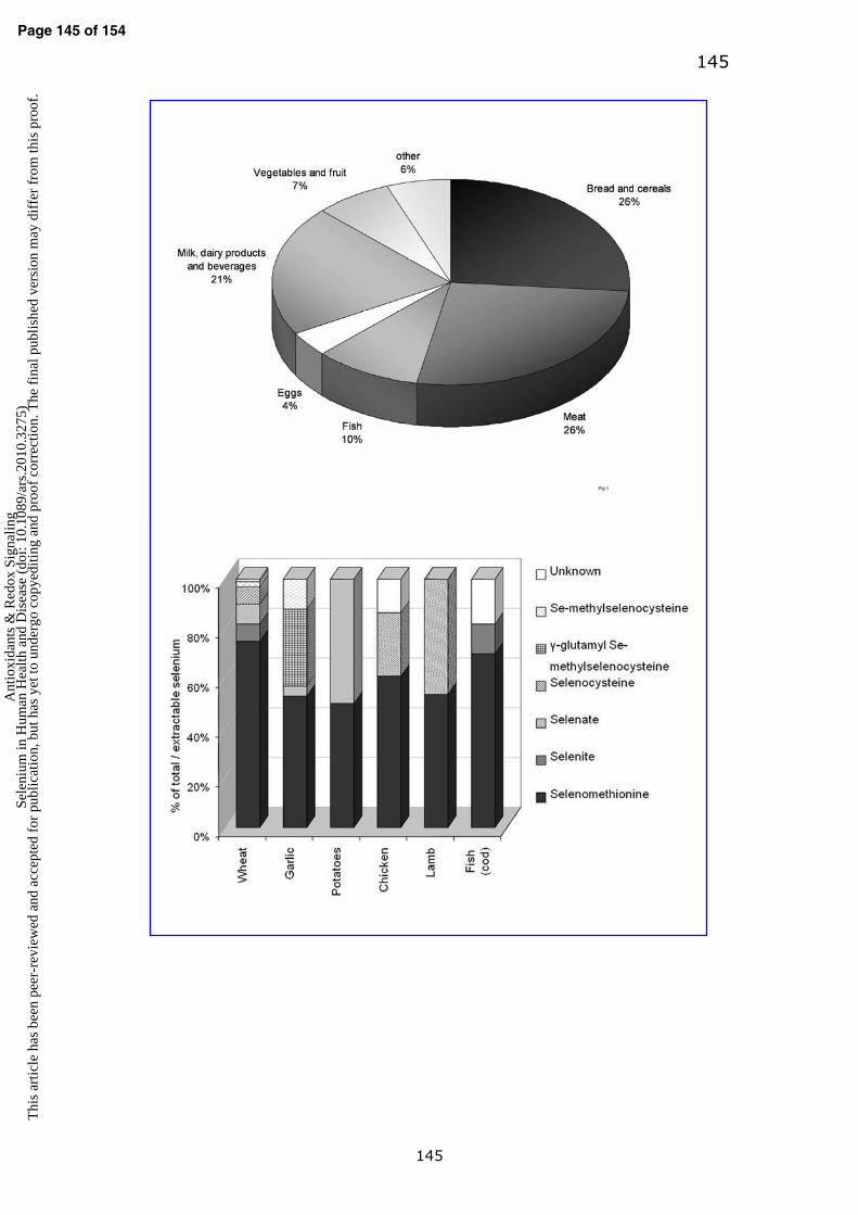
145
145
Page 145 of 154 A
ntio
xida
nts
& R
edox
Sig
nalin
gSe
leni
um in
Hum
an H
ealth
and
Dis
ease
(do
i: 10
.108
9/ar
s.20
10.3
275)
Thi
s ar
ticle
has
bee
n pe
er-r
evie
wed
and
acc
epte
d fo
r pu
blic
atio
n, b
ut h
as y
et to
und
ergo
cop
yedi
ting
and
proo
f co
rrec
tion.
The
fin
al p
ublis
hed
vers
ion
may
dif
fer
from
this
pro
of.
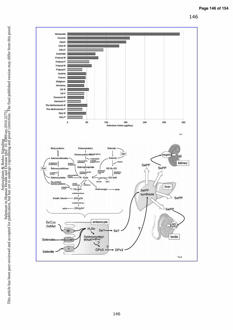
146
146
Page 146 of 154A
ntio
xida
nts
& R
edox
Sig
nalin
gSe
leni
um in
Hum
an H
ealth
and
Dis
ease
(do
i: 10
.108
9/ar
s.20
10.3
275)
Thi
s ar
ticle
has
bee
n pe
er-r
evie
wed
and
acc
epte
d fo
r pu
blic
atio
n, b
ut h
as y
et to
und
ergo
cop
yedi
ting
and
proo
f co
rrec
tion.
The
fin
al p
ublis
hed
vers
ion
may
dif
fer
from
this
pro
of.
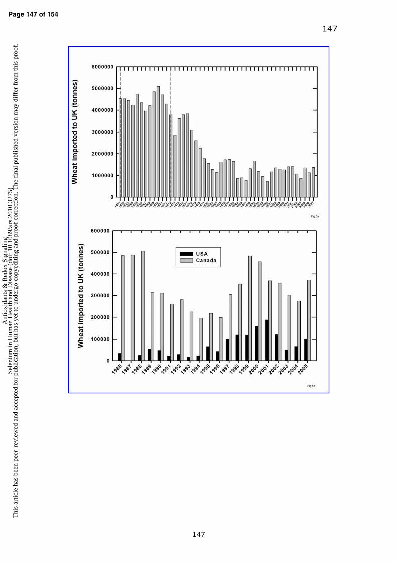
147
147
Page 147 of 154 A
ntio
xida
nts
& R
edox
Sig
nalin
gSe
leni
um in
Hum
an H
ealth
and
Dis
ease
(do
i: 10
.108
9/ar
s.20
10.3
275)
Thi
s ar
ticle
has
bee
n pe
er-r
evie
wed
and
acc
epte
d fo
r pu
blic
atio
n, b
ut h
as y
et to
und
ergo
cop
yedi
ting
and
proo
f co
rrec
tion.
The
fin
al p
ublis
hed
vers
ion
may
dif
fer
from
this
pro
of.

148
148
Page 148 of 154A
ntio
xida
nts
& R
edox
Sig
nalin
gSe
leni
um in
Hum
an H
ealth
and
Dis
ease
(do
i: 10
.108
9/ar
s.20
10.3
275)
Thi
s ar
ticle
has
bee
n pe
er-r
evie
wed
and
acc
epte
d fo
r pu
blic
atio
n, b
ut h
as y
et to
und
ergo
cop
yedi
ting
and
proo
f co
rrec
tion.
The
fin
al p
ublis
hed
vers
ion
may
dif
fer
from
this
pro
of.

149
149
Page 149 of 154 A
ntio
xida
nts
& R
edox
Sig
nalin
gSe
leni
um in
Hum
an H
ealth
and
Dis
ease
(do
i: 10
.108
9/ar
s.20
10.3
275)
Thi
s ar
ticle
has
bee
n pe
er-r
evie
wed
and
acc
epte
d fo
r pu
blic
atio
n, b
ut h
as y
et to
und
ergo
cop
yedi
ting
and
proo
f co
rrec
tion.
The
fin
al p
ublis
hed
vers
ion
may
dif
fer
from
this
pro
of.
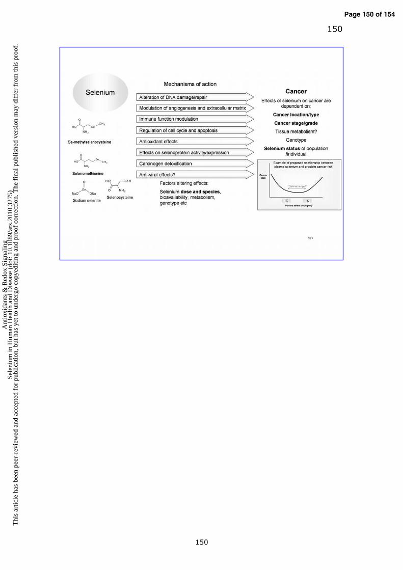
150
150
Page 150 of 154A
ntio
xida
nts
& R
edox
Sig
nalin
gSe
leni
um in
Hum
an H
ealth
and
Dis
ease
(do
i: 10
.108
9/ar
s.20
10.3
275)
Thi
s ar
ticle
has
bee
n pe
er-r
evie
wed
and
acc
epte
d fo
r pu
blic
atio
n, b
ut h
as y
et to
und
ergo
cop
yedi
ting
and
proo
f co
rrec
tion.
The
fin
al p
ublis
hed
vers
ion
may
dif
fer
from
this
pro
of.

151
151
Page 151 of 154 A
ntio
xida
nts
& R
edox
Sig
nalin
gSe
leni
um in
Hum
an H
ealth
and
Dis
ease
(do
i: 10
.108
9/ar
s.20
10.3
275)
Thi
s ar
ticle
has
bee
n pe
er-r
evie
wed
and
acc
epte
d fo
r pu
blic
atio
n, b
ut h
as y
et to
und
ergo
cop
yedi
ting
and
proo
f co
rrec
tion.
The
fin
al p
ublis
hed
vers
ion
may
dif
fer
from
this
pro
of.
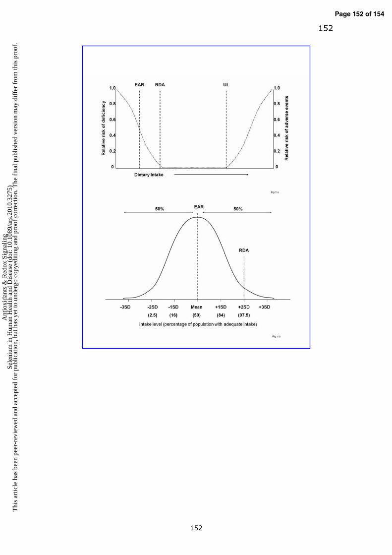
152
152
Page 152 of 154A
ntio
xida
nts
& R
edox
Sig
nalin
gSe
leni
um in
Hum
an H
ealth
and
Dis
ease
(do
i: 10
.108
9/ar
s.20
10.3
275)
Thi
s ar
ticle
has
bee
n pe
er-r
evie
wed
and
acc
epte
d fo
r pu
blic
atio
n, b
ut h
as y
et to
und
ergo
cop
yedi
ting
and
proo
f co
rrec
tion.
The
fin
al p
ublis
hed
vers
ion
may
dif
fer
from
this
pro
of.

153
153
Page 153 of 154 A
ntio
xida
nts
& R
edox
Sig
nalin
gSe
leni
um in
Hum
an H
ealth
and
Dis
ease
(do
i: 10
.108
9/ar
s.20
10.3
275)
Thi
s ar
ticle
has
bee
n pe
er-r
evie
wed
and
acc
epte
d fo
r pu
blic
atio
n, b
ut h
as y
et to
und
ergo
cop
yedi
ting
and
proo
f co
rrec
tion.
The
fin
al p
ublis
hed
vers
ion
may
dif
fer
from
this
pro
of.

154
154
Page 154 of 154A
ntio
xida
nts
& R
edox
Sig
nalin
gSe
leni
um in
Hum
an H
ealth
and
Dis
ease
(do
i: 10
.108
9/ar
s.20
10.3
275)
Thi
s ar
ticle
has
bee
n pe
er-r
evie
wed
and
acc
epte
d fo
r pu
blic
atio
n, b
ut h
as y
et to
und
ergo
cop
yedi
ting
and
proo
f co
rrec
tion.
The
fin
al p
ublis
hed
vers
ion
may
dif
fer
from
this
pro
of.