selenium in birds
TRANSCRIPT
University of Nebraska - LincolnDigitalCommons@University of Nebraska - Lincoln
USGS Staff -- Published Research US Geological Survey
2011
Selenium in BirdsHarry M. OhlendorfCH2MHILL
Gary H. HeinzU.S. Geological Survey
Follow this and additional works at: http://digitalcommons.unl.edu/usgsstaffpub
Part of the Geology Commons, Oceanography and Atmospheric Sciences and MeteorologyCommons, Other Earth Sciences Commons, and the Other Environmental Sciences Commons
This Article is brought to you for free and open access by the US Geological Survey at DigitalCommons@University of Nebraska - Lincoln. It has beenaccepted for inclusion in USGS Staff -- Published Research by an authorized administrator of DigitalCommons@University of Nebraska - Lincoln.
Ohlendorf, Harry M. and Heinz, Gary H., "Selenium in Birds" (2011). USGS Staff -- Published Research. 975.http://digitalcommons.unl.edu/usgsstaffpub/975

Selenium in Birds
Harry M. OhlendorfCH2MHILLSacramento, CA
Gary H. HeinzU.S. Geological SurveyPatuxent Wildlife Research CenterLaurel, MD
Published in Environmental Contaminants in Biota: Interpreting Tissue Concentrations, 2nd edition, ed. W. Nelson Beyer & James P. Meador (Boca Raton: CRC, 2011).
This chapter is a U.S. government work, not subject to copyright in the United States.

21 Selenium in Birds
Harry M. Ohlendorf Gary H. Heinz
CONTENTS
21. I Introduction .......................................................................................................................... 669 21.2 Dietary Requirements versus Toxicity ................................................................................. 671 2 1.3 Egg and Tissue Concentrations ............................................................................................. 676
21.3.1 Eggs .......................................................................................................................... 676 2 I .3. I. I Laboratory Studies ..................................................................................... 677 21.3. 1.2 Field Studies ............................................................................................... 681
21.3.2 Liver .......................................................................................................................... 683 21.3.2.1 Laboratory Studies ..................................................................................... 683 21.3.2.2 Field Studies ............................................................................................... 685
21.3.3 Kidney ....................................................................................................................... 686 21.3.4 Muscle ....................................................................................................................... 687 21.3.5 Blood ......................................................................................................................... 687 21.3.6 Integument/Feathers ................................................................................................. 689
2 1.4 Biomarkers ............................................................................................................................ 690 21.4.1 Biochemical .............................................................................................................. 690 21.4.2 MorphologicaL .......................................................................................................... 690
21.5 Interactions ........................................................................................................................... 691 21.6 Hormesis ............................................................................................................................... 693 Summary ........................................................................................................................................ 693 Acknowledgments .......................................................................................................................... 696 References ...................................................................................................................................... 696
21.1 INTRODUCTION
Selenium (Se) is a metalloid trace element that birds and other wildlife need in small amounts for good health. The main purpose of this chapter is to interpret tissue concentrations of Se. However, hecause food is the main source of Se accumulation for birds and other wildlife, and because dietary concentrations for effects on bird reproduction have been reported, we also provide interpretive information on Se in the diet.
Se deficiencies in domestic poultry and livestock occur in some parts of the world and must be corrected by additions of Se to the diet. However, the range of dietary concentrations that provides adequate but nontoxic amounts of Se is narrow compared with the ranges for most other essential trace elements.
In the 1930s, grains grown on seleniferous soils in South Dakota caused reproductive failure when fed to chickens (Gallus domesticus) (Poley and Moxon 1938). The most drastic incident of Se poisoning
669

670 Environmental Contaminants in Biota
in wild birds occurred at Kesterson Reservoir (located on the Kesterson National Wildlife Refuge) in California during the early and mid-1980s (Ohlendorf et al. 1986a, 1988, Ohlendorf 1989, 2002, Ohlendorf and Hothem 1995). Water used to irrigate crops in the San Joaquin Valley of California dissolved naturally occurring Se salts from the soil, and when the Se-Iaden subsurface water was drained from agricultural fields into Kesterson Reservoir, levels of Se that were toxic to birds accumulated in plants and animals used as foods by the birds. Reproductive failure and adult mortality Occurred. The findings at Kesterson Reservoir received extensive publicity and led to a series of laboratory and field studies (summarized in this chapter) that provide one of the best case studies in ecotoxicology during the past 30 years. The integrated field studies at Kesterson and related laboratory studies have been recognized as a "gold standard" in the field of ecotoxicology (Suter 1993). Similar problems of impaired bird reproduction were subsequently discovered elsewhere in the western United States, most notably in the Tulare Basin in California (Skorupa and Ohlendorf 1991, Skorupa 1998a).
High concentrations of Se in foods of wildlife are not limited to areas where soils are naturally high in Se. They also can be the result of the disposal of sewage sludge or fly ash, mining activity, or emissions from metal smelters (Robberecht et al. 1983, Wadge and Hutton 1986, Cappon 1991, Skorupa I 998a, Ratti et al. 2006, Wayland and Crosley 2006).
An assessment of the toxicity of Se is complicated by its occurrence in many different chemical forms, some differing greatly in their toxicity to birds. The four common oxidation states are selenide (-2), elemental Se (0), selenite (+4), and selenate (+6). Elemental Se is virtually insoluble in water and presents little risk to birds. Both selenite and selenate are toxic to birds, but organic selenides pose the greatest hazard. Among the organic selenides, selenomethionine has been shown to be highly toxic to birds and seems to be the form most likely to harm wild birds because it results in high bioaccumulation of Se in their eggs.
Much has been learned about Se toxicity to birds during the last 25 years; some of that information was summarized in the earlier edition by Heinz (1996). Other reviews in relation to exposure and effects of Se in birds are provided by Skorupa (l998a), O'Toole and Raisbeck (1998), U.S. DI (1998), Eisler (2000), Hoffman (2002), and Ohlendorf (2003). The purpose of this chapter is to identify the concentrations of Se in avian diets and in avian eggs and other tissues that are toxic, and to discuss how different chemical forms of Se and their interactions with other environmental contaminants can alter toxicity. We also present what are considered background (or no-effect) concentrations of Se from Se-normal areas, when available.
Background and reference area concentrations can be very useful for interpreting the possible toxic thresholds of a contaminant, especially when it is known with some certainty that the reference area has no known source of the contaminant in question. However, because some "background" concentrations of contaminants such as Se are reported from areas where the Se input is unknown, and may not, in fact, be what might be called "normal," "baseline," or "uncontaminated," they should be referred to as "reference area" samples, and a certain degree of caution must be exercised when using those concentrations as being synonymous with safe levels. The rigorous identification of safe levels of Se, or other contaminants, can really come only from the findings of controlled laboratory dosing studies and carefully designed field studies. In other words, merely because a contaminant like Se is at a level that has been reported from what are believed to be Se-normal areas does not, in itself, prove that the levels are safe.
The manner in which different authors present Se concentrations can be confusing, so it is important to understand the various ways results can be presented. Se concentrations typically are reported as micrograms per liter (J,lg/L) in most fluids (but sometimes J,lg/g or J,lg/dL in blood) and milligrams per kilogram (mg/kg), or micrograms per gram (J,lg/g) in soil, sediment, plant or animal tissues, and diets. Concentrations in soil, sediment, tissues, and diets can be expressed either on a wet-weight (or fresh-weight basis, which is considered to be synonymous) or a dry-weight basis. Although moisture loss during sample processing can be controlled fairly well in the laboratory, it is sometimes difficult to do so under field conditions. Therefore, reporting results on dry-weight basis helps ensure comparability of values.

Selenium in Birds 671
Conversion from one basis to the other is a function of the moisture content in the sample (which should be reported regardless of which basis is used), as follows:
. 100 Dry-weIght conc. = wet-weight conc. x (. ) .
100 - MOIsture percentage
In this chapter, we preferentially provide Se concentrations in diets and tissues on dry-weight basis (unless otherwise noted), and provide typical moisture content of eggs and tissues to enable readers to make conversions. When results were originally reported on wet-weight basis, the original concentrations are given in parentheses following the approximate dw concentration.
Se's ability to interact with other nutrients and environmental contaminants, especially other elements, also sometimes complicates an interpretation of toxic thresholds in tissues of birds. Although we do not attempt a comprehensive review to interpret critical levels of Se in the presence of elevated levels of other pollutants, we include a brief section on interactions, and the reader should be aware that such interactions exist.
21.2 DIETARY REQUIREMENTS VERSUS TOXICITY
In general, the diet is the most important exposure pathway for birds and, whenever possible, dietary concentrations should be included when reporting results or evaluating the effects observed in experimental or field studies. With the previously stated caution about "background" levels of Se in mind, mean background concentrations in diets of freshwater and terrestrial avian species are typically <3 mg/kg, with thresholds for reproductive impairment in the range of 3-8 mg/kg (Table 21.1).
TABLE 21.1 Published Assessment Values for Effects of Dietary or Tissue Concentrations of Se on Birds
Medium and
Level/Statusa
Adequate
To",-ic
Background
Reproductive
impairment
Concentration
(mg Se/kg, dw)
>5.0
<3.0
3-8
Effects
. " ,[;e~els ,are excessive bUt ': ''.~J~nsidere(! toxic to
':po, ,l;!y
, Reitiqed eg~ hatchability , iUtd~e~atogenic effects , ,iit~i\ibi:yo$fchicks None
Reduced egg
hatchability; potential
deformities in embryos/
chicks at upper end of
range
Comments Reference
Lower dietary concentrations Puis 1988 Me marginal or deficient, lind diets must be fortified
, , Poultry are relatively sensitive to effects of
selenium Poultry are relatively sensitive' to effects of selel)iutn'
Deficiencies associated with
lower concentrations have
not been reported in wild
birds
Sensitivity varies by species
and chemical form of Se in
diet
Puls1988
PnlsJ988
U.S. Dr 1998. Eisler
2000
U.S. Dr 1998, Eisler
2000
continued
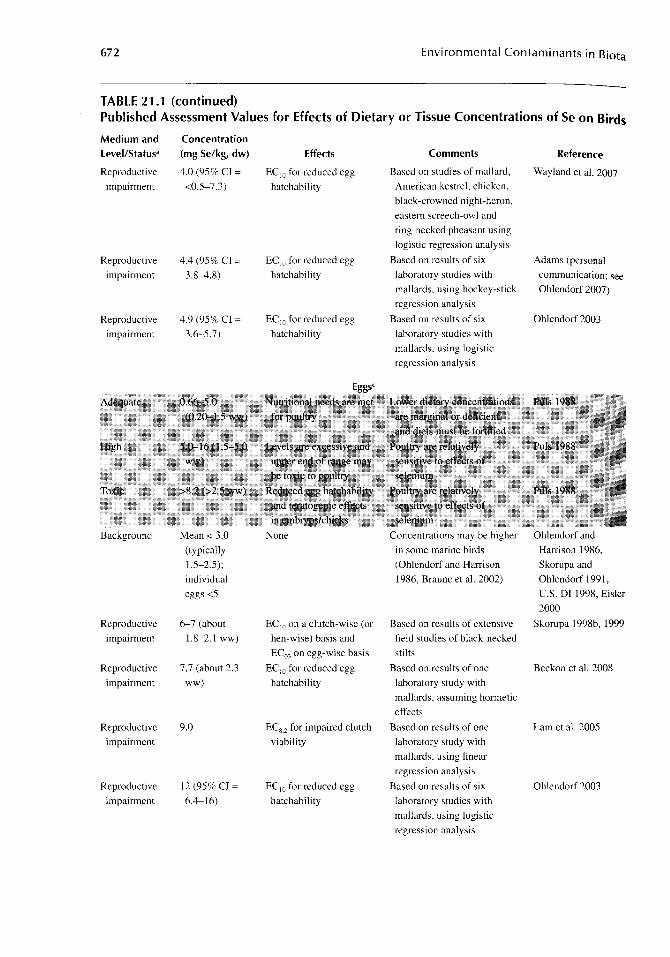
672 Environmental Contaminants in Biota
TABLE 21.1 (continued) Published Assessment Values for Effects of Dietary or Tissue Concentrations of Se on Birds
Medium and Level/Status'
Reproductive
impairment
Reproductive
impairment
Reproductive
impairment
Background
Reproductive
impairment
Reproductive
impairment
Reproductive
impairment
Reproductive
impairment
Concentration (mg Se/kg, dw)
4.0 (95% CI =
<0.5-7.3)
4.4 (95% CI =
3.8-4.8)
4.9 (95% CI =
3.6-5.7)
Mean < 3.0
(typically
1.5-2.5);
individual
eggs <5
6--7 (about
1.8-2.1 ww)
7.7 (about 2.3
ww)
9.0
12 (95% CI =
6.4-16)
Effects
EC lO for reduced egg
hatchability
EC lO for reduced egg
hatchability
EC IO for reduced egg
hatchability
None
EC lO on a clutch-wise (or
hen-wise) basis and
ECOJ on egg-wise basis
EC lO for reduced egg
hatchability
ECS2 for impaired clutch
viability
EC IO for reduced egg
hatchability
Comments
Based on studies of mallard,
American kestrel, chicken,
black-crowned night-heron,
eastern screech-owl and
ring-necked pheasant using
logistic regression analysis
Based on results of six
laboratory studies with
mallards, using hockey-stick
regression analysis
Based on results of six
laboratory studies with
mallards, using logistic
regression analysis
Concentrations may be higher
in some marine birds
(Ohlendorf and Harrison
1986, Braune et al. 2002)
Based on results of extensive
field studies of black-necked
stilts
Based on results of one
laboratory study with
mallards, assuming hormetic
effects
Based on results of one
laboratory study with
mallards, using linear
regression analysis
Based on results of six
laboratory studies with
mallards, using logistic
regression analysis
Reference
Wayland et al. 2007
Adams (personal
communication; see
Ohlendorf 2007)
Ohlendorf 2003
Ohlendorf and
Harrison 1986,
Skorupa and
Ohlendorf 1991,
U.S. DI 1998, Eisler
2000
Skorupa 1998b, 1999
Beckon et al. 2008
Lam et a1. 2005
Ohlendorf 2003
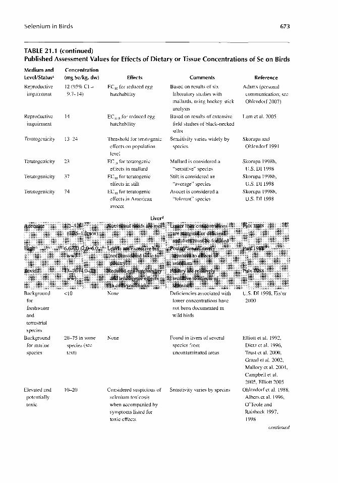
Selenium in Birds 673
TABLE 21.1 (continued) Published Assessment Values for Effects of Dietary or Tissue Concentrations of Se on Birds
Medium and Concentration level/Status' (mg Se/kg, dw) Effects Comments Reference
Reproductive 12 (95% CI = EC10 for reduced egg Based on results of six Adams (personal
impairment 9.7-14) hatchability laboratory studies with communication; see
mallards, using hockey-stick Ohlendorf 2007)
analysis
Reproductive 14 EC 11.8 for reduced egg Based on results of extensive Lam et al. 2005
impairment hatchability field studies of black-necked
stilts
Teratogenicity 13-24 Threshold for teratogenic Sensitivity varies widely by Skorupa and
effects on population species Ohlendorf 1991
level
Teratogenicity 23 EC 10 for teratogenic Mallard is considered a Skorupa 1998b,
effects in mallard "sensitive" species U.S. Dr 1998
Teratogenicity 37 EC IO for teratogenic Stilt is considered an Skorupa 1998b,
effects in stilt "average" species U.S. Dr 1998
Teratogenicity 74 EC 10 for teratogenic Avocet is considered a Skorupa 1998b,
effects in American "tolerant" species U.S. Dr 1998
avocet
liverd
Background <10 None Deficiencies associated with U.S. Dr 1998, Eisler
for lower concentrations have 2000
freshwater not been documented in
and wild birds
terrestrial
species
Background 20-75 in some None Found in livers of several Elliott et al. 1992,
for marine species (see species from Dietz et al. 1996,
species text) uncontaminated areas Trust et al. 2000,
Grand et al. 2002,
Mallory et al. 2004,
Campbell et al.
2005, Elliott 2005
Elevated and 10-20 Considered suspicious of Sensitivity varies by species Ohlendorf et al. 1988,
potentially selenium toxicosis Albers et al. 1996,
toxic when accompanied by O'Toole and
symptoms listed for Raisbeck 1997,
toxic effects 1998
continued
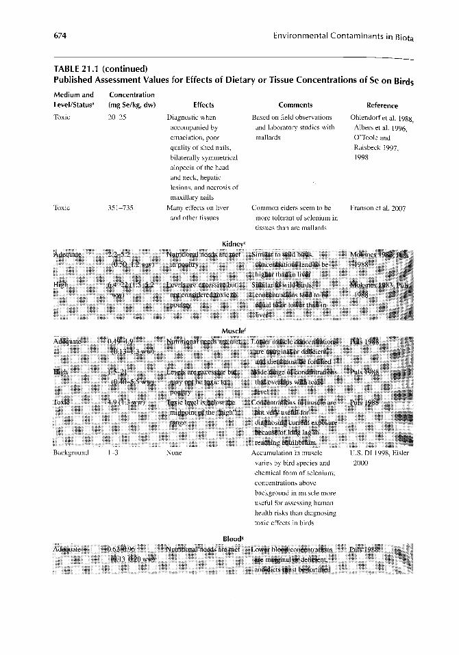
674 Environmental Contaminants in Biota
TABLE 21.1 (continued) Published Assessment Values for Effects of Dietary or Tissue Concentrations of Se on Birds
Medium and Level/Status·
Toxic
Toxic
Adequate.
Background
Adequate·
Concentration (mg Se/kg, dw) Effects Comments
20~25 Diagnostic when Based on field observations
and laboratory studies with
mallards
351~735
2.2-5.2 (0.50-1..2 ww)
4.9 (1.3 ww)
1~3
O.6Z...{}.96 (0.13-0.20 Ww)
accompanied by
emaciation, poor
quality of shed nails,
bilaterally symmetrical
alopecia of the head
and neck, hepatic
lesions, and necrosis of
maxillary nails
Many effects on liver
and other tissues
Kidney·
Nuttitiona! needs are mer
inpouitry
Muscle f
Common eiders seem to be
more tolerant of selenium in
tissues than are mallards
Similar .to wJldbirds,
. con~ntratioris tend to.be
.b:lgher!:bali in liver Similar to. wild Illrds,.
•. Nutntlona1needs. arem~t.. Low~ m:Usclect}nce~ttattoflS
Toxic leveUs below the . mic(poiat of i:he "high" range
None
Bloodg
·~~inal or d~JicieJ;it, . and dietsotustbe ibrtltied: W:iaerange of .concentriiii;qn$ ..... . that 6Vetlap~ With toXic ....
leVel . .. ..
not very useful for
diagnosing current eXposure because ofIong lag in reacIDl)geqcilibrium
Accumulatiol) in muscIe
varies by bird species and
chemical form of selenium;
concentrations above
background in muscle more
useful for assessing human
health risks than diagnosing
toxic effects in birds
Nutritional needs ate met Lower 1)ioodconcenttations are marginal or deficIent,
and diets mns! be fortified
Reference
Ohlendorf et al. 1988,
Albers et al. 1996, O'Toole and
Raisbeck 1997,
1998
Franson et al. 2007
U.S. or 1998, Eisler
2000

Selenium in Birds 675
TABLE 21.1 (continued) published Assessment Values for Effects of Dietary or Tissue Concentrations of Se on Birds
Medium and Level/Status'
Background
Provisional
threshold
warranting
further study
Background
Provisional
threshold
warranting
further study
Concentration (mg Se/kg, dw) Effects
0.48-1.9 None
(0.10-0.40
ww)
4.8 (1.0 ww)
1-4 (typically
1-2)
5
Interpretive relationship
to effects is limited, but
elevated levels
associated with effects
on reproduction or
survival
Feathersh
None
Interpretive relationship
to effects is limited, but
elevated levels
associated with
exposure when the
feathers were
developing
Comments
Deficiencies associated with
lower concentrations have
not been documented in
wild birds
Blood selenium
concentrations are good
indicator of current/recent
exposure, and especially
important for sampling
when animals should not be
sacrificed
Based on breast feathers;
concentrations in feathers
vary by type and reflect
exposure at the time feathers
were grown, rather than
current exposure
Feather selenium
concentrations are not good
indicator of current/recent
exposure, but may be useful
if limitations are understood
(see text)
Reference
U.S. DI 1998, Eisler
2000
Heinz et al. 1990,
Heinz and
Fitzgerald 1993a,
O'Toole and
Raisbeck 1997,
U.S. DI 1998,
Yamamoto et al.
1998, Santolo et al.
1999, Eisler 2000
Burger 1993,
Ohlendorf 1993,
U.S. DI 1998, Eisler
2000
Burger 1993,
Ohlendorf 1993,
U.S. DI 1998, Eisler
2000
" Typical moisture content (%) and approximate conversion factor are shown in footnotes for each medium. Values that are
shaded are based on domestic poultry rather than wild species.
b Variable moisture; laboratory diet typically -10%, but natural diet varies widely «10->90%).
, 65-80% moisture, varying with species and incubation stage; use 70% (i.e., factor of 3.3) for approximate conversion.
d 70% moisture; use factor of 3.3 for approximate conversion.
, 76-78% moisture, based on limited data; use factor of 4.3 for approximate conversion.
j 74% moisture; use factor of 3.8 for approximate conversion.
g 79% moisture in lab studies, variable under field conditions; use factor of 4.8 for approximate conversion.
h 10% moisture assumed (not well defined); use factor of 1.1 for approximate conversion.
For birds, as for most other animals, dietary Se requirements appear to be between 0.05 and 0.5 mg/kg (NAS-NRC 1976, 1983, Combs and Combs 1986, Oldfield 1990, 1998, Eisler 2000). Excess Se in the diet of female birds during the period just before egg-laying can result in the transfer of Se to the eggs or other tissues at harmful levels, although sensitivity to Se varies among species (Skorupa and Ohlendorf 1991, Ohlendorf 1996, Skorupa 1998a, 1998b). Detwiler (2002) analyzed field-collected eggs and conducted laboratory studies with chickens to determine partitioning of Se in eggs (to albumen, yolk, and embryo) and to identify toxicokinetic causes of species
676 Environmental Contaminants in Biota
variability in sensitivity to Se. As expected, differences among species, as well as those due to form of Se in the diet, are complex. Those complexities are not described in detail here, but readers may wish to read about them in Detwiler's (2002) work.
Ohlendorf (2003) used data from six laboratory studies with mallards (Anas platyrhynchos) (Heinz et al. 1987, 1989, Stanley et al. 1994, 1996, Heinz and Hoffman 1996, 1998) to calculate an EC]() (i.e., the "effective concentration" that caused a 10% effect; in this case, the dietary concentration that reduced hatching of eggs 10% below that of the control group in the same study) along with 95% confidence intervals (95% CI) for the mean Se concentration in the diet. The dietary ECIQ was calculated to be 4.9 mg Se/kg, with 95% CI of 3.6-5.7 mg Se/kg.
The EC IO of 4.9 mg Se/kg was estimated by fitting a logistic regression model to the available data. It should be noted, however, that the mallard studies used a "dry" diet that had about 10% moisture. Ohlendorf (2003) used the reported dietary Se concentrations without adjustment for that moisture content, but an upward adjustment of the values (by 11%; to about 5.4 mg/kg) would be appropriate to account for the moisture content of the duck diet.
Adams et al. (2003) used hockey-stick regression on data for egg Se concentrations and adverse effects in mallards to derive toxicity thresholds, such as ECIO values. On further analyses (as described in Ohlendorf 2007), they found a threshold to exist when dietary Se was plotted against egg inviability and duckling mortality (which incorporated the cumulative effects of fertilization success and hatchability plus survival of ducklings for 6,7, or 14 days after hatching, as reported for the different studies). The inflection point occurred at a dietary Se concentration of 3.9 mg/kg. The predicted EC IO was 4.4 mg Se/kg (just slightly above the inflection point), and the 95% CI around the predicted EC]() ranged from 3.8 to 4.8 mg Se/kg.
Wayland et al. (2007) used logistic regression to calculate ECIO values based on experimental studies of six species (mallard, American kestrel [Falco sparverius], domestic chicken, black-crowned night-heron [Nycticorax nycticorax], eastern screech-owl [Megascops asia] and ring-necked pheasant [Phasianus colchicus]). The ECIO was 4.0 mg Se/kg with 95% CI from <0.5 to 7.3 mg Se/kg. The effect of including several species was to widen the confidence limits substantially (compared to mallard EC IO), indicating a high degree of difference among species in sensitivity to Se.
Information on forms of Se in invertebrates (as potential diets for birds) is limited, but Andrahennadi et al. (2007) found variability in the Se speciation among aquatic insects that included mayflies (Ephemeroptera), stoneflies (Plecoptera), caddisflies (Trichoptera), and craneflies (Diptera) from streams in Alberta, Canada. Higher percentages of inorganic Se were found in primary consumers, detritivores, and filter feeders than in predatory insects. Among the organic forms, organic selenides constituted a major fraction in most organisms. A form of selenide, believed to represent selenomethionine, varied widely among aquatic insects (from 36% to 98% of the total Se), indicating a high degree of variability in bioaccumulation potential from diet to eggs. Nevertheless, the chemical forms of Se in aquatic foods of birds have received little study. It is likely that varying chemical forms of Se are present to some degree in plants and animals eaten by birds, yet the toxic concentrations of few Se compounds have been determined in birds.
Interpretive guidelines that have resulted from extensive testing with poultry are provided by PuIs (1988). The Se concentrations for diet (as well as those for eggs and other tissues) are helpful guidelines for wild birds as well as domestic poultry. Dietary Se concentrations of less than 0.30 mg/kg are considered to be below the range adequate for good adult health and reproduction, 3.0-5.0 mg/kg are high, and above 5.0 mg/kg are toxic (Table 21.1).
21.3 EGG AND TISSUE CONCENTRATIONS
21.3.1 EGGS
Mean background Se concentrations in eggs of freshwater and terrestrial birds are <3 mg/kg dw (typically 1.5-2.5 mg/kg dw); concentrations lower than about 0.66 mg/kg dw may indicate

Selenium in Birds 677
inadequate Se in the diet, and maximums for individual eggs are <S mg/kg dw (Table 21.1). Moisture content of eggs varies by stage of incubation (decreasing throughout incubation) and by species, but typical moisture content of field-collected eggs is usually 6S-80% (Ohlendorf and Hothem 1995). Fresh mallard eggs, such as those collected from laboratory studies, have about 70% moisture (Stanley et al. 1996). The latter value provides a reasonable conversion factor (3.3) for estimating from one basis to the other and, except where noted, is used in this chapter when Se concentrations in eggs were originally reported on wet-weight basis, but the moisture content of samples was not reported.
21.3.1.1 Laboratory Studies
In a wide variety of species, if one expresses both the diet and eggs on a dry-weight basis, Se concentrations in bird eggs range from roughly equal to about three or four times the concentrations in the diet of the female at the time of egg-laying (Heinz et al. 1987, 1989, Smith et al. 1988, Ohlendorf 1989, Stanley et al. 1994, 1996, Wiemeyer and Hoffman 1996, Santolo et al. 1999). However, Se transfer from diet to egg varies by species and the chemical form of Se in the diet.
When birds fed on Se-contaminated diets during the laying season, the exposure was quickly reflected in elevated levels of Se in eggs (Heinz 1993b, Latshaw et al. 2004, DeVink et al. 2008a). Similarly, when the birds were switched to a clean diet, Se concentrations in eggs declined quickly. When mallard hens were fed a diet containing IS mg Se/kg (as selenomethionine), levels peaked in eggs (to about 43-66 mg Se/kg dw; 13-20 mg Se/kg ww) after about 2 weeks on the treated diet and leveled off at a relatively low level «16 mg Se/kg dw; <S mg Se/kg ww) about 10 days after switching to an untreated diet (Heinz 1993b). The findings of this study and two others with ring-necked pheasants (Phasianus colchicus) (Latshaw et al. 2004) or lesser scaup (Aythya ajjinis) (DeVink et al. 2008a) summarized later have important implications for evaluation of field exposures, such as how quickly and for what duration Se exposure may adversely affect bird reproduction. Concentrations of Se in eggs are especially important because they provide the best samples for evaluating potential adverse reproductive effects (Skorupa and Ohlendorf 1991). Knowing Se concentrations in food items available to wild birds at a site also can be useful in assessing risks of reproductive effects, but relationships between the available food and concentrations that occur in eggs can vary widely on the basis of physiology and feeding ecology of the birds. Se speciation in the diet also may be important in this regard (i.e., plant vs. animal diets).
When ring-necked pheasants received feed that contained 9.3 mg Se/kg because of a feed mixing problem, severe effects occurred within 4 days (Latshaw et al. 2004). The rate of egg production decreased and bird aggression increased. About 12% of the hens died within a week; necropsy results were consistent with Se toxicity. After 8 days, the toxic feed was removed and replaced with fresh feed. Egg production, which had dropped by SO%, returned to normal within 10 days of feed replacement. Hatchability of eggs laid from days 8 to 14 after the pheasants received the toxic feed dropped to 3S%, and more than SO% of the embryos that survived to the point where they could be examined had deformed beaks and abnormal eyes. Hatchability of eggs laid 21-28 days after the hens had received the toxic feed (i.e., 13-20 days after it was replaced by new feed) was almost 80%. Similar to the study with mallards, this incident showed a rapid onset of effects and a rapid recovery in response to dietary Se concentrations.
To assess the possible effects of Se on reproduction and fitness (measured as body mass) of lesser scaup, captive scaup were fed a control diet or one supplemented with Se at 7.S or IS mg/kg for 30 days to simulate dietary exposure to Se during late spring migration (DeVink et al. 2008a). The treated feed was removed after 30 days, just before the birds began laying. There was no effect of Se on body mass, breeding probability, or clutch initiation dates. Se concentrations in the first eggs laid by these birds were 2S-30 mg/kg in the 7.S-mg/kg and 30-3S mg/kg in the IS-mg/kg treatment groups. Egg Se concentrations of both treatment groups decreased rapidly after the Se-supplemented feed was removed, and within 8 and 12 days, respectively, the egg Se concentration was less than 9 mg/kg dw. There was no significant intraclutch variation in egg Se deposition.
678 Environmental Contaminants in Biota
The embryo is the avian life stage most sensitive to Se (Poley et al. 1937, Poley and Moxon 1938 Heinz et al. 1987, 1989, Hoffman and Heinz 1988). Because it is the Se in the egg, rather than i~ the parent bird, that causes developmental abnormalities and death of avian embryos, Se in the egg gives the most sensitive measure for evaluating hazards to birds (Skorupa and Ohlendorf 1991). Given the rapid accumulation and loss patterns of Se in birds (Heinz et al. 1990, Heinz 1993b, Heinz and Fitzgerald 1993b, Latshaw et al. 2004), Se concentrations in eggs also probably best represent contamination of the local environment. Additional advantages of measuring Se in eggs are that eggs are frequently easier to collect than adult birds, the loss of one egg from a nest probably has little effect on a population, and the egg represents an integration of exposure of the adult female during the few days or weeks before egg-laying.
The concentration detected in eggs and the toxicity of that concentration seem to depend on the chemical form of the ingested Se. Organoselenium compounds are believed to be major forms in plants and animals. One organoselenium compound, selenomethionine, when fed to breeding mallards was more toxic to embryos than was selenocystine or sodium selenite (Heinz et al. 1989). Selenomethionine is a major form of Se in wheat seeds and soybean protein (Olson et al. 1970, Yasumoto et al. 1988). Hamilton et al. (1990) found selenomethionine to be an excellent model for Se poisoning in Chinook salmon (Oncorhynchus tshawytscha) when compared with the toxicity of Se that was biologically incorporated into mosquitofish (Gambusia affinis) collected at Kesterson Reservoir in California. Yamamoto et al. (1998) measured Se concentrations in blood and excreta of American kestrels fed either a selenomethionine-fortified diet or animals from Kesterson. They found no significant differences in concentrations or in accumulation and depuration of Se among experimental groups that received Se as selenomethionine or naturally incorporated in tissue of animals from Kesterson.
When mallards were fed a diet containing 10 mg Se/kg as selenomethionine (and about 10% moisture), reproductive success was significantly lower in the treated ducks than in controls, and a small sample of five eggs from the treated birds contained a mean of about 15 mg Se/kg dw (4.6 mg Se/kg ww) (Heinz et al. 1987). Because mallards were fed only one dietary concentration of Se in the form of selenomethionine, no safe level was established in this experiment. All that can be said is that the safe level in eggs was below about 15 mg Se/kg dw.
In a subsequent study, mallards were fed a diet containing about 10% moisture and 0, 1,2,4,8, or 16 mg/kg of added Se as selenomethionine (Heinz et al. 1989). The reproductive success of the groups fed 1, 2, or 4 mg Se/kg did not significantly differ from that of controls; mean Se concentrations in a sample of IS eggs from each of these groups were about 2.7, 5.3, and 11 mg/kg dw (0.83, 1.6, and 3.4 mg/kg ww). The group fed 8 mg Se/kg produced 57% as many healthy ducklings as the controls; the reduction in numbers was caused mainly by hatching failure and the early death of those that did hatch. A sample of 15 eggs from this group contained about 36 mg Se/kg dw (11 mg Se/kg ww). The group fed 16 mg Se/kg failed to produce any healthy young, and a sample of 10 of their eggs contained an average of about 59 mg Se/kg dw (18 mg Se/kg ww). Therefore, based on this study, the highest mean Se concentration in eggs not associated with reproductive impairment was about II mg/kg dw (3.4 mg/kg ww), and the lowest mean toxic concentration was 36 mg/kg dw (11 mg/kg ww).
Lam et al. (2005) subjected the data from this study with mallards (Heinz et al. 1989) to statistical analyses to estimate the threshold for effects on clutch viability. They normalized treatment response for control response and subjected the data to linear regression analysis, and then used a stepwise increment of 0.5-mg Se/kg concentration units followed by a one-tailed, one-sample t-test comparing the percentage of impairment of clutch viability (±95% CI) with zero to derive threshold effect levels of Se in eggs associated with impaired hatchability. They determined that 9 mg Se/kg was the lowest concentration in eggs at which clutch viability was significantly different than zero, and that the value represented an ECS.2 for effects. A recent paper by Beckon et al. (2008) used the mean response data from the same laboratory study with mallards (Heinz et al. 1989) to evaluate potential hormetic effects exhibited by the treatment groups, and found an ECIQ of 7.7 mg Se/kg (see later section on Hormesis).

Selenium in Birds 679
In another study, Heinz and Hoffman (1996) compared the toxicity of three forms of selenomethionine. In nature, selenomethionine occurs almost exclusively in the L form, which is one of the two stereoisomer forms it can take (Cukierski et al. 1989). The other stereoisomer is the D form, and in many feeding studies with birds a mixture of the two forms (seleno-oL-methionine) has been fed. In yeast, most of the Se is in the form of seleno-L-methionine (Beilstein and Whanger 1986), and in addition to being in the naturally occurring form, it is biologically incorporated into the yeast. Pairs of breeding mallards were fed 10 mg Se/kg in each of the three forms. The results suggested that seleno-oL-methionine and seleno-L-methionine were of similar toxicity and both were more toxic than the Se in selenized yeast, but the lower toxicity of selenized yeast may have been due to a lower bioavailability of the selenomethionine in the yeast. A sample of eggs from the pairs fed seleno-L-methionine contained a mean of about 30 mg Se/kg dw (8.9 mg Se/kg ww), which resulted in a severe reduction in reproductive success (6.4% hatching of fertile eggs compared to 41.3% for controls). Eggs from pairs fed the seleno-oL-methionine contained a mean of about 31 mg Se/kg dw (9.2 mg Se/kg ww), and hatching of fertile eggs was 7.6%. Eggs from the pairs fed the selenized yeast contained a mean of only about 22 mg Se/kg dw (6.6 mg Se/kg ww), and hatching success was 27.0%. Because even the 22 mg Se/kg derived from the selenized yeast had a profound effect on reproductive success a toxic threshold was not established, but was obviously well below 22 mg Se/kg. Three studies were conducted to evaluate the interactive effects of Se with arsenic (As) (Stanley et al. 1994), boron (B) (Stanley et al. 1996), or mercury (Hg) (Heinz and Hoffman 1998), which are described in a later section (Interactions).
Using the same approach as that described earlier for the dietary values associated with reduced egg hatchability in mallards, Ohlendorf (2003) found the EC]() in eggs was 12 mg Se/kg dw, with 95% CIs of 6.4-16 mg Se/kg dw. The EC]() of 12 mg Se/kg was estimated by fitting a logistic regression model to the results of the six laboratory studies with mallards mentioned earlier.
The ECw for mallard duckling mortality, as reported in Adams et al. (2003), ranged from 12 to 16 mg Se/kg dw in eggs. These EC]() values are based on a synthesis of the same six laboratory studies as mentioned earlier, but using the final endpoint of duckling mortality (the same effects data used in the dietary EC IO evaluation with hockey-stick regression above); the range of EC]() values reflects different statistical approaches for analyzing the data. Based on further analyses of those data, Adams (personal Communication; see Ohlendorf 2007) determined that the inflection point of the hockey stick occurred at an egg Se concentration of 9.8 mg/kg dw, with a predicted EC IO of about 12 mg/kg dw, which was comparable to that derived by Ohlendorf (2003). The 95% CI using hockey-stick regression was much narrower (9.7-14 mg/kg dw) than that derived by Ohlendorf using logistic regression (6.4-16 mg/kg dw). Given that there is a clear egg-Se threshold at which effects begin to be observed, a unimodal model, such as logistic regression, may result in exaggerated confidence intervals, particularly in the tails.
In a laboratory study designed to measure the lingering effects of an overwinter exposure to selenomethionine on reproduction, mallards were fed a diet containing 15 mg Se/kg for 21 weeks before the onset of laying (Heinz and Fitzgerald 1993b). Females began laying after various lengths of time off treatment. This experimental design was not ideal for determining the lowest concentration of Se in eggs associated with reproductive impairment, but the authors were able to make some general conclusions. Some eggs hatched when Se in eggs was as high as about 20-30 mg/kg dw (6-9 mg/kg ww), but other eggs failed to hatch when Se concentrations were estimated to be between 9.9 and 16 mg/kg dw (3 and 5 mg/kg ww). The authors concluded that the most logical reason why some embryos die while others survive when exposed to a given concentration of Se is that mallard embryos vary in their individual sensitivity to Se.
When black-crowned night-herons were fed a diet containing 10 mg Se/kg as selenomethionine (on close to a dry-weight basis) hatching success of fertile eggs was not reduced (Smith et al. 1988). The eggs of treated herons contained a mean concentration of about 11 mg Se/kg dw (3.3 mg Se/kg ww). The results from this study must be taken with some caution, however, because sample sizes were small (n = 5 pairs per group) and hatching success of fertile eggs of the control group was poor (32%).

680 Environmental Contaminants in Biota
Martin (1988) fed Japanese quail (Coturnix coturnix japonica) diets containing 5 or 8 mg Se/kg and chickens 10 mg Se/kg as selenomethionine, respectively. At 5 mg Se/kg, the hatching success of fertile quail eggs (56.4%) was lower than that of controls (76.4%); eggs from treated females contained about 23 mg Se/kg dw (7.1 mg Se/kg ww). At 8 mg Se/kg, the hatching of quail eggs was further decreased to 10.4% (compared with 75.1 % for controls in that trial), and Se in eggs averaged about 40 mg/kg dw (12 mg/kg ww). The hatching success of the chickens fed 10 mg Se/kg also was depressed (23.2% compared with 84.5% for controls), and Se in eggs averaged about 36 mg/kg dw (9.6 mg/kg ww; the conversion from ww to dw [3.8] was based on the contents of chicken eggs containing about 73.6% water [Romanoff and Romanoff 1949]). No-effect concentrations in the diet or eggs were not determined.
In another study with chickens, diets were supplemented with seleniferous grains in amounts to produce dietary concentrations of 2.5, 5, and 10 mg Se/kg (Moxon and Poley 1938, Poley and Moxon 1938). Modern statistical techniques were not applied to these data, and chemical analyses were different from those used today, but at 2.5 mg Se/kg in the diet, the hatching success of fertile eggs was no different from that of controls, and a sample of eggs contained Se at about 15 mg/kg dw in albumen and 3.2 mg/kg dw in yolk 1.75 mg/kg and 1.67 mg/kg ww, respectively; conversions from ww to dw here and below (multiply ww concentrations by 8.3 for albumen and by 1.9 for yolk) were based on the fact that chicken eggs are composed of about 55.8% albumen, 31.9% yolk, and 12.3% shell, and that the moisture content of albumen is about 87.9% while that of yolk is 48.7% (Romanoff and Romanoff 1949). At 5 mg Se/kg in the diet, the hatching of eggs was "slightly reduced," and Se in egg albumen and yolks averaged about 24 and 5.2 mg/kg dw (2.95 and 2.73 mg/kg ww), respectively. At 10 mg Se/kg, hatching decreased to zero, and albumen and yolks contained about 53 and 7.4 mg Se/kg dw (6.40 and 3.92 mg Se/kg ww), respectively. Based on the percentages of albumen and yolk in chicken eggs and the respective percentages of water in albumen and yolk, a Se threshold of about 10 mg/kg dw (3 mg/kg ww) in whole eggs was associated with reproductive impairment in the study where chickens were fed 5 mg Se/kg; this threshold is similar to the findings of more rigorous recent studies with mallards.
Harmful concentrations of Se in eggs may be of a different magnitude when another chemical form of Se, sodium selenite, is fed to birds. A diet containing 7 mg Se/kg as sodium selenite caused reproductive impairment in chickens but resulted in only about 7.2 and 3.8 mg Se/kg dw (0.87 and 2.02 mg Se/kg ww) in egg albumen and yolk (Ort and Latshaw 1978).
In another study with chickens, a diet containing 8 mg Se/kg as sodium selenite impaired reproduction, and whole eggs contained from about 5.5 to 7.1 mg/kg dw (1.46-1.86 mg/kg ww) of Se (Arnold et al. 1973). The chemical form of Se in chicken eggs seems to be different when sodium selenite rather than selenomethionine is fed (Latshaw 1975, Latshaw and Osman 1975).
In mallards, a dietary concentration of 25 mg Se/kg as sodium selenite impaired reproduction but resulted in a mean of only about 4.3 mg/kg dw (1.3 mg/kg ww) of Se in eggs (Heinz et al. 1987). Therefore, although higher dietary concentrations of sodium selenite than selenomethionine must be fed to mallards to harm reproduction, lower concentrations of Se in eggs are associated with harm.
Selenium also may affect egg fertility in some species, but egg fertility is not always reported from field or laboratory studies. Lack of reporting on fertility effects in some studies of Se effects in birds may be due in part to a general practice of simply including infertile eggs as inviable eggs (i.e., "infertility" effects may not be separated from "embryotoxic" effects in the overall measurement of hatchability). Failure to measure infertility as a separate endpoint may be due to the difficulty often associated with distinguishing infertile eggs from those containing embryos that have died very early in development. Nevertheless, decreased fertility is a distinct effect from embryotoxicity, particularly in that it can indicate a mechanism acting on adult, rather than embryonic, physiology. In American kestrels fed selenomethionine at 12 mg Se/kg, egg fertility was significantly reduced (by over 14%) compared to kestrels fed 6 mg Se/kg (Santolo et al. 1999). Results obtained in kestrels suggest that infertility may be an important factor contributing to the overall reproductive

Selenium in Birds 681
impairment in some species. However, in mallards (Heinz et al. 1987, Heinz and Hoffman 1996, 1998) and black-crowned night-herons (Smith et al. 1988) fed IO mg Se/kg as selenomethionine, egg fertility was not reduced compared with controls. Similarly, fertility was not affected in mallards fed diets containing Se at 7 mg/kg (Stanley et al. 1996) or 16 mg/kg (Heinz et al. 1989) as selenomethionine, but hatchability of fertile eggs was significantly reduced. Thus, effects on egg fertility in mallards and night-herons are not likely to be as ecologically significant as reduced hatchability.
21.3.1.2 Field Studies
Selenium concentrations in the eggs of marine species are variable, but may be higher than in freshwater or terrestrial birds, even in remote areas (Ohlendorf 1989). For example, eggs of three species (wedge-tailed shear water [Puffinus pacificus], red-footed booby [Sula sula], and sooty tern [Sterna /i~scata]) were sampled at four locations throughout the Hawaiian Archipelago, from Oahu to Midway (Ohlendorf and Harrison 1986). Mean Se concentrations varied only slightly by location, from about 4.4 to 5.3 mg/kg dw (1.1-1.4 mg/kg ww) for shearwaters, 5.0-6.1 mg/kg (0.76-0.92 mg/kg ww) for boobies, and 4.1-5.1 mg/kg (1.1-1.4 mg/kg ww) for terns, but all were higher than typical offreshwater species. Henny et al. (1995) predicted egg concentrations (21.3 or 29.2 mg Se/kg dw, based on different regressions) from liver concentrations in white-winged scoters (Melanitta fusca) (mean of 54 mg Se/kg dw for combined males and females; concentration not given separately for females) based on established liver-egg relationships for freshwater species (Henny and Herron 1989, Ohlendorf et al. 1990, Ohlendorf and Hothem 1995). However, they found that Se concentrations in eggs were only about 10% of the predicted concentrations, from 2.7 to 4.7 mg/kg dw.
Braune et al. (2002) analyzed eggs of glaucous gulls (Larus hyperboreus), black-legged kittiwakes (Rissa tridactyla), thick-billed murres (Uria lomvia), and black guillemots (Cepphus grylle) from the Canadian Arctic. Mean Se concentrations varied somewhat by species and location, with all means between 1.1 and 2.7 mg/kg dw except for kittiwakes (with means of 4.4 mg/kg at two locations), so kittiwakes were the only species with means greater than typical of freshwater and terrestrial birds.
Eggs of common eiders (Somateria mollissima) collected from five locations in the Baltic Sea near coastal Finland also had median Se concentrations (0.55 mg/kg ww; about 1.65 mg/kg dw) that were similar to background for freshwater and terrestrial birds (Franson et al. 2000). Thus, there seems to be no consistent difference between marine and other birds.
Using the results of extensive field studies of black-necked stilts (Himantopus mexican us), Skorupa (l998a, 1999) found a threshold of 6-7 mg Se/kg in eggs to be associated with impaired egg hatchability. That concentration is about equivalent to the EC IO on a clutch-wise (or hen-wise) basis and the EC01 on an egg-wise basis. Lam et a1. (2005) used the same statistical approach as described earlier for the laboratory study with mallards to estimate the threshold for effects on stilt clutch viability. They derived an EC11g of 14 mg Se/kg at which clutch viability was significantly impaired (i.e., greater than zero impairment). It should be noted that the background rate of clutch inviability (when Se concentrations in eggs are <6 mg/kg) is estimated at 8.7% (U.S. DI 1998).
Studying birds at Kesterson Reservoir in California. Ohlendorf et al. (I 986b) used logistic regression to estimate a 50% chance of embryo death or deformity in American coots (Fulica americana) when Se concentrations in eggs were about 18 mg/kg dw. The estimated Se concentration causing the same effect in black-necked stilts was 24 mg/kg. The value for eggs of eared grebes (Podiceps nigricollis) could not be calculated because even the lowest Se concentration detected in eggs (44 mg/kg) was embryotoxic. The logistic approach is best suited to estimate the 50% effect concentration, not the concentrations of Se in eggs at which embryo deaths and deformities begin for each species. These concentrations would obviously be somewhat lower than the 50% effect levels.
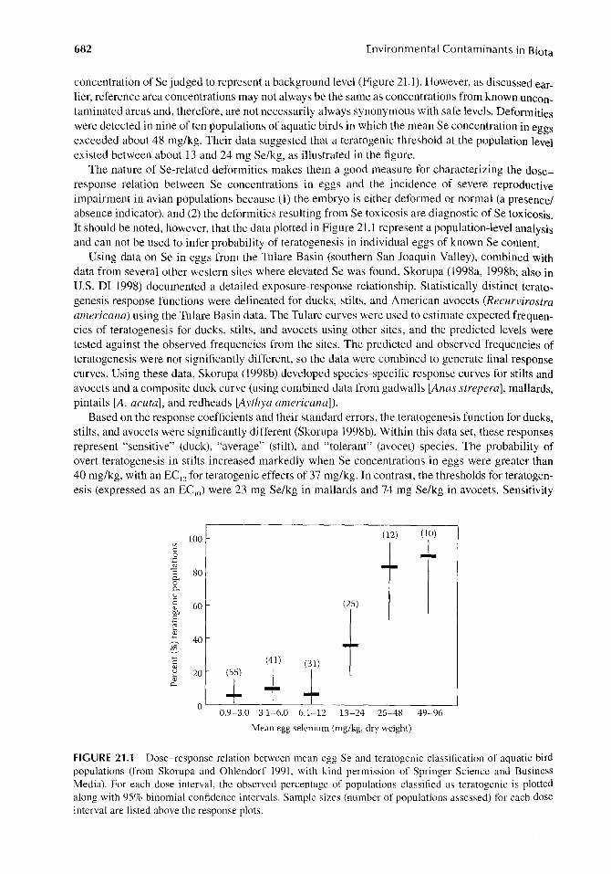
Skorupa and Ohlendorf (1991) examined the relation between Se concentrations in eggs of various aquatic bird species and reproductive impairment at the population level. Embryo deformities were detected in only 3 of 55 popUlations of birds that had a mean Se concentration of less than 3 mg/kg in eggs (and these deformities were not characteristic of those induced by Se); this is a
682 Environmental Contaminants in Biota
concentration of Se judged to represent a background level (Figure 21.1). However, as discussed earlier, reference area concentrations may not always be the same as concentrations from known uncontaminated areas and, therefore, are not necessarily always synonymous with safe levels. Deformities were detected in nine of ten populations of aquatic birds in which the mean Se concentration in eggs exceeded about 48 mg/kg. Their data suggested that a teratogenic threshold at the population level existed between about 13 and 24 mg Se/kg, as illustrated in the figure.
The nature of Se-related deformities makes them a good measure for characterizing the doseresponse relation between Se concentrations in eggs and the incidence of severe reproductive impairment in avian populations because (1) the embryo is either deformed or normal (a presence/ absence indicator), and (2) the deformities resulting from Se toxicosis are diagnostic of Se toxicosis. It should be noted, however, that the data plotted in Figure 21.1 represent a population-level analysis and can not be used to infer probability of teratogenesis in individual eggs of known Se content.
Using data on Se in eggs from the Tulare Basin (southern San Joaquin Valley), combined with data from several other western sites where elevated Se was found, Skorupa (1998a, 1998b; also in U.S. Dr 1998) documented a detailed exposure-response relationship. Statistically distinct teratogenesis response functions were delineated for ducks, stilts, and American avocets (Recurvirostra americana) using the Tulare Basin data. The Tulare curves were used to estimate expected frequencies of teratogenesis for ducks, stilts, and avocets using other sites, and the predicted levels were tested against the observed frequencies from the sites. The predicted and observed frequencies of teratogenesis were not significantly different, so the data were combined to generate final response curves. Using these data, Skorupa (l998b) developed species-specific response curves for stilts and avocets and a composite duck curve (using combined data from gadwalls [Anas strepera], mallards, pintails [A. acuta], and redheads [Aythya americana]).
Based on the response coefficients and their standard errors, the teratogenesis function for ducks, stilts, and avocets were significantly different (Skorupa 1998b). Within this data set, these responses represent "sensitive" (duck), "average" (stilt), and "tolerant" (avocet) species. The probability of overt teratogenesis in stilts increased markedly when Se concentrations in eggs were greater than 40 mg/kg, with an EC IO for teratogenic effects of 37 mg/kg. In contrast, the thresholds for teratogenesis (expressed as an EC IO) were 23 mg Se/kg in mallards and 74 mg Se/kg in avocets. Sensitivity
100 (12) (10)
'"
t c: 0 0.g -I-"3 80 c.. 0 c.. u '2 60 (25) <l)
bJl
t .8 e 1l 40 ~ <=: (41) (31) <l)
+ u 20 (55)
-I-.., <l)
-+ P-
O 0.9-3.0 3.1-6.0 6.1-12 13-24 25-48 49-96
Mean egg selenium (mg/kg, dry weight)
FIGURE 21.1 Dose-response relation between mean egg Se and teratogenic classification of aquatic bird populations (from Skorupa and Ohlendorf 1991, with kind permission of Springer Science and Business Media). For each dose interval, the observed percentage of populations classified as teratogenic is plotted along with 95% binomial confidence intervals. Sample sizes (number of populations assessed) for each dose interval are listed above the response plots.

Selenium in Birds 683
of these species to effects of Se on egg hatchability followed a similar pattern, with mallards being more sensitive than stilts, which are more sensitive than avocets (U.S. DI 1998).
21.3.2 LIVER
Background Se concentrations in livers of freshwater and terrestrial birds are <10 mg/kg dw (Table 21.1), while livers of marine birds from uncontaminated areas tend to have considerably higher Se concentrations (often 20 mg/kg or more; Dietz et al. 1996, Trust et al. 2000, Grand et al. 2002, Mallory et al. 2004, Elliott 2005). Typical moisture content is about 70% (Ohlendorf et al. 1990, Stanley et al. 1996).
21.3.2.1 Laboratory Studies In a manner similar to that for eggs, Se concentrations in the liver respond quickly when birds are placed on or taken off a Se-contaminated diet (Heinz et al. 1990). When mallards were fed a diet containing 10 mg Se/kg, Se concentrations in liver were predicted to reach 95% of equilibrium in 7.8 days; the rate of loss from liver also was rapid, with half-time of 18.7 days. Thus, Se concentrations measured in the livers of birds sampled outside the breeding season are not good predictors of potential reproductive effects. In laboratory studies of reproductive effects, livers of male mallards had higher concentrations of Se than those of females, probably because females excreted part of the Se they had accumulated through egg-laying (e.g., Heinz et al. 1987, 1989, Heinz and Hoffman 1998). Nevertheless, analysis of livers of either male or female field-collected birds can provide a useful indication of the relative level of exposure experienced by the population.
Laboratory studies have been conducted with mallards to determine the kinds of lesions and other measurements that can be used for diagnosis of Se toxicosis in birds (Albers et al. 1996, Green and Albers 1997, O'Toole and Raisbeck 1997, 1998). Dietary concentrations of added Se ranged from 10 to 80 mg/kg in these studies. Various hepatic lesions were associated with dietary exposures greater than 10 mg Se/kg, and Se concentrations in livers increased in response to the dietary levels. In general, ducks that received diets containing more than 20 mg Se/kg developed a number of lesions of the liver, and those receiving 40 mg/kg or more Se in their diets lost weight and had abnormal changes in the integument (described later) in addition to the liver. Lesions of the integument and liver, and weight loss, when corroborated by elevated Se concentrations in tissues (especially the liver), can be diagnostic of Se toxicosis in birds. It should be noted, however, that some birds died without exhibiting any significant morphological lesions even though they were emaciated. Although a clear threshold Se concentration in livers (or other tissues) for diagnosis of Se toxicity could not be defined, concentrations greater than 10 mg/kg were considered suspicious of Se toxicosis, particularly when accompanied by emaciation, poor quality (and sloughing) of nails, bilaterally symmetrical alopecia of the head and neck, toxic hepatic lesions, and necrosis of maxillary nails.
In laboratory studies with birds fed diets containing selenomethionine, when Se concentrations in the diet and in livers of mallards, night-herons, and eastern screech-owls were expressed on a dryweight basis, liver concentrations ranged from roughly equal to the dietary concentrations to about three times the dietary levels (Heinz et al. 1987, 1989, Smith et al. 1988, Stanley et al. 1994, 1996, Wiemeyer and Hoffman 1996). At Kesterson Reservoir, Se concentrations in livers of European starling (Sturnus vulgaris) nestlings (7.5 mg/kg) were only slightly higher than those in the invertebrates being fed to the chicks (6.2 mg/kg) by adults (Santolo 2007).
In a laboratory study, surviving mallard ducklings fed 40 mg Se/kg as selenomethionine had a mean Se concentration of about 224 mg/kg dw (68 mg/kg ww) in the liver, whereas ducklings that died had a mean of about 198 mg/kg dw (60 mg/kg ww) (Heinz et al. 1988). In another laboratory study, this time with adult male mallards fed 100 mg Se/kg as selenomethionine, the livers of survivors contained a mean of about 142 mg Se/kg dw (43 mg Se/kg ww), and the livers of birds that died contained a mean of about 125 mg Se/kg dw (38 mg Se/kg ww) (Heinz 1993a).
684 Environmental Contaminants in Biota
When adult male mallards were fed 32 mg Se/kg as selenomethionine, they accumulated an average of about 96 mg Se/kg dw (29 mg Se/kg ww) in their livers (Hoffman et al. 1991). One of 10 birds fed 32 mg Se/kg died, and others had hyperplasia of the bile duct and hemosiderin pigmentation of the liver and spleen. Various other sublethal effects, such as elevated plasma alkaline phosphatase activity and a change in the ratio of hepatic oxidized glutathione to reduced glutathione, Were observed in ducks with lower hepatic concentrations. At a dietary concentration of 8 mg Se/kg, which caused several of the physiological effects mentioned above, the mean concentration of Se in the liver was about 41 mg/kg dw (12.5 mg/kg ww).
Based on these laboratory studies, in which Se was present as selenomethionine in the diet and was the only element fed at toxic concentrations, mortality of young and adult mallards could occur when hepatic concentrations of Se reach roughly 66 mg/kg dw (20 or more mg/kg ww), and important sublethal effects are likely when the concentrations exceed about 33 mg/kg dw (10 mg/kg ww).
Using Se concentrations in adult female livers to predict when reproductive impairment OCcurs in birds is not nearly as good as using Se concentrations in eggs, because it is the Se in the egg that actually harms the embryo (Skorupa and Ohlendorf 1991). Extrapolating from liver to egg will introduce additional uncertainty above that already existing for the egg. However, in a controlled laboratory study, the correlation between Se concentrations in eggs and in the livers of laying females was demonstrated by feeding mallards selenomethionine (Egg Semg/ kg ww = -1.10 + 2.6 (Liver Semg/ kg ww); R2 = 0.83; p < .01; Heinz et al. 1989). Therefore, when Se concentrations in eggs are not available, the concentrations in the livers of females during the breeding season can be used to estimate whether reproduction might be impaired. When Se concentrations are known for both the eggs and livers of breeding females, judgments on the hazards of Se to reproduction should be based on Se in the egg.
In laboratory studies of reproduction, the livers of male mallards contained more Se than did the livers of females fed the same diets (Heinz et al. 1987, 1989). Because females may use the egg as a route of Se excretion unavailable to males, one would expect that, in the field, the lowest reproductive effect threshold of Se would be in the livers of laying females and that the livers of males would be less useful in predicting effects on reproduction, even if the males were collected during the breeding season and from the area where reproduction is of concern. The advantage of sampling laying females, however, may be more academic than practical. In nature, it is easier and more likely that a female would be collected before or after egg-laying, at which time the concentration of Se in her liver should be the same as in the liver of a male. If one collects breeding males in the wild or has reason to believe that the collected females were not collected during egg-laying, a lO-mg/kg dw (3-mg/kg ww) threshold concentration of Se in the liver would be on the low side (and would represent the upper end of background conditions); a value of about 13-20 mg Se/kg dw (4-6 mg Se/kg ww) might be more appropriate for freshwater birds. However, some marine species typically have higher hepatic Se concentrations even in remote areas (as noted previously), so these values would not be appropriate for those species.
Female mallards that were fed 10 mg Se/kg as selenomethionine had reduced reproductive success and a mean of about 16 mg Se/kg dw (4.7 mg Se/kg ww) in their livers (Heinz et al. 1987). Because no dietary concentrations below 10 mg/kg were used, a no-effect level of Se in the liver was not determined in this study.
A dietary concentration of 8 mg Se/kg as selenomethionine significantly reduced reproductive success of mallards, and livers of the treated females contained a mean of about 12 mg Se/kg dw (3.5 mg Se/kg ww) (Heinz et al. 1989). In the same study, reproductive success was not significantly different between females fed 4 mg Se/kg and controls, and livers contained a mean of about 7.9 mg Se/kg dw (2.4 mg Se/kg ww). Based on a regression equation of Se concentrations in female livers versus their eggs (Heinz et al. 1989), the threshold Se concentration of 10 mg/kg dw (3 mg/kg ww) in eggs corresponds to a Se value of about 5.3 mg/kg dw (1.6 mg/kg ww) in the liver. However, we do not know whether the data for this regression were linear in the lower end of the Se range. If the data were curvilinear, a value of 10 mg Se/kg dw (3 mg Se/kg ww) in eggs may correspond to a value of roughly 10 mg Se/kg dw (3 mg Se/kg ww) for the liver.

Selenium in Birds 685
In these laboratory studies with mallards, between 16 and 31 eggs were laid before each female was sacrificed. Depletion of Se through egg-laying, therefore, may have been greater in the laboratory than in nature where birds lay fewer eggs. If depletion of Se is greater by females in a laboratory study, the Se concentrations in the liver associated with reproductive impairment could be on the low side.
Separate studies were conducted to evaluate the interactive effects of Se with As (Stanley et al. 1994), B (Stanley et al. 1996), and Hg (Heinz and Hoffman 1998). The results of the interactions are described in more detail in a later section (Interactions); here we discuss only the effects of the Se treatment by itself. When Se was fed alone at dietary concentrations of 3.5 or 7.0 mg/kg in the B study, the mean Se concentration in livers of females was about 11 mg/kg dw (3.5 mg/kg diet) or 17 mg/kg (7 mg/kg diet) (3.2 and 5.1 mg/kg ww in liver). Hatching success was reduced in the 7-mg Se/kg treatment group when compared to controls and the 3.5-mg Se/kg treatment group. No embryonic deformities were found in that study; although Se reduced duckling weight, it did not affect duckling survival. When ducks were fed Se at 10 mg/kg in both the As and Hg studies, Se accumulated significantly in eggs and livers, reduced hatching success and duckling survival (or production per pair), and was teratogenic. In the As study, the mean Se concentration in livers of ducks receiving the 1O-mg/kg diet was 31 mg/kg in females and 34 mg/kg in males. In the Hg study, the mean Se concentration in livers of hens receiving the 1O-mg/kg diet was about 20 mg/kg dw (6.0 mg/kg ww), and in males it was about 32 mg/kg dw (9.6 mg/kg ww).
Franson et al. (2007) fed common eiders a diet containing 20 mg Se/kg as seleno-L-methionine or a diet that was started at 20 mg Se/kg and increased over time to 60 mg Se/kg. Among the ducks fed the 20-mg Se/kg diet, 57% exhibited lipidosis and hypertrophy of Kupffer cells in the liver. Among the ducks fed the 60-mg Se/kg diet, 83% exhibited cellular lipidosis and 100% had hypertrophy of Kupffer cells. One duck in the 60-mg Se/kg group died after 30 days and another was euthanized on day 32 after developing a staggering gait and a 35% weight loss. Selenium concentrations in livers averaged 351 mg/kg dw in the 20-mg/kg dietary group and 735 mg/kg dw in the 60-mg/kg dietary group. The authors of that study stated that the effects of Se generally were comparable to those seen in mallards fed similar dietary concentrations of selenomethionine; however, the eiders accumulated more Se in their livers than did the mallards. For example, in one study (O'Toole and Raisbeck 1997) mallards fed 60 mg Se/kg accumulated about 200 mg Se/kg dw (60.6 mg Se/kg ww) in liver versus the 735 mg Se/kg dw for the eiders fed 60 mg Se/kg in the Franson et al. (2007) study, leading the authors of the eider study to conclude that eiders, and probably other sea ducks, apparently have a higher adverse effects threshold of Se in tissues than do freshwater species.
21.3.2.2 Field Studies Selenium concentrations in the liver have been used to estimate both exposure and effects on birds. For example, livers of adult birds (coots, stilts, and ducks) collected from Kesterson Reservoir and reference areas showed time-period differences related to collection site and duration of exposure (Ohlendorf et al. 1990). In addition, Se concentrations in preftedging juvenile birds of some species were generally similar to those in livers of late-season adults. Geometric means for Se in adult stilts in 1983 were as follows: Kesterson Reservoir-41.8 mg/kg early, 94.4 mg/kg late nesting season; Volta Wildlife Area-1O.7 mg/kg early, 5.41 mg/kg late nesting season. Selenium concentrations in juveniles were 94.6 mg/kg at Kesterson and 4.l0 mg/kg at the Volta Wildlife Area.
Although accumulation in the liver is dose-dependent (Hoffman et al. 1991), the hepatic concentration is only an imprecise estimator of the pathological condition of a bird. The cutoff is not clear between Se concentrations in the livers of birds killed by Se poisoning and others exposed to high concentrations but collected alive. The livers of birds found dead at the Kesterson Reservoir contained 26-86 mg Se/kg, whereas the livers of birds shot there contained 38-85 mg Se/kg (Ohlendorf et al. 1988).
Selenium toxicosis effects in several species of aquatic birds found at Kesterson Reservoir in 1984-1986 were described previously (Ohlendorf et al. 1988, 1990, Ohlendorf 1989, 1996, Ohlendorf and Hothem 1995). Those birds exhibited many of the same signs of selenosis as those

686 Environmental Contaminants in Biota
later found in mallards (as described above), including hepatic lesions, alopecia, necrosis of the beak, and weight loss.
Livers of diving ducks (such as scoters [Melanitta spp.] and scaups [Aythya spp.]) from estuarine habitats have been found to contain higher concentrations of Se than other aquatic birds in the same habitats (Ohlendorf et al. 1986c, 1989, 1991, Henny et al. 1991). One possible reason for the higher concentrations of Se in these diving ducks is that they forage on benthic organisms, which bioaccumulate Se to a higher degree than foods of some other aquatic birds. However, many species of marine birds, including some that feed on planktonic crustaceans or other near-surface organisms, also tend to have higher hepatic Se concentrations than typical of freshwater birds (Elliott et al. 1992, Dietz et al. 1996, Campbell et al. 2005, Elliott 2005). Those include species such as Leach's storm-petrel (Oceanodroma leucorhoa), northern fulmar (Fulmarus glacialis), black-footed albatross (Diomedea nigripes), and black-legged kittiwake that have mean Se concentrations up to 75 mg/kg.
Based on field data, a very high risk of embryonic deformity exists when the mean Se Concentration in the livers of a popUlation of birds using nonmarine habitats (both sexes included and females not necessarily laying) exceeded about 30 mg/kg dw (U.S. Fish and Wildlife Service 1990). Populations with means below about 10 mg Se/kg dw generally did not have many deformed embryos. Some species of marine birds can accumulate high concentrations of Se in their livers without correspondingly high concentrations in their eggs (e.g., Henny et al. 1995, Braune et al. 2002, Campbell et al. 2005, DeVink et al. 2008b).
21.3.3 KIDNEY
Background Se concentrations in bird kidneys have not been clearly defined, and there is no consistent trend regarding liver/kidney ratios. Selenium concentrations in kidneys of birds from Se-normal areas were somewhat higher than those in the liver (liver/kidney ratios of less than 1), but concentrations in the two tissues were similar in birds from the Se-contaminated Kesterson Reservoir (Ohlendorf et al. 1988, 1990) and in the Imperial Valley of California (Koranda et al. 1979). Selenium concentrations in liver and kidneys of American coots from Kesterson Reservoir and the reference site (Volta Wildlife Area) were significantly correlated (r = 0.98). The average moisture content of kidneys was 76-78%, so a conversion factor of 4.3 can be used to estimate from wet-weight to dry-weight concentrations.
When chickens were fed 0.1 mg Se/kg as selenomethionine for 18 weeks, Se concentrations in kidneys (about 3.3 mg/kg dw; 0.77 mg/kg ww) were higher than those in the liver (about 2.0 mg/kg dw; 0.60 mg/kg ww), but when the diet contained 6 mg Se/kg the kidney and liver Se concentrations were essentially equal (both about 22 mg/kg dw; 5.2 and 6.6 mg/kg ww, but with different moisture contents assumed for kidney and liver) (Moksnes 1983).
In a study to determine body distribution of trace elements in black-tailed gulls (Larus crassirostris) nesting on Rishiri Island in Hokkaido Prefecture, Japan, Se concentrations in kidneys of both adults (6.9 mg/kg) and juveniles (6.5 mg/kg) were significantly (p < .001) higher than in livers (adults, 4.5 mg/kg; juveniles, 5.3 mg/kg) (Agusa et al. 2005).
In a laboratory study with mallards (Albers et al. 1996), Se concentrations in livers of surviving ducks were consistently higher than those in kidneys when the ducks were fed diets supplemented with Se at 0 (control), 10, 20, or 40 mg/kg. However, concentrations in the two tissues were more similar among the birds that died during the exposure period. When expressed on a dry-weight basis, Se concentrations in livers were about two or three times the dietary concentration, whereas those in kidneys averaged less than twice the dietary concentration.
Although concentrations of Se in kidneys representative of those diagnostic of harm to adult health or reproductive success are poorly understood, if one had no other information on Se values in tissues other than in kidneys, one could assume a roughly one-to-one correspondence between the concentration of Se in kidney and liver. In this way one could make a preliminary assessment of
Selenium in Birds 687
possible harm to birds, but this assessment would be weak compared to those based on concentrations in eggs or livers.
21.3.4 MUSCLE
Background Se concentrations in muscle tissues of birds are 1-3 mg/kg (Table 21.1). Average moisture content of mallard muscle in a laboratory study was 74% (Heinz et al. 1987).
As in eggs and liver, Se concentrations in muscle increase and decrease in response to changes in dietary exposure, but the changes occur more slowly (Heinz et al. 1990) and diagnostic concentrations for effects are not readily available. Heinz et al. (1990) fed female mallards 10 mg Se/kg as selenomethionine for 6 weeks, followed by 6 weeks off treatment, and measured Se in the liver and breast muscle. By 6 weeks, Se in breast muscle averaged about 24 mg/kg dw (6.3 mg/kg ww). Selenium in the liver had nearly peaked after about I week, whereas muscle was projected to reach a peak of about 30 mg Se/kg dw (8 mg Se/kg ww) after 81 days. Likewise, Se was eliminated faster from the liver than from breast muscle, indicating that the two tissues may contain similar concentrations of Se, but only after both reach equilibrium. This difference in accumulation and loss rates between tissues helps explain the variability observed in the muscle-liver relationships at Kesterson Reservoir and the reference site described below (Ohlendorf et al. 1990).
Selenium concentrations in breast muscle from juvenile ducks (Anas spp.) at Kesterson Reservoir and a reference site (Volta Wildlife Area) were measured because of concern about human consumption of ducks harvested in the vicinity of Kesterson (Ohlendorf et al. 1990). Mean Se concentrations were higher at Kesterson than the reference site, and were only slightly lower than those in livers of these birds. However, the relationship between muscle and liver (R2 = 0.69) of the ducks was considerably more variable than that between kidneys and livers of American coots from the two sites (R2 = 0.97). The predictive equation was:
Log Se in muscle = 0.22 + 0.65 log Se in liver.
When mallards were fed 10 mg Se/kg as selenomethionine in a laboratory study, females had similar concentrations of Se in the liver (about 16 mg/kg dw; 4.7 mg/kg ww) and breast muscle (about 19 mg/kg dw; 4.9 mg/kg ww), whereas males had much higher concentration in the liver (about 28 mg/kg dw; 8.6 mg/kg ww) than in breast muscle (about 12 mg/kg dw; 3.1 mg/kg ww) (Heinz et al. 1987). Because the females were laying eggs, they may have been using stores of Se from the liver to incorporate into eggs.
Fairbrother and Fowles (1990) reported more Se in breast muscle (about 22 mg/kg) than in the liver (about 16 mg/kg) of male mallards given drinking water containing 2.2 mg SelL (as selenomethionine) for 12 weeks. When chickens were fed 0.1 mg Se/kg as selenomethionine for 18 weeks, Se concentrations in breast muscle (about 1.1 mg/kg dw; 0.29 mg/kg ww) were about half of those in the liver (about 1.9 mg/kg dw; 0.60 mg/kg ww), but when fed 6 mg Se/kg in the diet nearly equal Se concentrations were reported in the breast muscle and liver (20 and 22 mg/kg dw; 5.4 and 6.6 mg/kg ww) (Moksnes 1983).
As was the case with I iver, much more Se was accumulated in muscle when ducks received an organic form of Se (selenomethionine) at 10 mg/kg than when fed a diet supplemented with an equivalent concentration of inorganic Se (selenite, which is used routinely, but at much lower concentrations, in poultry diets) (Heinz et al. 1987). Also, females that received the organic Se during the reproductive study accumulated significantly more Se in breast muscle than the males receiving the same treatment.
21.3.5 BLOOD
Background Se concentrations in whole blood of nonmarine birds are 0.1-0.4 mg/L on a wetweight basis (Table 21.1). However, marine birds inhabiting unpolluted areas often have higher

688 Environmental Contaminants in Biota
Se concentrations in their blood (e.g., Franson et al. 2000, Wayland et al. 2001, 2008, Grand et al. 2002), and similar findings were observed at Great Salt Lake, UT (Conover and Vest 2009).
Under uniform sampling conditions, the moisture content of blood is fairly uniform, but under field conditions the moisture content can vary substantially. For example, when mallard blood was sampled over a period of about 3 months by exsanguination in a laboratory study, the dry-weight content of blood averaged 21.70 ± 0.21% (mean ± SE) (Scanlon, 1982). In a laboratory study with kestrels (Yamamoto et al. 1998, Santolo et al. 1999, G. M. Santolo, personal communication), the dry-weight content of blood averaged 21.40 ± 0.11% (mean ± SE) with a range from 14% to 25%. However, when kestrels and other raptors were sampled in the field (Santolo and Yamamoto 1999, G. M. Santolo, personal communication), the dry-weight content of blood averaged 19.30 ± 0.14% (mean ± SE) with a range from 9% to 32%. In both the laboratory and field studies of kestrels (and other raptors), blood samples were taken in a consistent manner from the birds by the same investigators. However, there was much greater variability in moisture content of birds collected in the field (variance = 8.3) and than in the lab (variance = 2.2).
In experimental studies, Se concentrations in blood of mallards (Heinz et al. 1990, Heinz and Fitzgerald 1993a, O'Toole and Raisbeck 1997) and American kestrels (Yamamoto et al. 1998, Santolo et al. 1999) reflected dietary exposure levels. Mallards receiving Se (as selenomethionine) at dietary concentrations of 10, 25, or 60 mg/kg had blood-Se concentrations of about 22, 43, or 77 mg/L dw (4.5, 8.9, or 16 mg/L ww) (O'Toole and Raisbeck 1997). The concentration of Se in blood increased in a time- and dose-dependent manner and reached a plateau after 40 days.
When female mallards were fed increasingly high dietary concentrations of Se as selenomethionine (from 10 to 160 mg/kg over a period of 31 days), birds began to die at the end of the 31-day exposure (Heinz et al. 1990). Survivors contained means of about 60 mg Se/kg dw (12 mg Se/kg ww) in the blood on day 31, when their diet was switched to an untreated diet. Half-time for loss of Se from blood was 9.8 days, which was much faster than for muscle (23.9 days). In another study (Heinz and Fitzgerald 1993a), adult male mallards were fed 10,20,40, or 80 mg Se/kg as selenomethionine. Mortality began in the 40- and 80-mg Se/kg treatment groups during the third week on treatment, when samples of blood from surviving ducks in the same pens contained means of about 25 or 70 mg Se/kg dw (5 or 14 mg Se/kg ww). Blood-Se concentrations of the ducks fed lower-Se diets plateaued after 8 weeks at about 42 mg/kg dw (8.4 mg/kg ww) for the lO-mg/kg treatment group and 70 mg/kg dw (14 mg/kg ww) for the 20-mg/kg dietary concentration. However, samples of blood were not taken from any of the birds that died. Therefore, comparisons of Se concentrations between the dead and the survivors were not possible.
In American kestrels (Yamamoto et al. 1998), maximal blood concentrations, when expressed on a dry-weight basis, were about the same as those in the selenomethionine-supplemented diet. The Se concentration in blood after 77 days on treatment was 5.0 mg/kg for kestrels receiving the 5 mg/kg diet and 8.9 mg/kg for those receiving the 9 mg/kg dietary concentration. Selenium concentrations in blood returned to near the control concentrations in 28 days after the experimental diets were removed. Selenium concentrations in excreta of the kestrels were higher than those in blood during the treatment period, indicating that they excrete a substantial amount of the ingested Se.
To assess the possible effects of Se on reproduction and fitness (measured as body mass) of lesser scaup, captive scaup were fed a control diet or one supplemented with Se at 7.5 or 15 mg/kg for 30 days to simulate late spring migration (DeVink et al. 2008a). The treated feed was removed after 30 days, before the birds began laying. There was no effect of Se on body mass, breeding probability, or clutch initiation dates. Blood-Se concentrations differed between the treatment groups in proportion to dose, with mean Se concentrations in blood after 30 days on treatment (16.3 and 30.8 mg/kg) about twice the concentration in the diet. The half·lives for Se concentrations in blood were 22 days for the 7.5-mg/kg treatment group and 16 days for the 15-mg/kg treatment group.
When Franson et al. (2007) fed common eiders a diet containing 20 mg Se/kg as selenoL-methionine or a diet that was started at 20 mg Se/kg and increased over time to 60 mg Se/kg (as described in Liver section), the eiders accumulated high concentrations of Se in their blood.

Selenium in Birds 689
Within 35 days on the high-Se diet the eiders lost about 30% of their body mass and mean blood Se concentration was about 88 mg/kg (17.5 mg/kg ww). Body mass of the eiders on the 20-mg Se/kg diet was similar to that of controls, although mean blood Se in the 20-mg/kg group was about 70 mg/kg (14 mg/kg ww), which was higher than that of controls (about 2 mg/kg; <0.4 mg/kg ww).
Differences in the relationship between blood and liver Se concentrations may be attributed to more rapid initial elimination from liver than blood (Heinz et a1. 1990, Wayland et a1. 2001) and to binding of Se to inorganic mercury (IoHg) forming an inert Hg-Se protein with a long half-life (Scheuhammer et a1. 1998).
Selenium concentrations in wild-trapped birds can be measured in blood as a nonlethal approach for assessing exposure and, when combined with laboratory findings, can be interpreted as to whether exposures are potentially harmful. For example, Se concentrations were measured in terrestrial birds of several species from Kesterson Reservoir, the area surrounding that site, and several reference areas in California from 1994 to 1998 (Santolo and Yamamoto 1999). Except for loggerhead shrikes (Lanius iudovicianus), blood-Se was higher in birds from within Kesterson than in birds from other areas. For shrikes, the mean Se concentrations for birds from Kesterson (13 mg/kg dw) were not significantly different than those from nearby surrounding areas (8.5 mg/kg), although the maximum Se concentration at Kesterson (38 mg/kg) was more than twice the maximum for the surrounding area (16 mg/kg). Among species at Kesterson Reservoir, blood-Se concentration was higher in loggerhead shrikes and northern harriers (Circus cyaneus) than in the other species (hawks and owls) sampled. This difference among species is likely due to the differing sizes of foraging ranges of the various species (nesting harriers and young were sampled). Adult starlings collected from nest boxes within Kesterson had a mean Se concentration of 16 mg/kg in blood, and concentrations in eggs were significantly correlated with those in blood (Santolo 2007).
Based on the information available, we conclude that Se concentrations in blood can indicate recent dietary exposures of birds, but relationships vary among species, and concentrations in blood can not be clearly related to effects on reproduction or individual health and fitness.
21.3.6 INTEGUMENT/FEATHERS
Background concentrations of Se in feathers are 1-4 mg/kg, and are typically less than 2 mg/kg (Table 21.1), with moisture content of about 10%. As is the case for liver and other tissues, Se concentrations may be higher in the feathers of birds from areas with elevated levels of Hg, because of the interactions between these two elements. Analyses of feathers may provide useful information concerning exposures of birds to Se if they are considered carefully. It is important to recognize that the Se may have been deposited into the feathers at the time they were formed (which may have been months earlier and thousands of miles away from the sampling time and location), or the Se may be the result of external contamination (Goede and de Bruin 1984, 1985, 1986, Goede et a1. 1989, Burger 1993). Concentrations also may have been reduced through leaching. Different kinds of feathers from the same bird may contain different concentrations, depending partly on when and where the feathers were grown during the molt cycle.
Overall, feathers are not very useful for diagnosing potential harm in birds, especially because Se concentrations in them are not good indicators of current or recent exposure (unless, perhaps, while the feathers are growing) (Burger 1993, Ohlendorf 1993, U.S. DI 1998, Eisler 2000). However, a Se concentration of 5 mg/kg was identified as a threshold warranting further study (U.S. DI 1998).
Feather loss (bilateral alopecia) is one of the signs of chronic selenosis in birds that may be observed in the field when dietary concentrations are high (Ohlendorf et al. 1988, Ohlendorf 1996). As mentioned earlier, laboratory studies have been conducted with mallards to determine the kinds of lesions and other measurements that can be used for diagnosis of Se toxicosis in birds (Albers et al. 1996, Green and Albers 1997, O'Toole and Raisbeck 1997, 1998). In general, ducks that received diets containing more than 20 mg Se/kg developed a number of lesions of the integument.
690 Environmental Contaminants in Biota
Those receiving 40 mg/kg or more Se in their diets lost weight and had abnormal changes in the integument that involved structures containing hard keratin, such as feathers (alopecia/depterylation [i.e., feather loss]), beaks (necrosis), and nails (onychoptosis [sloughed or broken]). When corrobo_ rated by elevated Se concentrations in tissues (especially the liver), the observed integumentary and hepatic lesions, as well as weight loss, can serve for diagnosis of Se toxicosis in birds. It should be noted, however, that some birds died without exhibiting any significant morphological lesions even though they were emaciated.
In conclusion, Se concentrations in feathers can indicate exposure of birds at the time the feathers grew, but concentrations that may be diagnostic of problems have not been developed.
21.4 BIOMARKERS
21.4.1 BIOCHEMICAL
A number of studies have described physiological changes that are associated with Se exposure in field-collected or laboratory-exposed birds (Ohlendorf et al. 1988, Hoffman et al. 1989, 1991, 1998, Hoffman and Heinz 1998). These generally involved changes in measurements associated with liver pathology and glutathione metabolism (e.g., glycogen, protein, total sulfhydryl and protein-bound sulfhydryl concentrations; and glutathione peroxidase activity). In lesser scaup, results of a field study suggested that corticosterone release may be influenced by complex contaminant interactions in relation to body condition and body size (Pollock and Machin 2009). When cadmium concentrations were high and birds were in good body condition, there was a negative relationship between liver Se and corticosterone, but not in birds with poor body condition. The overall mean Se concentration in livers was 4.3 mg/kg, with no apparent difference between the two groups.
Wayland et al. (2002) found an inverse association between stress response (measured as corticosterone concentrations following capture) and Se in common eiders nesting in the Canadian Arctic in 1999. Following capture and blood sampling, the birds were placed in a flight pen on-site for 8 days to examine immune function. Cell-mediated immunity was positively related to hepatic Se (geometric means were 14.1 mg/kg in females, 32.1 mg/kg in males). The heterophil: lymphocyte ratio was inversely related to hepatic Se. In 1998, hepatic Se (geometric mean of 17.2 mg/kg in females) was positively related to body mass, abdominal fat mass, kidney mass, and liver mass.
Hoffman (2002) and Spallholz and Hoffman (2002) provide discussions of the mechanisms and role of Se toxicity and oxidative stress in aquatic birds. As dietary and tissue concentrations of Se increase, increases in plasma and hepatic glutathione peroxidase activities occur, followed by dosedependent increases in the ratio of hepatic oxidized to reduced glutathione, and ultimately hepatic lipid peroxidation. At a given tissue (or egg) Se concentration, one or more of these oxidative effects were associated with teratogenesis (at about 15 mg Se/kg dw [4.6 mg Se/kg ww] in eggs), reduced growth of ducklings (at about 50 mg Se/kg dw [15 mg Se/kg ww] in liver), diminished immune system (at about 16 mg Se/kg dw [5 mg Se/kg wwl in liver), and histopathological lesions (at about 96 mg Se/kg dw [29 mg Se/kg ww] in liver) in adults. These effects have been documented in field and laboratory studies, as reviewed by Hoffman (2002).
21.4.2 MORPHOLOGICAL
The characteristic reproductive effects of Se observed in both field and laboratory studies include reduced hatchability of eggs (due to embryo mortality) and a high incidence of embryo deformities (teratogenic effects) (Ohlendorf 1996,2003). Selenium-induced abnormalities are often multiple and include defects of the eyes (microphthalmia = abnormally small eyes; possible anophthalmia = missing eyes), feet or legs (amelia = absence of legs; ectrodactylia = absence of toes), beak (incomplete development of the lower beak, spatulate narrowing of the upper beak), brain (hydrocephaly = a swelling of the skull due to fluid accumulation in the brain; exencephaly = an opening in the skull
Selenium in Birds 691
that exposes the brain), and abdomen (gastroschisis = an opening of the gut wall, exposing the intestines and other internal organs). Most of these abnormalities are illustrated through photographs that have been published elsewhere (e.g., Ohlendorf et al. 1986a, 1988, Ohlendorf 1989, 1996, Ohlendorf and Hothem 1995, O'Toole and Raisbeck 1998).
Morphological changes in adult birds as a result of chronically consuming diets with excessive Se have been documented in field and laboratory studies, as described in earlier sections and other reviews (e.g., Ohlendorf 1989, 1996,2003, O'Toole and Raisbeck 1998, Eisler 2000). They include poor body condition (i.e., weight loss and loss of body lipids), feather loss, and histopathological changes in tissues. Tissue concentrations that cause these changes are not clear-cut, but effects are sometimes observed when hepatic Se is >10 mg/kg. American kestrels fed a diet containing Se at a concentration of 12 mg/kg lost lean body mass, suggesting that they were burning muscle mass as a result of this exposure (not seen in the lower treatment group fed 6 mg/kg); this may be the cause of wasting seen in other species (Yamamoto and Santolo 2000).
21.5 INTERACTIONS
The most studied interactions of Se with other environmental contaminants are between Se and Hg, where each may counteract the toxicity of the other (Cuvin-Aralar and Furness 1991) but also may increase bioaccumulation in tissues (e.g., Furness and Rainbow 1990, Heinz and Hoffman 1998). However, Se toxicity has also been reported to be reduced by elevated levels oflead (Donaldson and McGowan 1989), copper and cadmium (Hill 1974), silver (Jensen 1975), and arsenic (Thapar et a1. 1969, Stanley et al. 1994). Despite their common occurrence, biological effects of metal contaminant mixtures are poorly understood and difficult to predict.
Interactions between Se and vitamins A, C, and E, as well as sulfur-containing amino acids also have been documented (NAS-NRC 1976, 1983, Kishchak 1998, Eisler 2000). The interactions may be synergistic or antagonistic in terms of effects on uptake and metabolism, and the degree of interaction is affected by numerous factors. Thus, the topic of interactions is too complex to be addressed in detail in this review, and only a few examples of recent studies are discussed. Nevertheless, some of the interactions of Se with other chemicals can be important factors in the design of field or laboratory studies and in the evaluation of results, and they should be taken into consideration.
After adverse effects characteristic of Se toxicosis were observed in field studies at Kesterson Reservoir, California (described earlier), a series of laboratory studies was conducted, primarily with mallards, to help interpret the potential toxicity of different forms of Se, dietary sources of Se, and interactions with other dietary components including methionine, protein, and various trace elements that might be encountered in nature. Hamilton and Hoffman (2003) provide a review of the findings from the various laboratory studies, including Se concentrations in diets or tissues associated with the effects.
Here we summarize only the laboratory studies conducted to assess interactions with As (Stanley et al. 1994), B (Stanley et a1. 1996), and Hg (Heinz and Hoffman 1998) in addition to relevant field studies. Each of the laboratory studies involved varying levels of dietary exposures of breeding mallards to Se alone, one of the other elements alone, and Se in combination with the other chemical. In each study, Se and the other chemical caused significant adverse effects on reproduction when present alone in the diet at higher treatment levels, but the interactions varied by chemical. Antagonistic interactions between As and Se occurred whereby As reduced Se accumulation in duck livers and eggs, and reduced the effects of Se on hatching success and embryo deformities when dietary As concentrations were 100 or 400 mg/kg. As the authors noted, however, the importance of the observed As-Se interaction in the environment is unknown because As may not be present in bird food items at contaminated sites in the form used in the study (sodium arsenate).
There was little evidence of interaction between Band Se when ducks were fed the two chemicals in combination. When the diet contained 10 mg Se/kg plus 10 mg Hg/kg, the effects on reproduction were worse than for either Se or Hg alone, even though Se concentrations in eggs were

692 Environmental Contaminants in Biota
elevated only modestly by the presence of Hg. The lO-mg Se/kg diet produced a mean of about 25 mg Se/kg on a dw basis (7.6 mg Se/kg ww) in eggs, and reduced the hatching success of fertile eggs to 24.0% compared to 44.2% for controls. When 10 mg Hg/kg was fed along with the 10 mg Se/kg, Se concentrations in eggs rose only to about 31 mg/kg dw (9.3 mg/kg ww), but hatching success dropped to 1.4%. Either the embryotoxicity of the Se had been increased by the presence ofHg, the embryotoxicity of the Hg was added to that of the Se, or some combination of these synergistic effects had occurred. In any case, the 31 mg Se/kg measured in eggs was associated with a greaterthan-expected level of embryonic death were one to focus only on the Se in the eggs. In addition to the number of young produced per female being significantly reduced in the earlier study, the frequency of teratogenic effects was significantly increased by the combination of Hg and Se in the diet, and Hg enhanced the storage of Se in duck tissues. Female mallards fed the combination diet had about 1.5 times higher hepatic Se concentrations than those fed the Se-only diet, and male mallards fed the combination diet had almost 12 times the Se concentration of those fed the Se-only diet. In contrast to the synergistic effects on reproduction, the combined Se plus Hg diet was less toxic to adult male mallards than either Se or Hg alone. In male mallards fed only the 10 mg Se/kg diet, livers contained a mean of about 32 mg Se/kg dw (9.6 mg Se/kg ww), but when 10 mg Hg/ kg was also in the diet, male livers contained a mean of about 380 mg Se/kg (114 mg Se/kg ww). A value of 380 mg Se/kg in the liver of ducks would almost certainly be equated with severe harm, but the coexistence of about 217 mg Hg/kg (65 mg Hg/kg ww) in the livers seemingly nullified the toxicity of the Se. Likewise, the 217 mg Hg/kg is well above the level normally associated with harm in birds; in this study a level of about 237 mg Hg/kg (71 mg Hg/kg ww) was reported in the male mallards fed only the 10 mg Hg/kg, and Hg-induced toxicity and mortality were observed in this group of males. Obviously, the Hg and Se had conferred a mutually antagonistic effect on each other, but only as far as the adult birds were concerned.
Mercury and Se concentrations in the livers of various free-living carnivorous mammals often are highly correlated in a molar ratio of 1:1 (Scheu hammer 1987, Furness and Rainbow 1990, Cuvin-Aralar and Furness 1991, Eisler 2000). However, there is no consistent pattern for such a correlation in the livers of birds. For example, in diving ducks from San Francisco Bay, hepatic Hg and Se were correlated, but Se concentrations exceeded Hg concentrations by 6- to IS-fold on molar basis (Ohlendorf et al. 1986c, 1991). Elsewhere, Hg and Se concentrations were positively correlated in some bird livers, but not in others, or they were negatively correlated (see review by Ohlendorf 1993). These relationships may change as birds remain at the sampling location (due to differential accumulation and loss rates for Hg and Se), they may vary because of differing relative concentrations of the two elements, and other factors (such as the chemical forms present) also may complicate the patterns of bioaccumulation.
When there is a low concentration of Hg, a lower molar ratio is observed; however, at high Hg and Se concentrations in the liver, most Se binds Hg resulting in a Hg:Se ratio greater than 1.0 (Kim et al. 1996). For example, livers of black-footed albatross that contained total mercury (THg) concentrations over 100 mg/kg had an equivalent molar ratio of 1:1 between THg and Se, but such a relationship was unclear when birds had relatively low Hg levels. Studies by Henny et al. (2002) and Spalding et al. (2000) have shown high correlations of Se with IoHg on a molar basis in livers of fish-eating birds. As the THg concentration increased, the percentage present as methylmercury (MeHg) decreased. Those authors suggested that Se may contribute to the sequestration of IoHg, thereby reducing its toxicity. This conclusion would be consistent with the results of a Se-Hg interaction study with mallards by Heinz and Hoffman (1998) described earlier.
Recent work by Eagles-Smith et al. (2009) provides a useful understanding of Se-Hg relationships. They assessed the role of Se in demethylation of MeHg in the livers of adults and chicks of four waterbird species that commonly breed in San Francisco Bay (American avocets, black-necked stilts, Caspian terns [Hydroprogne caspia; formerly Sterna caspia], and Forster's terns [Sterna Jorsteri]). In adults (all species combined) there was strong evidence for a threshold model where demethylation of MeHg occurred above a hepatic THg concentration threshold of 8.51 ± 0.93 mg/kg,

Selenium in Birds 693
and there was a strong decline in percent MeHg values as THg concentrations increased above 8.51 mg/kg. Conversely, there was no evidence for a demethylation threshold in chicks, and they found that percent MeHg values declined linearly with increasing THg concentrations. For adults, they also found taxonomic differences in the demethylation responses, with avocets and stilts showing a higher demethylation rate than terns when concentrations exceeded the threshold, whereas terns had a lower demethylation threshold (7.48 ± 1.48 mg/kg) than avocets and stilts (9.91 ± 1.29 mg/kg). Selenium concentrations were positively correlated with IoHg in livers of birds above the demethylation threshold, but not below, suggesting that Se may act as a binding site for demethylated Hg and may reduce the potential for secondary toxicity.
Similar findings were reported by Scheuhammer et al. (2008) for common loons (Gavia immer) and bald eagles (Haliaeetus leucocephalus), although the thresholds were very different. In liver, both species had a wide range of THg concentrations, substantial demethylation of MeHg, and coaccumulation of Hg and Se. There were molar excesses of Se over Hg up to about 50-60 mg Hg/kg, above which there was an approximate 1:1 molar ratio of Hg:Se in both species. Thus, the amount of Se bound to Hg at any given concentration of THg is likely to vary among species, suggesting that the 8.5 mg Hg/kg threshold described earlier is not a universal one.
At this time it is not possible to enumerate what concentrations of Se need to be in eggs or tissues to cause harm when certain concentrations of other contaminants such as Hg are also present in the samples. Likewise, the concentrations of combinations of Se and other chemicals that would lead one to conclude that no harm from Se, or the other chemical, is likely to occur are unknown. However, when elevated concentrations of other contaminants, especially Hg, are found along with Se in eggs or tissues, caution should be exercised in interpreting the significance of the Se (and the other contaminant). When warranted and feasible (due to time and resource constraints), this caution would translate into conducting careful field studies at the contaminated site to determine if reproduction and adult health are normal, compared to an uncontaminated reference area.
21.6 HORMESIS
Selenium is an essential trace element for bird diets, as described earlier, and inadequate dietary levels ofbioavailable Se may result in low Se in eggs. When poultry diets contain Se concentrations of less than 0.30 mg/kg and eggs contain less than about 0.66 mg/kg dw (0.20 mg/kg ww), they are considered to be below the "adequate" range (PuIs 1988).
Consideration of the hormetic effects of Se may result in lowering of thresholds for diet and eggs described above. A recent paper by Beckon et al. (2008) used the mean response data for the control and five treatment levels from the mallard study by Heinz et al. (1989) to evaluate potential hormetic effects exhibited by the treatment groups. They concluded that the EC IO from that study was 7.7 mg Se/kg (although their Figure 5 says 7.3 mg Se/kg). Because Se concentrations in bird eggs may be used in setting site-specific water quality standards for Se (e.g., Great Salt Lake; State of Utah 2008), the difference in conclusions between the Ohlendorf (2003) and Beckon et al. (2008) results are important from a regulatory as well as scientific standpoint. Consequently, further analyses of the available data from the six studies with mallards (Heinz et al. 1987, 1989, Heinz and Hoffman 1996,1998, Stanley et al. 1994,1996) are underway by the authors of this chapter.
Summary
Selenium is an essential nutrient for birds, with a narrow range of concentrations between what is a beneficial diet « 3 mg/kg dw) and what represents a threshold for reproductive impairment (in the range of 3-8 mg/kg, depending on species and the form of Se in the diet). When birds eat a high-Se diet, Se levels in the diet are quickly reflected in concentrations in eggs, liver, and blood, but more slowly in muscle. Similarly, when birds are switched from a high-Se diet to one
694 Environmental Contaminants in Biota
with a lower concentration (or when they migrate from a high-Se area to a Se-normal area), the eggs, liver, and blood adjust relatively quickly to the lower concentrations.
Kidneys are not as useful as livers for diagnosing Se status of birds, although the concentrations in kidneys and livers are highly correlated. In Se-normal areas, concentrations in kidneys tend to be higher than those in the liver, but concentrations in the two tissues are similar in birds from high-Se areas. Feathers can be useful under some circumstances, but it is important to recognize that Se concentrations in feathers reflect the exposure of the bird when the feathers were developing, not their current exposure.
Background, elevated, and various effect levels of Se in bird diets, eggs, and various tissues are summarized in Table 21.1. We present there a range of effect concentrations because different techniques have been used to develop them, and the reader can select from the range of values those that are appropriate for the degree of protectiveness (conservatism) desired under a particular set of circumstances.
Based on our experience and review of the literature, we recommend the values presented in Table 21.2 as diagnostic levels for Se concentrations in eggs, livers, and diet to evaluate the probability that Se may be causing adverse effects in birds. Se concentrations in eggs and livers should be considered the primary diagnostic levels, complemented by Se levels in the diet and observed effects on egg hatchability or signs of toxicosis such as those described for liver or other tissues. As stated previously, when Se concentrations are known for both the eggs and livers of breeding females, judgments on the hazards of Se to reproduction should be based on Se in the egg.
Short of doing a time-consuming study of reproductive success, analysis of eggs is by far the best way to determine status of a population with respect to potential reproductive impairment. No single criterion is available for diagnosis of Se toxicosis in young or adult birds, but Se toxicosis is indicated when elevated Se concentrations in tissues (especially when greater than 20 mg/kg in the liver) are accompanied by emaciation, poor quality of shed nails, bilaterally symmetrical alopecia of the head and neck, toxic hepatic lesions, and necrosis of maxillary nails.
Regardless of which kind of sample is being analyzed (diet, egg, or other tissue), we highly recommend measuring moisture content of the samples and reporting those values along with the Se concentration. The literature contains a mixture of wet-weight and dry-weight concentrations in different media, and it is difficult to relate concentrations on one basis to the other without knowing the moisture content of the samples. This is important because moisture content varies by sample type and handling procedures.
Physiological changes associated with Se exposure in field-collected or laboratory-exposed birds generally involve changes in measurements associated with liver pathology and glutathione metabolism (e.g., glycogen, protein, total sulfhydryl and protein-bound sulfhydryl, concentrations; glutathione peroxidase activity). As dietary and tissue concentrations of Se increase, increases in plasma and hepatic glutathione peroxidase activities occur, followed by dose-dependent increases in the ratio of hepatic oxidized to reduced glutathione, and ultimately hepatic lipid peroxidation. At a given tissue (or egg) Se concentration, one or more of these oxidative effects were associated with teratogenesis (when Se concentrations in eggs reached about 15 mg/kg dw = 4.6 mg/kg ww), reduced growth of ducklings (at about 50 mg Se/kg dw = 15 mg Se/kg ww in liver), diminished immune system (at about 16 mg Se/kg dw = 5 mg Se/kg ww in liver) and histopathological lesions (about 96 mg Se/kg dw = 29 mg Se/kg ww in liver) in adults.
The characteristic reproductive effects of Se observed in both field and laboratory studies include reduced hatchability of eggs (due to embryo mortality) and high incidence of developmental abnormalities (due to teratogenesis). Se-induced abnormalities are often multiple and include defects ofthe eyes (microphthalmia and possible anophthalmia [i.e., abnormally small or missing eyes]), feet or legs (amelia and ectrodactylia [absence of legs or toes]), beak (incomplete development of the lower beak, spatulate narrowing of the upper beak), brain (hydrocephaly and

Selenium in Birds 695
TABLE 21.2 Recommended Assessment Values for Effects of Dietary or Tissue Concentrations of Se on Birds
Medium and Level/ Status/Effectsa
Background
Reproductive impairment
Reproductive impairment
Background
Reproductive impairment
Reproductive impairment
Teratogenicity
Teratogenicity
Background for freshwater
and terrestrial species
Background for some marine
species
Elevated and potentially
toxic in freshwater and
terrestrial species
Toxic
Concentration (mg Se/kg, dw)
<3.0
<4.0
>5.0
Comments
Dieth
Typical concentrations in diet items for birds; deficiencies associated
with low concentrations have not been reported in wild birds
Low probability for reduced egg hatchability; near background
concentration for many aquatic food-chain items (i.e., field diet,
typically <3.0 mg/kg); value based on studies of multiple species
Elevated probability for reduced egg hatchability in sensitive
species; effects down to this concentration may be measurable in
the laboratory but unlikely to be detectable in the field unless
dietary concentrations are considerably higher
Eggs' Mean < 3.0 (typically Concentrations may be higher in some marine birds
1.5-2.5); individual
eggs < 5
<8.0
>12
<20
>35
<10
20-75 in some species
(see text)
10-20
>20
Low probability for reduced egg hatchability, including effects in
sensitive species
Elevated probability for reduced egg hatchability in sensitive and
moderately sensitive species
Low probability for teratogenic effects in most species, and
threshold for statistically discernable incidence in sensitive
species such as mallard
Probability for teratogenic effects in species of "average"
sensitivity such as black-necked stilt
Liverd
Low probability of adverse effects in these species
Low probability of adverse effects in these species; must consider
species differences compared to freshwater and terrestrial species
Considered suspicious of selenium toxicosis when accompanied by
symptoms listed for toxic effects (see text); sensitivity varies by
species
Diagnostic of Se toxicosis when accompanied by emaciation, poor
quality of shed nails, bilaterally symmetrical alopecia of the head
and neck, hepatic lesions, and necrosis of maxillary nails; based
on field observations and laboratory studies with mallards
Notes: No specific recommendations are made for kidney, muscle, blood, or feathers, although each of them can indicate
levels of exposure. Kidney concentration is generally correlated with liver; muscle responds more slowly than eggs,
liver, or blood in reflecting current exposure; and feathers reflect exposure at the time they were growing rather than
the time of sampling.
" Typical moisture content (o/c) and approximate conversion factor are shown in footnotes for each medium.
h Variable moisture; laboratory diet typically -10%, but natural diet varies widely «10->90%).
, 65-80% moisture, varying with species and incubation stage; use 70% (i.e., factor of 3.3) for approximate conversion.
u 70% moisture; use factor of 3.3 for approximate conversion.

696 Environmental Contaminants in Biota
exencephaly [fluid accumulation in the brain and exposure of the brain]), and abdomen (gastroschisis [an open fissure of the abdomen]).
Selenium interacts with a number of other environmental contaminants and nutrients of interest for birds. The interactions of Se with Hg have been studied most extensively, but interactions with As also may be important. Se and Hg each may counteract or increase the toxicity of the other but also may increase bioaccumulation in tissues. Dietary Hg and Se together were more harmful to mallard reproduction than either element was alone, while they were less toxic to adult birds in combination than they were alone. Consequently, where Hg may be elevated, both Se and Hg should be evaluated. In a similar study of Se and inorganic As, interactions between As and Se were antagonistic, whereby As reduced Se accumulation in duck livers and eggs, and reduced the effects of Se on hatching success and embryo deformities.
Recent work on Se-Hg interactions has shown strong evidence for a threshold above which demethylation of MeHg occurred, and there was a strong decline in percent MeHg values as THg concentrations increased above the threshold. Conversely, there was no evidence for a demethylation threshold in chicks, and percent MeHg values declined linearly with increasing THg concentrations. For adults, there were taxonomic differences in the demethylation responses, with avocets and stilts showing a higher demethylation rate than terns when concentrations exceeded the threshold, whereas terns had a lower demethylation threshold than avocets and stilts. Selenium concentrations were positively correlated with IoHg in livers of birds above the demethylation threshold, but not below, suggesting that Se may act as a binding site for demethylated Hg and may reduce the potential for secondary toxicity.
In summary, the ecotoxicology of Se is complex, because of the variable chemical forms in which it occurs in the environment, its interactions with other environmental contaminants, and large differences in species sensitivity to the adverse effects of Se. The most likely effects to be observed in the field are reproductive impairment. which has been documented at a number ?f locations during the past 25 years or so. However, Se toxicosis and mortality of adult birds also has been observed and may occur when exposures are higher than those causing reproductive impairment. The assessment values for diet, eggs, and other tissues presented in Table 21.1 can be used to evaluate risks of adverse effects in birds.
ACKNOWLEDGMENTS
We appreciate the assistance of G. M. Santolo in providing some of the material for this chapter through our previous work, and for his helpful review of the draft. C. M. and T. W. Custer, M. Wayland, and W. N. Beyer also reviewed the manuscript and provided useful comments.
REFERENCES
Adams, W. J., K. V. Brix, M. Edwards, L. M. Tear, D. K. DeForest, and A. Fairbrother. 2003. Analysis of field and laboratory data to derive selenium toxicity thresholds for birds. Environ. Toxicol. Chem. 22:2020-2029.
Agusa, T., et al. 2005. Body distribution of trace elements in black-tailed gulls from Rishiri Island, Japan: Agedependent accumulation and transfer to feathers and eggs. Environ. Toxicol. Chem. 24:2107-2120.
Albers, P. H., D. E. Green, and C. 1. Sanderson. 1996. Diagnostic criteria for selenium toxicosis in aquatic birds: Dietary exposure, tissue concentrations, and macroscopic effects. I. Wild/. Dis. 32:468-485.
Andrahennadi, R., M. Wayland, and I. 1. Pickering. 2007. Speciation of selenium in stream insects using X-ray absorption spectroscopy. Environ. Sci. Technol. 41 :7683-7687.
Arnold, R. L., O. E. Olson, and C. W. Carlson. 1973. Dietary selenium and arsenic additions and their effects on tissue and egg selenium. Poult. Sci. 52:847-854.
Beckon. W. N., C. Parkins, A. Maximovich. and A. V. Beckon. 2008. A general approach to modeling biphasic relationships. Environ Sci. Technol. 42: 1308-1314.

Selenium in Birds 697
Beilstein, M. A., and P. D. Whanger. 1986. Deposition of dietary organic and inorganic selenium in rat erythrocyte proteins. 1. Nutr. 116: 1701-1710.
Braune, B. M., G. M. Donaldson, and K. A. Hobson. 2002. Contaminant residues in seabird eggs from the Canadian Arctic. II. Spatial trends and evidence from stable isotopes for intercolony differences. Environ. Pollut.117:133-145.
Burger, J. 1993. Metals in avian feathers: Bioindicators of environmental pollution. Rev. Environ. Toxicol. 5:203-311.
Campbell, L. M., R. J. Norstrom, K. A. Hobson, D. C. G. Muir, S. Backus, and A. T. Fisk. 2005. Mercury and other trace elements in a pelagic Arctic marine food web (Northwater Polynya, Baffin Bay). Sci. Total Environ. 351/352:247-263.
Cappon, C. J. 1991. Sewage sludge as a source of environmental selenium. Sci. Total Environ. 100: 177-205. Combs, G. F., Jr., and S. B. Combs. 1986. The role of selenium in nutrition. Orlando, FL: Academic Press, Inc. Conover, M. R., and J. L. Vest. 2009. Selenium and mercury concentrations in California gulls breeding on the
Great Salt Lake, Utah, USA. Environ. Toxicoi. Chem. 28:324-329. Cukierski, M. J., et al. 1989. 30-day oral toxicity study of L-selenomethionine in female long-tailed macaques
(Macacafascicularis). Fund. Appi. Toxicol. 13:26-39. Cuvin-Aralar, M. L. A., and R. W. Furness. 1991. Mercury and selenium interaction: A review. Ecotoxicol.
Environ. Sa! 21 :348-364. Detwiler, S. J. 2002. Toxicokinetics of selenium in the avian egg: Comparisons between species differing in
embryonic tolerance. PhD diss., University of California, Davis. DeVink, 1.-M. A., R. G. Clark, S. M. Slattery, and T. M. Scheuhammer. 2008a. Effects of dietary selenium on
reproduction and body mass of captive lesser scaup. Environ. Toxicol. Chem. 27:471-477. DeVink, J.-M. A., R. G. Clark, S. M. Slattery, and M. Wayland. 2008b. Is selenium affecting body condition and
reproduction in boreal breeding scaup, scoters, and ring-necked ducks? Environ. Poilut. 152: 116-122. Dietz, R., F. Riget, and P. Johansen. 1996. Lead, cadmium, mercury and selenium in Greenland marine animals.
Sci. Total Environ. 186:67-93. Donaldson, W. E., and C. McGowan. 1989. Lead toxicity in chickens: Interaction with toxic dietary levels of
selenium. Bioi. Trace Elem. Res. 20: 127-133. Eagles-Smith, C. A., J. T. Ackerman, J. Yee, and T. L. Adelsbach. 2009. Mercury demethylation in waterbird
livers: Dose-response thresholds and differences among species. Environ. Toxicol. Chem. 28:568-577. Eisler, R. 2000. Handbook of chemical risk assessment: Health hazards to humans, plants, and animals, Vol. 3,
pp. 1649-1705. Boca Raton, FL: Lewis Publishers. Elliott, 1. E. 2005. Trace metals, stable isotope ratios, and trophic relations in seabirds from the North Pacific
Ocean. Environ. Toxicoi. Chem. 24:3099-3105. Elliott, J. E., A. M. Scheuhammer, F. A. Leighton, and P. A. Pearce. 1992. Heavy metal and metallothionein
concentrations in Atlantic Canadian seabirds. Arch. Environ. Contam. Toxicoi. 22:63-73. Fairbrother, A., and J. Fowles. 1990. Subchronic effects of sodium selenite and selenomethionine on several
immune functions in mallards. Arch. Environ. Contam. Toxicoi. 19:836-844. Franson, J. c., T. Hollmen, R. H. Poppenga, M. Hario, M. Kilpi, and M. R. Smith. 2000. Selected trace ele
ments and organochlorines: Some findings in blood and eggs of nesting common eiders (Somateria mollissima) from Finland. Environ. Toxicoi. Chem. 19:1340-1347.
Franson, 1. C., et al. 2007. Effects of dietary selenium on tissue concentrations, pathology, oxidative stress, and immune function in common eiders (Somateria mollissima). 1. Toxicoi. Environ. Health (Part A) 70:861-874.
Furness, R. W., and P. S. Rainbow. 1990. Heavy metals in the marine environment. Boca Raton, FL: CRC Press. Goede, A. A., and M. de Bruin. 1984. The use of bird feather parts as a monitor for metal pollution. Environ.
Poilut. (Ser. B) 8:281-298. Goede, A. A., and M. de Bruin. 1985. Selenium in a shore bird, the dunlin, from the Dutch Waddenzee. Mar.
Poilut. Bull. 16: 115-117. Goede, A. A., and M. de Bruin. 1986. The use of bird feathers for indicating heavy metal pollution. Environ.
Monit. Assess. 7:249-256. Goede, A. A., T. Nygard, M. de Bruin, and E. Steinnes. 1989. Selenium, mercury, arsenic and cadmium in the
lifecycle of the dunlin, Calidris alpina, a migrant wader. Sci. Total Environ. 78:205-218. Grand, 1. B., J. C. Franson, P. L. Flint, and M. R. Petersen. 2002. Concentrations of trace elements in eggs and
blood of spectacled and common eiders on the Yukon-Kuskokwim Delta, Alaska, USA. Environ. Toxicoi. Chem.21:1673-1678.
Green, D. E., and P. H. Albers. 1997. Diagnostic criteria for selenium toxicosis in aquatic birds: Histologic lesions. 1. Wildi. Dis. 33:385-404.

698 Environmental Contaminants in Biota
Hamilton, S. J., and D. J. Hoffman. 2003. Trace element and nutrition interactions in wildlife. In Handbook of eeotoxieology, 2nd ed., eds. D. J. Hoffman, B. A. Rattner, G. A. Burton, Jr., and J. Cairns, Jr., pp. 1197-1235. Boca Raton, FL: Lewis Publishers.
Hamilton, S. J., K. J. Buhl, N. L. Faerber, R. H. Wiedmeyer, and F. A. Bullard. 1990. Toxicity of organic selenium in the diet to chinook salmon. Environ. Toxieo!. Chem. 9:347-358.
Heinz, G. H. 1993a. Re-exposure of mallards to selenium after chronic exposure. Environ. Toxieol. Chem. 12: 1691-1694.
Heinz, G. H. 1993b. Selenium accumulation and loss in mallard eggs. Environ. Toxieo!. Chem. 12:775-778. Heinz, G. H. 1996. Selenium in birds. In Environmental contaminants in wildlife: Interpreting environ
mental contaminants in animal tissues, eds. W. N. Beyer, G. H. Heinz, and A. W. Redmon-Norwood, pp. 447-458. Boca Raton, FL: Lewis Publishers.
Heinz, G. H., and M. A. Fitzgerald. 1993a. Overwinter survival of mallards fed selenium. Arch. Environ. Contam. Toxieo!. 25:90-94.
Heinz, G. H., and M. A. Fitzgerald. 1993b. Reproduction of mallards following overwinter exposure to selenium. Environ. Pollut. 81:117-122.
Heinz, G. H., and D. J. Hoffman. 1996. Comparison ofthe effects of seleno-L-methionine, seleno-DL-methionine, and selenized yeast on reproduction of mallards. Environ. Pollut. 91: 169-175.
Heinz, G. H., and D. J. Hoffman. 1998. Methylmercury chloride and selenomethionine interactions on health and reproduction in mallards. Environ. Toxieol. Chem. 17: 139-145.
Heinz, G. H., D. J. Hoffman, A. J. Krynitsky, and D. M. G. Weller. 1987. Reproduction in mallards fed selenium. Environ. Toxieo!. Chem. 6:423-433.
Heinz, G. H., D. J. Hoffman, and L. G. Gold. 1988. Toxicity of organic and inorganic selenium to mallard ducklings. Arch. Environ. Con tam. Toxieol. 17:561-568.
Heinz, G. H., D. 1. Hoffman, and L. G. Gold. 1989. Impaired reproduction of mallards fed an organic form of selenium. J. Wild!. Manage. 53:418-428.
Heinz, G. H., G. W. Pendleton, A. J. Krynitsky, and L. G. Gold. 1990. Selenium accumulation and elimination in mallards. Arch. Environ. Contam. Toxieol. 19:374-379.
Henny, C. 1., and G. B. Herron. 1989. Selenium, mercury, and white-faced ibis reproduction at Carson Lake, Nevada. J. Wild!. Manage. 53: \032-1045.
Henny, C. J., L. J. Blus, R. A. Grove, and S. P. Thompson. 1991. Accumulation of trace elements and organochlorines by surf scoters wintering in the Pacific Northwest. Northwest Nat. 72:43-60.
Henny, C. J., D. D. Rudis, T. J. Roffe, and E. Robinson-Wilson. 1995. Contaminants and sea ducks in Alaska and the circumpolar region. Environ. Health Perspeet. 103:41-49.
Henny, C. J., E. F. Hill, D. J. Hoffman, M. G. Spalding, and R. A. Grove. 2002. Nineteenth century mercury: Hazard to wading birds and cormorants of the Carson River, Nevada. Eeotoxieology 11 :213-231.
Hill, C. H. 1974. Reversal of selenium toxicity in chicks by mercury, copper, and cadmium. J. Nutr. 104:593-598.
Hoffman, D. J. 2002. Role of selenium toxicity and oxidative stress in aquatic birds. Aquat. Toxieo!. 57: 11-26. Hoffman, D. J., and G. H. Heinz. 1988. Embryotoxic and teratogenic effects of selenium in the diet of mallards.
J. Toxieol. Environ. Health 24:477-490. Hoffman, D. J., and G. H. Heinz. 1998. Effects of mercury and selenium on glutathione metabolism and oxida
tive stress in mallard ducks. Environ. Toxieo!. Chem. 17: 161-166. Hoffman, D. J., G. H. Heinz, and A. J. Krynitsky. 1989. Hepatic glutathione metabolism and lipid peroxidation
in response to excess dietary selenomethionine and selenite in mallard ducklings. J. Toxieo!. Environ. Health 27:263-271.
Hoffman, D. J., G. H. Heinz, L. J. LeCaptain, C. M. Bunck, and D. E. Green. 1991. Subchronic hepatotoxicity of selenomethionine ingestion in mallard ducks. J. Toxieol. Environ. Health 32:449-464.
Hoffman, D. J., H. M. Ohlendorf, C. M. Marn, and G. W. Pendleton. 1998. Association of mercury and selenium with altered glutathione metabolism and oxidative stress in diving ducks from the San Francisco Bay region, USA. Environ. Toxieol. Chem. 17:167-172.
Jensen, L. S. 1975. Modification of a selenium toxicity in chicks by dietary silver and copper. J. Nutr. 105:769-775.
Kim, E. Y., K. Saeki, S. Tanabe, H. Tanaka, and R. Tatsukawa. 1996. Specific accumulation of mercury and selenium in seabirds. Environ. Pollut. 94:261-265.
Kishchak, 1. T. 1998 Supplementation of selenium in the diets of domestic animals. In Environmental chemistry of selenium, eds. W. T. Frankenberger, Jr., and R. A. Engberg, pp. 143-152. New York, NY: Marcel Dekker.

Selenium in Birds 699
Koranda, J. 1., M. Stuart, S. Thompson, and C. Conrado. 1979. Biogeochemical studies of wintering waterfowl in the Imperial and Sacramento Valleys, Report UCID-18288, Lawrence Livermore Laboratory, University of California, Livermore, California.
Lam, 1. C. W., S. Tanabe, M. H. W. Lam, and P. K. S. Lam. 2005. Risk to breeding success of waterbirds by contaminants in Hong Kong: Evidence from trace elements in eggs. Environ. Pol/ut. 135 :481-490.
Latshaw, J. D. 1975. Natural and selenite selenium in the hen and egg. 1. Nutr. 105:32-37. Latshaw, J. D., and M. Osman. 1975. Distribution of selenium in egg white and yolk after feeding natural and
synthetic selenium compounds. Poult. Sci. 54: 1244-1252. Latshaw, 1. D., T. Y. Morishita, C. F. Sarver, and J. Thilsted. 2004. Selenium toxicity in breeding ring-necked
pheasants (Phasianus colchicus). Avian Dis. 48:935-939. Mallory, M. L., B. M. Braune, M. Wayland, H. G. Gilchrist, and D. L. Dickson. 2004. Contaminants in common
eiders (Somateria mollissima) of the Canadian Arctic. Environ. Rev. 12: 197-218. Martin, P. F. 1988. The toxic and teratogenic effects of selenium and boron on avian reproduction. M.S. Thesis,
University of California, Davis. Moksnes, K. 1983. Selenium deposition in tissues and eggs of laying hens given surplus of selenium as sele
nomethionine. Acta Vet. Scand. 24:34-44. Moxon, A. L., and W. E. Poley. 1938. The relation of selenium content of grains in the ration to the selenium
content of poultry carcass and eggs. Poult. Sci. 17:77-80. National Academy of Sciences-National Research Council (NAS-NRC). 1976. Selenium. Committee on
Medical and Biologic Effects of Environmental Pollutants, NRC. National Academy Press, Washington, D.C.
National Academy of Sciences-National Research Council (NAS-NRC). 1983. Selenium in Nutrition. Subcommittee on Selenium, Committee on Animal Nutrition, Board on Agriculture, NRC. National Academy Press, Washington, D.C.
Ohlendorf, H. M. 1989. Bioaccumulation and effects of selenium in wildlife. In Selenium in agriculture and the environment, ed. L. W. Jacobs, pp. 133-177. Special Publication 23. American Society of Agronomy and Soil Science Society of America, Madison, WI.
Ohlendorf, H. M. 1993. Marine birds and trace elements in the temperate North Pacific. In The status, ecology, and conservation of marine birds of the North Pacific, eds. K. Vermeer, K. T. Briggs, K. H. Morgan, and D. Siegel-Causey, pp. 232-240. Can. Wildl. Servo Spec. Publ., Ottawa.
Ohlendorf, H. M. 1996. Selenium. In Noninfectious diseases of wildlife, Second Edition, eds. A. Fairbrother, L. N. Locke, and G. L. Hoff, pp. 128-140. Ames, IA: Iowa State University Press.
Ohlendorf, H. M. 2002. The birds of Kesterson Reservoir: A historical perspective. Aquatic Toxicol. 57: 1-10. Ohlendorf, H. M. 2003. Ecotoxicology of selenium. In Handbook of ecotoxicology, 2nd., eds. D. J. Hoffman,
B. A. Rattner, G. A. Burton Jr., and J. C. Cairns Jr., pp. 465-500. Boca Raton, FL: Lewis Publishers. Ohlendorf, H. M. 2007. Threshold values for selenium in Great Salt Lake: Selections by the science panel. Final
Technical Memorandum. Prepared by CH2M HILL for the Great Salt Lake Science Panel. February 28; available at http://www.deq.utah.govlIssues/GSL_WQSC/selenium.htm
Ohlendorf, H. M., and C. S. Harrison. 1986. Mercury, selenium, cadmium and organochlorines in eggs of three Hawaiian seabird species. Environ. PoUut. (Series B) 11: 169-191.
Ohlendorf, H. M., and R. L. Hothem. 1995. Agricultural drainwater effects on wildlife in central California. In Handbook of ecotoxicology, eds. D. J. Hoffman, B. A. Rattner, G. A. Burton, Jr., and J. Cairns, Jr., pp. 577-595. Boca Raton, FL: Lewis Publishers.
Ohlendorf, H. M., D. J. Hoffman, M. K. Saiki, and T. W. Aldrich. 1986a. Embryonic mortality and abnormalities of aquatic birds: Apparent impacts of selenium from irrigation drainwater. Sci. Total Environ. 52:49-63.
Ohlendorf, H. M., R. L. Hothem, C. M. Bunck, T. W. Aldrich, and J. F. Moore. 1986b. Relationships between selenium concentrations and avian reproduction. Trans. N. Am. Wildl. Nat. Resour. Con! 51 :330-342.
Ohlendorf, H. M., R. W. Lowe, P. R. Kelly, and T. E. Harvey. 1986c. Selenium and heavy metals in San Francisco Bay diving ducks. 1. Wildl. Manage. 50:64-71.
Ohlendorf, H. M., A. W. Kilness, 1. L. Simmons, R. K. Stroud, D. J. Hoffman, and J. F. Moore. 1988. Selenium toxicosis in wild aquatic birds. 1. Toxicol. Environ. Health 24:67-92.
Ohlendorf, H. M., K. C. Marois, R. W. Lowe, T. E. Harvey, and P. R. Kelly. 1989. Environmental contaminants and diving ducks in San Francisco Bay. In Selenium and agricultural drainage: implications for San Francisco Bay and the California environment, Proceedings of the Fourth Selenium Symposium, Berkeley, CA, March 21, 1987, ed. A. Q. Howard, pp. 60-69. Sausalito, CA: The Bay Institute of San Francisco.
700 Environmental Contaminants in Biota
Ohlendorf, H. M., R. L. Hothem, C. M. Bunck, and K. C. Marois. 1990. Bioaccumulation of selenium in birds at Kesterson Reservoir, California. Arch. Environ. Contam. Toxicol. 19:495-507.
Ohlendorf, H. M., K. C. Marois, R. W. Lowe, T E. Harvey, and P. R. Kelly. 1991. Trace elements and organochlorines in surf scoters from San Francisco Bay, 1985. Environ. Monit. Assess. 18: 105-122.
Oldfield, J. E. 1990. Selenium: Its uses in agricUlture, nutrition & health, and environment. Special Publication of Selenium-Tellurium Development Association, Inc., Darien, Connecticut.
Oldfield, J. E. 1998. Environmental implications of uses of selenium with animals. In Environmental chemistry of selenium, eds. W. T Frankenberger, Jr. and R. A. Engberg, pp. 129-142. New York: Marcel Dekker.
Olson, O. E., E. J. Novacek, E. I. Whitehead, and 1. S. Palmer. 1970. Investigations on selenium in wheat. Phytochemistry (Oxf) 9: 1181-1188.
Ort, J. F, and J. D. Latshaw. 1978. The toxic level of sodium selenite in the diet of laying chickens. 1. Nutr. 108: J J 14- J 120.
O'Toole, D., and M. F Raisbeck. J 997. Experimentally induced selenosis of adult mallard ducks: Clinical signs, lesions, and toxicology. Vet. Patho/' 34:330-340.
O'Toole, D., and M. F Raisbeck. 1998. Magic numbers, elusive lesions: Comparative pathology and toxicology of selenosis in waterfowl and mammalian species. In Environmental chemistry of selenium, eds. W. T Frankenberger, Jf. and R. A. Engberg, pp. 355-395. New York: Marcel Dekker.
Poley, W. E., and A. L. Moxon. 1938. Tolerance levels of seleniferous grains in laying rations. Poult. Sci. 17:72-76.
Poley, W. E., A. L. Moxon, and K. W. Franke. J 937. Further studies of the effects of selenium poisoning on hatchability. Poult. Sci. 16:219-225.
Pollock, B., and K. L. Machin. 2009. Corticosterone in relation to tissue cadmium, mercury and selenium concentrations and social status of male lesser scaup (Aythya affinis). Ecotoxicology 18:5-14.
Puis, R. 1988. Mineral levels in animal health: Diagnostic data. Clearbrook, British Columbia, CAN: Sherpa International.
Ratti, J. T, A. M. Moser, E. O. Garton, and R. Miller. 2006. Selenium levels in bird eggs and effects on avian reproduction. 1. Wild/. Manage. 70:572-578.
Robberecht, H., H. Deelstra, D. Vanden Berghe, and R. Van Grieken. 1983. Metal pollution and selenium distributions in soils and grass near a non-ferrous plant. Sci. Total Environ. 29:229-241.
Romanoff, A. L., and A. J. Romanoff. 1949. The avian egg. New York, NY: John Wiley & Sons, Inc. Santolo, G. M. 2007. Selenium accumulation in European starlings nesting in a selenium-contaminated envi
ronment. Condor 109:863-870. Santolo, G. M., and J. T Yamamoto. 1999. Selenium in blood of predatory birds from Kesterson Reservoir and
other areas of California. 1. Wild/. Manage. 63: 1273-1281 . Santolo, G. M., J. T Yamamoto, J. M. Pisenti, and B. W. Wilson. 1999. Selenium accumulation and effects on
reproduction in captive American kestrels fed selenomethionine. 1. Wild/. Manage. 63:502-511. Scanlon, P. F. 1982. Wet and dry weight relationships of mallard (Anas platyrhynchos) tissues. Bull. Environ.
Contam. Toxico/. 29:615-617. Scheuhammer, A. M. 1987. The chronic toxicity of aluminium, cadmium, mercury, and lead in birds: A review.
Environ. Pollut. 46:263-295. Scheuhammer, A. M., A. H. K. Wong, and D. Boyd. 1998. Mercury and selenium accumulation in common
loons (Cavia immer) and common mergansers (Mergus merganser) from eastern Canada. Environ. Toxico/. Chem. 17:197-201.
Scheuhammer, A. M., et al. 2008. Relationships among mercury, selenium, and neurochemical parameters in common loons (Cavia immer) and bald eagles (Haliaeetus leucocephalus). Ecotoxicology 17 :93-101.
Skorupa, J. P. 1998a. Selenium poisoning of fish and wildlife in nature: Lessons from twelve real world experiences. In Environmental chemistry of selenium, eds. W. T Frankenberger, Jr. and R. A. Engberg, pp. 315-354. New York, NY: Marcel Dekker.
Skorupa, J. P. I 998b. Risk assessment for the biota database of the National Irrigation Water Quality Program. Prepared for the National Irrigation Water Quality Program, U.S. Department of the Interior, Washington, DC. April.
Skorupa, J. P. 1999. Beware of missing data and undernourished statistical models: Comment on Fairbrother et al.'s critical evaluation. Hum. Eco/. Risk Assess. 5: 1255-1262.
Skorupa, J. P., and H. M. Ohlendorf. 1991. Contaminants in drainage water and avian risk thresholds. In The economics and management of water and drainage in agriculture, eds. A. Dinar and D. Zilberman, pp. 345-368. Norwell, MA: Kluwer Academic Publishers.
Selenium in Birds 701
Smith, G. J., G. H. Heinz, D. J. Hoffman, J. W. Spann, and A. J. Krynitsky. 1988. Reproduction in blackcrowned night-herons fed selenium. Lake Reservoir Manage. 4: 175-180.
Spalding, M. G., P. C. Frederick, H. C. McGill, S. N. Bouton, and L. R. McDowell. 2000. Methylmercury accumulation in tissues and effects on growth and appetite in captive great egrets. 1. Wild/. Dis. 36:411-422.
Spallholz, J. E., and D. J. Hoffman. 2002. Selenium toxicity: Cause and effects in aquatic birds. Aquat. Toxico/. 57:27-37.
Stanley, T. R., Jf., J. W. Spann, G. J. Smith, and R. Rosscoe. 1994. Main and interactive effects of arsenic and selenium on mallard reproduction and duckling growth and survival. Arch. Environ. Contam. Toxicol. 26:444-451.
Stanley, T. R., Jf., G. J. Smith, D. J. Hoffman, G. H. Heinz, and R. Rosscoe. 1996. Effects of boron and selenium on mallard reproduction and duckling growth and survival. Environ. Toxicol. Chem. 15: 1124-1132.
State of Utah. 2008. Utah Administrative Code; Rule R317-2, Standards of Quality for Waters of the State, and Rule R317-2-14, Numeric Criteria, October 22.
Suter II, G. W. 1993. Ecological risk assessment. Boca Raton, FL: Lewis Publishers. Thapar, N. T., E. Guenthner, C. W. Carlson, and O. E. Olson. 1969. Dietary selenium and arsenic additions to
diets for chickens over a life cycle. Poult. Sci. 48:1988-1993. Trust, K. A., K. T. Rummel, A. M. Scheuhammer, I. L. Brisbin Jr., and M. J. Hooper. 2000. Contaminant expo
sure and biomarker responses in spectacled eiders (Somateria fischeri) from St. Lawrence Island, Alaska. Arch. Environ. Contam. Toxicol. 38: 107-113.
U.S. Department of the Interior (U.S. DI). 1998. Guidelines for interpretation of the biological effects of selected constituents in biota, water, and sediment. National Irrigation Water Quality Program Information Report No.3. USDI, Denver, CO. November.
U.S. Fish and Wildlife Service. 1990. Summary report: Effects of irrigation drainwater contaminants on wildlife, pp. 1-38. U.S. Fish and Wildlife Service, Patuxent Wildlife Research Center, Laurel, MD.
Wadge, A., and M. Hutton. 1986. The uptake of cadmium, lead, and selenium by barley and cabbage grown on soils amended with refuse incinerator fly ash. Plant Soil. 96:407-412.
Wayland, M., and R. Crosley. 2006. Selenium and other trace elements in aquatic insects in coal mine-affected streams in the Rocky Mountains of Alberta, Canada. Arch. Environ. Contam. Toxicol. 50:511-522.
Wayland, M., A. J. Garcia-Fernandez, E. Neugebauer, and H. G. Gilchrist. 2001. Concentrations of cadmium, mercury and selenium in blood, liver and kidney of common eider ducks from the Canadian Arctic. Environ. Monit. Assess. 71 :255-267.
Wayland, M., H. G. Gilchrist, T. Marchant, J. Keating, and J. E. Smits. 2002. Immune function, stress response, and body condition in Arctic-breeding common eiders in relation to cadmium, mercury, and selenium concentrations. Environ. Res. 90:47-60.
Wayland, M., R. Crosley, and E. Woods worth. 2007. A dietary assessment of selenium risk to aquatic birds on a coal mine affected stream in Alberta, Canada. Hum. Eco/. Risk Assess. 13:823-842.
Wayland, M., K. L. Drake, R. T. Alisauskas, D. K. Kellett, J. Traylor, C. Swoboda, and K. Mehl. 2008. Survival rates and blood metal concentrations in two species of free-ranging North American sea ducks. Environ. Toxico/. Chem. 27:698-704.
Wiemeyer, S. J., and D. J. Hoffman. 1996. Reproduction in eastern screech-owls fed selenium. 1. Wildl. Manage. 60:332-341.
Yamamoto, J. T., and G. M. Santolo. 2000. Body condition effects in American kestrels fed selenomethionine. 1. Wild/. Dis. 36:646-652.
Yamamoto, J. T., G. M. Santolo, and B. W. Wilson. 1998. Selenium accumulation in captive American kestrels (Falco 5parverius) fed selenomethionine and naturally incorporated selenium. Environ. Toxicol. Chem. 17:2494-2497.
Yasumoto, K., T. Suzuki, and M. Yoshido. 1988. Identification of selenomethionine in soybean protein. 1. Agric. Food Chem. 36:463-467.

Continental Field-Vole
By C. G. Specht, from The Royal Natural History, edited by Richard Lydekker, Frederick Warne & Co., London, 1893-94.