selective inhibition of fast skeletal muscle myosin as a
TRANSCRIPT

CONFIDENTIAL
Selective Inhibition of Fast Skeletal Muscle Myosin as a Novel Therapeutic Strategy for Muscular Dystrophy
Alan J Russell PhD, CSO, Edgewise Therapeutics
New Directions in Biology and Disease of Skeletal Muscle Conference
June 28, 2021

Forward‐Looking StatementsThis presentation contains forward‐looking statements that involve substantial risks and uncertainties of Edgewise Therapeutics, Inc. (“Edgewise” or the “Company”). All statements otherthan statements of historical facts contained in this presentation, including statements regarding our future financial condition, results of operations, business strategy and plans, andobjectives of management for future operations, as well as statements regarding industry trends, are forward‐looking statements. In some cases, you can identify forward‐looking statementsby terminology such as “estimate,” “intend,” “may,” “plan,” “potentially,” “will,” or the negative of these terms or other similar expressions.
We have based these forward‐looking statements largely on our current expectations and projections about future events and trends that we believe may affect our financial condition,results of operations, business strategy, and financial needs. These forward‐looking statements are subject to a number of risks, uncertainties, and assumptions, including, among otherthings: negative impacts of the COVID‐19 pandemic on Edgewise’s operations, including clinical trials; risks associated with the process of discovering, developing, and commercializing drugsthat are safe and effective for use as human therapeutics and operating as an early clinical stage company; Edgewise’s ability to develop, initiate, or complete preclinical studies and clinicaltrials for, obtain approvals for, and commercialize any of its product candidates; changes in Edgewise’s plans to develop and commercialize EDG‐5506 or any other product candidates; thepotential for clinical trials of EDG‐5506 or any other product candidates to differ from preclinical, preliminary, or expected results; Edgewise’s ability to enroll patients in its ongoing and futureclinical trials; operating results and business generally; Edgewise’s ability to raise funding it will need to continue to pursue its business and product development plans; regulatorydevelopments in the United States and foreign countries; Edgewise’s reliance on third parties, contract manufacturers, and contract research organizations; Edgewise’s ability to obtain andmaintain intellectual property protection for its product candidates; risks associated with access to capital and credit markets; the loss of key scientific or management personnel; competitionin the industry in which Edgewise operates; Edgewise’s ability to develop a proprietary drug discovery platform to build a pipeline of product candidates; general economic and marketconditions; and other risks. These risks are not exhaustive. New risk factors emerge from time to time, and it is not possible for our management to predict all risk factors, nor can we assessthe impact of all factors on our business or the extent to which any factor, or combination of factors, may cause actual results to differ materially from those contained in, or implied by, anyforward‐looking statements. You should not rely upon forward‐looking statements as predictions of future events. Although we believe that the expectations reflected in the forward‐lookingstatements are reasonable, we cannot guarantee future results, levels of activity, performance, or achievements. Except as required by law, we undertake no obligation to update publicly anyforward‐looking statements for any reason after the date of this presentation.
This presentation also contains estimates and other statistical data made by independent parties and by us relating to market size and growth and other data about our industry. This datainvolves a number of assumptions and limitations, and you are cautioned not to give undue weight to such estimates. In addition, projections, assumptions, and estimates of our futureperformance and the future performance of the markets in which we operate are necessarily subject to a high degree of uncertainty and risk.
This presentation concerns product candidates that are under clinical investigation, and which have not yet been approved for marketing by the U.S. Food and Drug Administration (FDA). It iscurrently limited by federal law to investigational use, and no representation is made as to its safety or effectiveness for the purposes for which it is being investigated.
NB. EDG‐5506 is an investigational drug and is not approved in any territory.

Mutations in Dystrophin or Other Proteins of the Sarcoglycan Complex Lead to a Family of Severe Myopathies
3
DMD: non‐functional dystrophin
• 12K‐15K patients in US• Muscle damage from birth; functional deficit by 4‐6 yrs. • Nearly all patients will be wheelchair‐bound by early teen yrs.• Death by respiratory/cardiac failure at 20‐30 yrs. old
BMD: partially functional protein
• 4K‐5K patients in US• Later onset versus DMD, typically 8‐15 yrs.• Variable progression for mobility (late 30s) and cardiomyopathy
sarcoglycan complex
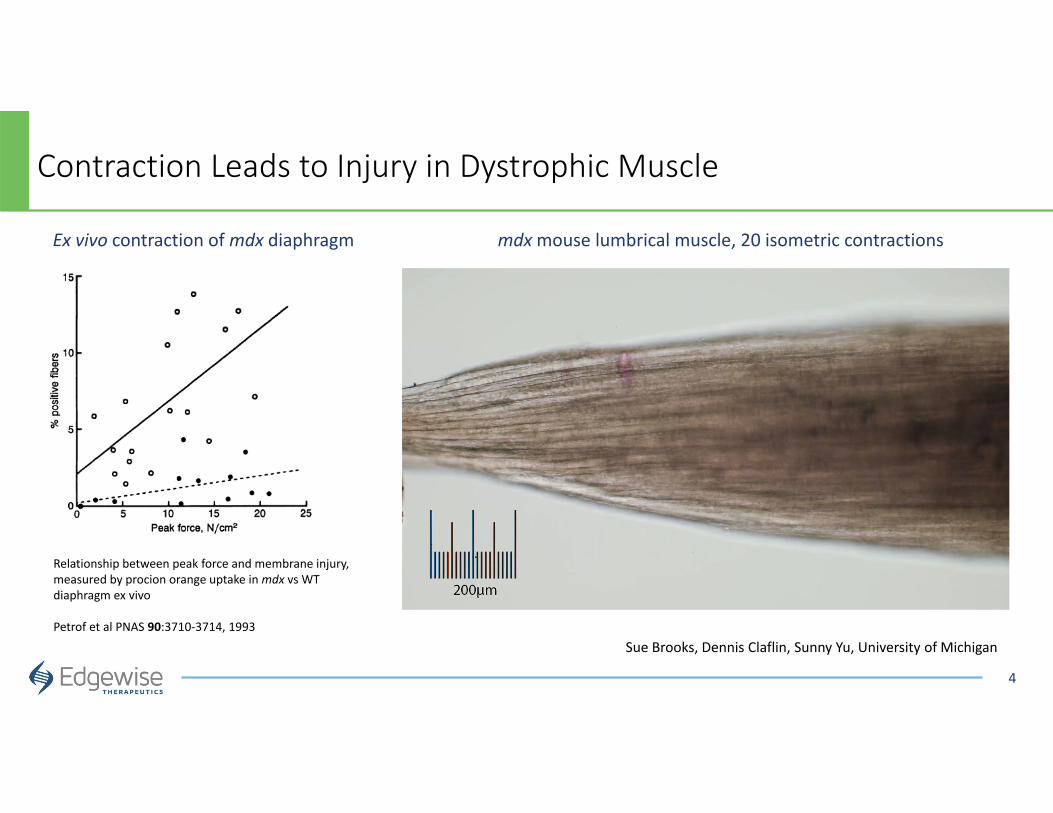
Contraction Leads to Injury in Dystrophic Muscle
4
mdxmouse lumbrical muscle, 20 isometric contractions
Sue Brooks, Dennis Claflin, Sunny Yu, University of Michigan
Relationship between peak force and membrane injury, measured by procion orange uptake in mdx vs WT diaphragm ex vivo
Petrof et al PNAS 90:3710‐3714, 1993
Ex vivo contraction of mdx diaphragm

Contraction of Dystrophic Muscle Leads to Membrane Stress, Calcium Entry, Hypercontraction, and Necrosis
Ca2+Ca2+
Ca2+
Ca2+
Ca2+
5Sue Brooks, Dennis Claflin, Sunny Yu, University of Michigan
Dystrophin connects the membrane to the contractile
machinery
With contraction, dystrophin anchors the
membrane
• No Dystrophin• Normal muscle
organization but no dystroglycan complex
• No Dystrophin• Contraction
causes un-controlled membrane stress and muscle damage

Skeletal Muscle is Comprised of Slow (Type I) and Fast (Type II) Fibers
Type I: Slow/Cardiac
Type II a: Fast Fatigue-Resistant
Type II x/d: Fast Fatigable
A Twitch
BB Unfused tetanic force
C Fatigability
Top row (A) shows tension developed during single twitches. The middle row (B) shows the tension developed during an unfused tetanus. Thebottom row (C) shows the degree to which each fiber type can sustain force during continuous stimulation.
Key: Electrical stimulus
50% 35% 15%
6
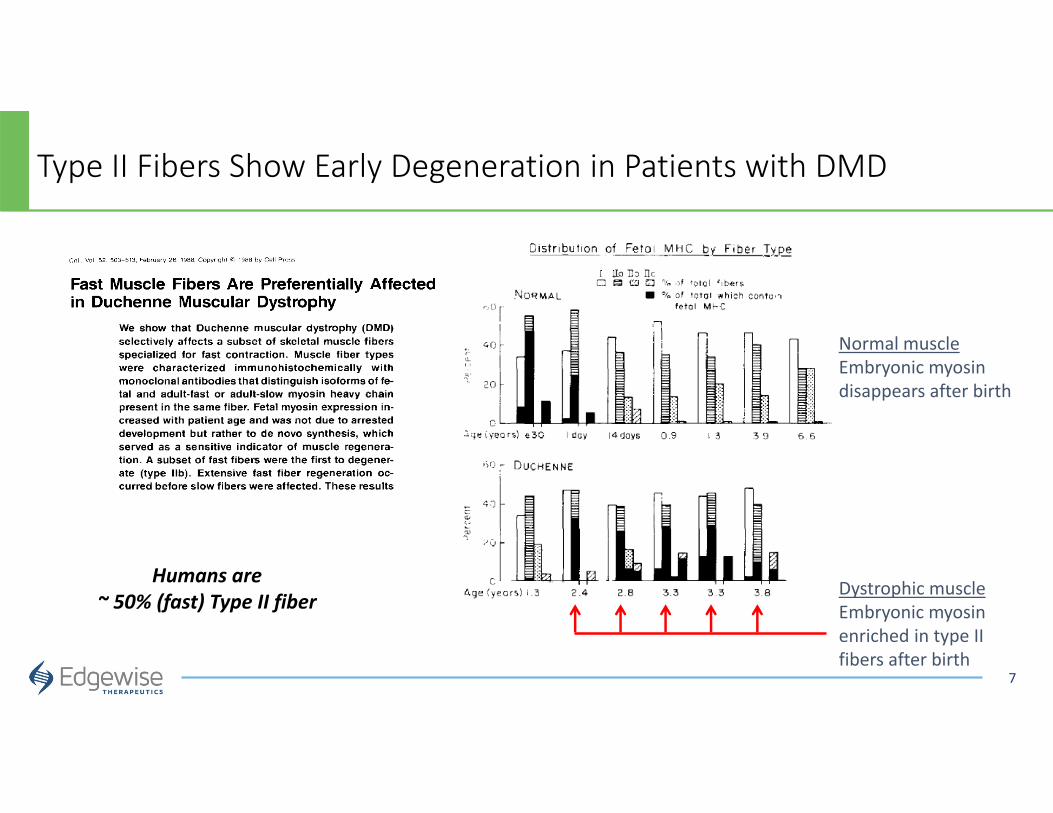
Type II Fibers Show Early Degeneration in Patients with DMD
7
Humans are ~ 50% (fast) Type II fiber
Dystrophic muscleEmbryonic myosin enriched in type II fibers after birth
Normal muscleEmbryonic myosin disappears after birth
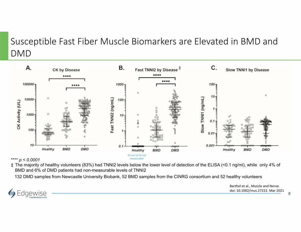
Susceptible Fast Fiber Muscle Biomarkers are Elevated in BMD and DMD
8
132 DMD samples from Newcastle University Biobank, 52 BMD samples from the CINRG consortium and 52 healthy volunteers
43 out of 52 not measurable**** p < 0.0001
‡ The majority of healthy volunteers (83%) had TNNI2 levels below the lower level of detection of the ELISA (<0.1 ng/ml), while only 4% of BMD and 6% of DMD patients had non-measurable levels of TNNI2
‡
Barthel et al., Muscle and Nerve. doi: 10.1002/mus.27222. Mar 2021

9
Therapeutic Hypothesis: Selectively limit contraction in susceptible fast muscle fibers
Reduce muscle contraction in susceptible type II (fast) muscle fibers to a level sufficient to prevent muscle breakdown and improve dystrophic muscle health
• Demonstrate that other types of striated muscle contraction (e.g. cardiac) are not impaired
• Demonstrate that protective levels of muscle inhibition do not impair strength or coordination
• Demonstrate that long‐term protection improves muscle health
X

10
The Target: Fast Skeletal Muscle Myosin
MyosinHydrolyzes ATP to bind actin and generate force
11
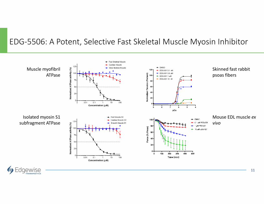
EDG‐5506: A Potent, Selective Fast Skeletal Muscle Myosin Inhibitor
456789
0
20
40
60
80
100
120
pCa
DMSOEDG-001 0.1 MEDG-001 0.6 MEDG-001 1 MEDG-001 10 M
Forc
e (%
Fm
ax)
Skinned fast rabbit psoas fibers
Mouse EDL muscle ex vivo
Muscle myofibril ATPase
Isolated myosin S1 subfragment ATPase

12
Low Levels of Inhibition are Required to Protect mdxMuscle ex vivo
DMD muscle (mdxmouse) 0.3 M EDG‐5506DMD muscle (mdxmouse) no treatment
One hour incubation reduces maximal force by 15%Contraction‐induced injuries completely preventedSuffers extensive contraction‐induced injuries
Claflin, Su and Brooks. U Michigan

13
Treatment is Associated with Reduced Calcium Entry

14
Protection with EDG‐5506 Also Present after Eccentric Contraction ex vivo
WT mdx mdx5 M EDG-5506
Ecce
ntric
con
trac
tion
peak
forc
e de
clin
e (p
erce
nt)
Deficit in WT muscle
Lengthening contraction of mouse EDL muscle ex vivo
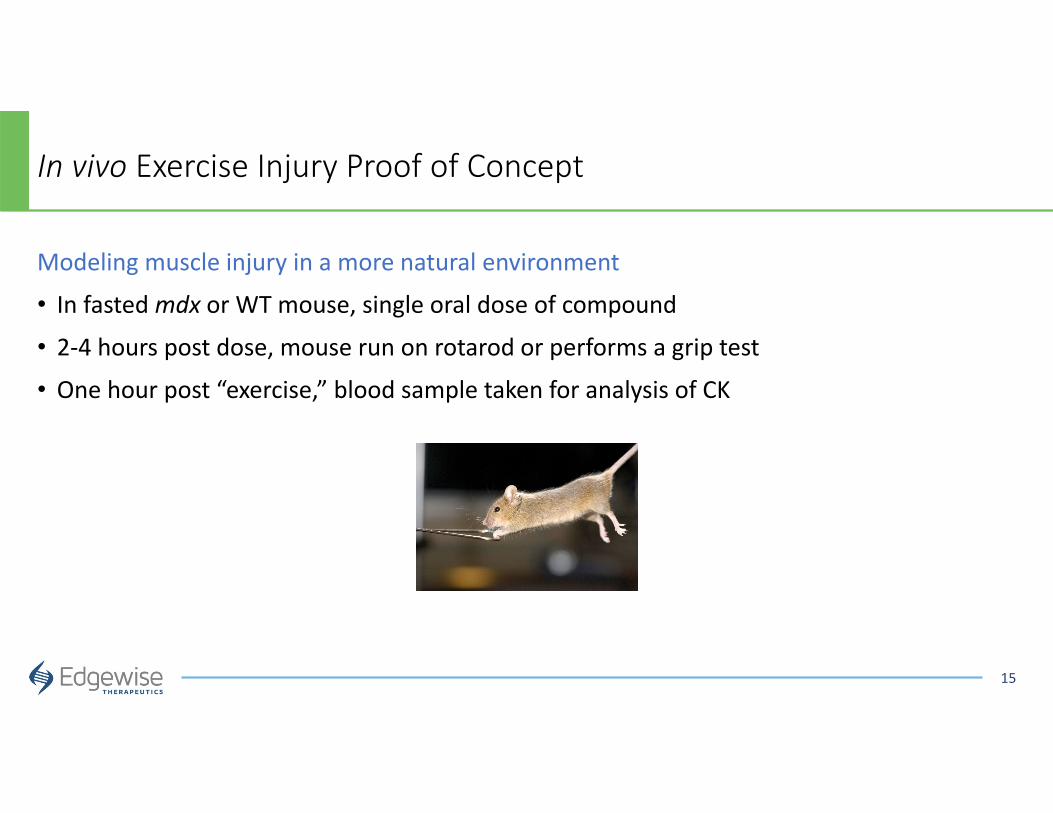
Modeling muscle injury in a more natural environment
• In fasted mdx or WT mouse, single oral dose of compound
• 2‐4 hours post dose, mouse run on rotarod or performs a grip test
• One hour post “exercise,” blood sample taken for analysis of CK
15
In vivo Exercise Injury Proof of Concept
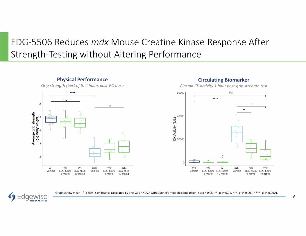
EDG‐5506 Reduces mdxMouse Creatine Kinase Response After Strength‐Testing without Altering Performance
16Graphs show mean +/‐ 1 SEM. Significance calculated by one‐way ANOVA with Dunnet's multiple comparison: ns: p > 0.05, **: p <= 0.01, ***: p <= 0.001, ****: p <= 0.0001.
Circulating BiomarkerPlasma CK activity 1 hour post‐grip strength test
ns
***
Physical PerformanceGrip strength (best of 5) 4 hours post‐PO dose

EDG‐5506 Protects Muscles from Membrane Disruption in mdxMice
17Graph shows mean +/‐ 1 SEM. Significance calculated by one‐way ANOVA with Dunnet's multiple comparison (****0.0001)
✱✱✱✱
✱✱✱✱
Ex vivo Dye Uptake by Contracting Muscle In vivo Dye Uptake by Leaky Muscle Fibers
Red = fluorescent procion orange. Taken up by leaky muscle fibersGreen = wheat germ agglutinin (outlines fibers)
1 M EDG-5506 5 M EDG-5506
Evans blue uptake in non‐exercised mdx mice after 3 weeks of treatment

18
Eight Weeks of Dosing with EDG‐5506 Reduces Diaphragm Fibrosis
WTmdxvehicle
mdxEDG‐5506 1 mg/kg
mdxEDG‐5506 3 mg/kg
Bars 200 m
WTmd
x Veh
icle
mdx E
DG-5506
1mg/kg
mdx E
DG-5506
3mg/kg
Picr
osiri
us P
ositi
ve A
rea
(%)
✱✱✱✱
✱
• 8 weeks dosing of mdxmice starting at 5 weeks• EDG‐5506 given at 1 or 3 mg/kg PO by gavage

EDG‐5506’s Impact on Cardiac Fibrosis is a Significant Finding Since Cardiac Myopathy is a Common Driver of Mortality in DMD and BMD
19
EDG‐5506 Led to a Reduction in the Incidence of Cardiac Hypertrophy*
15 Months Dosing of DBA/2J mdxMice Demonstrated Lower Cardiac Fibrosis Compared to Vehicle Controls*
12 months
15 months
Control EDG-5506
* Graph shows mean +/‐ SEM. Significance calculated by one‐way ANOVA with Dunnet's multiple comparison (*<0.05; **<0.01; ***<.001; ****<0.0001)
20
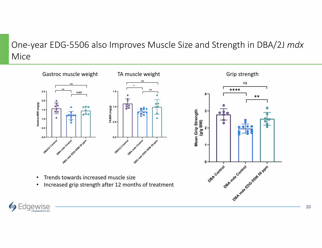
One‐year EDG‐5506 also Improves Muscle Size and Strength in DBA/2J mdxMice
DBA Control
DBA mdx C
ontrol
DBA mdx E
DG-5506
50 ppm
0
1
2
3
4
Mea
n G
rip S
treng
th(g
/g B
W)
ns✱✱✱✱
✱✱
DBA/2J Contro
lDBA m
dx Contro
l
DBA mdx E
DG-5506
50 ppm
0.0
0.5
1.0
1.5
TA/B
W (m
g/g)
ns
**ns
DBA/2J Contro
lDBA m
dx Contro
l
DBA mdx E
DG-5506
50 ppm
0.0
0.5
1.0
1.5
2.0
2.5
Gas
troc/
BW (m
g/g)
ns
**0.067
• Trends towards increased muscle size• Increased grip strength after 12 months of treatment
Gastroc muscle weight TA muscle weight Grip strength
21
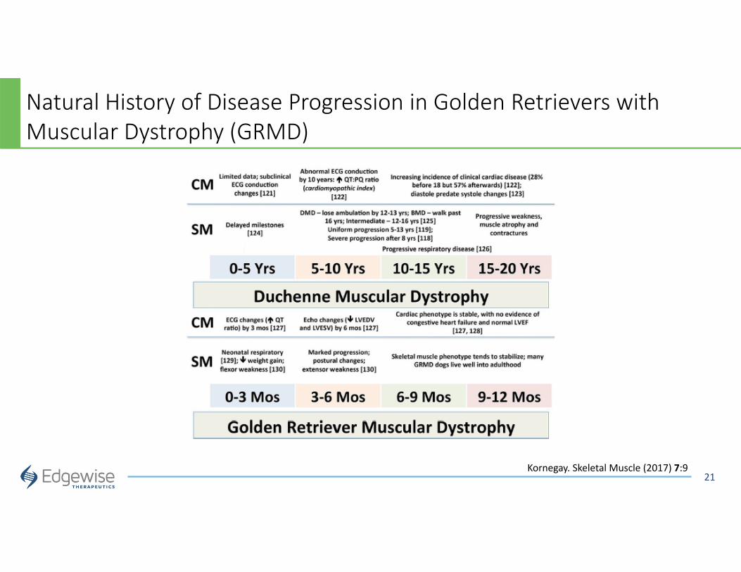
Natural History of Disease Progression in Golden Retrievers with Muscular Dystrophy (GRMD)
Kornegay. Skeletal Muscle (2017) 7:9

EDG‐5506 Decreases Creatine Kinase and Increases Habitual Activity in a GRMD
22
Circulating Biomarker Throughout StudyPlasma CK across ALL GRMD dogs at 7 months*
Daily Average ActivityGRMD dogs at 18 months*
Vehicl
e Bas
eline
EDG-5506
Dosing
Washout
Vehicl
e Rep
eat
0
5000
10000
15000
20000
25000
ns
✱
✱✱✱✱
✱✱✱
✱✱✱✱
ns
Pre-Dose
Baseli
neVeh
icle B
aseli
neEDG-55
06 Dosin
g
Washout
Daily
Ave
rage
Act
ivity
(Fitb
ark
poin
ts)
✱✱✱✱
✱✱✱
Pete Nghiem, Alexis Rutledge. Texas A&M

EDG‐5506 Treatment Reverses Proteome Signatures Associated with the Dystrophic State
23
1 Hathout Y, et. al., Sci Rep, 2019
Significance calculated by one‐way ANOVA with Tukey’s multiple comparison correction. * p<0.05, **p<0.01, ***p<0.001, ****p<0.0001
* Proteins selected by overlap between GRMD and published DMD signature biomarkers1: 40 increased and 9 decreased.
* *
EDG-5506 Treatment Post-Treatment EDG-5506 Treatment Post-Treatment
GRMD Increased* GRMD Decreased*
1.3K SOMAscan® plasma analysis

Clinical Progression with EDG‐5506
Single ascending doses in healthy participants:
Completed
Multiple ascending doses (14 days) in
healthy participants: Ongoing
Multiple doses (14 days) in participants
with BMD at levels well tolerated in healthy
participants
N= ~ 130 N= ~ 16
Study Objectives • To assess safety of single and multiple oral doses of EDG‐5506 in healthy participants and in participants with
BMD• To assess pharmacokinetics of single and multiple oral doses of EDG‐5506 in healthy participants and in
participants with BMD• To assess blood biomarkers in participants with BMD
Up to 130 participants Up to 12 participants
*Review *Review
24
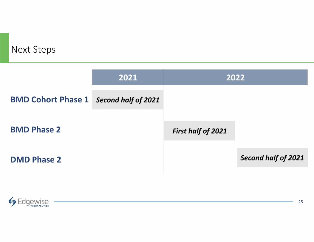
2021 2022
BMD Cohort Phase 1
BMD Phase 2
DMD Phase 2
First half of 2021
Next Steps
Second half of 2021
25
Second half of 2021
