selective decrease of components of the creatine kinase
TRANSCRIPT

Selective Decrease of Components of the Creatine KinaseSystem and ATP Synthase Complex in Chronic ChagasDisease CardiomyopathyPriscila Camillo Teixeira1,2,3, Ronaldo Honorato Barros Santos4, Alfredo Inacio Fiorelli4, Angelina
Morand Bianchi Bilate1, Luiz Alberto Benvenuti5, Noedir Antonio Stolf4, Jorge Kalil1,2,3, Edecio Cunha-
Neto1,2,3*
1 Laboratory of Immunology, Heart Institute, School of Medicine, University of Sao Paulo, Sao Paulo, Brazil, 2 Division of Clinical Immunology and Allergy, School of
Medicine, University of Sao Paulo, Sao Paulo, Brazil, 3 Institute for Investigation in Immunology (iii), INCT, Sao Paulo, Brazil, 4 Division of Surgery, Heart Institute, School of
Medicine, University of Sao Paulo, Sao Paulo, Brazil, 5 Division of Pathology, Heart Institute, School of Medicine, University of Sao Paulo, Sao Paulo, Brazil
Abstract
Background: Chronic Chagas disease cardiomyopathy (CCC) is an inflammatory dilated cardiomyopathy with a worseprognosis than other cardiomyopathies. CCC occurs in 30 % of individuals infected with Trypanosoma cruzi, endemic inLatin America. Heart failure is associated with impaired energy metabolism, which may be correlated to contractiledysfunction. We thus analyzed the myocardial gene and protein expression, as well as activity, of key mitochondrialenzymes related to ATP production, in myocardial samples of end-stage CCC, idiopathic dilated (IDC) and ischemic (IC)cardiomyopathies.
Methodology/Principal Findings: Myocardium homogenates from CCC (N = 5), IC (N = 5) and IDC (N = 5) patients, as well asfrom heart donors (N = 5) were analyzed for protein and mRNA expression of mitochondrial creatine kinase (CKMit) andmuscular creatine kinase (CKM) and ATP synthase subunits aplha and beta by immunoblotting and by real-time RT-PCR.Total myocardial CK activity was also assessed. Protein levels of CKM and CK activity were reduced in all threecardiomyopathy groups. However, total CK activity, as well as ATP synthase alpha chain protein levels, were significantlylower in CCC samples than IC and IDC samples. CCC myocardium displayed selective reduction of protein levels and activityof enzymes crucial for maintaining cytoplasmic ATP levels.
Conclusions/Significance: The selective impairment of the CK system may be associated to the loss of inotropic reserveobserved in CCC. Reduction of ATP synthase alpha levels is consistent with a decrease in myocardial ATP generationthrough oxidative phosphorylation. Together, these results suggest that the energetic deficit is more intense in themyocardium of CCC patients than in the other tested dilated cardiomyopathies.
Citation: Teixeira PC, Santos RHB, Fiorelli AI, Bilate AMB, Benvenuti LA, et al. (2011) Selective Decrease of Components of the Creatine Kinase System and ATPSynthase Complex in Chronic Chagas Disease Cardiomyopathy. PLoS Negl Trop Dis 5(6): e1205. doi:10.1371/journal.pntd.0001205
Editor: Rodrigo Correa-Oliveira, Rene Rachou Research Center, Brazil
Received November 11, 2010; Accepted May 1, 2011; Published June 28, 2011
Copyright: � 2011 Teixeira et al. This is an open-access article distributed under the terms of the Creative Commons Attribution License, which permitsunrestricted use, distribution, and reproduction in any medium, provided the original author and source are credited.
Funding: This research was supported by the Brazilian Council for Scientific and Technological Development - CNPq and the Sao Paulo State Research FundingAgency - FAPESP. PCT is recipient of a Sao Paulo State Research Funding Agency - FAPESP fellowship. ECN and JK are recipients of Brazilian Council for Scientificand Technological Development - CNPq productivity awards. The funders had no role in study design, data collection and analysis, decision to publish, orpreparation of the manuscript.
Competing Interests: The authors have declared that no competing interests exist.
* E-mail: [email protected]
Introduction
Chagas disease is a significant cause of morbidity and mortality
in Central and South America, affecting about 13 million people
[1]. The disease is caused by infection with the intracellular
protozoan parasite Trypanosoma cruzi. About 30 % of infected
patients develop chronic Chagas disease cardiomyopathy (CCC),
an inflammatory cardiomyopathy that occurs decades after the
initial infection. One-third of CCC patients further progress to a
particularly aggressive, life-threatening dilated cardiomyopathy;
CCC is a major indication of heart failure in Latin America [2,3].
Clinical progression, length of survival and overall prognosis are
significantly worse in CCC patients when compared to patients
with dilated cardiomyopathy of non-inflammatory etiology, like
idiopathic dilated or ischemic cardiomyopathies (IDC or IC,
respectively) [4–7]. Due to migration from endemic countries, an
estimated 300,000 people with Chagas disease are living in the
USA, where a significant number of cases of CCC are expected
per year [8]. The pathogenesis of CCC is unclear, and multiple
mechanisms have been proposed (Reviewed in [9]). The most
characteristic histopathological lesions in cardiac patients with
CCC are consistent with inflammation and a myocardial
remodeling process: T cell/macrophage-rich myocarditis, hyper-
trophy, and fibrosis with cardiomyocyte damage [10,11]. The
local cytokine production profile shows a T1-type response, with
interferon-gamma-induced chemokines [12–15]. As the currently
licensed anti-T. cruzi drugs may not be effective in preventing the
progression of heart lesions of CCC [16], treatment is only
www.plosntds.org 1 June 2011 | Volume 5 | Issue 6 | e1205
CORE Metadata, citation and similar papers at core.ac.uk
Provided by PubMed Central

supportive. In patients with refractory heart failure, the only
available treatment is heart transplantation [17]. The absence of
alternative treatment for CCC is a consequence of limited
knowledge about the pathogenesis.
Energy metabolism imbalances have been reported in dilated
cardiomyopathies and heart failure [18]. Since the heart consumes
more energy than any other organ, impairments in energy
production could lead to a mechanical failure of the heart, and
disturbances in electrical conduction [18]. Mitochondrial oxidative
phosphorylation is essential for the production of energy for
cardiac function. This system comprises the oxidative phosphor-
ylation complex, which includes the electron transport chain
(complexes I to IV) and the F1FO ATP synthase (complex V). In
aerobic tissues, most ATP is synthesized via the mitochondrial
F1FO ATP synthase complex. Studies have shown that certain
components of oxidative phosphorylation may be impaired in
heart failure [18]. Patients with IDC or IC show a reduced
myocardial activity of complex III when compared to controls
[19]. In IDC patients, a decreased activity of myocardial
cytochrome c oxidase (complex IV) was observed [20]. With the
progression of dilated cardiomyopathy, higher levels of spatial and
functional heterogeneity within mitochondrial populations are
observed, indicative of mitochondrial damage [21]. It has been
reported that mitochondrial damage leads to loss of mitochondrial
function, impairing energy production and cell physiology, and to
the enhancement of pathologic function, producing oxidative-,
calcium-, apoptosis-mediated myocyte injury [22].
Creatine kinases (CK) are also key enzymes of energy metabolism,
which connect mitochondrial ATP-producing and cytosolic ATP-
consuming process, and are thus of central importance to the cellular
energy homeostasis [23]. This system acts as an energy buffer, in
which mitochondrial creatine kinase (CKMit) catalyzes the transfer
of high energy phosphate bond from ATP to creatine to form
phosphocreatine and ADP. Phosphocreatine, a molecule smaller
than ATP, diffuses rapidly from the mitochondria to the myofibrils,
where myofibrillar creatine kinase (MM, MB, BB dimers, formed by
CKM, the muscular isoform, and CKB, the brain isoform) catalyzes
ATP production from phosphocreatine, generating free creatine,
which diffuses back into mitochondria [18,23]. Impaired ATP
transfer and utilization may limit contractile function by means of a
decrease in the average cytoplasmic ATP concentration [18]. Most
of the components of the CK system are down-regulated in heart
failure, with levels of creatine, phosphocreatine, CKMit and CKM
all significantly reduced in animal models and in humans [24,25].
CK deficiency in isolated hearts may cause a decline of over 70 % in
ATP delivery to myofibrils, leading to a blunted contractile reserve
[26].
Proteomic profiling of myocardium from CCC patients revealed
that 27 % of identified proteins belong to energy metabolism
pathways [27]. Using gene expression profiling, our group found
differential expression of a significant number of genes involved in
oxidative phosphorylation and lipid catabolism in myocardial
samples from CCC patients, but not in samples from patients with
dilated cardiomyopathy, when compared to samples from subjects
without cardiomyopathy [15]. Genetic profiling studies showed
that hearts of T. cruzi-infected mice have shown a decreased
expression of oxidative phosphorylation enzymes [28]. Likewise,
biochemical and histochemical analysis revealed a reduced activity
of the respiratory chain complexes in hearts of T. cruzi-infected
mice [29]. Proteomic analysis of myocardial samples from acutely
T. cruzi-infected Syrian hamsters showed an up-regulation of the
energy metabolism proteins glutamate oxaloacetate transaminase
1 and pyruvate dehydrogenase b, that may be associated with a
high ATP demand after T. cruzi infection [30].
Inflammatory cytokines, which are present in the CCC myocar-
dium and induce local signaling [12–15], have been reported to alter
the energy metabolism. IFN-gamma was shown to inhibit the
mitochondrial oxidative metabolism [31] and increase the rate of
cardiac ATP depletion in cardiomyocytes [32]. Additionally, studies
with cultured human skeletal muscle cells demonstrated that IFN-
gamma treatment could inhibit CK activity [33].
Thus, the evidence is consistent with the hypothesis that the
myocardium of patients with CCC could present an impaired
energy metabolism. In order to test this hypothesis, we compared
the protein and mRNA expression of CKM, CKMit, and the
alpha and beta subunits of the catalytic F1 domain of ATP
synthase complex (ATPa and ATPb, respectively) in myocardial
samples from CCC, with that of dilated cardiomyopathies of other
etiologies, and healthy hearts from organ donors. We also
measured total creatine kinase enzymatic activity in the same
sample groups.
Methods
Samples of human myocardiumMyocardial samples were obtained from left ventricular-free
wall heart tissue from end-stage heart failure patients at the
moment of heart transplantation. Samples from 5 CCC (at least 2
positive results in 3 independent anti-T. cruzi serology tests –
ELISA immunoassay, indirect immunofluorescence assay and
indirect hemagglutination test), 5 IDC (dilated cardiomyopathy in
the absence of ischemic disease, negative serology for Chagas
disease) and 5 coronary angiography-proven IC patients were
collected (Table 1). Left ventricular free wall samples were also
obtained from healthy hearts of organ donors, which were not
used for transplantation for technical reasons. The protocol was
approved by the Institutional Review Board of the University of
Sao Paulo School of Medicine and written informed consent was
obtained from the patients. Samples were cleared from pericar-
dium and fat, quickly frozen in liquid nitrogen and stored at
270uC. Protein homogenates were obtained using lysing solution
(1:10 w/v) containing 7 mol/L urea, 10 mmol/L Tris, 5 mmol/L
magnesium acetate and 4 % CHAPS, pH 8.0, by mechanical
homogenization (PowerGen, Fisher Scientific). For experiments
measuring the creatine kinase enzyme activity, 20 mg of tissue was
Author Summary
Chronic Chagas disease cardiomyopathy (CCC) affectsmillions in endemic areas and is presenting in growingnumbers in the USA and European countries due tomigration currents. Clinical progression, length of survivaland overall prognosis are significantly worse in CCCpatients when compared to patients with dilated cardio-myopathy of non-inflammatory etiology. Impairment ofenergy metabolism seems to play a role in heart failuredue to cardiomyopathies. Herein, we have analyzedenergy metabolism enzymes in myocardium samples ofCCC patients comparing to other non-inflammatorycardiomyopathies. We found that myocardial tissue fromCCC patients displays a significant reduction of bothmyocardial protein levels of ATP synthase alpha andcreatine kinase enzyme activity, in comparison to controlheart samples, as well as idiopathic dilated cardiomyop-athy and ischemic cardiomyopathy. Our results suggestthat CCC myocardium displays a selective energetic deficit,which may play a role in the reduced heart functionobserved in such patients.
Energy Metabolism Enzymes in CCC Myocardium
www.plosntds.org 2 June 2011 | Volume 5 | Issue 6 | e1205
lysed in solution (1:20 w/v) containing 0.32 mol/L sucrose,
10 mmol/L HEPES and 1 mmol/L EDTA, pH 7.4, by mechan-
ical homogenization. The homogenate was then sonicated for
three cycles of 10 s each to 10 Watts (60 Dismembrator Sonic,
Fisher Scientific), centrifuged at 12,000 g for 30 min. Superna-
tants were collected and stored at 270uC. Protein quantification
was performed with the Bradford method (BioRad).
Myocardial histopathologyThe samples from myocardial tissue (left ventricular free wall)
were fixed in buffered formalin solution (pH 7.2), embedded in
paraffin, and cut into 5 mm sections. Sections were stained with
hematoxylin-eosin (H&E) and picrosirius red.
Analysis of protein expression by ImmunoblottingExtracts of myocardial samples containing 30 mg of protein were
heated for 5 min at 95uC, and subjected to one-dimensional
electrophoresis (SDS-PAGE) using 12.5 % polyacrylamide gel and
the vertical electrophoresis system Ruby SE600 (GE Healthcare).
After electrophoresis, proteins were transferred from gel to a
nitrocellulose membrane using the TE Semi-Dry Transfer Unit (GE
Healthcare). The nitrocellulose membranes were incubated with
monoclonal antibodies to proteins involved in energy metabolism:
anti- F1FO ATP synthase alpha (ATPa) and anti- F1FO ATP
synthase beta (ATPb) (Molecular Probes), anti-mitochondrial
creatine kinase (CKMit) and anti- muscle creatine kinase (CKM)
(Santa Cruz) and polyclonal anti- glyceraldehyde 3-phosphate
dehydrogenase (GAPDH) (R & D Systems). Each membrane was
subjected to incubation with compatible secondary antibodies
conjugated with peroxidase, developed using ECL Plus Western
Blotting Detection Reagents (GE Healthcare) and detection using
X-ray equipment. Analysis of densitometry was performed using the
program ImageQuant TL (GE Healthcare).
Analysis of mRNA expression by real-time reversetranscriptase (RT)-PCR
Total RNA from left ventricle samples was isolated using the
RNeasy Fibrous tissue kit (Quiagen). Contaminating DNA was
removed by treatment with RNase-free DNase I. cDNA was
obtained from 5 mg total RNA using Super-script IITM reverse
transcriptase (Invitrogen). mRNA expression was analyzed by real-
time quantitative reverse transcriptase (RT)-PCR with SYBR
Green I PCR Master Mix (Applied Biosystems) and 250 nM of
sense and anti-sense primers using the ABI Prism 7500 Real Time
PCR System (Applied Biosystems). The following primers were
designed using Primer Express software version 3.0 (Applied
Biosystems): GAPDH (M33197): (F) 59-TGGTCTCCTCTGA-
CTTCAACA-39, (R) 59-AGCCAAATTCGTTGTCATACC-39;
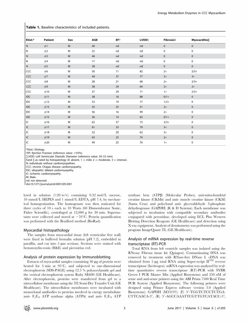
Table 1. Baseline characteristics of included patients.
Etiol.* Patient Sex AGE EF{ LVDD{ Fibrosis1 Myocarditis||
N #1 M 40 nd nd 0 0
N #2 M 22 nd nd 0 0
N #3 M 46 nd nd 0 0
N #4 M 17 nd nd 0 0
N #5 M 28 nd nd 0 0
CCC #6 M 50 11 82 2+ 2/3+
CCC #7 M 49 37 77 3+ 3+
CCC #8 M 28 21 68 2+ 2/3+
CCC #9 M 58 29 64 2+ 2+
CCC #10 M 57 29 71 1+ 2/3+
IDC #11 M 38 16 88 0/1+ 0
IDC #12 M 53 19 77 1/2+ 0
IDC #13 M 55 25 51 3+ 0
IDC #14 M 56 16 99 2+ 0
IDC #15 M 36 14 62 0/1+ 0
IC #16 M 62 37 75 2/3+ 0
IC #17 M 61 33 79 3+ 0
IC #18 M 52 20 62 3+ 0
IC #19 M 63 25 74 2+ 0
IC #20 M 49 25 76 1+ 0
*Etiol.: Etiology.{EF: Ejection Fraction (reference value: $55%).{LVDD: Left Ventricular Diastolic Diameter (reference value: 39–53 mm).1and || as rated by histopatology (0: absent, 1 +: mild, 2 +: moderate, 3 +: intense).N: individuals without cardiomyopathies.CCC: chronic Chagas disease cardiomyopathy.IDC: idiopathic dilated cardiomyopathy.IC: ischemic cardiomyopathy.M: Male.nd: not detected.doi:10.1371/journal.pntd.0001205.t001
Energy Metabolism Enzymes in CCC Myocardium
www.plosntds.org 3 June 2011 | Volume 5 | Issue 6 | e1205

ATPa (NM_001001937): (F) 59-TCTTCAAAAGACTGGGAC-
TGCTGA-39, (R) 59-AAGACACGCCCAGTTTCTTCAAG-39;
ATPb (NM_001686): (F) 59-GCCCAGCATTTGGGTGAGA-39,
(R) 59-GATTGGTGCACCAGAATCCAGT-39; CKM (NM_0-
01824): (F) 59-GCTCTCTGTGGAAGCTCTCAACA-39, (R) 59-
GATGAGCTGCTGCTGCTCCT-39; CKMit (NM_001825): (F)
59-TGACGAGGAGTCCTATGAGGTGTT-39, (R) 59-AGATC-
CGTTGTGTGCTTCATCAC-39. After every PCR, an ampli-
con melting point curve was obtained. This yielded a single peak
with the expected temperature provided by Primer Express
software, confirming the specificity of the PCR. GAPDH mRNA
expression was used for normalization. The amount of mRNA
in the left ventricles samples was calculated using the 2-DCt
method [34].
Measurement of the creatine kinase enzymatic activityThe enzymatic activity measurements of CK in the myocardial
samples were performed using the CK-NAC kit (Doles). Basically,
this method is a kinetic system where CK catalyzes the
transphosphorylation reaction of ADP to ATP. A series of coupled
enzymatic reactions produce NADH in a concentration directly
proportional to the enzymatic activity of CK in the sample. The
analyses were performed using a UV/Vis U-2001 spectropho-
tometer (Hitachi), monitoring the increase in absorbance of
NADH per minute at the wavelength of 340 nm at 37uC, using
a thermostatic bath (MultiTemp III, GE Healthcare). The
measurement of enzymatic activity is given in international units
(U); one unit of CK is the amount of enzyme that oxidizes 1 mmol/
L of NADH per minute. Values were normalized by the amount of
protein present in the sample.
Statistical analysisAll statistical analyses were performed with GraphPad Prism 4.0
software (GraphPad Software). Descriptive statistics are given as
average and standard deviation. The non-parametric Newman-
Keuls test was used for comparison between the groups. P-values
,0.05 were considered as statistically significant.
Results
While myocardial sections from all 3 cardiomyopathy groups
displayed cardiomyocyte hypertrophy and fibrosis upon histo-
patholological analysis, myocarditis associated with a predominant
lymphocytic infiltration was only observed among CCC heart
lesions (Figure 1, Table 1). No significant differences were found in
age, ejection fraction (EF) or left ventricular diastolic diameter
(LVDD) among the three cardiomyopathy groups.
Figures 2 A and B show the differential protein expression of the
ATP-synthase, subunits alpha (ATPa) and beta (ATPb), respec-
tively. Representative immunoblots are depicted in Figure S1. The
ATPa was 18 % less expressed in CCC myocardium when
compared to myocardial samples from individuals without
cardiomyopathies (p,0.01). In contrast, IC myocardium showed
an increase of 25 % (p,0.001) in ATPa when compared to
control samples, while in IDC there was no significant reduction of
ATPa levels (5 %, p = ns) in comparison to the control group. In
the comparison between cardiomyopathy groups, ATPa levels in
CCC were 34 % lower than in IC myocardium (p,0.01); and 13
% lower than those found in IDC myocardium (p,0.05). There
was no significant decrease of ATPb in CCC when compared to
control samples (9 %, p = ns). However, we observed increased
expression of ATPb in IC and IDC myocardium when compared
to the control group [32 % (p,0.001) and 10 % (p,0.05),
respectively]. In the comparison between cardiomyopathy groups,
we found that CCC myocardium samples express significantly less
ATPb than IC or IDC myocardium [31 % (p,0.001) and 17 %
(p,0.01), respectively].
We have also detected significant protein expression differences
among enzymes of the creatine kinase system. The expression of
CKM (Figure 2C) was decreased in samples from patients with
CCC (33 %, p,0.01), IDC (23 %, p,0.05) and IC (24 %,
p,0.05) when compared to the control group. Of note, in the
comparison between cardiomyopathy groups, the average CKM
expression was most decreased among CCC patients (13 % and 12
% reduction when compared to IDC and IC, respectively),
although the difference was not statistically significant. The protein
expression of CKMit (Figure 2D) was decreased in samples from
patients with CCC and IDC, when compared to the control group
(16 % and 4 %, respectively), but the differences failed to achieve
statistical significance. In contrast, samples from patients with IC
showed an increased expression of CKMit when compared to the
control group (13 %; p = ns). In the comparison between
cardiomyopathy groups, the protein levels of CKMit were
decreased in samples from patients with CCC when compared
to samples from patients with IC and IDC [26 % (p,0.01) and 13
% (p = ns), respectively].
We also analyzed the mRNA expression of the enzymes tested
above, ATPa, ATPb, CKM and CKMit. Figure 3 shows the
values of relative quantification of mRNA expression of these
enzymes. We found that mRNA expression of CKM was
decreased in samples from patients with CCC and IDC when
compared to samples from subjects without cardiomyopathy [78
% (p,0.05) and 69 % (p,0.05)]. Also, the mRNA expression of
CKMit was reduced in CCC samples (75 %, p,0.05) when
compared to samples from subjects without cardiomyopathy. The
average expression of mRNAs for all 4 enzymes was reduced in
the 3 cardiomyopathies when compared to control samples, and
samples from CCC patients showed the lowest expression levels.
However, due to high interindividual variation of expression
within each group, most of the comparisons were not statistically
significant. Interestingly, mRNA levels of ATPa and ATPb were
not increased in samples from patients with IC in comparison to
samples from control group, as seen in the analysis of protein
expression. In the comparison between cardiomyopathy groups,
none of the enzymes analyzed showed significant differences in
mRNA expression for ATPa and ATPb.
In order to evaluate whether the differential protein expression
of enzymes of the creatine kinase system observed above had an
impact on myocardial enzyme activity, we compared total creatine
kinase activity among groups (Figure 4). The creatine kinase
enzyme activity was reduced in samples from patients with CCC
(59 %, p,0.01), IDC (35 %, p,0.01) and IC (31 %, p,0.01)
when compared to samples from the control group (Figure 4). Of
interest, creatine kinase enzyme activity was significantly reduced
in myocardial samples from patients with CCC, when compared
to IDC and IC patients [37 % (p,0.05) and 41 % (p,0.05),
respectively].
Discussion
In this paper, we observed a reduced expression of CKM, a key
enzyme in myocardial energetic metabolism, in several dilated
cardiomyopathies. Most importantly, we found that CCC
myocardium shows significantly reduced levels of protein expres-
sion of ATP synthase alpha subunit and total creatine kinase
enzyme activity, when compared to IDC or IC.
We observed that the myocardial creatine kinase system shows
impaired function in patients with all forms of cardiomyopathy.
Energy Metabolism Enzymes in CCC Myocardium
www.plosntds.org 4 June 2011 | Volume 5 | Issue 6 | e1205

Energy Metabolism Enzymes in CCC Myocardium
www.plosntds.org 5 June 2011 | Volume 5 | Issue 6 | e1205

The reduced myocardial protein expression of CKM, observed in
all cardiomyopathy groups, was reflected in the reduced total
creatine kinase activity. This has been previously described for
IDC and IC [35]. The reduced protein expression of CKM was
probably due to transcriptional regulation at least in the cases of
CCC and IDC, since CKM mRNA expression was also
significantly reduced in samples from patients of such groups,
when compared to samples from individuals without cardiomy-
opathy. Animals genetically deficient in CKM develop myocardial
hypertrophy and left ventricular dilation [36], as well as higher
susceptibility to mitochondrial damage and cardiac disturbances in
calcium homeostasis after ischemia and reperfusion [37]. Myo-
cardial ATP flux through the CK system was shown to be reduced
by 50 % in patients with heart failure [38]. Since the CK reaction
is the prime source of the myocardial energy reserve, the deficit in
ATP flux through CK may contribute to the pathogenesis of heart
failure [39]. The finding that average CKM and CKMit levels
from CCC samples were the lowest among all groups, and that
total CK activity in CCC samples was significantly lower than that
of the other cardiomyopathy groups, indicates that CCC patients
may show a stronger functional impairment in the CK system than
other etiologies of dilated cardiomyopathy. It is likely that the
significantly reduced myocardial expression of CKMit, observed in
comparison to IC, - and to a lesser degree also in comparison to
control group - may have contributed to the reduction in the total
CK activity observed in CCC samples. Regarding the discrepancy
between the significantly reduced CKMit mRNA levels and the
less prominent reduction of CKMit protein levels observed in
CCC, it could be due to an increased stability of this protein. The
loss of CK activity in isolated hearts has been reported to cause a
decline in ATP delivery to myofibrils, leading to a blunted
contractile reserve [26]. Reduced energy reserve via creatine
kinase, as indicated by reduced phosphocreatine/ATP ratios,
limits cardiac performance during metabolic stress conditions [40].
Figure 2. Analysis of differential protein expression of energy metabolism enzymes by immunoblotting. (A) ATPa: ATP synthase alphasubunit, (B) ATPb: ATP synthase beta subunit, (C) CKM: creatine kinase M and (D) CKMit: mitochondrial creatine kinase. The densitometric values ofeach protein for each sample were normalized by the values of GAPDH. The horizontal lines show statistically significant changes: *p,0.05; **p,0.01;***p,0.001.doi:10.1371/journal.pntd.0001205.g002
Figure 1. Histopathological features of myocardial samples. Slides of hematoxilin-eosin- (left column) and picrosirius red- (right column)stained myocardial sections of representative patients with CCC, IDC and IC and individuals without cardiomyopathies (N). Myocardial hypertrophycharacterized by fiber and nuclear enlargement is evident in the CCC, IDC and IC groups. Lymphocytic myocarditis is present only in the CCC group.Interstitial fibrosis stained red in picrosirius stain is present in the CCC, IDC and IC groups.doi:10.1371/journal.pntd.0001205.g001
Energy Metabolism Enzymes in CCC Myocardium
www.plosntds.org 6 June 2011 | Volume 5 | Issue 6 | e1205

Significantly, CCC patients have been reported to display an
impaired myocardial contractile response to dobutamine [41]. It is
thus possible that this reduced contractile reserve is a consequence
of the significant derangement in CK activity reported here in
CCC myocardium.
A correlation has been reported between decreased total CK
activity and LV dysfunction [42]. However, samples from CCC
patients studied here showed lower total CK activity than those of
IDC or IC patients, despite the fact that LV dysfunction status was
similar in CCC, IC and IDC patients. This may indicate that there
are disease-specific factors that induce a stronger reduction in CK
activity in CCC, when compared to non-inflammatory cardiomy-
opathies. While the creatine kinase system may buffer transient
changes in ATP levels, the rate of oxidative ATP synthesis must be
closely matched to the rate of consumption. Most myocardial ATP
is generated through the mitochondrial oxidative phosphorylation
(complex I-V). In our study, we also found changes in the protein
expression of ATPa and ATPb (belonging to the F1 subunit of
ATP synthase complex - complex V). The finding that ATPa was
only reduced in CCC myocardium, but not in IC and IDC,
indicates that these patients could be at a greater impairment in
cardiac ATP supply, as suggested by studies in animal models of
heart failure [43]. However, in our study, patients with IC showed
elevated protein levels of ATPa and ATPb, and patients with
IDC showed elevated levels of ATPb when compared to the
Figure 3. mRNA expression of energy metabolism enzymes by real time RT-PCR. (A) ATPa: ATP synthase alpha subunit, (B) ATPb: ATPsynthase beta subunit, (C) CKM: creatine kinase M and (D) CKMit: mitochondrial creatine kinase. The horizontal lines show statistically significantchanges: *p,0.05.doi:10.1371/journal.pntd.0001205.g003
Figura 4. Analysis of creatine kinase enzymatic activity. Thevalues were normalized by the amount of protein from each sample.The horizontal lines show statistically significant changes: *p,0.05;**p,0.01.doi:10.1371/journal.pntd.0001205.g004
Energy Metabolism Enzymes in CCC Myocardium
www.plosntds.org 7 June 2011 | Volume 5 | Issue 6 | e1205

myocardium of subjects without cardiomyopathy. Significantly, a
study showed an increase in mRNA of oxidative phosphorylation
components in chronic ischemia due to severe atherosclerosis [44].
In vivo measurements in normal hearts subjected to adrenergic
stress have shown that ATP production by oxidative phosphor-
ylation increases with the demand, while ATP production by the
CK system remains unchanged [39]. Authors thus suggest that the
ratio of ATP production by the CK system to oxidative
phosphorylation decreases upon demand; in addition, the ratio
may be even lower in resting hearts from heart failure patients, due
to a decreased CK flux [39]. Since a reduced CK flux may be one
of the most prominent metabolic abnormalities in heart failure
[39], the findings of selectively reduced CK activity - and perhaps
also oxidative phosphorylation activity - may suggest that energy
production in CCC myocardium can be especially restricted in
situations of increased demand.
Inflammation associated to the significant lymphocytic infiltrate
may play an important role in multiple steps of CCC pathogenesis.
Inflammatory cytokines such as IFN-gamma and TNF-alpha,
abundantly produced in the inflammatory milieu of CCC heart
tissue, are known to induce gene expression changes in
cardiomyocytes [12,45,46], and may directly influence energy
metabolism. It has been shown that in vitro treatment with IFN-
gamma inhibited the oxidative metabolism [31], and increased the
rate of ATP depletion in cardiomyocytes [32]. Additionally,
studies with cultured human skeletal muscle cells demonstrated
that IFN-gamma treatment could inhibit the CK activity [33].
In summary, we reported that CK activity and ATPa levels are
significantly reduced in CCC myocardium when compared to
IDC and IC samples. If confirmed by studies with a higher
number of samples, one could hypothesize that these findings
could contribute to the contractile dysfunction, loss of inotropic
reserve and worse outcome of CCC when compared to
cardiomyopathies of non-inflammatory etiology. In vivo analysis
of CK flux rate and ATP synthesis though oxidative phosphor-
ylation may allow further validation of the present findings.
Supporting Information
Figure S1 Representative immunoblotting of the pro-teins. ATPa: ATP synthase alpha; ATPb: ATP synthase beta;
CKMit: mitochondrial creatine kinase; CKM: creatine kinase M;
GAPDH: glyceraldehyde-3-phosphate dehydrogenase, used for
normalization.
(TIF)
Acknowledgments
The authors thank Drs. Leo K. Iwai and Maristela Hernandez for helpful
discussion, and Ms. Andreia C. Kuramoto for technical assistance.
Author Contributions
Conceived and designed the experiments: PCT ECN. Performed the
experiments: PCT RHBS AIF AMBB LAB. Analyzed the data: PCT ECN
AIF LAB JK. Contributed reagents/materials/analysis tools: PCT ECN
LAB RHBS AIF NAS JK. Wrote the paper: PCT ECN.
References
1. Schofield CJ, Jannin J, Salvatella R (2006) The future of Chagas disease control.
Trends Parasitol 22: 583–588.
2. Dias E, Laranja FS, Miranda A, Nobrega G (1956) Chagas’ disease; a clinical,
epidemiologic, and pathologic study. Circulation 14: 1035–1060.
3. Dias JC (2000) [Epidemiological surveillance of Chagas disease]. Cad SaudePublica 16 Suppl 2: 43–59.
4. Mady C, Cardoso RH, Barretto AC, da Luz PL, Bellotti G, et al. (1994) Survivaland predictors of survival in patients with congestive heart failure due to Chagas’
cardiomyopathy. Circulation 90: 3098–3102.
5. Bocchi EA (1994) [Update on indications and results of the surgical treatment ofheart failure]. Arq Bras Cardiol 63: 523–530.
6. Bestetti RB, Muccillo G (1997) Clinical course of Chagas’ heart disease: acomparison with dilated cardiomyopathy. Int J Cardiol 60: 187–193.
7. Silva CP, Del Carlo CH, Oliveira Junior MT, Scipioni A, Strunz-Cassaro C,et al. (2008) Why do patients with chagasic cardiomyopathy have worse
outcomes than those with non-chagasic cardiomyopathy? Arq Bras Cardiol 91:
358–362.
8. Bowling J, Walter EA (2009) Recognizing and meeting the challenge of Chagas
disease in the USA. Expert Rev Anti Infect Ther 7: 1223–1234.
9. Marin-Neto JA, Cunha-Neto E, Maciel BC, Simoes MV (2007) Pathogenesis of
chronic Chagas heart disease. Circulation 115: 1109–1123.
10. Pereira Barretto AC, Mady C, Arteaga-Fernandez E, Stolf N, Lopes EA, et al.(1986) Right ventricular endomyocardial biopsy in chronic Chagas’ disease. Am
Heart J 111: 307–312.
11. Higuchi ML, De Morais CF, Pereira Barreto AC, Lopes EA, Stolf N, et al.
(1987) The role of active myocarditis in the development of heart failure in
chronic Chagas’ disease: a study based on endomyocardial biopsies. Clin Cardiol10: 665–670.
12. Abel LC, Rizzo LV, Ianni B, Albuquerque F, Bacal F, et al. (2001) ChronicChagas’ disease cardiomyopathy patients display an increased IFN-gamma
response to Trypanosoma cruzi infection. J Autoimmun 17: 99–107.
13. Cunha-Neto E, Kalil J (2001) Heart-infiltrating and peripheral T cells in thepathogenesis of human Chagas’ disease cardiomyopathy. Autoimmunity 34:
187–192.
14. Gomes JA, Bahia-Oliveira LM, Rocha MO, Martins-Filho OA, Gazzinelli G,
et al. (2003) Evidence that development of severe cardiomyopathy in humanChagas’ disease is due to a Th1-specific immune response. Infect Immun 71:
1185–1193.
15. Cunha-Neto E, Dzau VJ, Allen PD, Stamatiou D, Benvenutti L, et al. (2005)Cardiac gene expression profiling provides evidence for cytokinopathy as a
molecular mechanism in Chagas’ disease cardiomyopathy. Am J Pathol 167:305–313.
16. Teixeira AR, Cunha Neto E, Rizzo LV, Silva R (1990) Trypanocidal nitroarene
treatment of experimental Trypanosoma cruzi infection does not prevent
progression of chronic-phase heart lesions in rabbits. J Infect Dis 162: 1420.
17. Bocchi EA, Fiorelli A (2001) The paradox of survival results after heart
transplantation for cardiomyopathy caused by Trypanosoma cruzi. First
Guidelines Group for Heart Transplantation of the Brazilian Society of
Cardiology. Ann Thorac Surg 71: 1833–1838.
18. Neubauer S (2007) The failing heart--an engine out of fuel. N Engl J Med 356:
1140–1151.
19. Jarreta D, Orus J, Barrientos A, Miro O, Roig E, et al. (2000) Mitochondrial
function in heart muscle from patients with idiopathic dilated cardiomyopathy.
Cardiovasc Res 45: 860–865.
20. Quigley AF, Kapsa RM, Esmore D, Hale G, Byrne E (2000) Mitochondrial
respiratory chain activity in idiopathic dilated cardiomyopathy. J Card Fail 6:
47–55.
21. Murry CE, Richard VJ, Reimer KA, Jennings RB (1990) Ischemic precondi-
tioning slows energy metabolism and delays ultrastructural damage during a
sustained ischemic episode. Circ Res 66: 913–931.
22. Lesnefsky EJ, Moghaddas S, Tandler B, Kerner J, Hoppel CL (2001)
Mitochondrial dysfunction in cardiac disease: ischemia--reperfusion, aging,
and heart failure. J Mol Cell Cardiol 33: 1065–1089.
23. Carvajal K, Moreno-Sanchez R (2003) Heart metabolic disturbances in
cardiovascular diseases. Arch Med Res 34: 89–99.
24. Lygate CA, Fischer A, Sebag-Montefiore L, Wallis J, ten Hove M, et al. (2007)
The creatine kinase energy transport system in the failing mouse heart. J Mol
Cell Cardiol 42: 1129–1136.
25. Ventura-Clapier R, Garnier A, Veksler V (2004) Energy metabolism in heart
failure. J Physiol 555: 1–13.
26. Liao R, Nascimben L, Friedrich J, Gwathmey JK, Ingwall JS (1996) Decreased
energy reserve in an animal model of dilated cardiomyopathy. Relationship to
contractile performance. Circ Res 78: 893–902.
27. Teixeira PC, Iwai LK, Kuramoto AC, Honorato R, Fiorelli A, et al. (2006)
Proteomic inventory of myocardial proteins from patients with chronic Chagas’
cardiomyopathy. Braz J Med Biol Res 39: 1549–1562.
28. Garg N, Popov VL, Papaconstantinou J (2003) Profiling gene transcription
reveals a deficiency of mitochondrial oxidative phosphorylation in Trypanosoma
cruzi-infected murine hearts: implications in chagasic myocarditis development.
Biochim Biophys Acta 1638: 106–120.
29. Vyatkina G, Bhatia V, Gerstner A, Papaconstantinou J, Garg N (2004) Impaired
mitochondrial respiratory chain and bioenergetics during chagasic cardiomy-
opathy development. Biochim Biophys Acta 1689: 162–173.
Energy Metabolism Enzymes in CCC Myocardium
www.plosntds.org 8 June 2011 | Volume 5 | Issue 6 | e1205
30. Bilate AM, Cunha-Neto E (2008) Chagas disease cardiomyopathy: current
concepts of an old disease. Rev Inst Med Trop Sao Paulo 50: 67–74.
31. Luss H, Watkins SC, Freeswick PD, Imro AK, Nussler AK, et al. (1995)
Characterization of inducible nitric oxide synthase expression in endotoxemic
rat cardiac myocytes in vivo and following cytokine exposure in vitro. J Mol Cell
Cardiol 27: 2015–2029.
32. Wang D, McMillin JB, Bick R, Buja LM (1996) Response of the neonatal rat
cardiomyocyte in culture to energy depletion: effects of cytokines, nitric oxide,
and heat shock proteins. Lab Invest 75: 809–818.
33. Kalovidouris AE, Plotkin Z, Graesser D (1993) Interferon-gamma inhibits
proliferation, differentiation, and creatine kinase activity of cultured human
muscle cells. II. A possible role in myositis. J Rheumatol 20: 1718–1723.
34. Schmittgen TD, Livak KJ (2008) Analyzing real-time PCR data by the
comparative C(T) method. Nat Protoc 3: 1101–1108.
35. Nascimben L, Ingwall JS, Pauletto P, Friedrich J, Gwathmey JK, et al. (1996)
Creatine kinase system in failing and nonfailing human myocardium.
Circulation 94: 1894–1901.
36. Nahrendorf M, Spindler M, Hu K, Bauer L, Ritter O, et al. (2005) Creatine
kinase knockout mice show left ventricular hypertrophy and dilatation, but
unaltered remodeling post-myocardial infarction. Cardiovasc Res 65: 419–427.
37. Spindler M, Meyer K, Stromer H, Leupold A, Boehm E, et al. (2004) Creatine
kinase-deficient hearts exhibit increased susceptibility to ischemia-reperfusion
injury and impaired calcium homeostasis. Am J Physiol Heart Circ Physiol 287:
H1039–1045.
38. Ingwall JS (1982) Phosphorus nuclear magnetic resonance spectroscopy of
cardiac and skeletal muscles. Am J Physiol 242: H729–744.
39. Weiss RG, Gerstenblith G, Bottomley PA (2005) ATP flux through creatine
kinase in the normal, stressed, and failing human heart. Proc Natl Acad Sci U S A102: 808–813.
40. Tian R, Nascimben L, Kaddurah-Daouk R, Ingwall JS (1996) Depletion of
energy reserve via the creatine kinase reaction during the evolution of heartfailure in cardiomyopathic hamsters. J Mol Cell Cardiol 28: 755–765.
41. Acquatella H, Perez JE, Condado JA, Sanchez I (1999) Limited myocardialcontractile reserve and chronotropic incompetence in patients with chronic
Chagas’ disease: assessment by dobutamine stress echocardiography. J Am Coll
Cardiol 33: 522–529.42. Ingwall JS, Atkinson DE, Clarke K, Fetters JK (1990) Energetic correlates of
cardiac failure: changes in the creatine kinase system in the failing myocardium.Eur Heart J 11(Suppl B): 108–115.
43. Ning XH, Zhang J, Liu J, Ye Y, Chen SH, et al. (2000) Signaling and expressionfor mitochondrial membrane proteins during left ventricular remodeling and
contractile failure after myocardial infarction. J Am Coll Cardiol 36: 282–287.
44. Corral-Debrinski M, Stepien G, Shoffner JM, Lott MT, Kanter K, et al. (1991)Hypoxemia is associated with mitochondrial DNA damage and gene induction.
Implications for cardiac disease. JAMA 266: 1812–1816.45. Reis MM, Higuchi Mde L, Benvenuti LA, Aiello VD, Gutierrez PS, et al. (1997)
An in situ quantitative immunohistochemical study of cytokines and IL-2R+ in
chronic human chagasic myocarditis: correlation with the presence ofmyocardial Trypanosoma cruzi antigens. Clin Immunol Immunopathol 83:
165–172.46. Schaub MC, Hefti MA, Harder BA, Eppenberger HM (1997) Various
hypertrophic stimuli induce distinct phenotypes in cardiomyocytes. J Mol Med75: 901–920.
Energy Metabolism Enzymes in CCC Myocardium
www.plosntds.org 9 June 2011 | Volume 5 | Issue 6 | e1205