selection of differentiating cells by different levels of...
TRANSCRIPT
3849RESEARCH ARTICLE
INTRODUCTIONNeural precursor cells (NPCs) produce neurons at a period called theneurogenic phase during mammalian brain development (Temple,2001; Hirabayashi and Gotoh, 2005). Neocortical NPCssuccessively produce different types of neurons depending on thestage at which they differentiate, and this contributes to cortical layerformation (Molyneaux et al., 2007). A subset of NPCs is selected todifferentiate into neurons at a given time point during the neurogenicphase, whereas other NPCs maintain their undifferentiated state. Theratio between undifferentiated and differentiating NPCs should thusbe a key factor in determining the type and correct number ofneurons produced at each stage. However, it remains largely unclearby what mechanisms NPCs are selected to undergo neuronaldifferentiation in the developing mammalian brain. The prevailingview is that the ratio of asymmetric to symmetric cell divisioncontributes to the proportion of NPCs differentiating into neurons.This model assumes that asymmetric (vertical/oblique) cell divisionproduces one differentiating and one undifferentiated daughter celldue to an asymmetric distribution of cell fate determinants, whereassymmetric (planar) cell division at the apical surface of theventricular zone mainly produces two undifferentiated daughtercells (Chenn and McConnell, 1995; Götz and Huttner, 2005).However, perturbation of the ratio of vertical/oblique to planardivision does not necessarily alter the rate of neuronal production(Konno et al., 2008), indicating that other mechanisms are also likelyto contribute to the selection of differentiating cells. It is not knownwhether cell-cell interactions are important for the selection ofdifferentiating cells in the mammalian brain.
The Notch signaling pathway plays a central role in themaintenance of the undifferentiated state of NPCs in the mammaliancentral nervous system (CNS) (Gaiano and Fishell, 2002; Yoon andGaiano, 2005; Louvi and Artavanis-Tsakonas, 2006), and has beenimplicated in the selection of differentiating cells in a number ofsystems (Artavanis-Tsakonas et al., 1995; Beatus and Lendahl, 1998;Greenwald, 1998). Notch is a transmembrane receptor that is activatedby the binding of ligands (delta-like 1, 3, 4 and jagged 1, 2 inmammals) presented by neighboring cells, and thus mediatessignaling generated by cell-cell interactions. This triggers the cleavageof the intracellular domain of Notch, which then translocates to thenucleus and binds to Rbpj, converting it from a transcriptionalsuppressor to an activator. During neurogenesis, Notch activation ofRbpj induces the expression of basic helix-loop-helix (bHLH) Hesproteins, which suppress proneural bHLH transcriptional regulators,such as Neurogenins and Mash1, and, thereby, suppress neuronaldifferentiation (Kageyama et al., 2005). Notch signaling has beenproposed to contribute to binary cell fate specification from anequipotent/homogeneous population through a mechanism calledlateral inhibition (Heitzler and Simpson, 1991; Muskavitch, 1994;Wilkinson et al., 1994; Artavanis-Tsakonas et al., 1995; Chitnis, 1995;Heitzler et al., 1996; Lewis, 1996; Beatus and Lendahl, 1998;Greenwald, 1998). This mechanism is based on feedback wherebyNotch activation suppresses the expression of its ligand, Delta. If theexpression levels of Delta are slightly different among cells, thisdifference is amplified because Delta-expressing cells receive fewerNotch signals and express more Delta, while the surrounding cellsreceive more Notch signals and express less Delta. This amplificationultimately segregates the equipotent/homogeneous cell populationinto two distinct cell populations: Delta-positive Notch-inactivated(differentiating) cells and Delta-negative Notch-activated(undifferentiated) cells. This binary cell fate specification by theNotch-Delta lateral inhibitory signaling pathway was firstdemonstrated in Drosophila neuroectoderm (Heitzler and Simpson,1991; Heitzler et al., 1996) and C. elegans gonad (Wilkinson et al.,1994), and then in other systems such as chick and Xenopus retina
Selection of differentiating cells by different levels of delta-like 1 among neural precursor cells in the developing mousetelencephalonDaichi Kawaguchi1, Takeshi Yoshimatsu1, Katsuto Hozumi2 and Yukiko Gotoh1,*
During the neurogenic phase of mammalian brain development, only a subpopulation of neural precursor cells (NPCs)differentiates into neurons. The mechanisms underlying this selection remain unclear. Here we provide evidence that the Notch-Delta pathway plays an important role in this selection in the developing mouse telencephalon. We found that the expressionpatterns of the Notch ligand delta-like 1 (Dll1) and of the active form of Notch1 were mutually exclusive and segregated intodistinct NPC subpopulations in the ventricular zone of the telencephalon. When Dll1 was overexpressed in a small, but not a large,proportion of NPCs, these cells underwent neuronal differentiation in vitro and in vivo. This Dll1-induced neuronal differentiationdid not occur when cells were plated at lower densities in an in vitro culture. Importantly, conditional deletion of the Dll1 gene in asmall proportion of NPCs reduced neurogenesis in vivo, whereas deletion in a large proportion promoted premature neurogenesis.These results support the notion that different levels of Dll1 expression determine the fate of NPCs through cell-cell interactions,most likely through the Notch-Delta lateral inhibitory signaling pathway, thus contributing to the selection of differentiating cells.
KEY WORDS: Dll1, Notch, Lateral inhibition, Cell-cell interaction, Neural precursor cell, Telencephalon
Development 135, 3849-3858 (2008) doi:10.1242/dev.024570
1Institute of Molecular and Cellular Biosciences, University of Tokyo, 1-1-1 Yayoi,Bunkyo-ku, Tokyo 113-0032, Japan. 2Department of Immunology, Tokai UniversitySchool of Medicine, 143 Shimokasuya, Isehara, Kanagawa 259-1193, Japan.
*Author for correspondence (e-mail: [email protected])
Accepted 3 October 2008 DEVELO
PMENT
3850
(Dorsky et al., 1997; Henrique et al., 1997). It might also function inthe mammalian CNS given that the proneural bHLH genes (Mash1and neurogenin 1/2) and anti-neural bHLH Hes genes positively andnegatively regulate, respectively, the expression of delta-like 1 (Dll1)(Casarosa et al., 1999; Castro et al., 2006; Hatakeyama andKageyama, 2006), and that Dll1 exhibits non-homogeneous (so-calledsalt-and-pepper) expression patterns in the developing mouse CNS(Lindsell et al., 1996). However, the causal relationship between theexpression levels of Dll1 and cell fate in these systems has not beendemonstrated. Thus, it is unclear whether the Notch-Delta lateralinhibitory system operates in uncommitted NPCs to determine whichNPCs become neurons during the neurogenic phase in the mammalianCNS. Although numerous studies have reported the requirement ofNotch ligands for the maintenance of undifferentiated NPCs, it is notclear whether Notch ligands are required for neuronal differentiation(via cell-cell interactions) in the CNS.
In this study, we found that expression of Dll1 and activation ofNotch1 occur in different cells in a mutually exclusive manner in theventricular zone of the embryonic mouse telencephalon. Importantly,by the overexpression and conditional deletion of Dll1, we found thatdifferent levels of Dll1 expression can determine the proportion ofdifferentiating cells among NPCs through cell-cell interactions. Inparticular, Dll1 deletion in a small proportion of NPCs revealed aprerequisite role of Dll1 in neuronal differentiation. These resultsstrongly support the notion that lateral inhibition of Notch signalingindeed operates in the developing mammalian brain and contributesto the selection of differentiating cells among uncommitted NPCs.
MATERIALS AND METHODSMiceFloxed Dll1 mice (kind gift of Dr J. Lewis) (Hozumi et al., 2004) werecrossed with mice expressing Cre under the control of the nestin promoterand enhancer (nestin-Cre mice) (kind gift of Dr R. Kageyama) (Isaka et al.,1999). Jcl:ICR strain mice (ICR mice) were purchased from CLEA Japan.All mice were maintained according to the protocol approved by the AnimalCare and Use Committee of the University of Tokyo.
Expression constructs and antibodiesThe plasmids pMX-enhanced green fluorescent protein (EGFP) (pMX-GFP), pMX-IRES-EGFP (pMX-IG) and pMX-SV40-puro were kindlyprovided by Dr T. Kitamura (University of Tokyo, Tokyo, Japan). The Crerecombinase construct was kindly provided by Dr S. Kato (Universityof Tokyo, Tokyo, Japan). HA-tagged rat Dll1 was inserted into theEcoRI/SnaBI sites of pMX-IG (pMX-Dll1-IG). The plasmids pMX-SV40-GFP and pMX-Cre-SV40-GFP have been described previously (Yoshimatsuet al., 2006). HA-tagged rat Dll1 intracellular domain (DICD) (Cys560 toVal714) was amplified by PCR from the HA-tagged rat Dll1 construct,verified by DNA sequencing, and then inserted into the BamHI/EcoRI sitesof pMX-IG (pMX-DICD-IG). Recombinant retroviruses were producedusing the pMX vectors as described previously (Hirabayashi et al., 2004).
Antibodies used in immunocytochemistry and immunohistochemistrywere: mouse monoclonal antibodies to β III-tubulin (TuJ1) (Babco) at 1:1000,nestin (BD Pharmingen) at 1:200, Gfap (Chemicon) at 1:500, phosphorylatedhistone H3 (pH3) (Cell Signaling Technology) at 1:1000, Pcna (Ab-1,Oncogene) at 1:500 and BrdU (BD Biosciences) at 1:50; and rabbit polyclonalantibodies to Dll1 (H-265, Santa Cruz Biotechnology) at 1:100, cleavedNotch1 (Val 1744, Cell Signaling Technology) at 1:100, Sox2 (Chemicon) at1:1000, Pax6 (Chemicon) at 1:1000, Tbr2 (Chemicon) at 1:1000 and GFP(MBL) at 1:1000. Alexa Fluor-labeled secondary antibodies, TO-PRO-3 andHoechst 33342 (for nuclear staining) were from Molecular Probes.
Primary NPC culture and immunostainingPrimary NPCs were prepared from the dorsal cerebral cortex of ICR mouseembryos at E12.5 (E1 was defined as 12 hours after detection of the vaginalplug) as described previously (Hirabayashi et al., 2004). The cells werecultured in medium comprising a 1:1 (v/v) mixture of Dulbecco’s modified
Eagle’s medium and F12 medium (Gibco) supplemented with B27(Invitrogen) and with or without human FGF2 (R&D Systems). To obtainNPC-enriched populations, we plated the dissociated neuroepithelium directlyon non-coated 100-mm dishes in culture medium containing FGF2 (20 ng/ml)or both FGF2 (20 ng/ml) and Egf (20 ng/ml, Upstate), and cultured the cellsfor 3 days (neurosphere culture). The resulting neurospheres were thendissociated and plated on poly-D-lysine-coated dishes in culture mediumcontaining FGF2 (20 ng/ml) to yield an NPC culture. For retroviral infection,cells were mixed with recombinant viruses for 18 hours, washed withphosphate-buffered saline (PBS) and then incubated in the absence or presenceof a low dose of FGF2 (2 ng/ml). Clonal analysis was performed as describedpreviously (Hirabayashi et al., 2004). For immunostaining, cells were fixedwith 4% paraformaldehyde in PBS, permeabilized with 0.1% Triton X-100for 10 minutes, incubated with primary antibodies overnight and then withsecondary antibodies for 30 minutes, and mounted in Mowiol (Calbiochem).
ImmunohistochemistryImmunohistochemistry was performed as described previously (Hirabayashiet al., 2004; Yoshimatsu et al., 2006). For staining with anti-Dll1 and anti-cleaved Notch1, antigen retrieval was performed by autoclave treatment ofsections for 5-10 minutes at 105°C in 0.01 M sodium citrate buffer (pH 6.0) andTarget Retrieval Solution (TRS) (Dako), respectively. The TSA Kit (MolecularProbes) was used for signal amplification of Dll1 or cleaved Notch1 staining.Staining with anti-Dll1 and anti-cleaved Notch1 was performed using the ABCKit (Vector Laboratories) and TSA Kit. After antigen retrieval in TRS, thesamples were incubated sequentially with anti-cleaved Notch1, withbiotinylated secondary antibody, with ABC reagent (ABC Kit) and then withtyramide-biotin (TSA Kit). The samples were then subjected to antigen retrievalby autoclaving for 10 minutes at 105°C in 0.01 M sodium citrate buffer (pH6.0) and incubated with anti-Dll1, HRP-conjugated secondary antibody, thenwith streptavidin-Alexa Fluor 488 (for detection of cleaved Notch1) andtyramide-Alexa Fluor 555 (TSA Kit) (for detection of Dll1). The fluorescenceimages were obtained with a confocal laser microscope (LSM510, Zeiss).
In situ hybridizationIn situ hybridization on frozen brain sections was performed essentially asdescribed previously (Nomura and Osumi, 2004). The digoxigenin-labeledantisense riboprobe for detecting mouse Dll1 corresponds to a ~1.6 kbregion spanning exons 8 and 11.
Retrovirus infection in uteroThe protocol for retrovirus infection in utero was a modification of a methodfor in utero electroporation (Tabata and Nakajima, 2001). At E12.5, micewere anaesthetized and the uterine horns were exposed. Recombinantretrovirus suspension (0.5-1.0 μl) with Fast Green (0.01%) was injected intothe cerebral ventricles of each littermate. The uterine horns were returned tothe abdominal cavity to allow the embryos to continue normal development.Two or three days after the operation, the embryos were harvested and thebrains examined by immunohistochemical analysis.
BrdU labelingFor in vivo labeling of BrdU, a single injection of BrdU (50 mg/kg,intraperitoneally) was performed 30 minutes prior to sacrifice. BrdU-positive cells were detected by immunohistochemistry as described above.
Statistical analysisQuantitative data are presented as the mean ± s.e.m. from representativeexperiments. The experiments were repeated at least three times with similarresults. Values were compared using the unpaired Student’s t-test. P<0.05was considered statistically significant.
RESULTSDll1 is expressed in a subpopulation ofundifferentiated NPCs in the mouse embryonictelencephalonWhereas previous reports have shown that Dll1 mRNA is expressedonly in postmitotic cells in the developing mouse telencephalon(Campos et al., 2001), a recent report has demonstrated the
RESEARCH ARTICLE Development 135 (23)
DEVELO
PMENT
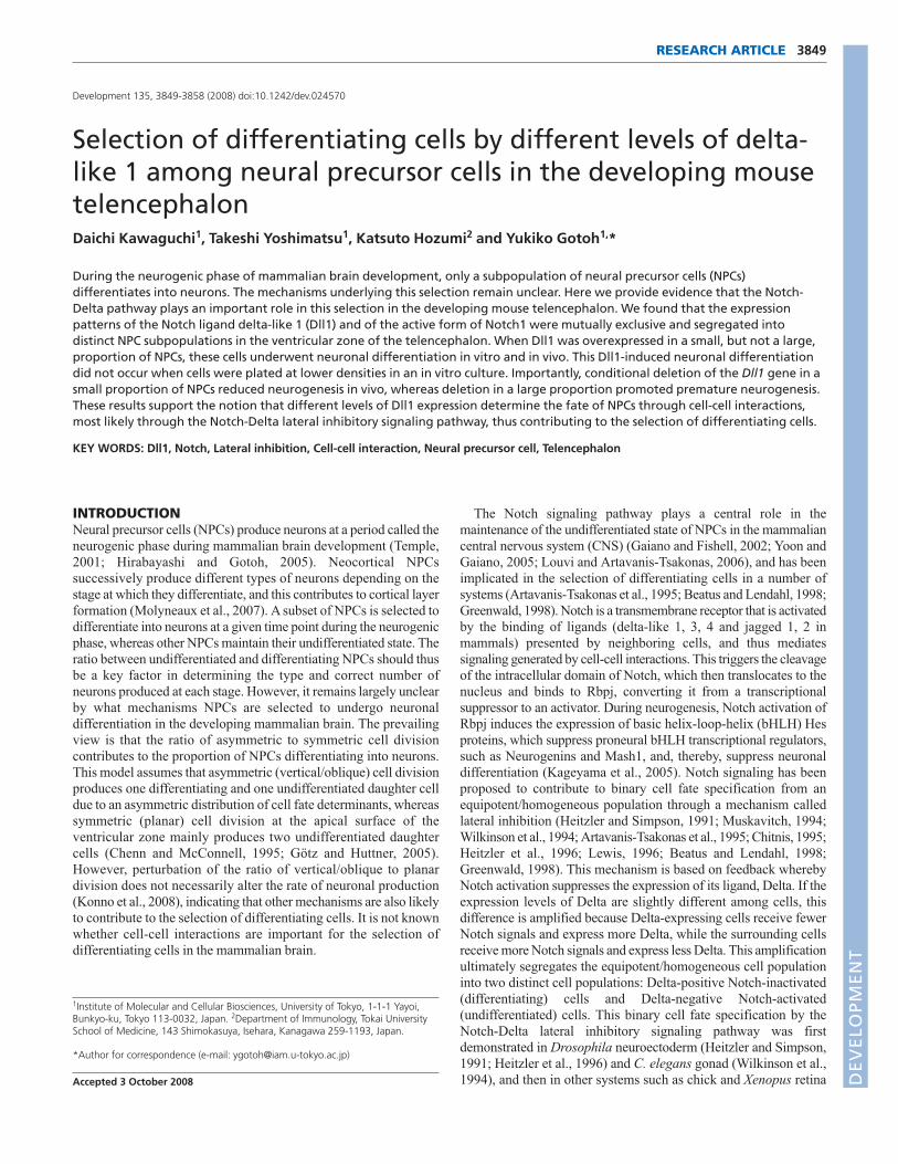
oscillatory expression of Dll1 mRNA at low levels in proliferatingNPCs (Shimojo et al., 2008). In this study, we investigated theexpression of Dll1 protein in the developing mouse telencephalonby immunohistochemical analyses. Dll1-immunopositive cells weredetected in a subset of cells in the ventricular zone (VZ), theintermediate zone (IZ) and the cortical plate (CP) of neocortex andin the ganglionic eminences at E13.5 (Fig. 1A). At later stages, suchas at E16.5, Dll1-expressing cells were found in the VZ and IZ, butat very low levels in the CP of the neocortex (Fig. 1B). Theexpression pattern of Dll1 protein was similar to that of Dll1 mRNA(Fig. 1C,D). Importantly, most of the Dll1-expressing cells in the VZwere co-immunostained with antibodies to the proliferation markerPcna and to the undifferentiated NPC markers nestin and Sox2 (Fig.1E-P). Consistent with this, Dll1-expressing cells in the VZ weremostly devoid of the neuronal marker β III-tubulin (TuJ1; Tubb3),whereas most of the Dll1-expressing cells in the IZ (and those in theCP at E13.5) were TuJ1-positive (Fig. 1Q-T). These results suggestthat Dll1 is expressed in a subset of undifferentiated NPCs in the VZ,as well as in immature neurons localized at the IZ and CP.
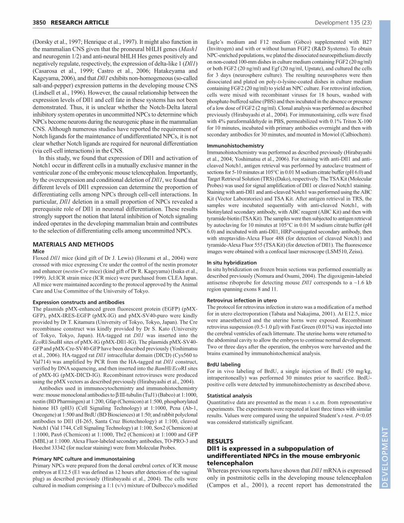
Mutually exclusive expression patterns of Dll1and active Notch1It has been reported that NPCs localized at the VZ areheterogeneous in terms of their Notch activity, as revealed byimmunostaining with anti-cleaved Notch1 (active Notch1)
antibody, which recognizes the presenilin/γ-secretase-catalyzedcleavage site of Notch1, and by the use of a reporter gene thatmonitors the activity of Notch signaling (Tokunaga et al., 2004; DelMonte et al., 2007; Mizutani et al., 2007). We confirmed that onlya subpopulation of NPCs in the VZ was immunostained for activeNotch1 (Fig. 2B,F). However, a negative correlation between theNotch activity and Dll1 expression in the developing mouse brainhas not been demonstrated, although this would provide keyevidence to support the role of the Notch-Delta lateral inhibitorysystem in this system of differentiation. We thus carried outimmunohistochemistry of active Notch1 and Dll1 together, andfound that their expression patterns in the VZ of the telencephalonwere segregated into distinct cells in a mutually exclusive manner(Fig. 2A-H). In the VZ of the neocortex at E13.5, 73.8% of Dll1-negative cells were positive for active Notch1 within the nucleus,whereas only 24.7% of Dll1-positive cells were positive for activeNotch1 within the nucleus (Fig. 2I). Dll1 expression and Notch1activation might have a rough correlation with the cell cycle, asactive Notch1- and Dll1-expressing cells tended to localize basallyand apically in the VZ, respectively (Fig. 2A-H). However, thepercentage of active Notch1-positive cells was significantly loweramong Dll1-positive cells than among Dll1-negative cells in boththe basal and apical halves of the VZ (see Fig. S1 in thesupplementary material). These results suggest that the mutuallyexclusive patterns of active Notch1 and Dll1 expression might be
3851RESEARCH ARTICLENPC fate choice by Dll1 in mouse brain
Fig. 1. Expression of Dll1 in the developing mousetelencephalon. (A,B,E-T) Brain sections from ICR mouseembryos at E13.5 (A,E-T) or E16.5 (B) were immunostainedwith anti-Dll1 alone (A,B) or together with anti-Pcna (E-H), anti-nestin (I-L), anti-Sox2 (M-P) or anti-β III-tubulin (TuJ1) (Q-T).(C,D) Dll1 mRNA in sections of E13.5 ICR mouse brain detectedby in situ hybridization. Higher magnifications of the boxedregion in C,G,K,O and S are shown in D,H,L,P and T,respectively. NCX, neocortex. Scale bars: 200μm in A-C; 20μmin D-T.
DEVELO
PMENT
3852
established at different phases of the cell cycle. This negativecorrelation between active Notch1 and Dll1 expression stronglysupports the existence of the Notch-Delta lateral inhibitory systemin the developing mouse telencephalon.
Dll1 expression regulates neuronal fatespecification in vitroIt has been proposed that Dll1 expressed in immature neuronssuppresses excess neuronal differentiation by activating Notch inthe neighboring NPCs (Henrique et al., 1997; Campos et al., 2001).However, the expression of Dll1 in undifferentiated NPCs, asobserved in Fig. 1, suggests an additional role of Dll1, i.e. inneuronal fate specification among NPCs through the lateralinhibitory system. If this is the case, ectopic expression of Dll1 ina subpopulation of NPCs should lead to neuronal differentiation ofthese cells, as ectopic Dll1 activates Notch signaling and reducesDll1 expression in the surrounding cells, resulting in a reduction ofNotch signaling in the Dll1-expressing cells (Heitzler and Simpson,1991; Dorsky et al., 1997; Henrique et al., 1997). We examined thishypothesis by introducing Dll1 into an in vitro culture of NPCs.NPC cultures (see Materials and methods) prepared from E12.5mouse neocortex were infected with retroviruses encoding eithergreen fluorescent protein (GFP) alone, or GFP together with Dll1,at a low titer so that only a small number (less than 0.1%) of thecells were infected. Under this condition, the fate of each infectedclone could be traced as a cluster of GFP-positive cells (Fig. 3A).The expression of Dll1 significantly increased the proportion ofclones containing only TuJ1-positive neurons (pure TuJ1-positiveclones) among GFP-positive clones (Fig. 3B). The expression ofDll1 also increased the percentage of TuJ1-positive cells amongGFP-positive cells (Fig. 3C). The expression of Dll1 did not appearto alter the size of the clones (see Fig. S2 in the supplementarymaterial). Since the stage of this culture roughly corresponds to theneurogenic phase, the percentage of cells expressing the astroglialmarker Gfap among infected cells was small (~7%, regardless ofDll1 expression) (Fig. 3D), indicating that the Dll1-inducedneuronal differentiation shown in Fig. 3B was not due to asuppression of an alternate (astroglial) fate of NPCs. Thecontribution of cell death was also negligible in these cultures (lessthan 2% among GFP-positive cells).
The fate switch of NPCs from neurogenic to astrogliogenic canbe recapitulated in an in vitro culture. When Dll1 was overexpressedin a subpopulation of NPCs prepared from a 12 days in vitro (DIV)culture of E12.5 neocortical neuroepithelial cells, which correspondto the astrogliogenic phase, the proportion of clones containing onlyGfap-positive astrocytes was reduced and the proportion of clonescontaining only TuJ1-positive neurons was increased (Fig. 3E,F).This suggests that high levels of Dll1 were sufficient to change theastrogliogenic fate into the neurogenic fate when expressed at theastrogliogenic phase. Interestingly, the levels of Dll1 mRNA werereduced at around the onset of the astrogliogenic phase (Campos etal., 2001; Irvin et al., 2004). This reduction might contribute to thesuppression of neurogenesis during the astrogliogenic phase.
Dll1-induced neuronal differentiation requirescell-cell interactions among NPCsIf neuronal differentiation induced by Dll1 expression can beascribed to the lateral inhibition model, it should be dependent oncell-cell interaction and the difference in Dll1 levels between thecells. To examine this, we introduced Dll1 into a large proportionof NPCs by retroviral infection at a high titer (more than 70% ofNPCs were infected under this condition), so that there would beno major differences in Dll1 levels among the cells. In this case,the expression of Dll1 did not promote neuronal differentiation(Fig. 4A). We further examined whether cell-cell interactions areessential for Dll1-induced neuronal differentiation by reducingthe cell density. We introduced Dll1 into a small proportion ofNPCs by retroviral infection at a low titer, as in Fig. 3, butchanged the seeding cell density. At a lower cell density(0.26�105 cells/cm2 at seeding), the expression of Dll1 no longerincreased the percentage of pure TuJ1-positive clones amongGFP-positive clones (Fig. 4B). These results together support theidea that Dll1 induces neuronal differentiation of NPCs via cell-cell interaction, consistent with the lateral inhibition model.
Reduction of the cell density did not significantly increase thepercentage of TuJ1-positive clones among GFP-positive clones incontrol cultures (Fig. 4B), suggesting the existence of a trans-acting signal that promotes neuronal differentiation in a non-cell-autonomous manner. Such a mechanism might involve Wntsignaling, as Wnt ligands, including Wnt7a, were expressed in
RESEARCH ARTICLE Development 135 (23)
Fig. 2. Dll1 expression and Notch1 activation are segregatedinto distinct cells in the VZ of the developing mousetelencephalon. (A-H) Brain sections from ICR mouse embryos atE13.5 were immunostained with anti-active Notch1 (actN1) andanti-Dll1 and TO-PRO-3 (nuclear staining). Higher magnifications ofthe boxed regions in C and G are shown in D and H, respectively.Arrowheads in D,H indicate Dll1-positive actN1-negative cells in theVZ. (I) The proportions of active Notch1-positive cells among Dll1-negative or Dll1-positive cells in the VZ of the neocortex weredetermined by immunohistochemical analysis. Data are the mean ±s.e.m. of values from three sections of each brain, and similar resultswere obtained from six independent brains. *P<0.0005. NCX,neocortex; GE, ganglionic eminence. Scale bars: 20μm.
DEVELO
PMENT

this culture (data not shown), and the prevention of Wnt signalingsuppresses the neuronal differentiation of NPCs cultured undersimilar conditions (Hirabayashi et al., 2004).
The intracellular domain of Dll1 does not affectneuronal differentiationDll1 is cleaved at a transmembrane site upon Notch binding, and theintracellular domain of Dll1 has been shown to translocate into thenucleus and transmit some signals cell-autonomously (Bland et al.,2003; Ikeuchi and Sisodia, 2003; LaVoie and Selkoe, 2003; Six et al.,2003; Kolev et al., 2005). A recent study has shown that expressionof the Dll1 intracellular domain (DICD) promotes neurogenesis cell-autonomously in P19 cells (Hiratochi et al., 2007). However, we
found that overexpression of DICD, even when expressed in a smallnumber of NPCs, did not induce neuronal differentiation in primarytelencephalic NPC culture (Fig. 4C) or in vivo (see Fig. S3 in thesupplementary material). These results again suggest that neuronaldifferentiation induced by Dll1 expression is not due to a cell-autonomous mechanism.
Different levels of Dll1 expression regulateneuronal fate specification in the developingmouse neocortexIn addition to the in vitro experiments, we wished to examine theeffects of Dll1 expression on NPC fate in vivo. We thereforeintroduced Dll1 into a small number of NPCs in the telencephalonby injecting retroviruses encoding GFP alone, or both GFP and Dll1,into the telencephalic ventricle at E12.5. Two days after infection,the numbers of GFP-positive cells localized at each area [VZ,subventricular zone (SVZ), IZ or CP] were determined. Whencontrol retroviruses were introduced, 26.8±2.7% of GFP-positivecells were found within the VZ 2 days after infection. When Dll1-expressing retroviruses were introduced, the percentage of GFP-positive cells within the VZ was markedly reduced (5.3±2.1%) (Fig.5A-C). Dll1 expression also decreased the percentage of GFP-positive cells located in the SVZ, and increased the percentages ofGFP-positive cells in the IZ and CP. All of the GFP-positive cells inthe IZ and CP were negative for the NPC marker Pax6, and morethan 97% of these cells were positive for TuJ1 in both control andDll1-expression samples. In fact, the expression of Dll1 significantlyincreased the proportion of TuJ1-positive cells among GFP-positive
3853RESEARCH ARTICLENPC fate choice by Dll1 in mouse brain
Fig. 3. Dll1 overexpression in a small proportion of NPCs inducesneuronal differentiation in vitro. Primary NPCs were prepared from(A-D) 3-day sphere cultures [3 days in vitro (3 DIV)] or (E,F) 12-daysphere cultures (fourth passage, 12 DIV) of the neocortical cells ofE12.5 ICR mice. The NPCs were infected with a retrovirus encoding GFP(pMX-GFP, control), or both GFP and Dll1 (pMX-Dll1-IG), at a low titerand subjected to clonal analysis (see Materials and methods). (A) Arepresentative GFP-positive clone stained with anti-GFP, TuJ1 andHoechst. After incubation for 2 days in the presence of a low dose ofhuman FGF2 (2 ng/ml) (B-D) or in the absence of FGF2 (E,F), cells werestained with anti-GFP and TuJ1 or anti-GFP and anti-Gfap. Thepercentage of clones containing only TuJ1-positive (B,E) or only Gfap-positive (D,F) cells among GFP-positive clones was then determined byimmunocytochemical analysis. The percentage of TuJ1-positive cellsamong GFP-positive cells was also determined by immunocytochemistry(C). *P<0.02, **P<0.01, ***P<0.0001. Scale bar: 50μm in A.
Fig. 4. Dll1 overexpression induces neuronal differentiation in anon-cell-autonomous manner. (A) Primary NPCs were prepared from3-day sphere cultures (3 DIV) of the neocortical cells of E12.5 ICR mice,infected with a retrovirus encoding GFP (pMX-GFP, control), or bothGFP and Dll1 (pMX-Dll1-IG), at a high titer and analyzed as in Fig. 3C.(B) Primary NPCs were prepared from 3-day sphere cultures (3 DIV) ofthe neocortical cells of E12.5 ICR mice and plated at different celldensities (0.26�, 0.52� and 1.04�105 cells/cm2). These cells wereinfected with a retrovirus encoding GFP (pMX-GFP, control), or bothGFP and Dll1 (pMX-Dll1-IG), at a low titer and analyzed as in Fig. 3B(clonal analysis). *P<0.05. (C) Primary NPCs were prepared from 3-daysphere cultures (3 DIV) of the neocortical cells of E12.5 ICR mice andinfected with a retrovirus encoding GFP (pMX-GFP, control), or bothGFP and the delta-like 1 intracellular domain (pMX-DICD-IG), at a lowtiter and analyzed as in Fig. 3B (clonal analysis).
DEVELO
PMENT

3854
cells in the whole neocortex (Fig. 5D). These results stronglysuggest that Dll1 expression in a small population of cells at the VZpromotes neuronal differentiation in the developing neocortex.
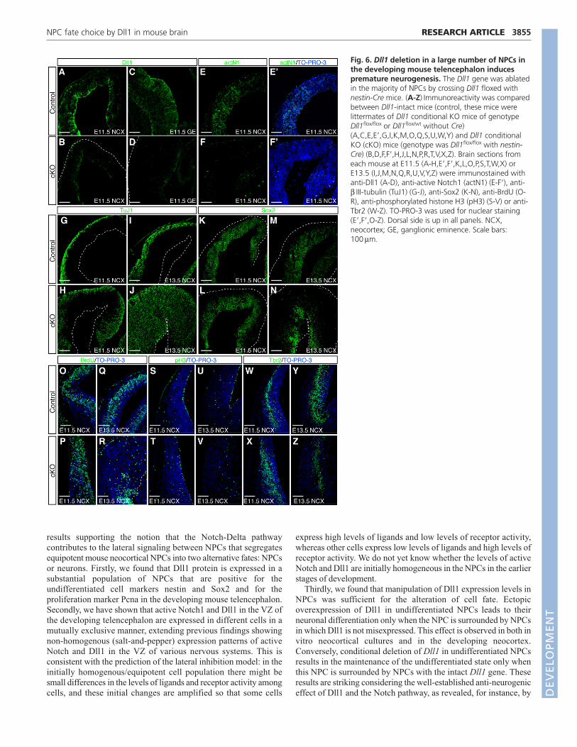
Conditional deletion of Dll1 results in aberrantneuronal differentiation in vivoIt has previously been reported that deletion of Dll1 results inembryonic lethality at E11.5, accompanying hyperplasia of the CNS(Hrabe de Angelis et al., 1997) and increased early production ofGABA-positive neurons at ganglionic eminences (Yun et al., 2002).However, it has remained unclear whether Dll1 is a major Notchligand in other parts of the telencephalon such as the neocortex,especially given that other Notch ligands such as jagged 2 and Dll3are also expressed in the developing neocortex (Luo et al., 1997;Campos et al., 2001; Irvin et al., 2004). Thus, to avoid the embryoniclethality at early developmental stages, we utilized a conditionaldeletion of the Dll1 gene in the CNS. Conditional deletion wasachieved by crossing mice carrying both Dll1 alleles flanked by loxPsequences (Dll1flox/flox) (Hozumi et al., 2004) with mice harboring atransgene of Cre recombinase under the control of the nestinpromoter and enhancer (Isaka et al., 1999). In these mice, Dll1immunoreactivity was already decreased at E11.5 in telencephalon(Fig. 6A-D). By E13.5, the Dll1-deficient neocortex became thickerthan the control neocortex in the radial axis, but thinner in thetangential axis (Fig. 6I,J,M,N, and data not shown), with a slightincrease in cell death (data not shown). The levels of active (cleaved)Notch1 in NPCs were significantly reduced in the Dll1 conditional
KO mice at E11.5 (Fig. 6E,E�,F,F�). These mice exhibited robustpremature neurogenesis in the neocortex as well as in the subcorticalregions by E13.5, as detected by an increase in TuJ1-positive cellsand a reduction in Sox2-positive cells (Fig. 6G-N). We also observeda transient increase of Tbr2 (Eomes)-positive basal progenitors atE11.5 and a decrease at E13.5 in the lateral region of Dll1-deficientneocortex (Fig. 6W-Z, see also Fig. 6O-V showing the aberrantlocation of mitotic cells), probably owing to the prematureneurogenesis and subsequent exhaustion of NPCs. At E16.5, fewerNPCs were found in the VZ of the Dll1 conditional KO mice ascompared with the control mice (data not shown), also suggestingthe exhaustion of NPCs owing to the premature neurogenesis. Sincethese phenotypes are consistent with the premature neurogenesisobserved in mice defective in the canonical Notch pathway (e.g.Notch1- and Rbpj-deficient mice) (de la Pompa et al., 1997), theseresults suggest that Dll1 is a major ligand for the Notch receptor inthe developing neocortex and that it contributes to the maintenanceof the undifferentiated state of NPCs.
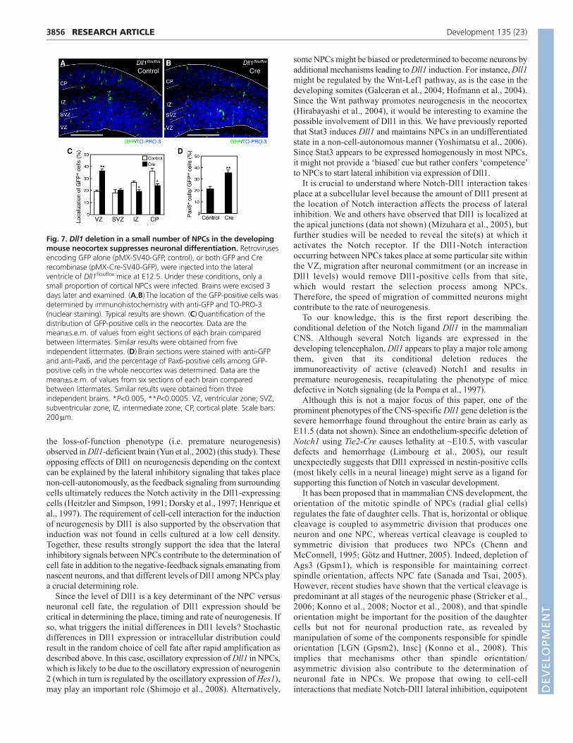
Given that Dll1 is a major Notch ligand in the developingtelencephalon, we next examined whether different levels ofendogenous Dll1 among undifferentiated NPCs could determine thefate of NPCs in vivo, and whether the Notch-Delta lateral inhibitorysystem indeed operates in vivo. We deleted the Dll1 gene in a smallproportion of NPCs in vivo by injecting retroviruses harboring GFPalone, or GFP with Cre recombinase, into the telencephalic ventricleof Dll1flox/flox mice at E12.5. When examined 3 days after infection,expression of Cre recombinase increased the percentage of GFP-positive cells within the VZ (from 19.2% to 36.3%) and reduced thatof GFP-positive cells within the IZ and CP (IZ, from 26.7% to19.3%; CP, from 36.1% to 26.2%) (Fig. 7A-C). Since more than96% of GFP-positive cells in the VZ were Pax6-positive and TuJ1-negative in both control and Cre-infected samples, this resultindicates that the reduction of endogenous Dll1 in a small proportionof NPCs suppressed the neuronal differentiation of these cells. Infact, the deletion of Dll1 significantly increased the proportion ofPax6-positive cells among GFP-positive cells in the wholeneocortex (Fig. 7D). Since the reduction of endogenous Dll1 in allNPCs dramatically promoted neuronal differentiation (Fig. 6), theseresults strongly suggest that different levels of endogenous Dll1regulate the neuronal differentiation of NPCs via cell-cellinteraction, most likely through the lateral inhibitory system.
DISCUSSIONA classical view of the role of the Notch pathway in Drosophilaneuroectoderm is that it selects differentiating cells based on a‘competition’ between equipotent progenitor cells throughamplifying differences in Notch ligand levels (Heitzler andSimpson, 1991; Heitzler et al., 1996). By contrast, it has beenproposed that the Notch pathway in the developing vertebrate CNSmainly contributes to the negative-feedback signal emanating fromnascent neurons to suppress excess neuronal differentiation ofNPCs. This view was supported by the observation that Dll1 mRNAis found predominantly in nascent neurons/intermediate progenitors(neuron-committed progenitors), but is scarce in NPCs in thedeveloping retina, spinal cord and telencephalon (Henrique et al.,1997; Campos et al., 2001; Minaki et al., 2005; Kawaguchi et al.,2008). A recent study of mind bomb 1, an activator of Dll1, alsosupports this view because of its expression in nascentneurons/intermediate progenitors (Yoon et al., 2008). However,another study has recently revealed low-level expression of Dll1mRNA in NPCs (Shimojo et al., 2008), although its role in NPCdifferentiation remained unclear. In this study, we present a series of
RESEARCH ARTICLE Development 135 (23)
Fig. 5. Dll1 overexpression in a small number of NPCs in thedeveloping mouse neocortex induces neuronal differentiation.Retroviruses encoding GFP alone (pMX-GFP, control), or both GFP andDll1 (pMX-Dll1-IG), were injected into the lateral ventricle of ICR miceat E12.5. Under these conditions, only a small proportion of corticalNPCs were infected. Brains were excised 2 days later and examined.(A,B) The location of the GFP-positive cells was determined byimmunohistochemistry with anti-GFP and TO-PRO-3 (nuclear staining).Typical results are shown. (C) Quantification of the distribution of GFP-positive cells in the neocortex. Data are the mean±s.e.m. of values fromeight sections of each brain compared between littermates. Similarresults were obtained from seven independent littermates. (D) Brainsections were stained with anti-GFP and TuJ1, and the percentage ofTuJ1-positive cells among GFP-positive cells in the whole neocortex wasdetermined. Data are the mean±s.e.m. of values from eight sections ofeach brain compared between littermates. Similar results were obtainedfrom three independent brains. *P<0.01, **P<0.0001. VZ, ventricularzone; SVZ, subventricular zone; IZ, intermediate zone; CP, cortical plate.Scale bars: 200μm.
DEVELO
PMENT
results supporting the notion that the Notch-Delta pathwaycontributes to the lateral signaling between NPCs that segregatesequipotent mouse neocortical NPCs into two alternative fates: NPCsor neurons. Firstly, we found that Dll1 protein is expressed in asubstantial population of NPCs that are positive for theundifferentiated cell markers nestin and Sox2 and for theproliferation marker Pcna in the developing mouse telencephalon.Secondly, we have shown that active Notch1 and Dll1 in the VZ ofthe developing telencephalon are expressed in different cells in amutually exclusive manner, extending previous findings showingnon-homogenous (salt-and-pepper) expression patterns of activeNotch and Dll1 in the VZ of various nervous systems. This isconsistent with the prediction of the lateral inhibition model: in theinitially homogenous/equipotent cell population there might besmall differences in the levels of ligands and receptor activity amongcells, and these initial changes are amplified so that some cells
express high levels of ligands and low levels of receptor activity,whereas other cells express low levels of ligands and high levels ofreceptor activity. We do not yet know whether the levels of activeNotch and Dll1 are initially homogeneous in the NPCs in the earlierstages of development.
Thirdly, we found that manipulation of Dll1 expression levels inNPCs was sufficient for the alteration of cell fate. Ectopicoverexpression of Dll1 in undifferentiated NPCs leads to theirneuronal differentiation only when the NPC is surrounded by NPCsin which Dll1 is not misexpressed. This effect is observed in both invitro neocortical cultures and in the developing neocortex.Conversely, conditional deletion of Dll1 in undifferentiated NPCsresults in the maintenance of the undifferentiated state only whenthis NPC is surrounded by NPCs with the intact Dll1 gene. Theseresults are striking considering the well-established anti-neurogeniceffect of Dll1 and the Notch pathway, as revealed, for instance, by
3855RESEARCH ARTICLENPC fate choice by Dll1 in mouse brain
Fig. 6. Dll1 deletion in a large number of NPCs inthe developing mouse telencephalon inducespremature neurogenesis. The Dll1 gene was ablatedin the majority of NPCs by crossing Dll1 floxed withnestin-Cre mice. (A-Z) Immunoreactivity was comparedbetween Dll1-intact mice (control, these mice werelittermates of Dll1 conditional KO mice of genotypeDll1flox/flox or Dll1flox/wt without Cre)(A,C,E,E�,G,I,K,M,O,Q,S,U,W,Y) and Dll1 conditionalKO (cKO) mice (genotype was Dll1flox/flox with nestin-Cre) (B,D,F,F�,H,J,L,N,P,R,T,V,X,Z). Brain sections fromeach mouse at E11.5 (A-H,E�,F�,K,L,O,P,S,T,W,X) orE13.5 (I,J,M,N,Q,R,U,V,Y,Z) were immunostained withanti-Dll1 (A-D), anti-active Notch1 (actN1) (E-F�), anti-β III-tubulin (TuJ1) (G-J), anti-Sox2 (K-N), anti-BrdU (O-R), anti-phosphorylated histone H3 (pH3) (S-V) or anti-Tbr2 (W-Z). TO-PRO-3 was used for nuclear staining(E�,F�,O-Z). Dorsal side is up in all panels. NCX,neocortex; GE, ganglionic eminence. Scale bars:100μm.
DEVELO
PMENT
3856
the loss-of-function phenotype (i.e. premature neurogenesis)observed in Dll1-deficient brain (Yun et al., 2002) (this study). Theseopposing effects of Dll1 on neurogenesis depending on the contextcan be explained by the lateral inhibitory signaling that takes placenon-cell-autonomously, as the feedback signaling from surroundingcells ultimately reduces the Notch activity in the Dll1-expressingcells (Heitzler and Simpson, 1991; Dorsky et al., 1997; Henrique etal., 1997). The requirement of cell-cell interaction for the inductionof neurogenesis by Dll1 is also supported by the observation thatinduction was not found in cells cultured at a low cell density.Together, these results strongly support the idea that the lateralinhibitory signals between NPCs contribute to the determination ofcell fate in addition to the negative-feedback signals emanating fromnascent neurons, and that different levels of Dll1 among NPCs playa crucial determining role.
Since the level of Dll1 is a key determinant of the NPC versusneuronal cell fate, the regulation of Dll1 expression should becritical in determining the place, timing and rate of neurogenesis. Ifso, what triggers the initial differences in Dll1 levels? Stochasticdifferences in Dll1 expression or intracellular distribution couldresult in the random choice of cell fate after rapid amplification asdescribed above. In this case, oscillatory expression of Dll1 in NPCs,which is likely to be due to the oscillatory expression of neurogenin2 (which in turn is regulated by the oscillatory expression of Hes1),may play an important role (Shimojo et al., 2008). Alternatively,
some NPCs might be biased or predetermined to become neurons byadditional mechanisms leading to Dll1 induction. For instance, Dll1might be regulated by the Wnt-Lef1 pathway, as is the case in thedeveloping somites (Galceran et al., 2004; Hofmann et al., 2004).Since the Wnt pathway promotes neurogenesis in the neocortex(Hirabayashi et al., 2004), it would be interesting to examine thepossible involvement of Dll1 in this. We have previously reportedthat Stat3 induces Dll1 and maintains NPCs in an undifferentiatedstate in a non-cell-autonomous manner (Yoshimatsu et al., 2006).Since Stat3 appears to be expressed homogenously in most NPCs,it might not provide a ‘biased’ cue but rather confers ‘competence’to NPCs to start lateral inhibition via expression of Dll1.
It is crucial to understand where Notch-Dll1 interaction takesplace at a subcellular level because the amount of Dll1 present atthe location of Notch interaction affects the process of lateralinhibition. We and others have observed that Dll1 is localized atthe apical junctions (data not shown) (Mizuhara et al., 2005), butfurther studies will be needed to reveal the site(s) at which itactivates the Notch receptor. If the Dll1-Notch interactionoccurring between NPCs takes place at some particular site withinthe VZ, migration after neuronal commitment (or an increase inDll1 levels) would remove Dll1-positive cells from that site,which would restart the selection process among NPCs.Therefore, the speed of migration of committed neurons mightcontribute to the rate of neurogenesis.
To our knowledge, this is the first report describing theconditional deletion of the Notch ligand Dll1 in the mammalianCNS. Although several Notch ligands are expressed in thedeveloping telencephalon, Dll1 appears to play a major role amongthem, given that its conditional deletion reduces theimmunoreactivity of active (cleaved) Notch1 and results inpremature neurogenesis, recapitulating the phenotype of micedefective in Notch signaling (de la Pompa et al., 1997).
Although this is not a major focus of this paper, one of theprominent phenotypes of the CNS-specific Dll1 gene deletion is thesevere hemorrhage found throughout the entire brain as early asE11.5 (data not shown). Since an endothelium-specific deletion ofNotch1 using Tie2-Cre causes lethality at ~E10.5, with vasculardefects and hemorrhage (Limbourg et al., 2005), our resultunexpectedly suggests that Dll1 expressed in nestin-positive cells(most likely cells in a neural lineage) might serve as a ligand forsupporting this function of Notch in vascular development.
It has been proposed that in mammalian CNS development, theorientation of the mitotic spindle of NPCs (radial glial cells)regulates the fate of daughter cells. That is, horizontal or obliquecleavage is coupled to asymmetric division that produces oneneuron and one NPC, whereas vertical cleavage is coupled tosymmetric division that produces two NPCs (Chenn andMcConnell, 1995; Götz and Huttner, 2005). Indeed, depletion ofAgs3 (Gpsm1), which is responsible for maintaining correctspindle orientation, affects NPC fate (Sanada and Tsai, 2005).However, recent studies have shown that the vertical cleavage ispredominant at all stages of the neurogenic phase (Stricker et al.,2006; Konno et al., 2008; Noctor et al., 2008), and that spindleorientation might be important for the position of the daughtercells but not for neuronal production rate, as revealed bymanipulation of some of the components responsible for spindleorientation [LGN (Gpsm2), Insc] (Konno et al., 2008). Thisimplies that mechanisms other than spindle orientation/asymmetric division also contribute to the determination ofneuronal fate in NPCs. We propose that owing to cell-cellinteractions that mediate Notch-Dll1 lateral inhibition, equipotent
RESEARCH ARTICLE Development 135 (23)
Fig. 7. Dll1 deletion in a small number of NPCs in the developingmouse neocortex suppresses neuronal differentiation. Retrovirusesencoding GFP alone (pMX-SV40-GFP, control), or both GFP and Crerecombinase (pMX-Cre-SV40-GFP), were injected into the lateralventricle of Dll1flox/flox mice at E12.5. Under these conditions, only asmall proportion of cortical NPCs were infected. Brains were excised 3days later and examined. (A,B) The location of the GFP-positive cells wasdetermined by immunohistochemistry with anti-GFP and TO-PRO-3(nuclear staining). Typical results are shown. (C) Quantification of thedistribution of GFP-positive cells in the neocortex. Data are themean±s.e.m. of values from eight sections of each brain comparedbetween littermates. Similar results were obtained from fiveindependent littermates. (D) Brain sections were stained with anti-GFPand anti-Pax6, and the percentage of Pax6-positive cells among GFP-positive cells in the whole neocortex was determined. Data are themean±s.e.m. of values from six sections of each brain comparedbetween littermates. Similar results were obtained from threeindependent brains. *P<0.005, **P<0.0005. VZ, ventricular zone; SVZ,subventricular zone; IZ, intermediate zone; CP, cortical plate. Scale bars:200μm.
DEVELO
PMENT

NPCs can differentiate into neurons at a certain rate even withoutasymmetric division. This is based on the finding that differentialDll1 expression levels between NPCs are sufficient fordetermining the neuronal fate. It is also conceivable thatasymmetric division (caused by an asymmetric inheritance of fatedeterminants, if any) can bias the outcome of lateral inhibitionbetween adjacent cells. However, because Dll1-deficient NPCssurrounded by NPCs with an intact Dll1 gene are less likely toundergo neurogenesis, the Notch-Dll1 lateral inhibitorymechanism might be dominant over fate determination byasymmetric division and could be prerequisite for neuronaldifferentiation.
We thank Cancer Research UK (CRUK) and Dr Julian Lewis for Dll1 floxedmice; Dr Ryoichiro Kageyama for nestin-Cre mice; Drs Toshio Kitamura andShigeaki Kato for plasmids; Dr Marc Lamphier for critical reading of themanuscript; and members of the Gotoh laboratory for discussion. D.K. is aresearch fellow of the Japan Society for the Promotion of Science (JSPS). Thiswork was supported by Grants-in-Aid from the Ministry of Education, Culture,Sports, Science and Technology (MEXT) of Japan, and for SORST from theJapan Science and Technology Agency, and for JSPS fellows from JSPS. Thiswork was also supported in part by the Global COE Program (Integrative LifeScience Based on the Study of Biosignaling Mechanisms), MEXT, Japan.
Supplementary materialSupplementary material for this article is available athttp://dev.biologists.org/cgi/content/full/135/23/3849/DC1
ReferencesArtavanis-Tsakonas, S., Matsuno, K. and Fortini, M. E. (1995). Notch
signaling. Science 268, 225-232.Beatus, P. and Lendahl, U. (1998). Notch and neurogenesis. J. Neurosci. Res. 54,
125-136.Bland, C. E., Kimberly, P. and Rand, M. D. (2003). Notch-induced proteolysis and
nuclear localization of the Delta ligand. J. Biol. Chem. 278, 13607-13610.Campos, L. S., Duarte, A. J., Branco, T. and Henrique, D. (2001). mDll1 and
mDll3 expression in the developing mouse brain: role in the establishment of theearly cortex. J. Neurosci. Res. 64, 590-598.
Casarosa, S., Fode, C. and Guillemot, F. (1999). Mash1 regulates neurogenesisin the ventral telencephalon. Development 126, 525-534.
Castro, D. S., Skowronska-Krawczyk, D., Armant, O., Donaldson, I. J.,Parras, C., Hunt, C., Critchley, J. A., Nguyen, L., Gossler, A., Gottgens, B.et al. (2006). Proneural bHLH and Brn proteins coregulate a neurogenicprogram through cooperative binding to a conserved DNA motif. Dev. Cell 11,831-844.
Chenn, A. and McConnell, S. K. (1995). Cleavage orientation and theasymmetric inheritance of Notch1 immunoreactivity in mammalianneurogenesis. Cell 82, 631-641.
Chitnis, A. B. (1995). The role of Notch in lateral inhibition and cell fatespecification. Mol. Cell. Neurosci. 6, 311-321.
de la Pompa, J. L., Wakeham, A., Correia, K. M., Samper, E., Brown, S.,Aguilera, R. J., Nakano, T., Honjo, T., Mak, T. W., Rossant, J. et al. (1997).Conservation of the Notch signalling pathway in mammalian neurogenesis.Development 124, 1139-1148.
Del Monte, G., Grego-Bessa, J., Gonzalez-Rajal, A., Bolos, V. and De LaPompa, J. L. (2007). Monitoring Notch1 activity in development: evidence for afeedback regulatory loop. Dev. Dyn. 236, 2594-2614.
Dorsky, R. I., Chang, W. S., Rapaport, D. H. and Harris, W. A. (1997).Regulation of neuronal diversity in the Xenopus retina by Delta signalling. Nature385, 67-70.
Gaiano, N. and Fishell, G. (2002). The role of notch in promoting glial and neuralstem cell fates. Annu. Rev. Neurosci. 25, 471-490.
Galceran, J., Sustmann, C., Hsu, S. C., Folberth, S. and Grosschedl, R. (2004).LEF1-mediated regulation of Delta-like1 links Wnt and Notch signaling insomitogenesis. Genes Dev. 18, 2718-2723.
Götz, M. and Huttner, W. B. (2005). The cell biology of neurogenesis. Nat. Rev.Mol. Cell Biol. 6, 777-788.
Greenwald, I. (1998). LIN-12/Notch signaling: lessons from worms and flies.Genes Dev. 12, 1751-1762.
Hatakeyama, J. and Kageyama, R. (2006). Notch1 expression isspatiotemporally correlated with neurogenesis and negatively regulated byNotch1-independent Hes genes in the developing nervous system. Cereb. Cortex16 Suppl. 1, i132-i137.
Heitzler, P. and Simpson, P. (1991). The choice of cell fate in the epidermis ofDrosophila. Cell 64, 1083-1092.
Heitzler, P., Bourouis, M., Ruel, L., Carteret, C. and Simpson, P. (1996). Genesof the Enhancer of split and achaete-scute complexes are required for aregulatory loop between Notch and Delta during lateral signalling in Drosophila.Development 122, 161-171.
Henrique, D., Hirsinger, E., Adam, J., Le Roux, I., Pourquie, O., Ish-Horowicz,D. and Lewis, J. (1997). Maintenance of neuroepithelial progenitor cells byDelta-Notch signalling in the embryonic chick retina. Curr. Biol. 7, 661-670.
Hirabayashi, Y. and Gotoh, Y. (2005). Stage-dependent fate determination ofneural precursor cells in mouse forebrain. Neurosci. Res. 51, 331-336.
Hirabayashi, Y., Itoh, Y., Tabata, H., Nakajima, K., Akiyama, T., Masuyama,N. and Gotoh, Y. (2004). The Wnt/beta-catenin pathway directs neuronaldifferentiation of cortical neural precursor cells. Development 131, 2791-2801.
Hiratochi, M., Nagase, H., Kuramochi, Y., Koh, C. S., Ohkawara, T. andNakayama, K. (2007). The Delta intracellular domain mediates TGF-beta/Activinsignaling through binding to Smads and has an important bi-directional functionin the Notch-Delta signaling pathway. Nucleic Acids Res. 35, 912-922.
Hofmann, M., Schuster-Gossler, K., Watabe-Rudolph, M., Aulehla, A.,Herrmann, B. G. and Gossler, A. (2004). WNT signaling, in synergy withT/TBX6, controls Notch signaling by regulating Dll1 expression in the presomiticmesoderm of mouse embryos. Genes Dev. 18, 2712-2717.
Hozumi, K., Negishi, N., Suzuki, D., Abe, N., Sotomaru, Y., Tamaoki, N.,Mailhos, C., Ish-Horowicz, D., Habu, S. and Owen, M. J. (2004). Delta-like 1is necessary for the generation of marginal zone B cells but not T cells in vivo.Nat. Immun. 5, 638-644.
Hrabe de Angelis, M., McIntyre, J., 2nd. and Gossler, A. (1997). Maintenanceof somite borders in mice requires the Delta homologue DII1. Nature 386, 717-721.
Ikeuchi, T. and Sisodia, S. S. (2003). The Notch ligands, Delta1 and Jagged2, aresubstrates for presenilin-dependent “gamma-secretase” cleavage. J. Biol. Chem.278, 7751-7754.
Irvin, D. K., Nakano, I., Paucar, A. and Kornblum, H. I. (2004). Patterns ofJagged1, Jagged2, Delta-like 1 and Delta-like 3 expression during late embryonicand postnatal brain development suggest multiple functional roles inprogenitors and differentiated cells. J. Neurosci. Res. 75, 330-343.
Isaka, F., Ishibashi, M., Taki, W., Hashimoto, N., Nakanishi, S. andKageyama, R. (1999). Ectopic expression of the bHLH gene Math1 disturbsneural development. Eur. J. Neurosci. 11, 2582-2588.
Kageyama, R., Ohtsuka, T., Hatakeyama, J. and Ohsawa, R. (2005). Roles ofbHLH genes in neural stem cell differentiation. Exp. Cell Res. 306, 343-348.
Kawaguchi, A., Ikawa, T., Kasukawa, T., Ueda, H. R., Kurimoto, K., Saitou,M. and Matsuzaki, F. (2008). Single-cell gene profiling defines differentialprogenitor subclasses in mammalian neurogenesis. Development 135, 3113-3124.
Kolev, V., Kacer, D., Trifonova, R., Small, D., Duarte, M., Soldi, R., Graziani, I.,Sideleva, O., Larman, B., Maciag, T. et al. (2005). The intracellular domain ofNotch ligand Delta1 induces cell growth arrest. FEBS Lett. 579, 5798-5802.
Konno, D., Shioi, G., Shitamukai, A., Mori, A., Kiyonari, H., Miyata, T. andMatsuzaki, F. (2008). Neuroepithelial progenitors undergo LGN-dependentplanar divisions to maintain self-renewability during mammalian neurogenesis.Nat. Cell Biol. 10, 93-101.
LaVoie, M. J. and Selkoe, D. J. (2003). The Notch ligands, Jagged and Delta, aresequentially processed by alpha-secretase and presenilin/gamma-secretase andrelease signaling fragments. J. Biol. Chem. 278, 34427-34437.
Lewis, J. (1996). Neurogenic genes and vertebrate neurogenesis. Curr. Opin.Neurobiol. 6, 3-10.
Limbourg, F. P., Takeshita, K., Radtke, F., Bronson, R. T., Chin, M. T. and Liao,J. K. (2005). Essential role of endothelial Notch1 in angiogenesis. Circulation111, 1826-1832.
Lindsell, C. E., Boulter, J., diSibio, G., Gossler, A. and Weinmaster, G. (1996).Expression patterns of Jagged, Delta1, Notch1, Notch2, and Notch3 genesidentify ligand-receptor pairs that may function in neural development. Mol.Cell. Neurosci. 8, 14-27.
Louvi, A. and Artavanis-Tsakonas, S. (2006). Notch signalling in vertebrateneural development. Nat. Rev. Neurosci. 7, 93-102.
Luo, B., Aster, J. C., Hasserjian, R. P., Kuo, F. and Sklar, J. (1997). Isolation andfunctional analysis of a cDNA for human Jagged2, a gene encoding a ligand forthe Notch1 receptor. Mol. Cell. Biol. 17, 6057-6067.
Minaki, Y., Mizuhara, E., Morimoto, K., Nakatani, T., Sakamoto, Y., Inoue, Y.,Satoh, K., Imai, T., Takai, Y. and Ono, Y. (2005). Migrating postmitotic neuralprecursor cells in the ventricular zone extend apical processes and form adherensjunctions near the ventricle in the developing spinal cord. Neurosci. Res. 52,250-262.
Mizuhara, E., Nakatani, T., Minaki, Y., Sakamoto, Y., Ono, Y. and Takai, Y.(2005). MAGI1 recruits Dll1 to cadherin-based adherens junctions and stabilizesit on the cell surface. J. Biol. Chem. 280, 26499-26507.
Mizutani, K., Yoon, K., Dang, L., Tokunaga, A. and Gaiano, N. (2007).Differential Notch signalling distinguishes neural stem cells from intermediateprogenitors. Nature 449, 351-355.
Molyneaux, B. J., Arlotta, P., Menezes, J. R. and Macklis, J. D. (2007). Neuronalsubtype specification in the cerebral cortex. Nat. Rev. Neurosci. 8, 427-437.
3857RESEARCH ARTICLENPC fate choice by Dll1 in mouse brain
DEVELO
PMENT
3858
Muskavitch, M. A. (1994). Delta-notch signaling and Drosophila cell fate choice.Dev. Biol. 166, 415-430.
Noctor, S. C., Martinez-Cerdeno, V. and Kriegstein, A. R. (2008). Distinctbehaviors of neural stem and progenitor cells underlie cortical neurogenesis. J.Comp. Neurol. 508, 28-44.
Nomura, T. and Osumi, N. (2004). Misrouting of mitral cell progenitors in thePax6/small eye rat telencephalon. Development 131, 787-796.
Sanada, K. and Tsai, L. H. (2005). G protein betagamma subunits and AGS3control spindle orientation and asymmetric cell fate of cerebral corticalprogenitors. Cell 122, 119-131.
Shimojo, H., Ohtsuka, T. and Kageyama, R. (2008). Oscillations in notchsignaling regulate maintenance of neural progenitors. Neuron 58, 52-64.
Six, E., Ndiaye, D., Laabi, Y., Brou, C., Gupta-Rossi, N., Israel, A. and Logeat,F. (2003). The Notch ligand Delta1 is sequentially cleaved by an ADAM proteaseand gamma-secretase. Proc. Natl. Acad. Sci. USA 100, 7638-7643.
Stricker, S. H., Meiri, K. and Götz, M. (2006). P-GAP-43 is enriched in horizontalcell divisions throughout rat cortical development. Cereb. Cortex 16 Suppl. 1,i121-i131.
Tabata, H. and Nakajima, K. (2001). Efficient in utero gene transfer system tothe developing mouse brain using electroporation: visualization of neuronalmigration in the developing cortex. Neuroscience 103, 865-872.
Temple, S. (2001). The development of neural stem cells. Nature 414, 112-117.
Tokunaga, A., Kohyama, J., Yoshida, T., Nakao, K., Sawamoto, K. andOkano, H. (2004). Mapping spatio-temporal activation of Notch signalingduring neurogenesis and gliogenesis in the developing mouse brain. J.Neurochem. 90, 142-154.
Wilkinson, H. A., Fitzgerald, K. and Greenwald, I. (1994). Reciprocal changesin expression of the receptor lin-12 and its ligand lag-2 prior to commitment in aC. elegans cell fate decision. Cell 79, 1187-1198.
Yoon, K. and Gaiano, N. (2005). Notch signaling in the mammalian centralnervous system: insights from mouse mutants. Nat. Neurosci. 8, 709-715.
Yoon, K. J., Koo, B. K., Im, S. K., Jeong, H. W., Ghim, J., Kwon, M. C., Moon,J. S., Miyata, T. and Kong, Y. Y. (2008). Mind bomb 1-expressing intermediateprogenitors generate notch signaling to maintain radial glial cells. Neuron 58,519-531.
Yoshimatsu, T., Kawaguchi, D., Oishi, K., Takeda, K., Akira, S., Masuyama,N. and Gotoh, Y. (2006). Non-cell-autonomous action of STAT3 inmaintenance of neural precursor cells in the mouse neocortex. Development133, 2553-2563.
Yun, K., Fischman, S., Johnson, J., Hrabe de Angelis, M., Weinmaster, G.and Rubenstein, J. L. (2002). Modulation of the notch signaling by Mash1and Dlx1/2 regulates sequential specification and differentiation ofprogenitor cell types in the subcortical telencephalon. Development 129,5029-5040.
RESEARCH ARTICLE Development 135 (23)
DEVELO
PMENT