seelens mf intraocular lens - hanita · pdf fileseelens mf intraocular lens 4 august 2011...
TRANSCRIPT

1. HANITA LENSES
Clinical Evaluation
Phase 1 Report
(9-12 month follow up)
SeeLens MF
Intraocular Lens
4 August 2011
Version 2

Page 1 of 21
CONTENTS
1. OBJECTIVES 2
2. EFFICACY AND SAFETY ASSESSMENTS 2
3. MEDICAL DEVICE SPECIFICATION AND ADMINISTRATION 3
4. METHODS 6
5. STATISTICAL METHODS 7
6. RESULTS (3 MONTHS) 7
7. RESULTS (9 TO 12 MONTHS) 14
8. INTRA-OPERATIVE COMPLICATIONS 16
9. POSTOPERATIVE COMPLICATIONS 16
10. ADVERSE EVENTS 18
11. CONCLUSIONS: 19

Page 2 of 21
1. OBJECTIVES
This report describes clinical experience with the SeeLens MF, Hanita Lenses’ novel multifocal
lens for cataract patients. It is a post CE mark authorization, prospective, non-comparative
study.
The clinical evaluation aims were:
to evaluate visual acuity,
Establish the A- constant of the IOL
This report summarizes the results of the 3 month investigation and the results of a 9 to 12
month follow up of 8 bilateral implanted eyes (4 patients) and 10 unilateral implanted eyes (10
patients) implanted with SeeLens MF.
The key safety and efficacy parameters are:
Best Corrected Visual Acuity (BCVA)
2. EFFICACY AND SAFETY ASSESSMENTS
The efficacy and safety assessments were determined as defined by and according to the ISO
11979 directive. The following are the demands required by the directive:
1. Post Operative BCVA of at least 6/12 (20/40) within 88% of patients' population. For the
"best cases" patients, BCVA 6/12 (20/40) or better, for at least 94% of the patients.
(Requirements defined by ISO 11979-7 2006 for a sample size of 100 patients).
2. IOL related Post Operative complication and Adverse Events equal to or less then the
allowed rate defined by ISO 11979-7 2006.

Page 3 of 21
3. MEDICAL DEVICE SPECIFICATION AND ADMINISTRATION
Seelens MF is a multifocal apodized diffractive aspheric, foldable, one piece lens.
The intraocular lens is a CE-marked medical device. Table 1 summarizes the lens specifications
SeeLens MF Specifications
Optic Diameter 6.0 mm
Power range
+15 to +35 (15-30D in 0.5D increments, 30-35D in 1D increments)
Addition Power +3D
Optic design Apodized diffractive aspheric Multifocal IOL
Lens design 360° Double square edge
Haptic angulation
5°
Material Hydrophilic Acrylic 25% water content
Light transmission
UV blocker and violet light filter 2% transmission @400nm 90% transmission @460nm
Refractive index
1.462 (35°C)
Nd-YAG laser Compatible
Estimated A constant
118.6
Placement Capsular bag
TABLE 1: SEELENS MF SPECIFICATIONS

Page 4 of 21
An increasingly important goal of modern cataract and implant surgery is to obtain the most
desirable outcome for the patients, thus contributing to spectacle-free vision and highest quality
of life.
The ideal state of the human phakic eye without any refractive error is known as emmetropia;
Rays of light perfectly focused from an infinitely distant object onto the fovea without
accommodation1.
Refractive power of the eye is determined by three main parameters: power(s) of the cornea,
power of the crystalline lens and axial length of the eye. Incompatibility between these
parameters leads to various types of refractive errors known as myopia and hyperopia.
The natural crystalline lens has the ability to accommodate in order to maintain a clear image
(focus) of an object whatever the distance from the eye. At about the age of 40, the lens becomes
less flexible and accommodation is lost gradually2, making close-range activities increasingly
difficult. This is called presbyopia. Once presbyopia has been diagnosed reading glasses or
corrective contact lenses are necessary to maintain near vision.
With age, a normal crystalline lens opacifies (cataract) disabling the eye in generating a clear,
well contrasted image. The only therapeutic solution to this problem is surgical replacement of
the crystalline lens with an intraocular lens (cataract surgery).
New technologies in IOLs optic designs provide for better options for cataract patients to correct
their visual deficits and to live their lives without visual aids.
The clinical demand for a solution to presbyopia is very high, as presbyopia afflicts the majority
of the world’s adult population.
Implantation of a bifocal IOL, like SeeLens MF, with a bifocal aspheric refractive/diffractive
structure, pupil size dependence and asymmetrical light distribution provides for a satisfactory
1 Diepes H. 2004, Refraktionsbestimmung
2 Lang G.K., Spraul C.W. (1998) Augenheilkunde

Page 5 of 21
full range of vision, a high level of uncorrected and corrected distance, intermediate and near
acuity and improved contrast sensitivity. Furthermore the SeeLens MF should allow for
independence of spectacles, thus enhancing patients’ satisfaction.
The optical performance of bifocal IOLs restores near, distance and intermediate vision for high
patient satisfaction. With such optical performances, patients may benefit from independence
from visual aids.
The SeeLens MF is designed for micro-incision cataract surgery (MICS), through sub-2mm
incisions.
In order to confirm these statements, a post-marketing study is initiated: Clinical experience on
the implantation of the SeeLens MF IOL. The main purpose of this study is to evaluate visual
acuity and contrast sensitivity of patients receiving the new bifocal IOL.
SeeLens MF is an apodized diffractive bifocal lens; so that the IOL is dependent on pupil size,
different proportions of the light energy are directed to each focus of the lens. Figure 1 below
shows the relative energy distribution at different pupil diameters.
FIGURE 1 SEELENS MF HAS A FAR FOCUS, AND A NEAR FOCUS AT +3D ADDITION POWER.
The optic of the IOL is designed for the highest possible MTF in the well established Arizona eye
model, which takes into account the negative spherical aberration required by the IOL in order
to lower the positive spherical aberration of the human cornea.
The Hanita Lenses intraocular lens hydrophilic material has been in use at Hanita Lenses for
more than 11 years and effectively verified its outstanding long-term behaviour in the market in

Page 6 of 21
terms of biocompatibility, transparency and stability of the visual function and centration. A
foldable and highly adaptable implant for all bag conformations, the SeeLens MF displays
outstanding tensile strength for maximum resistance during insertion, and offer controlled
unfolding for rapid visual recovery.
Surgical Procedure
The phacoemulsification procedure and the lens implantation was performed following the
instructions of use from the device’s manufacturers, and surgeon’s technique.
The SeeLens MF has directionality; such that the leading haptic must point left. The lens is dialed
clockwise.
4. METHODS
1. Vision and refractive performance:
Refraction for distance was measured in order to assess the A constant of the IOL.
2. Visual acuity will be measured using an ETDRS chart for distance at room illumination.
All results will be expressed in decimal (distance) or Jaeger (near) values.
3. Objective investigator’s opinion of the lens and the patient’s satisfaction.
2 centers were included in this evaluation, each one included total of 10 eyes of 10 patients.
A bilateral implantation was optional, as considered by the surgeon, as long as there were 10
patients included.
Surgeon Least number of eyes
Prof. Roberto Belluci, Italy. 10
Prof. Jan Novak, Czech Republic. 10

Page 7 of 21
Dr. Luis Paparo 8
The study was started in December 2010.
5. STATISTICAL METHODS
The following analyses were used to describe the data in this report:
Descriptive statistics: continuous variables are described with mean ± standard deviation (SD),
median, minimum and maximum. Nominal scale variables are described with absolute and
relative (percents) frequencies. Ordinal variables are described with means ± SD and
frequencies of the ordinal grades.
All analyses were done using Excel 2007 statistics tool package.
6. RESULTS (3 MONTHS)
Patients that were enrolled in the study were not consistently targeted for emmetropia and were not necessarily capable of achieving a flawless retinal image. This was because the purpose of this study was to evaluate the safety and the A-constant of the SeeLens MF.
6.1. POSTOPERATIVE BE ST CORRECTED VISUAL ACUITY (BCVA)
Best Corrected Visual Acuity (BCVA) was reported in the pre-operative and the three month follow-up visit. Pre-operative BCVA results are shown on Graph 2, and Post-operative BCVA results are shown on Graph 3.
Figure 2: Pre-operative BCVA distribution (Novak data, n=22)

Page 8 of 21
FIGURE 2 NOVAK RESULTS
Pre op data was unavailable from Prof. Belluci.
FIGURE 3 NOVAK AND BELLUCI RESULTS
Figure 3: 3-month Post-operative BCVA distribution (Novak: n=12, Belluci: n=17)
Two patients had macular problems (0.2 and 0.6 BCVA).
As shown in graph 3, postoperative (3 months) BCVA of 6/9 or better was reported in 86% of the eyes.
BCVA of 6/12 or better was achieved by 96.5% of the eyes (n=29) after 3 months.

Page 9 of 21
Dr. Paparo measured distance vision in 4 bilateral patients (8 eyes)
FIGURE 4 PAPARO RESULTS
6.2. EVALUATION OF A-CONST
Postoperative refractive deviation, calculated by Spherical Equivalent (SE), was reported in the follow- up visits, and optimized with the Novak and Belluci data. The results have shown that the A-const should be updated to 118.6.
6.3. INTERMEDIATE VISUAL ACUITY
Visual acuity was measured at 80cm, after 3 months (Novak). The average uncorrected intermediate visual acuity (UIVA) was 0.815.

Page 10 of 21
FIGURE 5 NOVAK RESULTS
The full range of near (40cm), intermediate (63cm and 100cm) was measured by Dr. Paparo. This was performed using Colenbrander mixed contrast visual acuity charts (ETDRS), which have optotypes matched for the true distance from the patient’s eyes. This is different from a defocus curve which uses only the usual distance ETDRS optotypes.
FIGURE 6 PAPARO RESULTS
The following chart (figure 7) shows the logMAR visual acuity at different distances, using ETDRS charts with suitable optotypes for each distance (40cm, 63cm, 100cm, and distance vision).

Page 11 of 21
FIGURE 7 – PAPARORESULTS
Below is the poster presented by Dr. Paparo at the PAAO 2011 conference.

Page 12 of 21

Page 13 of 21
6.4. NEAR VISUAL ACUITY
Visual acuity was measured for near vision, after 3 months (Novak). Binocular near visual acuity was J1.5.
FIGURE 8 - NOVAK RESULTS
FIGURE 9 PAPARO RESULTS (VALUES IN DECIMAL NOTATION BECAUSE THE RESULTS ARE OFF THE JAEGER NUMBER SCALE)

Page 14 of 21
7. RESULTS (9 TO 12 MONTHS)
Patients that were enrolled in the study were not consistently targeted for emmetropia and were not necessarily capable of achieving a flawless retinal image. This was because the purpose of this study was to evaluate the safety and the A-constant of the SeeLens MF.
The following graphs show the distribution of monocular (eyes) and binocular (patients) visual acuity. The results are presented in decimal values and Jaeger numbers. All data for these follow up visits from Prof. Novak.
1 patient (Novak) with J6 had ARMD.

Page 15 of 21
Bilateral patients (n=4, 8 eyes) Far UCVA binocular Far BCVA monocular with MF IOL DCNVA Jaeger No
average 0.90 0.92 1
std 0.12 0.15 0
Unilateral patients (n=10, 10
eyes) Far UCVA binocular
Far BCVA monocular with MF IOL
DCNVA Jaeger No
average 0.90 0.88 2.25
std 0.14 0.14 2.50
FIGURE 10 -NOVAK RESULTS
Results of competitor lenses (bilateral only) taken from: [1] Alfonso J, Fernández-Vega L, Señaris A, Montés-Micó R. Prospective study of the Acri.LISA bifocal intraocular lens. J Cataract Refract Surg 2007; 33:1930-5 [2] Apodized diffractive versus refractive multifocal intraocular lenses: Optical and visual evaluation Beata Z_ elichowska, MD, Marek Re˛kas, PhD, Andrzej Stankiewicz, PhD, Alejandro Cervin˜o, PhD,Robert Monte´s-Mico´ , PhD J Cataract Refract Surg 2008; 34:2036–2042 [3] Optical analysis, reading performance, and quality-of-life evaluation after implantation of a diffractive multifocal intraocular lens. Jorge L. Ali_o, MD, PhD, Ana B. Plaza-Puche, MSc, David P. Pi~nero, PhD, Francisco Amparo, MD, Ram_on Jim_enez, MSc, Jose L. Rodríguez-Prats, MD, Jaime Javaloy, MD, Vanessa Pongo, MD; J Cataract Refract Surg 2011; 37:27–37

Page 16 of 21
8. INTRA-OPERATIVE COMPLICATIONS
No intra-operative complications were reported. No IOL-related adverse events were observed. Two patients had macular problems prior to surgery. One patient suffered from amblyopia prior to surgery.
9. POSTOPERATIVE COMPLICATIONS
No evidence of post operative infection or excessive inflammation was reported in any of the patients. Patients were asked as to perception of halos, glare and their subjective feeling about their vision. The grading of visual phenomena was 0 for no halos or glare, 1 for existence of tolerable or non-severe halos and/or glare, and 2 for problems with halos or glare.

Page 17 of 21
Most patients reported some halos and a few reported glare. One eye of one patient out of 12 eyes had problems with visual phenomena. All patients asked expressed satisfaction with their vision, 6/8 (75%) patients asked felt their subjective vision was “excellent”. The SeeLens MF IOL position as seen through a slit lamp is presented in Image 1.
IMAGE 3: THE SEELENS MF IOL AS SEEN THROUGH A SLIT LAMP; 6 DAYS POST OPERATIVELY (NOVAK)
Page 18 of 21
Image 3 shows that the SeeLens MF is well centered. A typical clear cornea and healthy conjunctiva can be observed, as was seen in all SeeLens MF implants 3 months & 1 month post operatively. Implantation was performed by Dr. J. Novak, Regional Hospital, Pardubice, Czech Republic.
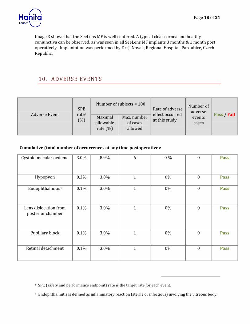
10. ADVERSE EVENTS
3 SPE (safety and performance endpoint) rate is the target rate for each event.
4 Endophthalmitis is defined as inflammatory reaction (sterile or infectious) involving the vitreous body.
Adverse Event SPE rate3 (%)
Number of subjects = 100 Rate of adverse effect occurred at this study
Number of adverse events cases
Pass / Fail Maximal
allowable rate (%)
Max. number of cases allowed
Cumulative (total number of occurrences at any time postoperative):
Cystoid macular oedema
3.0% 8.9% 6 0 % 0 Pass
Hypopyon
0.3% 3.0% 1 0% 0 Pass
Endophthalmitis4
0.1% 3.0% 1 0% 0 Pass
Lens dislocation from posterior chamber
0.1% 3.0% 1 0% 0 Pass
Pupillary block
0.1% 3.0% 1 0% 0 Pass
Retinal detachment
0.1% 3.0% 1 0% 0 Pass

Page 19 of 21
TABLE 7: ADVERSE EVENTS AS DEFINED BY ISO 11979-7 2006:
As shown in table 7, 100% of 37 implanted eyes were not reported with any adverse events.
Thus, it can be concluded that safety of SeeLens MF is in accordance to the ISO 11979-7 2006.
11. CONCLUSIONS:
3 months results
The detailed data from the current study on 37 eyes shows the following benefits of the SeeLens MF IOL:
Optical performance is in accordance to ISO 11979-7 2006 requirements.
The IOL A constant using IOL MASTER and SRK/T is set at 118.6.
Very good safety profile as reflected by a very low rate of intra- and post-operative
complications at 3 months.
5 Excludes posterior capsulotomies.
Secondary surgical intervention5
0.1% 4.2% 2 0% 0 Pass
Persistent (total number of occurrences at 3 months postoperative):
Corneal stroma oedema
0.1% 3.0% 1 0% 0 Pass
Cystoid macular oedema
0.1% 4.2% 2 2.5% 0 Pass
Iritis
0.1% 3.0% 1 0% 0 Pass
Raised IOP req. treatment 0.1% 4.2% 2 0% 0 Pass

Page 20 of 21
Dr. Paparo patients were enrolled according to the Phase 2 recommended patient selection
criteria, and following the optimization of the A constant. This resulted in excellent
uncorrected visual acuity and emmetropic refractive predictability, an excellent defocus
curve and high patient satisfaction.
9-12 months results
As the A constant was not optimized at the time of the first implantations, correction was
necessary in order to evaluate near vision. When corrected for distance all patients
implanted bilaterally achieved J1.
Binocular uncorrected vision for distance was excellent (0.9±0.12) for bilaterally implanted
patients.
Contrast sensitivity in photopic conditions (bilateral patients only) was comparable to other
multifocal IOLs.