section a. musculoskeletal conditions - veterans · web viewm21-1mr, part iii, subpart iv,...
TRANSCRIPT
M21-1MR, Part III, Subpart iv, Chapter 4, Section A
Section A. Musculoskeletal Conditions
Overview
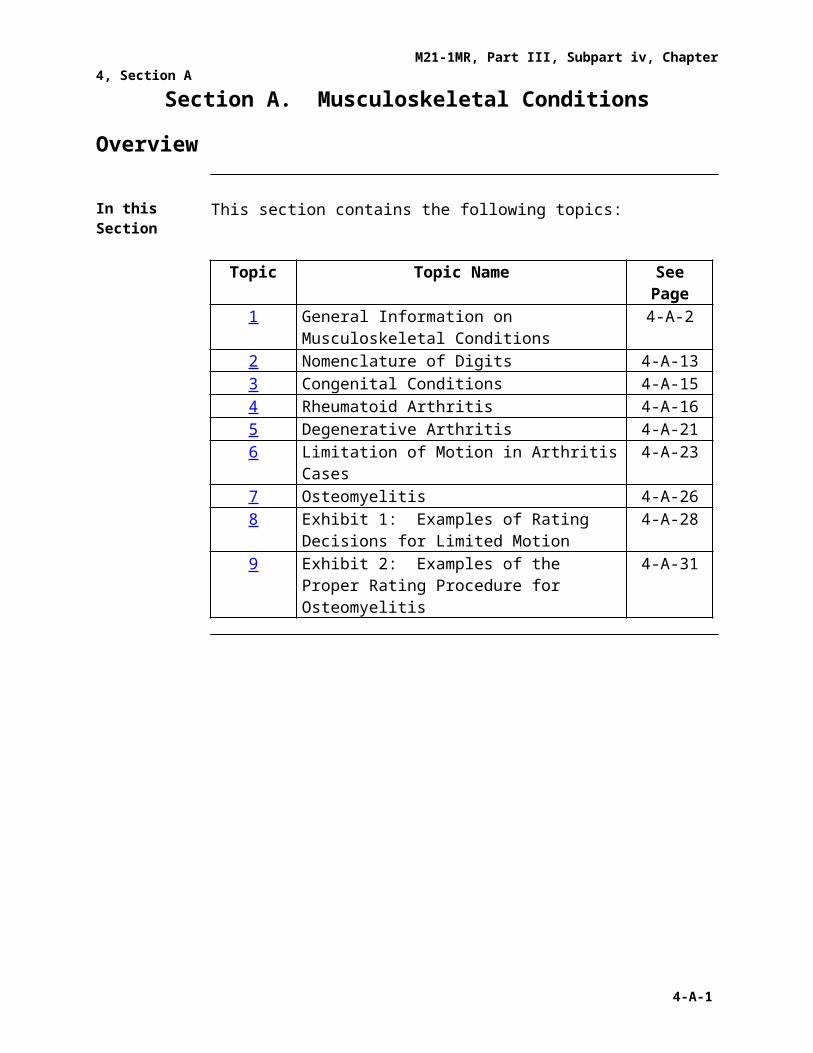
In this Section This section contains the following topics:
Topic Topic Name See Page1 General Information on Musculoskeletal
Conditions4-A-2
2 Nomenclature of Digits 4-A-133 Congenital Conditions 4-A-154 Rheumatoid Arthritis 4-A-165 Degenerative Arthritis 4-A-216 Limitation of Motion in Arthritis Cases 4-A-237 Osteomyelitis 4-A-268 Exhibit 1: Examples of Rating Decisions for
Limited Motion4-A-28
9 Exhibit 2: Examples of the Proper Rating Procedure for Osteomyelitis
4-A-31
4-A-1
M21-1MR, Part III, Subpart iv, Chapter 4, Section A
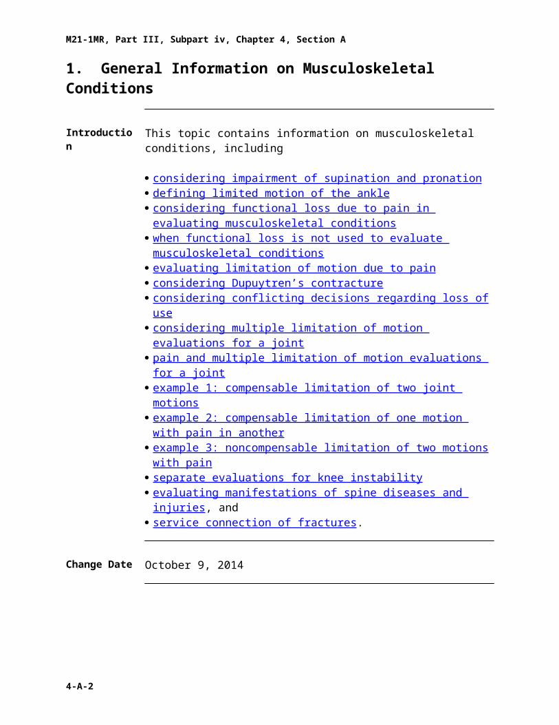
1. General Information on Musculoskeletal Conditions
Introduction This topic contains information on musculoskeletal conditions, including
· considering impairment of supination and pronation · defining limited motion of the ankle · considering functional loss due to pain in evaluating musculoskeletal
conditions· when functional loss is not used to evaluate musculoskeletal conditions · evaluating limitation of motion due to pain · considering Dupuytren’s contracture · considering conflicting decisions regarding loss of use · considering multiple limitation of motion evaluations for a joint · pain and multiple limitation of motion evaluations for a joint · example 1: compensable limitation of two joint motions · example 2: compensable limitation of one motion with pain in another · example 3: noncompensable limitation of two motions with pain · separate evaluations for knee instability · evaluating manifestations of spine diseases and injuries , and· service connection of fractures .
Change Date October 9, 2014
a. Considering Impairment of Supination and Pronation
When preparing ratings involving impairment of pronation and supination, bear in mind the following facts:
· full pronation is the position of the hand flat on a table· full supination is the position of the hand palm up, and· when examining limitation of pronation, the- arc is from full supination to full pronation, and- middle of the arc is the position of the hand, palm vertical to the table.
Assign the lowest 20 percent evaluation when pronation cannot be accomplished through more than the first three-quarters of the arc from full supination.
Do not assign a compensable evaluation for both limitation of pronation and limitation of supination of the same extremity.
Reference: For information on painful motion, see· 38 CFR 4.59 , and· M21-1MR, Part III, Subpart iv, 4.A.1.c .
Continued on next page
4-A-2

M21-1MR, Part III, Subpart iv, Chapter 4, Section A
1. General Information on Musculoskeletal Conditions, Continued
b. Defining Limited Motion of the Ankle
Under diagnostic code (DC) 5271, moderate limitation of ankle motion will be present when there is less than 15 degrees dorsiflexion or less than 30 degrees plantar flexion. Marked limitation of motion is demonstrated when there is less than five degrees dorsiflexion or less than 10 degrees plantar flexion.
c. Considering Functional Loss Due to Pain in Evaluating Musculoskeletal Conditions
Functional loss due to pain is a factor in the evaluation of musculoskeletal conditions under any DC that involves limitation of motion.
It is the responsibility of the examining physician to assess how pain and other factors related to functional impairment equate to limitation of motion. The examiner should either
· report this additional functional loss as range of motion in degrees, or· indicate that he/she cannot determine, without resort to mere speculation,
whether any of these factors cause additional functional loss, and provide the rationale for this opinion.
Notes:· The pain may be caused by the actual joint, connective tissues, nerves, or
muscles.· The medical nature of the particular disability determines whether the DC is
based on limitation of motion.· Per Jones (M.) v. Shinseki, 23 Vet.App. 382 (2010), VA may only accept a
medical examiner’s conclusion that an opinion would be speculative if- the examiner has explained the basis for such an opinion, identifying what
facts cannot be determined, or- the basis for the opinion is otherwise apparent in VA’s review of the
evidence.
References: For more information on· functional loss, see- 38 CFR 4.40 - DeLuca v. Brown , 8 Vet.App. 202 (1995)· disability of the joints, see 38 CFR 4.45, and· painful motion, see 38 CFR 4.59.
Continued on next page
4-A-3

M21-1MR, Part III, Subpart iv, Chapter 4, Section A
1. General Information on Musculoskeletal Conditions, Continued
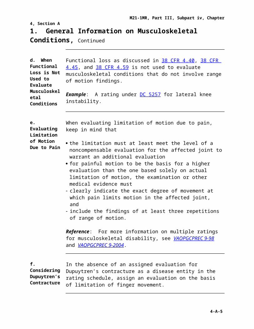
d. When Functional Loss is Not Used to Evaluate Musculoskeletal Conditions
Functional loss as discussed in 38 CFR 4.40, 38 CFR 4.45, and 38 CFR 4.59 is not used to evaluate musculoskeletal conditions that do not involve range of motion findings.
Example: A rating under DC 5257 for lateral knee instability.
e. Evaluating Limitation of Motion Due to Pain
When evaluating limitation of motion due to pain, keep in mind that
· the limitation must at least meet the level of a noncompensable evaluation for the affected joint to warrant an additional evaluation
· for painful motion to be the basis for a higher evaluation than the one based solely on actual limitation of motion, the examination or other medical evidence must
- clearly indicate the exact degree of movement at which pain limits motion in the affected joint, and
- include the findings of at least three repetitions of range of motion.
Reference: For more information on multiple ratings for musculoskeletal disability, see VAOPGCPREC 9-98 and VAOPGCPREC 9-2004.
f. Considering Dupuytren’s Contracture
In the absence of an assigned evaluation for Dupuytren’s contracture as a disease entity in the rating schedule, assign an evaluation on the basis of limitation of finger movement.
g. Considering Conflicting Decisions Regarding Loss of Use
Forward the claims folder to the Director, Compensation Service (211B), for an advisory opinion under M21-1MR, Part III, Subpart vi, 1.A.2.a to resolve a conflict if
· the Insurance Center determines loss of use of two extremities prior to rating consideration involving the same issue, and
· the determination conflicts with the proposed rating decision.
Note: This issue will generally be brought to the attention of the Rating Veterans Service Representative (RVSR) as a result of the type of personal injury, correspondence, or some indication in the claims folder that the insurance activity is involved.
Continued on next page
1. General Information on Musculoskeletal Conditions, Continued
4-A-4
M21-1MR, Part III, Subpart iv, Chapter 4, Section A
h. Considering Multiple Limitation of Motion Evaluations for a Joint
In VAOPGCPREC 9-2004 Office of General Counsel held that separate evaluations under DC 5260 (limitation of knee flexion) and DC 5261 (limitation of knee extension) can be assigned without pyramiding. Despite the fact that knee flexion and extension both occur in the same plane of motion, limitation of flexion (bending the knee) and limitation of extension (straightening the knee) represent distinct disabilities.
Important:· The same principle and handling apply only to- qualifying elbow movement diagnostic codes, flexion (DC 5206), extension
(DC 5207), and impairment of either supination or pronation (DC 5213).- qualifying hip movement diagnostic codes, extension (DC 5251), flexion
(DC 5252), and abduction, adduction or rotation (DC 5253).· Always ensure that multiple evaluations do not violate the amputation rule
in 38 CFR 4.68.
Note:· The Federal Circuit has definitively ruled that multiple evaluations for the
shoulder under DC 5201 are not permitted. In Yonek v. Shinseki, 22 F.3d 1355 (Fed. Cir. 2013) the court held that a Veteran is entitled to a single rating under DC 5201 even though a shoulder disability results in limitation of motion in both flexion (raising the arm in front of the body) and abduction (raising the arm away from the side of the body).
References:· for more information on pyramiding of evaluations, see- 38 CFR 4.14 , and- Esteban v. Brown , 6 Vet.App. 259 (1994),· for information on painful motion in multiple evaluations for joint limitation
of motion, see M21-1MR, Part III, Subpart iv, 4.A.1.i, and· for an example of actual limitation of motion of two knee motions, see
M21-1MR, Part III, Subpart iv, 4.A.1.j .
Continued on next page
1. General Information on Musculoskeletal Conditions, Continued
i. Pain and Multiple Limitation of Motion Evaluations for a Joint
Be aware of the following when considering the role of pain in evaluations for multiple motions of a single joint:
· When either of two qualifying joint motions is actually limited to a compensable degree and there is painful but otherwise noncompensable limitation of the complementary movement, only one compensable evaluation can be assigned.
- Mitchell v. Shinseki , 25 Vet. App. 32 (2011) reinforced that painful motion
4-A-5

M21-1MR, Part III, Subpart iv, Chapter 4, Section A
is the equivalent of limited motion only based on the specific language and structure of DC 5003, not for the purpose of DC 5260 and 5261. For arthritis, if one motion is actually compensable under its 52XX-series DC, then a 10 percent rating under DC 5003 is not available and the complementary motion cannot be treated as limited at the point where it is painful.
- 38 CFR 4.59 does not permit separate compensable evaluations for each painful joint motion. It only provides that VA policy is to recognize actually painful motion as entitled to at least the minimum compensable rating for the joint.
· When each qualifying joint motion is painful but motion is not actually limited to a compensable degree under its applicable 52XX-series DC, only one compensable evaluation can be assigned.
- Assigning multiple compensable evaluations for pain is pyramiding.- A joint affected by arthritis established by x-ray may be evaluated 10
percent disabling under DC 5003.- For common joint conditions that are not rated under the arthritis criteria
such as a knee strain or chondromalacia patella, a 10 percent evaluation can be assigned for the joint based on pain on motion under 38 CFR 4.59.
References:· for more information on pyramiding of evaluations, see- 38 CFR 4.14 , and- Esteban v. Brown , 6 Vet.App. 259 (1994)· for more information on assigning multiple evaluations for a single joint,
see M21-1MR, Part III, Subpart iv, 4.A.1.h, and· for examples of rating where one or both joint motions are not actually
limited to a compensable degree but there is painful motion, see M21-1MR, Part III, Subpart iv, 4.A.1.k and M21-1MR, Part III, Subpart iv, 4.A.1.l.
Continued on next page
1. General Information on Musculoskeletal Conditions, Continued
j. Example 1: Compensable Limitation of Two Joint Motions
Situation: Evaluation of chronic knee strain with the following examination findings:
· Flexion is limited to 45 degrees.· Extension is limited by 10 degrees.· There is no pain on motion.· There is no additional limitation of flexion or extension on additional
repetitions or during flare-ups.
Result: Assign a 10 percent evaluation under DC 5260 and a separate 10 percent evaluation under DC 5261.
4-A-6

M21-1MR, Part III, Subpart iv, Chapter 4, Section A
Explanation: Each rating warrants a separate evaluation and the ratings are for distinct disability.
k. Example 2: Compensable Limitation of One Motion With Pain in Another
Situation: Evaluation of knee tenosynovitis with the following examination findings:
· Flexion is limited to 45 degrees with pain at that point and no additional loss with repetitive motion.
· Extension is full to the 0 degree position, but active extension was limited by pain to 5 degrees.
Result: Assign one 10 percent evaluation under DC 5260.
Explanation:· Flexion is compensable under DC 5260 but extension remains limited to a
noncompensable degree under DC 5261.· Under Mitchell, the painful extension could only considered limited for the
purpose of whether a 10 percent evaluation can be assigned for the joint under DC 5003, which is not applicable in this example because a compensable evaluation was already assigned for flexion under DC 5260.
· 38 CFR 4.59 does not support a separate compensable evaluation for painful extension. The regulation states that the intention of the rating schedule is to recognize actually painful joints due to healed injury as entitled to at least the minimum compensable rating for the joint, not for each painful movement.
· If the fact pattern involved chondromalacia patella or a knee strain rather than tenosynovitis the result would be the same.
Continued on next page
1. General Information on Musculoskeletal Conditions, Continued
l. Example 3: Noncompensable Limitation of Two Motions With Pain
Situation: Evaluation of knee arthritis shown on x-ray with the following examination findings:
· Flexion is limited to 135 degrees with pain at that point.· Extension is full to the 0 degree position with pain at that point.· There is no additional loss of flexion or extension on repetitive motion.
Result: Assign one 10 percent evaluation for the knee under DC 5003.
Explanation:· There is limitation of major joint motion to a noncompensable degree under
DC 5260 and 5261, x-ray evidence of arthritis and satisfactory evidence of
4-A-7

M21-1MR, Part III, Subpart iv, Chapter 4, Section A
painful motion. Painful motion is limited motion for the purpose of applying DC 5003. Therefore a 10 percent evaluation is warranted for the joint.
· Assigning two compensable evaluations, each for pain, would be pyramiding.
· Neither DC 5003 nor 38 CFR 4.59 permits separate 10 percent evaluations for painful flexion and extension; they provide for a 10 percent rating for a joint.
· If the fact pattern involved chondromalacia patella or a knee strain rather than arthritis you would still assign a 10 percent evaluation, not separate evaluations. However the authority would be 38 CFR 4.59 and you should use DC 5260 rather than DC 5003.
Continued on next page
4-A-8

M21-1MR, Part III, Subpart iv, Chapter 4, Section A
1. General Information on Musculoskeletal Conditions, Continued
m. Separate Evaluations for Knee Instability
An evaluation for knee instability may be assigned in addition to any evaluation(s) assigned based on limitation of knee motion. Office of General Counsel has issued Precedent Opinions that an evaluation under DC 5257 does not pyramid with ratings based on limitation of motion.
DC 5257 refers to subluxation or lateral instability, but VBA policy is that ratings based on posterior or anterior instability are also permitted and do not pyramid with evaluations based on limitation of motion.
Notes:· Evaluations under DC 5258 (dislocated semilunar cartilage) and DC 5259
(symptomatic removal of semilunar cartilage) can pyramid with evaluations under DC 5260 or DC5261 for limitation of knee flexion or extension. Therefore, separate compensable evaluations are not permitted.
· Although DC 5258 refers to “dislocated” cartilage and “locking” of the knee the rating criteria contemplate limitation of motion of the knee through functional impairment with use (namely pain and effusion).
· DC 5259 provides for a compensable evaluation for a “symptomatic” knee post removal of the cartilage. VAOPGCPREC 9-98 states “DC 5259 requires consideration of 38 CFR 4.40 and 4.45 because removal of semilunar cartilage may result in complications producing loss of motion.”
Continued on next page
4-A-9

M21-1MR, Part III, Subpart iv, Chapter 4, Section A
1. General Information on Musculoskeletal Conditions, Continued
m. Separate Evaluations for Knee Instability (continued)
· Do not rate instability separately from a total knee replacement. The 30 percent and 100 percent evaluations under DC 5055 are minimum and maximum evaluations and, as such, encompass all identifiable residuals post knee replacement – including limitation of motion, instability, and functional impairment. The 60 percent and intermediate evaluations by their plain text provide the exclusive methods by which residuals can be evaluated at 40 or 50 percent and contemplate instability. Post arthroplasty, there may be instability with weakness (giving way) and pain. Note that the only way to obtain an evaluation in excess of 30 percent under DC 5262 (one of the specified bases for an intermediate rating under DC 5055) is if there is nonunion with loose motion and need for a brace. This clearly suggests instability is incorporated in the intermediate criteria.
Important: RVSRs are reminded to pay close attention to the combined evaluation of the knee disability prior to replacement surgery and to follow all required due process and protected evaluation procedures.
References: For more information on· pyramiding and separating individual decisions in a rating decision, see
M21-1MR, Part III, Subpart iv, 6.C.11· separate evaluation of knee instability, see- VAOPGCPREC 23-97 , and- VAOPGCPREC 9-98 · due process issues pertinent to knee replacements including- change of diagnostic code for a protected disability evaluation, see
38 CFR 3.951 , and M21-1MR, Part III, Subpart iv, 8.C.8 , and
- reduction procedures that would apply prior to assignment of a post surgical minimum evaluation lower than the running award rate, see 38 CFR 3.105, and M21-1MR, Part III, Subpart iv, 8.D.11 .
Continued on next page
4-A-10

M21-1MR, Part III, Subpart iv, Chapter 4, Section A
1. General Information on Musculoskeletal Conditions, Continued
n. Evaluating Manifestations of Spine Diseases and Injuries
Evaluate diseases and injuries of the spine based on the criteria listed in the General Rating Formula for Diseases and Injuries of the Spine (General Rating Formula). Under this criteria, evaluate conditions based on chronic orthopedic manifestations (e.g. painful muscle spasm or limitation of motion), and any associated neurological manifestations (e.g. footdrop, muscle atrophy, or sensory loss) by assigning separate evaluations for the orthopedic and neurological manifestations.
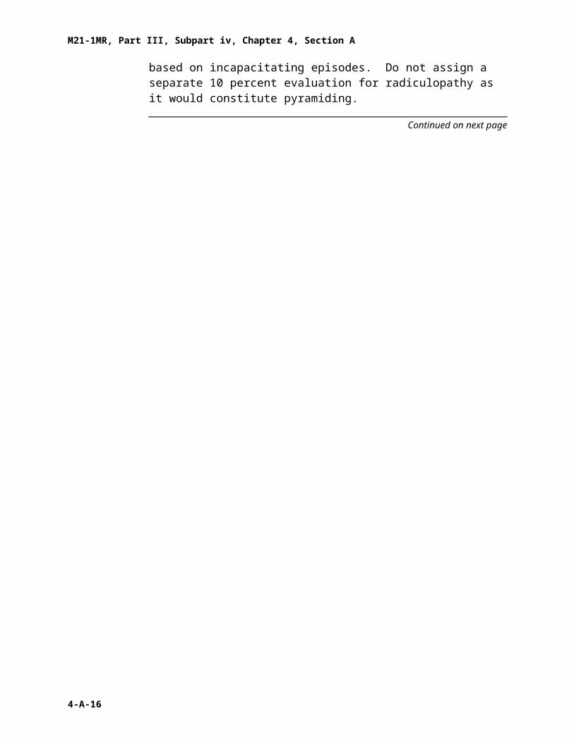
Evaluate intervertebral disc syndrome under DC 5243 either based on the General Rating Formula or the Formula for Rating Intervertebral Disc Syndrome Based on Incapacitating Episodes (Incapacitating Episode Formula), whichever formula results in the higher evaluation when all disabilities are combined under 38 CFR 4.25.
Note: If an evaluation is assigned based on incapacitating episodes, a separate rating may not be assigned for radiculopathy or any other associated objective neurological abnormality as it would constitute pyramiding.
Example: A Veteran’s degenerative disc disease of the lumbar spine warrants a 20 percent based on limitation of motion under the General Rating Formula and his mild radiculopathy of the left lower extremity warrants a 10 percent rating. However, the Veteran has incapacitating episodes of four weeks duration over the past 12 months, which would result in a 40 percent evaluation based on the Incapacitating Episode Formula. In such circumstances, assign only the 40 percent evaluation based on incapacitating episodes. Do not assign a separate 10 percent evaluation for radiculopathy as it would constitute pyramiding.
Continued on next page
4-A-11

M21-1MR, Part III, Subpart iv, Chapter 4, Section A
1. General Information on Musculoskeletal Conditions, Continued
o. Service Connection of Fractures
Decision makers must not automatically grant service connection for fracture or fracture residuals based on a mere service treatment record (STR) reference to a fracture.
Where service connection of a fracture or fracture residuals is claimed, service connection may be established when sufficient evidence, such as X-rays, a surgical report, casting, or a physical evaluation board report, documents the fracture.
Service connection may be established for a healed fracture even without current residual limited motion or functional impairment of a joint.
Assign a DC consistent with the location of the fracture. The fracture will be rated as noncompensable in the absence of any disabling manifestations.
Reference: For more information about unclaimed chronic disabilities found in STRs, see M21-MR, Part III, Subpart iv, 6.B.6.
4-A-12

M21-1MR, Part III, Subpart iv, Chapter 4, Section A
2. Nomenclature of Digits
Introduction This topic contains information on the nomenclature of digits, including
· specifying injured digits and phalanges , and· identifying the digits of the hand and foot .
Change Date December 13, 2005
a. Specifying Injured Digits and Phalanges
Follow the guidelines listed below to accurately specify the injured digits of the upper and lower extremities.
· Each digit, except the thumb and the great toe, includes three phalanges- the proximal phalanx (closest to the wrist or ankle)- the middle phalanx, and- the distal phalanx (closest to the tip of the finger or toe).· The joint between the proximal and middle phalanges is called the proximal
interphalangeal (PIP) joint.· The joint between the middle and distal phalanges is called the distal
interphalangeal (DIP) joint.· The thumb and great toe each have only two phalanges, the proximal
phalanx and the distal phalanx. Therefore, each thumb and each great toe has only a single joint, called the interphalangeal (IP) joint.
· The joints connecting the phalanges in the hands to the metacarpals are the metacarpophalangeal (MCP) joints.
· The joints connecting the phalanges in the feet to the metatarsals are the metatarsophalangeal (MTP) joints.
Note: If the location of the injury is unclear, obtain x-rays to clarify the exact point of injury.
Continued on next page
2. Nomenclature of Digits, Continued
b. Identifying the Digits of the Hand and Foot
Use the table below to correctly identify the digits of the hand and foot.
Note: Designate either right or left for the digits of the hand or foot.
If the extremity is the … Then identify the digit as the …
4-A-13

M21-1MR, Part III, Subpart iv, Chapter 4, Section A
hand · thumb· index· long· ring, or· little.
Note: Do not use numerical designations for either the fingers or joints of the fingers.
foot · first or great toe· second· third· fourth, or· fifth.
4-A-14

M21-1MR, Part III, Subpart iv, Chapter 4, Section A
3. Congenital Conditions
Introduction This topic contains information on congenital conditions, including
· recognizing variations in development and appearance , and· considering notable defects .
Change Date December 13, 2005
a. Recognizing Variations in Development and Appearance
Individuals vary greatly in their musculoskeletal development and appearance. Functional variations are often seen and can be attributed to
· the type of individual, and· his/her inherited or congenital variations from the normal.
b. Considering Notable Defects
Give careful attention to congenital or developmental defects such as
· absence of parts· subluxation (partial dislocation of a joint)· deformity or exostosis (bony overgrowth) of parts, and/or· accessory or supernumerary (in excess of the normal number) parts.
Note congenital defects of the spine, especially
· spondylolysis· spina bifida· unstable or exaggerated lumbosacral joints or angle, or· incomplete sacralization.
Notes:· Do not automatically classify spondylolisthesis as a congenital condition,
although it is commonly associated with a congenital defect.· Do not overlook congenital diastasis of the rectus abdominus, hernia of the
diaphragm, and the various myotonias.
Reference: For more information on congenital or developmental defects, see 38 CFR 4.9.
4-A-15
M21-1MR, Part III, Subpart iv, Chapter 4, Section A
4. Rheumatoid Arthritis
Introduction This topic contains information about rheumatoid arthritis, including
· characteristics of rheumatoid arthritis · periods of flares and remissions of rheumatoid arthritis · clinical signs of rheumatoid arthritis · radiologic changes in rheumatoid arthritis · disability factors associated with rheumatoid arthritis , and· points to consider in the rating decision .
Change Date December 29, 2007
a. Characteristics of Rheumatoid Arthritis
The following are characteristics of rheumatoid arthritis, also diagnosed as atrophic or infectious arthritis, or arthritis deformans:
· the onset- occurs before middle age, and- may be acute, with a febrile attack, and· the symptoms include a usually laterally symmetrical limitation of
movement- first affecting proximal interphalangeal and metacarpophalangeal joints- next causing atrophy of muscles, deformities, contractures, subluxations,
and- finally causing fibrous or bony ankylosis (abnormal adhesion of the bones
of the joint).
Important: Marie-Strumpell disease, also called rheumatoid spondylitis or ankylosing spondylitis, is not the same disease as rheumatoid arthritis. Rheumatoid arthritis and Marie-Strumpell disease have separate and distinct clinical manifestations and progress differently.
Continued on next page
4. Rheumatoid Arthritis, Continued
4-A-16
M21-1MR, Part III, Subpart iv, Chapter 4, Section A
b. Periods of Flares and Remissions in Rheumatoid Arthritis
The symptoms of rheumatoid arthritis come and go, depending on the degree of tissue inflammation. When body tissues are inflamed, the disease is active. When tissue inflammation subsides, the disease is inactive (in remission).
Remissions can occur spontaneously or with treatment, and can last weeks, months, or years. During remissions, symptoms of the disease disappear, and patients generally feel well. When the disease becomes active again (relapse), symptoms return.
Note: The return of disease activity and symptoms is called a flare. The course of rheumatoid arthritis varies from patient to patient, and periods of flares and remissions are typical.
c. Clinical Signs of Rheumatoid Arthritis
The table below contains information about the clinical signs of rheumatoid arthritis.
Stage of Disease
Symptoms
Initial · periarticular and articular swelling, often free fluid, with proliferation of the synovial membrane, and
· atrophy of the muscles
Note: Atrophy is increased to wasting if the disease is unchecked.
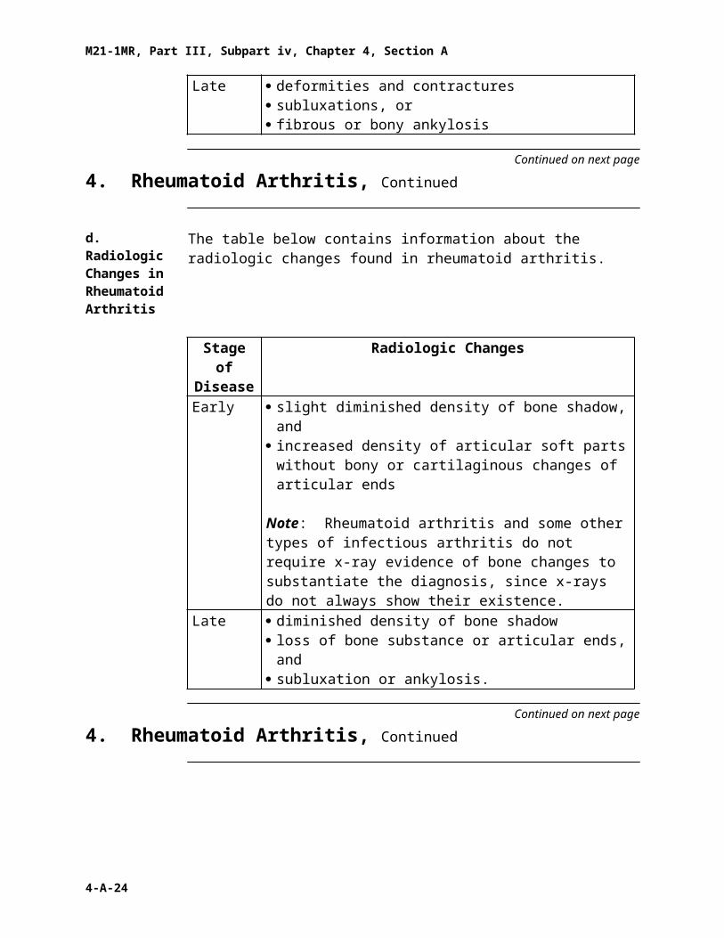
Late · deformities and contractures· subluxations, or· fibrous or bony ankylosis
Continued on next page
4. Rheumatoid Arthritis, Continued
d. Radiologic Changes in Rheumatoid Arthritis
The table below contains information about the radiologic changes found in rheumatoid arthritis.
Stage of Disease
Radiologic Changes
4-A-17

M21-1MR, Part III, Subpart iv, Chapter 4, Section A
Early · slight diminished density of bone shadow, and· increased density of articular soft parts without bony or
cartilaginous changes of articular ends
Note: Rheumatoid arthritis and some other types of infectious arthritis do not require x-ray evidence of bone changes to substantiate the diagnosis, since x-rays do not always show their existence.
Late · diminished density of bone shadow· loss of bone substance or articular ends, and· subluxation or ankylosis.
Continued on next page
4. Rheumatoid Arthritis, Continued
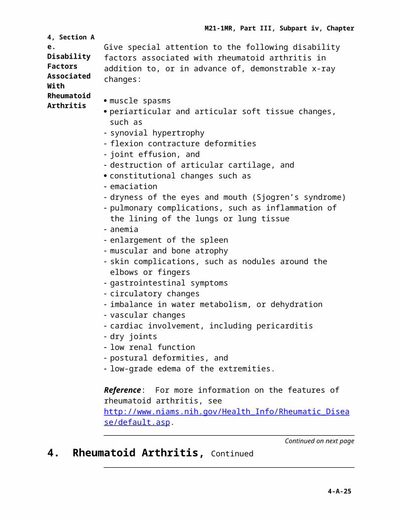
e. Disability Factors Associated With Rheumatoid Arthritis
Give special attention to the following disability factors associated with rheumatoid arthritis in addition to, or in advance of, demonstrable x-ray changes:
· muscle spasms· periarticular and articular soft tissue changes, such as- synovial hypertrophy- flexion contracture deformities- joint effusion, and- destruction of articular cartilage, and· constitutional changes such as- emaciation- dryness of the eyes and mouth (Sjogren’s syndrome)- pulmonary complications, such as inflammation of the lining of the lungs or
lung tissue- anemia- enlargement of the spleen- muscular and bone atrophy- skin complications, such as nodules around the elbows or fingers- gastrointestinal symptoms- circulatory changes- imbalance in water metabolism, or dehydration- vascular changes- cardiac involvement, including pericarditis- dry joints- low renal function- postural deformities, and- low-grade edema of the extremities.
Reference: For more information on the features of rheumatoid arthritis, see http://www.niams.nih.gov/Health_Info/Rheumatic_Disease/default.asp.
4-A-18

M21-1MR, Part III, Subpart iv, Chapter 4, Section A
Continued on next page
4. Rheumatoid Arthritis, Continued
f. Points to Consider in the Rating Decision
In the rating decision, note the presence of joints affected by any of the following:
· synovial hypertrophy or joint effusion· severe postural changes; scoliosis; flexion contracture deformities· ankylosis or limitation of motion of joint due to bony changes, and/or· destruction of articular cartilage.
4-A-19

M21-1MR, Part III, Subpart iv, Chapter 4, Section A
5. Degenerative Arthritis
Introduction This topic contains information about degenerative arthritis, including
· characteristics of degenerative arthritis · diagnostic symptoms of degenerative arthritis · radiologic changes in degenerative arthritis · symptoms of degenerative arthritis of the spine , and· points to consider in the rating decision .
Change Date December 13, 2005
a. Characteristics of Degenerative Arthritis
The following are characteristics of degenerative arthritis, also diagnosed as osteoarthritis or hypertrophic arthritis:
· The onset generally occurs after the age of 45.· It has no relation to infection.· It is asymmetrical (more pronounced on one side of the body than the
other).· There is limitation of movement in the late stages only.
b. Diagnostic Symptoms of Degenerative Arthritis
Diagnostic symptoms of degenerative arthritis include
· the presence of Heberden’s nodes or calcific deposits in the terminal joints of the fingers with deformity
· ankylosis, in rare cases· hyperostosis and irregular, notched articular surfaces of the joints· destruction of cartilage· bone eburnation, and· the formation of osteophytes.
Note: The flexion contracture deformities and severe constitutional symptoms described under rheumatoid arthritis do not usually occur in degenerative arthritis.
Continued on next page
5. Degenerative Arthritis, Continued
c. Radiologic Changes in Degenerative Arthritis
The table below contains information about the radiologic changes found in degenerative arthritis.
4-A-20

M21-1MR, Part III, Subpart iv, Chapter 4, Section A
Stage Radiologic ChangesEarly delicate spicules of calcium at the articular margins without
· diminished density of bone shadow, and· increased density of articular of parts.
Late · ridging of articular margins· hyperostosis· irregular, notched articular surfaces, and· ankylosis only in the spine.
d. Symptoms of Degenerative Arthritis of the Spine
Degenerative arthritis of the spine and pelvic joints is characterized clinically by the same general characteristics as arthritis of the major joints except that
· limitation of spine motion occurs early· chest expansion and costovertebral articulations are not usually affected· referred pain is commonly called “intercostal neuralgia” and “sciatica,”
and· localized ankylosis may occur if spurs on bodies of vertebrae impinge.
e. Points to Consider in the Rating Decision
Degenerative and traumatic arthritis require x-ray evidence of bone changes to substantiate the diagnosis.
Reference: For more information on considering x-ray evidence when evaluating arthritis, see DC 5003.
4-A-21

M21-1MR, Part III, Subpart iv, Chapter 4, Section A
6. Limitation of Motion in Arthritis Cases
Introduction This topic contains information on limitation of motion due to arthritis, including
· conditions compensable under other diagnostic codes · conditions not compensable under other diagnostic codes · reference for rating decisions involving limitation of motion · arthritis previously rated as a single disability · using DCs 5013 through 5024 in rating decisions , and· considering the effects of a change of diagnosis in arthritis cases .
Change Date December 13, 2005
a. Conditions Compensable Under Other Diagnostic Codes
For a joint or group of joints affected by degenerative arthritis, use the diagnostic code which justifies the assigned evaluation.
Example: When the compensable requirements for limited motion of a joint are met under a code other than DC 5003, hyphenate that code in the conclusion with a preceding “5003-.” Then list the appropriate code, such as DC 5261, limited extension of the knee, 10 percent, creating the code DC “5003-5261.”
Exception: If other joints affected by arthritis are compensably evaluated in the same rating, use only the code appropriate to these particular joints which support the assigned evaluation and omit the modifying “5003.”
b. Conditions Not Compensable Under Other Diagnostic Codes
Whenever limited motion is noncompensable under codes appropriate to a particular joint, assign 10 percent under DC 5003 for each major joint or group of minor joints affected by limited or painful motion as prescribed under DC 5003.
If there is no limited or painful motion, but there is x-ray evidence of degenerative arthritis, assign under DC 5003 either a 10 percent evaluation or a 20 percent for occasional incapacitating exacerbations, based on the involvement of two or more major joints or two or more groups of minor joints.
Important: Do not combine under 38 CFR 4.25 a 10 or 20 percent rating that is based solely on x-ray findings with ratings that are based on limited or painful motion.
Continued on next page
6. Limitation of Motion in Arthritis Cases, Continued
4-A-22

M21-1MR, Part III, Subpart iv, Chapter 4, Section A
c. Reference: Rating Decisions Involving Limitation of Motion
For more information on rating decisions involving limitation of motion, see M21-1MR, Part III, Subpart iv, 4.A.8.
d. Arthritis Previously Rated as a Single Disability
The RVSR may encounter cases where arthritis of multiple joints is rated as a single disability.
Use the information in the table below to handle cases where arthritis was previously rated as a single disability.
If … Then …· the separate evaluation of the
arthritic disability results in no change in the combined degree previously assigned, and
· a rating is required
rerate using the current procedure with the same effective date as previously assigned.
rerating the arthritic joint separately results in an increased combined evaluation
apply 38 CFR 3.105(a) to retroactively increase the assigned evaluation.
rerating the arthritic joint separately results in a reduced combined evaluation
· request an examination, and· if still appropriate, propose reduction
under 38 CFR 3.105(a) and 38 CFR 3.105(e).
Exception: Do not apply 38 CFR 3.105(a) if the assigned percentage is protected under 38 CFR 3.951.
Reference: For more information on protected ratings, see M21-1MR, Part III, Subpart iv, 8.C.
Continued on next page
6. Limitation of Motion in Arthritis Cases, Continued
e. Using DCs 5013 Through 5024 in Rating Decisions
Use the table below to rate cases that use DCs 5013 through 5024.
If the DC of the case is … Then …gout under DC 5017 rate the case as rheumatoid arthritis, 5002.
4-A-23
M21-1MR, Part III, Subpart iv, Chapter 4, Section A
· 5013 through 5016, and· 5018 through 5024
evaluate the case according to the criteria for limited motion or painful motion under DC 5003, degenerative arthritis.
Note: The provisions under DC 5003 regarding a compensable minimum evaluation of 10 percent for limited or painful motion apply to these diagnostic codes and no others.
Reference: For more information on 10 and 20 percent ratings based on x-ray findings, see 38 CFR 4.71a, DC 5003, Note (2).
f. Considering the Effects of a Change in Diagnosis in Arthritis Cases
A change of diagnosis among the various types of arthritis, particularly if joint disease has been recognized as service-connected for several years, has no significant bearing on the question of service connection.
Note: In older individuals, the effects of more than one type of joint disease may coexist.
Reference: For information on rating rheumatoid arthritis, see38 CFR 4.71a, DC 5002, Arthritis rheumatoid.
4-A-24
M21-1MR, Part III, Subpart iv, Chapter 4, Section A
7. Osteomyelitis
Introduction This topic contains information about osteomyelitis, including
· requiring constitutional symptoms · historical ratings · assigning historical ratings , and· the reasons to discontinue a historical rating .
Change Date December 13, 2005
a. Requiring Constitutional Symptoms
Constitutional symptoms are a prerequisite to the assignment of either the 100 percent or 60 percent evaluations under DC 5000.
Since both the 60 and 100 percent evaluations are based on constitutional symptoms, neither is subject to the amputation rule.
Reference: For more information on the amputation rule, see 38 CFR 4.68.
b. Historical Ratings
Both the 10 percent evaluation and that part of the 20 percent evaluation that is based on “other evidence of active infection within the last five years” are
· historical ratings, and· based on recurrent episodes of osteomyelitis.
Note: The 20 percent historical evaluation based on evidence of active infection within the past five years must be distinguished from the 20 percent evaluation authorized when there is a discharging sinus.
Continued on next page
7. Osteomyelitis, Continued
4-A-25

M21-1MR, Part III, Subpart iv, Chapter 4, Section A
c. Assigning Historical Ratings
An initial episode of active osteomyelitis is not a basis for either of the historical ratings.
Assign the historical rating as follows:
· When the first recurrent episode of osteomyelitis is shown- assign a 20 percent historical evaluation, and- extend the evaluation for five years from the date of examination showing
the osteomyelitis to be inactive.· Assign a closed rating at the expiration of the five-year extension.· Assign the 10 percent historical evaluation only if there have been two or
more recurrences of active osteomyelitis following the initial infection.
d. Reasons to Discontinue Historical Ratings
Do not discontinue the historical rating, even if treatment includes saucerization, sequestrectomy, or guttering, because the osteomyelitis is not considered cured.
Exception: If there has been removal or radical resection of the affected bone· consider osteomyelitis cured, and· discontinue the historical rating.
4-A-26

M21-1MR, Part III, Subpart iv, Chapter 4, Section A
8. Exhibit 1: Examples of Rating Decisions for Limited Motion
Introduction This exhibit contains three examples of ratings for limited motion in arthritis cases.
Change Date December 13, 2005
a. Example 1 Situation: The Veteran has residuals of degenerative arthritis with limitation of abduction of the right shoulder (major) to 90 degrees and limitation of flexion of the right knee to 45 degrees.
Coded Conclusion:1. SC (VE INC)5003-5201 Degenerative arthritis, right shoulder (dominant)20% from 12-14-03
5260 Degenerative arthritis, right knee10% from 12-14-03
COMB 30% from 12-14-03
Rationale: The shoulder and knee separately meet compensable requirements under diagnostic codes 5201 and 5260, respectively.
b. Example 2 Situation: The Veteran has X-ray evidence of degenerative arthritis of both knees without
· limited or painful motion of any of the affected joints, or· incapacitating episodes.
Coded Conclusion:1. SC (PTE INC)5003 Degenerative arthritis of the knees, x-ray evidence10% from 12-30-01
Rationale: There is no limited or painful motion in either joint, but there is x-ray evidence of arthritis in more than one joint to warrant a 10 percent evaluation under DC 5003.
Continued on next page
8. Exhibit 1: Examples of Rating Decisions for Limited Motion, Continued
4-A-27

M21-1MR, Part III, Subpart iv, Chapter 4, Section A
c. Example 3 Situation: The Veteran has X-ray evidence of degenerative arthritis of the right knee without limited or painful motion.
Coded Conclusion:1. SC (PTE INC)5003 Degenerative arthritis, right knee, x-ray evidence only0% from 12-30-01
Rationale: There is no limited or painful motion in the right knee or x-ray evidence of arthritis in more than one joint to warrant a compensable evaluation under DC 5003.
4-A-28

M21-1MR, Part III, Subpart iv, Chapter 4, Section A
9. Exhibit 2: Examples of the Proper Rating Procedure for Osteomyelitis
Introduction This exhibit contains eight examples of the proper procedure for rating osteomyelitis.
Change Date December 13, 2005
a. Example 1 Situation: The Veteran was diagnosed with osteomyelitis in service, with discharging sinus. At separation from service the osteomyelitis was inactive with no involucrum or sequestrum. There is no evidence of recurrence.
Result: As there has been no recurrence of active osteomyelitis following the initial episode in service, the historical evaluation of 20 percent is not for application. The requirements for a 20 percent evaluation based on activity are not met either.
Coded Conclusion:1. SC (PTE INC)5000 Osteomyelitis, right tibia0% from 12-2-93
b. Example 2 Situation: Same facts as in Example 1, but the Veteran had a discharging sinus at the time of separation from service.
Result: The Veteran meets the criteria for a 20 percent evaluation based on a discharging sinus. Schedule a future examination to ascertain the date of inactivity.
Coded Conclusion:1. SC (PTE INC)5000 Osteomyelitis, right tibia, active20% from 12-2-93
Continued on next page
9. Exhibit 2: Examples of the Proper Rating Procedure for Osteomyelitis, Continued
4-A-29

M21-1MR, Part III, Subpart iv, Chapter 4, Section A
c. Example 3 Situation: Same facts as in Example 2. Subsequent review examination reveals the sinus tract was healed and there is no other evidence of active infection.
Result: Since the Veteran has not had a recurrent episode of osteomyelitis since service, a historical rating of 20 percent is not for application. Take rating action under 38 CFR 3.105(e).
Coded Conclusion:1. SC (PTE INC)5000 Osteomyelitis, right tibia, inactive20% from 12-2-930% from 3-1-95
d. Example 4 Situation: Same facts as in Example 2. The Veteran is hospitalizedJuly 2l, 1996, with active osteomyelitis of the right tibia shown with discharging sinus. There is no involucrum, sequestrum, or constitutional symptom. Upon release from the hospital the discharging sinus is still present.
Result: Assign the 20 percent evaluation based on evidence showing draining sinus from the proper effective date. Schedule a future examination to ascertain date of inactivity.
Coded Conclusion:1. SC (PTE INC)5000 Osteomyelitis, right tibia, active0% from 3-1-9520% from 7-21-96
Continued on next page
9. Exhibit 2: Examples of the Proper Rating Procedure for Osteomyelitis, Continued
e. Example 5 Situation: Same facts as in Example 3. A routine future examination was conducted on July 8, 1997, showing the osteomyelitis to be inactive. There was no discharging sinus, no involucrum, sequestrum, or constitutional symptom. The most recent episode of active osteomyelitis (July 21, 1996) constitutes the first “recurrent” episode of active osteomyelitis.
Result: Continue the previously assigned 20 percent evaluation, which was granted on the basis of discharging sinus as a historical evaluation for 5 years from the examination showing inactivity.
Coded Conclusion:1. SC (PTE INC)
4-A-30

M21-1MR, Part III, Subpart iv, Chapter 4, Section A
5000 Osteomyelitis, right tibia, inactive20% from 7-21-960% from 7-8-02
f. Example 6 Situation: Same facts as in Example 4. In October 1999, the Veteran was again found to have active osteomyelitis with a discharging sinus, without involucrum, sequestrum, or constitutional symptoms.
Result: Continue the 20 percent evaluation. Rerating is necessary to remove the future reduction to 0 percent, and to schedule a future examination to establish the date of inactivity.
Coded Conclusion:1. SC (PTE INC)5000 Osteomyelitis, right tibia, active20% from 7-21-96
Continued on next page
9. Exhibit 2: Examples of the Proper Rating Procedure for Osteomyelitis, Continued
g. Example 7 Situation: Same facts as in Example 5. A review examination was conducted on April 8, 2000. The examination showed the discharging sinus was inactive, and there was no other evidence of active osteomyelitis. The most recent episode of osteomyelitis (October 1999) constitutes the second "recurrent" episode of active osteomyelitis.
Result: The historical evaluations of 20 and 10 percent both apply.
Coded Conclusion:1. SC (PTE INC)5000 Osteomyelitis, right tibia, inactive20% from 7-21-9610% from 4-8-05
Continued on next page
9. Exhibit 2: Examples of the Proper Rating Procedure for Osteomyelitis, Continued
4-A-31

M21-1MR, Part III, Subpart iv, Chapter 4, Section A
h. Example 8 Situation: Same facts as in Example 6. The Veteran was hospitalized June 10, 2002, with a recurrent episode of active osteomyelitis. A radical resection of the right tibia was performed and at hospital discharge (June 21, 2002), the osteomyelitis was shown to be cured.
Result: Assign a temporary total rating of 100 percent under paragraph 30 with a 1-month period of convalescence. Following application of 38 CFR 3.105(e), reduce the evaluation for osteomyelitis to 0 percent as a rating for osteomyelitis will not be applied following cure by removal or radical resection of the affected bone.
Coded Conclusion:1. SC (PTE INC)5000 Osteomyelitis, right tibia, P.O.20% from 7-21-96100% from 6-10-02 (Par. 30)20% from 8-1-020% from 10-1-02
4-A-32