sebastian lucas dept of histopathology st thomas’hospital ... · dept of histopathology st...
TRANSCRIPT

Bacterial & parasitic infections
Sebastian Lucas
Dept of Histopathology
St Thomas’ Hospital
London SE1

HIV disease
Biliary tract
infections
Bacterial &
Parasitic infections
Other viral
infections
Hepatitis A-x
HBV
HCV
Post-Tx
infections
Crypto-
sporidiosis
CMV
EBV

Hepatobiliary parasitesLiver• Leishmania spp
• Trypanosoma cruzi
• Entamoeba histolytica
• Toxoplasma gondii
• Plasmodium falciparum
• Balantidium coli
• Strongyloides stercoralis
• Angiostrongylus spp
• Enterobius vermicularis
• Ascaris lumbricoides
• Baylisascaris
• Toxocara canis
• Gnathostoma spp
• Capillaria hepatica
• Schistosoma spp
• Echinococcus granulosus & multilocularis
• pentasomes
Biliary tree & GB
• microsporidia spp
• Cryptosporidium spp
• Ascaris
• Fasciola hepatica
• Clonorchis sinensis
• Opisthorcis viverrini
• Dicrocoelium
• Echinococcus granulosus
Gutierrez: ‘Diagnostic Pathology ofParasitic Infections’, Oxford, 2000


What is this?
Both are the same
parasite

What is this?
Both are the same
parasite
Echinococcus multilocularis
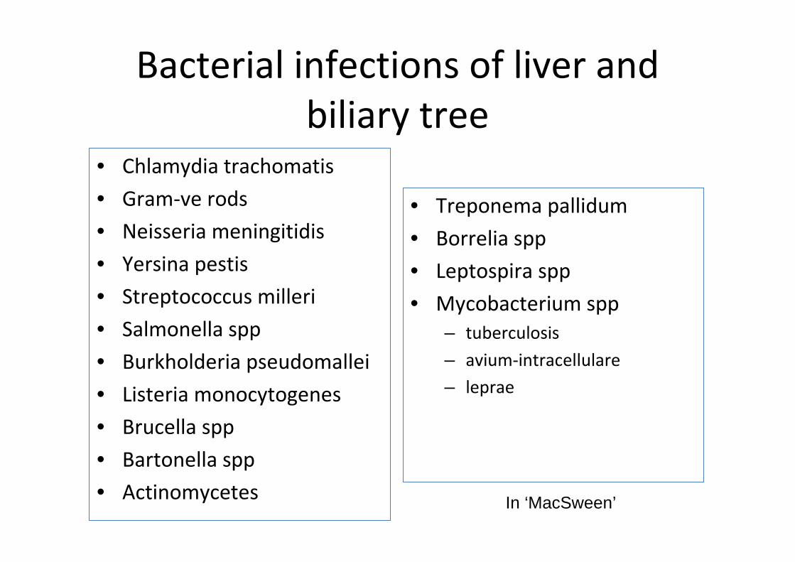
Bacterial infections of liver and
biliary tree• Chlamydia trachomatis
• Gram-ve rods
• Neisseria meningitidis
• Yersina pestis
• Streptococcus milleri
• Salmonella spp
• Burkholderia pseudomallei
• Listeria monocytogenes
• Brucella spp
• Bartonella spp
• Actinomycetes
• Treponema pallidum
• Borrelia spp
• Leptospira spp
• Mycobacterium spp
– tuberculosis
– avium-intracellulare
– leprae
In ‘MacSween’

2 manifestations of a classic bacterial infection

Bacteria & parasites
3 case studies
• Problem solving
• What it is
• What is the treatment
What you need to know
• What can happen
– differential diagnosis
• How it can look
pathologically
• What can be done to prove
or exclude the infection
• Who you can ask for help

Summary
• Anything can happen
– International travel
• ‘Unde venis?’
– Immunosuppression
• Acquired
• Iatrogenic
• Increasing reliance on molecular diagnostics
• Close collaboration with microbiologists and
ID clinicians

Case 1F20
Daughter of UK farmer
No travel abroad
Acute lymphoblastic leukaemia @ 12 years
2 months post haemopoietic stem cell transplant
Total colectomy for mucormycosis
Not on steroids or cyclosporin
Fever; liver failure
Liver biopsy
“Is this toxoplasmosis?”


x1,000

Small blue dots
• Toxoplasma
• Trypanosoma cruzi
• Leishmania
• Cryptosporidium
• microsporidia
• Bacterial cocci
– Staphylococcus
• Fungi
– Histoplasma capsulatum
– Candida spp
– Penicillium marneffei

Toxoplasma gondii

x1,000

Leish vs Histo
kinetoplast

Leish histo & cyto

Giemsa & Gram = protozoa, eccentric nucleus gram+

ZN (carbol fuchsin)+ and PAS+ polar dots

Case
• Not fungi
• Not bacterial
• Protozoa
– Not leishmania or toxoplasma
• Microsporidia
– Encephalitozoon sp
– Pleistophora sp

How to confirm
microsporidia?
Electron microscopy
Faecal analysis (acid fast
stain)
PCR on tissue or faeces
[no tissue remained in the
biopsy block]
PCR = Encephalitozoon
cuniculi
Patient died despite
therapy

EM identification

EM (thanks to Alan Curry, Manchester
HPA)

Microsporidia
• Parasitic protozoa of invertebrates & vertebrates
• Well known to vet pathologists
• Virtually unknown to human pathology until HIV/AIDS
• 1985: Enterocytozoon bieneusi – gut
• 1987: Encephalitozoon intestinalis – gut, systemic infection
• et seq: many other species - Pleistophora, E.cuniculi
• Treatment? albendazole + intraconazole

An autopsy case - cholangitis

Autopsy case of disseminated microsporidiosis

microsporidiosis

Case solved
• H&E histopathology
– Supportive special stains
• Exclusion of alternatives (special stain)
• Examination of corroborative material
• Molecular diagnostics
• Electron microscopy
• In the meantime, treatment started
– albendazole + itraconazole

Case 2
• Female Briton age 74
• Scan of liver shows a mass
• ? Tumour
• Partial liver resection 17 x 13 x 7cm
• Well circumscribed mass comprising two
adjacent nodules 2m diameter

Liver resection – H&E

Eosinophilic necrotic nodules
(liver, lymph node, spleen)
• Worm infections
• Tumour
– Hodgkin disease
• Drug reaction
• Idiopathic allergy / immunopathology
• NB: no useful published literature
– ‘Focal eosinophilic necrosis’ (FEN) mentioned in the radiological imaging literature from Asia

Liver resection

Liver resection – H&E

Liver resection

Liver resection

Liver resection – Charcot-Leyden crystals

Liver resection – what is this?

Case 2
• Original diagnosis
– Hydatid cyst
• …but hydatid serology negative
• Review of biopsy……

Your [Swiss path soc] diagnoses
• Amoebic abscess 14
• Fungal abscess 3
• Worm
– Hydatid 9
– Enterobius 7
– Schistosomiasis 4
– Onchocerca 1
– Fasciola 1
– Dicrocoelium 1

Liver resection

Liver resection – necrotic worm

Liver resection - ?testis

Liver resection
spines

Case 2
• Parasites most often seen in liver
– Hydatid cyst
– Schistosomiasis
– Enterobius vermicularis (pinworm)
– Visceral larva migrans
– Fasciola hepatica
– Clonorchis sinensis


Hydatid cyst

Old hydatid cyst in liver

Calcareous corpuscles & laminated membrane

Observations
• PRESENT
• Worm fragments
– Not scolices
• ?testis present
• Small cuticle spines
• ABSENT
• Laminated hydatid
membrane
• Ova
• Small larvae
• Intestine visible
• Calcareous corpuscles
(cestode)

Case 2
• Parasites most often seen in liver
– Hydatid cyst
– Schistosomiasis
– Enterobius vermicularis (pinworm)
– Visceral larva migrans
– Fasciola hepatica
– Clonorchis sinensis

Enterobius – simulating a liver metastasis

Enterobius vermicularis
Ala

Visceral larva migrans
Toxocara canis

Schistosome pair in a liver biopsy
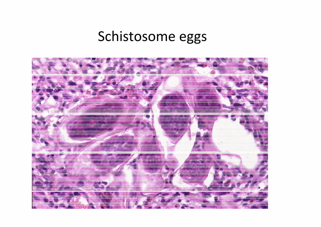
Schistosome eggs

Clonorchis in a liver biopsy


Case 2
• Parasites most often seen in liver
– Hydatid cyst
– Schistosomiasis
– Enterobius vermicularis (pinworm)
– Visceral larva migrans
– Fasciola hepatica
– Clonorchis sinensis
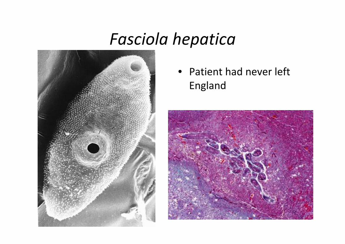
Fasciola hepatica
• Patient had never left
England

Fasciola hepatica

Fasciola invading liver

Fasciola

Serology
Hydatid –ve
Fasciola –ve
(but may be negative if infection is dead)

Our diagnosis[Swiss vet pathologist agreed]
Degenerate Fasciola hepatica worm

Life cycle of Fasciola hepatica

Case 3
Dangers of exotic holidays


Case 3
• Male 59 years
• British caucasian
• No previous travel history
• Jamaica 2-week holiday
• In second week, felt unwell
• One week after return, very ill
Case 3
• On admission
• Renal failure
• Liver failure
– Bilirubin up to 448
• Diagnosis: septic shock
• Much serology, blood culture (all -ve)

Case 3
• Intensive Care
• Support for lungs, liver and kidney
• Antibiotics
• WBC rose to 25,000
• Diagnosis: ?chronic myelomonocytic
leukaemia

Case 3
• Died one week after admission
• = 3 weeks after starting illness
• Tests results available
– HIV, HAV, HBV, HCV – negative
• Awaited
– dengue, yellow fever, CMV, EBV

Case 3
• Autopsy 5 days after death
– Ht 198cm, Wt 94kg
• Jaundice & oedema
• Petechiae around skin punctures
• Lungs – solid and red – ARDS
• Kidneys: 245 & 310gm – enlarged, soft, yellow,
petechiae++
• Spleen 430gm - soft

Case 3
• Liver 1840gm
• Soft, yellow, not cirrhotic, no haemorrhages,
no focal lesions, possibly collapsing
• Bile duct & gall bladder - normal


Case 3
• Autopsy liver tissue and pre-mortem blood
• Leptospira interrogans DNA
– (Micropathology Lab, Coventry, UK)
• IHC for Leptospira spp
– Dr Sherif Zaki, CDC, Atlanta, USA


Leptospira taxonomy:
determined by DNA reassociation
13 named species
Species Selected serovars
L.interrogans Icterohaemorrhagiae, Copenhageni, Australis, Caniola, etc
L.noguchi Panama, Pomona
L.borgpetersenii Ballum, Hardjo, Javanica
L.germospecies 1 Sichuan
L.germospecies 2-4 Non-pathogens
L.biflexa Non-pathogens

Leptospira scanning EM

Tip of the iceberg
Asymptomatic
Symptomatic infection
Severe illnessWeil’s disease

Case 3 Lung - ALI

Manifestation of leptospirosis

Summary
• Anything can happen
– International travel
• ‘Unde venis?’
– Immunosuppression
• Acquired
• Iatrogenic
• Increasing reliance on molecular diagnostics
• Close collaboration with microbiologists and
ID clinicians