screw xation in thoracolumbar fracture minimally invasive
TRANSCRIPT
Page 1/17
Hidden blood loss and its in�uencing factors afterminimally invasive percutaneous transpedicularscrew �xation in thoracolumbar fractureXin Yue
First A�liated Hospital of Dalian Medical UniversityKaige Mao
Dalian Medical UniversityMing Yang
First A�liated Hospital of Dalian Medical UniversityWentao Zhang
First A�liated Hospital of Dalian Medical UniversityZhonghai Li ( [email protected] )
First A�liated Hospital of Dalian Medical University https://orcid.org/0000-0003-4735-1193
Research article
Keywords: Hidden blood loss (HBL), Risk factors, Minimally invasive percutaneous transpedicular screw�xation (MIPTSF), Multiple regression analysis, Complication
Posted Date: April 28th, 2021
DOI: https://doi.org/10.21203/rs.3.rs-445719/v1
License: This work is licensed under a Creative Commons Attribution 4.0 International License. Read Full License

Page 2/17
AbstractObjective: This study aimed to investigate the amount of hidden blood loss (HBL) and its in�uencingfactors after minimally invasive percutaneous transpedicular screw �xation (MIPTSF) in thoracolumbarfracture.
Summary of Background Data: MIPTSF is generally accepted as a minimally invasive treatment forthoracolumbar fracture. However, HBL caused by this procedure is usually disregarded.
Materials and Methods: Between October 2017 and December 2020, a total of 146 patients (106 malesand 40 females, age range 21–59 years) were retrospectively examined, and their clinical andradiological data were recorded and analyzed. The Pearson or Spearman correlation analysis was usedto investigate an association between patient’s characteristics and HBL. Multivariate linear regressionanalysis was performed to elucidate the related clinical or radiological factors of HBL.
Results: A substantial amount of HBL (164.00 ±112.02 ml, 40.65% of TBL) occurred after transpedicularscrew internal �xation. Multivariate linear regression analysis revealed that HBL was positivelyassociated with total blood loss (TBL) (P=.000), percentage of vertebral height loss (VHL) (P=.000),percentage of vertebral height restoration (VHR) (P=.000), numbers of fractured vertebrae (P=.013), andnumbers of �xed vertebral segments (P=.002).
Conclusion: A large amount of HBL was incurred in patients undergoing MIPTSF in thoracolumbarfracture. More importantly, TBL, percentage of VHL, percentage of VHR, the numbers of fracturedvertebrae and �xed vertebral segments were independent risk factors for HBL.
BackgroundThe thoracolumbar spine is one of the area’s most commonly affected by spinal fractures 1,2. However,conventional open posterior pedicle screw �xation causes increased intraoperative bleeding, a higherinfection rate, postoperative back pain, delayed functional rehabilitation, and aggravation of posteriorligamentous complex (PLC) injury 3–5. With advances in surgical techniques and instrumentation, thepercutaneous approach has been successfully applied for pedicle screw �xation to treat thoracolumbarfracture 6. The percutaneous approach allows spine surgeons to insert pedicle screws and rods and toconnect them percutaneously through small skin incisions. Moreover, this system avoids thedisadvantages of conventional surgical treatment, minimizes soft tissue injury, reduces intraoperativeblood loss, and results in better postoperative pain scores than other approaches 7,8. According to pastclinical experience, MIPTSF is associated with a relatively low perioperative blood loss because of smallincision, reduced muscular dissection, and short operative time 9. But, the patients with thoracolumbarfractures tend to have a lower postoperative HB level than anticipated after surgery despite the apparentlysatisfactory perioperative management of blood loss. Previous studies examined only the volume of
Page 3/17
visible blood loss in the perioperative period. However, hidden blood loss (HBL) penetrating tissues,retained in a dead space, and lost due to hemolysis is often disregarded by orthopedic surgeons 10.
Hidden blood loss (HBL) is not usually recognized by general assessment because of its invisibility 11.HBL may exacerbate postoperative hemoglobin drop, affect postoperative outcomes, such as medicalcomplications, increased blood transfusion risks, and prolonged postoperative rehabilitation 12. The issueof HBL has been noted in other �elds of orthopedic surgery. The concept of HBL was �rst put forward bySehat in 2000 11. Sehat et al. reported that the proportion of HBL was 50% of the TBL in total kneearthroplasty. Xu et al reported the mean hidden loss calculated with our recommendable method was362.8ml and 47% of total loss in lumbar fusion surgery 13. However, few studies have considered HBL inMIPTSF surgery during treatment of AO type A1-A3 thoracolumbar fractures with no neurologicalsymptoms. Therefore, we retrospectively reviewed medical data of patients who underwent MIPTSF inour department in an attempt to evaluate HBL and identi�ed the in�uencing factors of HBL.
Methods
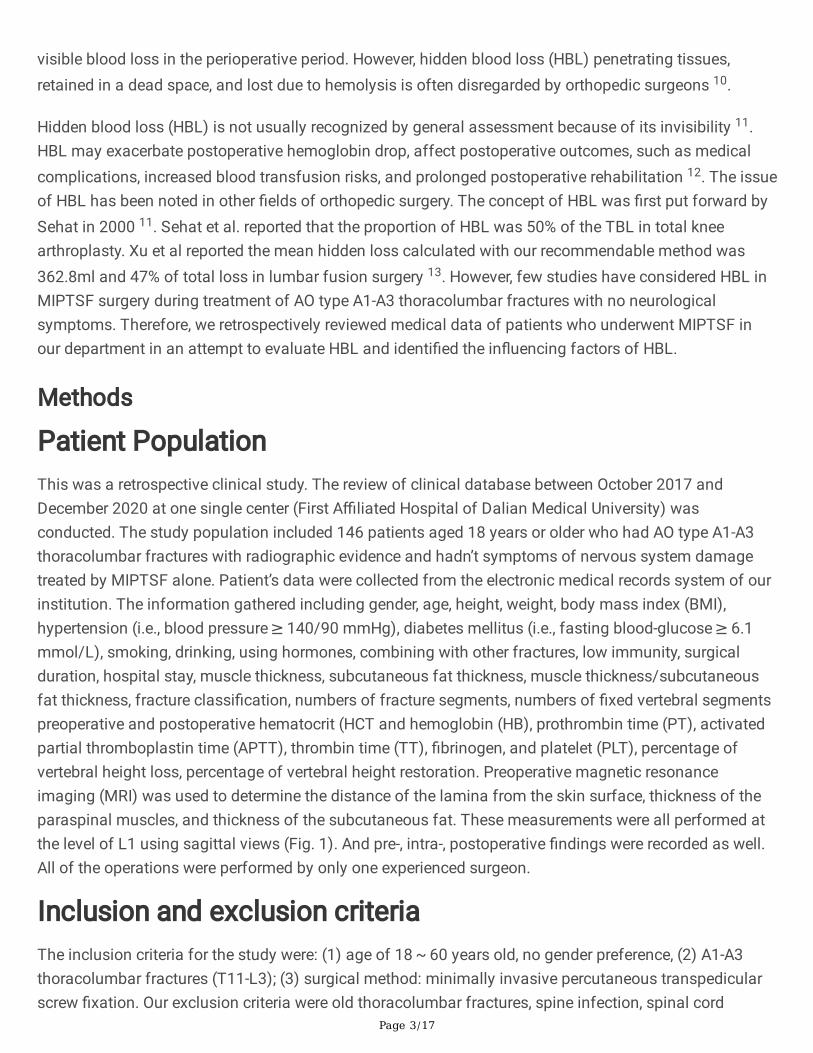
Patient PopulationThis was a retrospective clinical study. The review of clinical database between October 2017 andDecember 2020 at one single center (First A�liated Hospital of Dalian Medical University) wasconducted. The study population included 146 patients aged 18 years or older who had AO type A1-A3thoracolumbar fractures with radiographic evidence and hadn’t symptoms of nervous system damagetreated by MIPTSF alone. Patient’s data were collected from the electronic medical records system of ourinstitution. The information gathered including gender, age, height, weight, body mass index (BMI),hypertension (i.e., blood pressure ≥ 140/90 mmHg), diabetes mellitus (i.e., fasting blood-glucose ≥ 6.1mmol/L), smoking, drinking, using hormones, combining with other fractures, low immunity, surgicalduration, hospital stay, muscle thickness, subcutaneous fat thickness, muscle thickness/subcutaneousfat thickness, fracture classi�cation, numbers of fracture segments, numbers of �xed vertebral segmentspreoperative and postoperative hematocrit (HCT and hemoglobin (HB), prothrombin time (PT), activatedpartial thromboplastin time (APTT), thrombin time (TT), �brinogen, and platelet (PLT), percentage ofvertebral height loss, percentage of vertebral height restoration. Preoperative magnetic resonanceimaging (MRI) was used to determine the distance of the lamina from the skin surface, thickness of theparaspinal muscles, and thickness of the subcutaneous fat. These measurements were all performed atthe level of L1 using sagittal views (Fig. 1). And pre-, intra-, postoperative �ndings were recorded as well.All of the operations were performed by only one experienced surgeon.
Inclusion and exclusion criteriaThe inclusion criteria for the study were: (1) age of 18 ~ 60 years old, no gender preference, (2) A1-A3thoracolumbar fractures (T11-L3); (3) surgical method: minimally invasive percutaneous transpedicularscrew �xation. Our exclusion criteria were old thoracolumbar fractures, spine infection, spinal cord

Page 4/17
compression syndrome, severe cardiopulmonary comorbidity, major coagulopathy, and patients withsymptoms of nervous system damage, liver cirrhosis or uremia.
Management of blood lossNo patient received blood transfusion throughout the assessment period. All of the patients underwent afull blood count, including HCT, and HB before the surgery and 2 or 3 days after the surgery forcalculation of blood loss. No drainage was typically placed in any of the patients. There was little visibleblood loss after surgery, therefore, postoperative blood loss could be ignored.
Calculation of hidden blood lossFirstly, patient’s blood volume (PBV) was estimated in accordance with the formula of Nadler et al14. PBV(L) = k1 × height(m)3 + k2 ×weight(kg)2 + k3; where k1 = 0.3669, k2 = 0.03219 and k3 = 0.6041 for males,and k1 for females. = 0.3561, k2 = 0.03308 and k3 = 0.1833.
Secondly, according to the method of Gross et al. 15, the TBL was calculated based on the HCT level andthe PBV, as follows: TBL (mL) = PBV (L) × (HCTpre – HCTpost) / HCTave, where HCTpre is the initialpreoperative HCT, HCTpost is the HCT on the second or third day postoperatively, and HCTave is theaverage of the Hctpre and the Hctpost.
Finally, the method of Sehat et al. was used to calculate the HBL, as follows: HBL (mL) = TBL (mL) − VBL(mL) 11. Since no drainage was typically placed in any of the patients, intraoperative blood loss wasequal to VBL, VBL was given by the measured suction loss and blood loss in swabs, and recorded by theanesthetists.
The de�nition of anemiaAccording to the World Health Organization, anemia is characterized by HB levels of < 120 g/L for womenand < 130 g/L for men) 16.
Calculation of the percentage of vertebral height loss andrestorationAll of the included cases were examined using plain radiographs. The predicted height of each fracturedvertebra was calculated according to the average height of the two adjacent vertebrae. And the anteriorvertebral height loss and restoration was measured according to the affected vertebral body. Thepercentages of vertebral height loss (VHL) and vertebral height restoration (VHR) were calculated with thefollowing equations 17:
VBHave = (VBHa + VBHb) / 2
VHL (%) = (VBHave − VBHpre ) / VBHave × 100%,
VHR (%)= (VBHpost − VBHpre) / VBHave × 100%,
Page 5/17
where VBHave is the average height of the 2 adjacent vertebrae, and VBHpre is the preoperative anteriorvertebral body height, and VBHpost is the postoperative anterior vertebral body height. (Fig. 2)
Statistical analysisAll of the independent variables were incorporated into the model using the method of “Enter.” Dataanalyses were performed with the SPSS 23.0 software. (International Business Machines Corporation,Armonk, NY). A chi-squared test was adopted to compare the preoperative and postoperative incidence ofanemia. Pearson’s correlation (used for the normal data), Spearman’s correlation analysis (used for thenon-normal data), and multivariate linear regression analysis were performed to evaluate the in�uencingfactors associated with HBL. In all analyses, P < 0.05 was taken to indicate statistical signi�cance.
ResultsA total of 146 patients were reviewed retrospectively in this study. Among these patients were 106 malesand 40 females, with a mean age of 42.31 (range 21-59) years. Table 1 summarizes the demographicand clinical characteristics. The mean muscle thickness was 31.61±7.84 mm, while the meansubcutaneous fat thickness was 19.84±6.19 mm. The mean preoperative HCT and HB were 38.23±4.41%and 124.86±14.36g/l. The mean postoperative HCT and HB were 35.18±4.51% and 103.92±13.67g/l. Themean PBV was 4.87±0.71L. The mean HBL was 164.00±112.02ml, 40.65% of TBL, indicating aconsiderable amount of HBL, which was much higher than we had expected. The mean VBL was239.45±130.17ml. The mean TBL was 403.45±182.25ml. 76 patients suffered from preoperative anemia,and 56 patients developed anemia after surgery (Fig. 3). There were signi�cant differences between pre-and postoperative HCT (P < 0.001) and HB (P < 0.001) (Table 2).
The Pearson or Spearman correlation analysis for HBL found the following parameters with a P < 0.05(Table 3): TBL (P = 0.000), BMI (P = 0.000), muscle thickness (P = 0.000), subcutaneous fat thickness (P= 0.000), surgical duration (P = 0.000), PT (P = 0.000), APTT (P = 0.000), TT (P = 0.038), diabetes mellitus(P=.048), fracture classi�cation (type A1-A3) (P=.000), percentage of vertebral height loss (P=.000),percentage of vertebral height restoration (P=.000), numbers of fractured vertebrae (P=.000), andnumbers of �xed vertebral segments (P=.000).
Next, we performed multiple and stepwise linear regression analysis to explore the association betweenHBL and the in�uential factors mentioned earlier. The TBL (P=.000), percentage of vertebral height loss(P=.000), percentage of vertebral height restoration (P=.000), numbers of fractured vertebrae (P=.013),and numbers of �xed vertebral segments (P=.002) were independent risk factors for HBL (Table 4). Theresults indicated that other factors were not signi�cantly correlated with HBL.
Discussion

Page 6/17
Studies on HBL after orthopedic surgery have mostly focused on total hip arthroplasty (THA), total kneearthroplasty (TKA), and ALIF/PLIF surgery 18. In a work on anterior/posterior lumbar fusion surgery(ALIF/PLIF), HBL was approximately 40% of TBL 12,18. Chen et al. 19 reviewed and analyzed of thepatients undergoing conventional posterior open approach, the average HBL was 382 ± 153.8mL; and theaverage HBL of patients undergoing percutaneous approach was 240.0 ± 65.1mL. In our study, asubstantial amount of HBL (164.00 ± 112.02ml, 40.65% of TBL) occurred after MIPTSF., the obtainedamount was much greater than that of visible intraoperative blood loss. Some studies suggest that forpatients undergoing total hip replacement, HBL is positively correlated with changes in BMI, bloodtransfusion, incision length, preoperative and postoperative HCT, and negatively correlated with age 20.Nevertheless, there have been no previous studies regarding the in�uential factors correlated to the HBLduring the MIPTSF of AO type A1-A3 thoracolumbar fractures. In this study, we investigated and identi�edthe risk factors of HBL following this surgery by multivariate linear regression analysis. The resultsproposed that the TBL, percentage of vertebral height loss, percentage of vertebral height restoration,numbers of fractured vertebrae, and numbers of �xed vertebral segments were positive independent riskfactors for HBL.
Our statistical analysis showed that the patients who had massive TBL suffered from more HBL thanthose who have little TBL. TBL was the independent risk factor, which may have to do with PBV, becauseTBL is calculated by multiplying PBV by changes of HCT and subtracting the IBL according to the Grossformula 15,which might relate to the patient’s weight and height. However, BMI had not been identi�ed asa risk factor in our study, although body mass index was also calculated by weight and height. Based oncollected data in our study, it was easy to �nd that HBL is directly related to a large amount of blood loss.
HBL during orthopedic surgery is generally accepted as being due to blood in�ltration into tissuecompartments and loss due to hemolysis 21,22. Our study found that the percentage of vertebral heightloss and the percentage of vertebral height restoration were correlated with HBL. Vertebra involvescancellous bone, and its blood supply is abundant. The expansion of vertebral cavity will cause internalbleeding. The recovery of fractured vertebral body height may lead to enlarged cavity, and the spacearound vertebral body may be enlarged. We suspect that the blood would seep into these fracture spaces,leading to an increase in HBL 23. Vertebral cavity and muscle space also provide storage cavity for HBL.
In our study, the numbers of fractured vertebrae and numbers of �xed vertebral segments were positivelyrelated to HBL, as Chen et al. guessed 19. A previous study proposed that he number of fracturedvertebrae was the risk factor of HBL in percutaneous kyphoplasty surgery 24. Ju et al. held that ALIF wasassociated with substantial perioperative HBL, and the inclusion of L4/5 in the procedure were signi�cantrisk factors for increased blood loss 18. However, we found that the number of �xed segments was anindependent risk factor for hidden blood loss, and the fracture level was not included in our data. We willfurther explore the relationship between fracture level and hidden blood loss in the future.

Page 7/17
Our previous studies had shown that muscle thickness is also an independent risk factor for hidden bloodloss in spinal surgery 25, thicker muscle may be associated with larger penetrable tissue compartments,allowing blood to ooze into the tissue cavity 26. Jiang et al. 27 found that posterior cervical soft tissuewas positively correlated with both TBL and HBL in the expansive open-door laminoplasty (EOLP). Butthe muscle thickness or subcutaneous fat thickness was not clari�ed as a risk factor in this study. Wethink that this might be related to the less muscle damage caused by minimally invasive surgery.Therefore, we still need to further study the relationship between muscle thickness and HBL in the settingof spine surgery.
Excessive blood loss can increase the possibility of blood transfusion, which is associated withtransfusion reactions, anaphylactic reaction, infections and delayed recovery 28. Furthermore, excessiveblood loss can prolong the hospitalization time and increase the use of medication 29. The TBL,percentage of vertebral height loss, percentage of vertebral height restoration, numbers of fracturedvertebrae, and numbers of �xed vertebral segments should be correctly understood before operation toensure the safety of patient treatment.
ConclusionConsequently, MIPTSF is associated with substantial HBL. More importantly, the total blood loss,percentage of vertebral height loss, percentage of vertebral height restoration, numbers of fracturedvertebrae, and numbers of �xed vertebral segments were independent risk factors for HBL. It is importantfor surgeons to be aware of HBL, to avoid complications related to blood loss. Accurate perioperative HBLassessment can help prevent complications and improve rehabilitation.
AbbreviationsMIPTSFM: Minimally invasive percutaneous transpedicular screw �xation; PLC: Posterior ligamentouscomplex; PBV: Patient’s blood volume; VBL: Visible blood loss; HBL: Hidden blood loss; TBL: Total bloodloss; Hct: Hematocrit; Hb: Hemoglobin; PT: Prothrombin time; APTT: Activated partial thromboplastintime; TT, Thrombin time; PLT, Platelet; MRI, Magnetic resonance imaging; VHL: Vertebral height loss; VHR:Vertebral height restoration; THA: Total hip arthroplasty; TKA: Total knee arthroplasty; ALIF: Anteriorlumbar fusion; PLIF: Posterior lumbar fusion.
DeclarationsAcknowledgements
We would like to thank all the participants in the studies.
Authors’ contributions

Page 8/17
YX contributed to the study design, the writing of the paper, and drafting of the manuscript. LZHperformed the surgeries and participated in the design of the study. KGM, YM and WTZ collected andanalyzed the data. LZH reviewed and edited the manuscript. All authors read and approved the �nalmanuscript.
Funding
This study was supported by LiaoNing Revitalization Talents Program (XLYC1807131), the Science andTechnology Innovation Foundation of Dalian (2020JJ27SN070). The funders had no role in the studydesign, data collection and analysis, decision to publish, or preparation of the manuscript.
Availability of data and materials
All data used and analyzed during this study are available from the corresponding author uponreasonable request.
Ethics approval and consent to participate
This research was approved by the ethics committee of the First A�liated Hospital of Dalian MedicalUniversity. And agreement to participate was given by the participants. Because of the retrospectivenature of the study, informed consent was waived.
Consent for publication
Written informed consent for publication of their clinical details and/or clinical images was obtainedfrom the patient/parent/guardian/relative of the patient.
Competing interests
The authors declare that they have no competing interests.
References1. Tian NF, Wu YS, Zhang XL, et al. Fusion versus nonfusion for surgically treated thoracolumbar burst
fractures: a meta-analysis. PloS one 2013;8:e63995.
2. Fernández-de Thomas RJ, De Jesus O. Thoracolumbar Spine Fracture. StatPearls. Treasure Island(FL): StatPearls Publishing Copyright © 2020, StatPearls Publishing LLC., 2020.
3. Kim DY, Lee SH, Chung SK, et al. Comparison of multi�dus muscle atrophy and trunk extensionmuscle strength: percutaneous versus open pedicle screw �xation. Spine 2005;30:123-9.
4. Gnanenthiran SR, Adie S, Harris IA. Nonoperative versus operative treatment for thoracolumbar burstfractures without neurologic de�cit: a meta-analysis. Clinical orthopaedics and related research2012;470:567-77.

Page 9/17
5. Wu H, Fu C, Yu W, et al. The options of the three different surgical approaches for the treatment ofDenis type A and B thoracolumbar burst fracture. European journal of orthopaedic surgery &traumatology : orthopedie traumatologie 2014;24:29-35.
�. Wang H, Zhou Y, Li C, et al. Comparison of Open Versus Percutaneous Pedicle Screw Fixation Usingthe Sextant System in the Treatment of Traumatic Thoracolumbar Fractures. Clinical spine surgery2017;30:E239-e46.
7. Pannu CD, Farooque K, Sharma V, et al. Minimally invasive spine surgeries for treatment ofthoracolumbar fractures of spine: A systematic review. Journal of clinical orthopaedics and trauma2019;10:S147-s55.
�. Ansar MN, Hashmi SM, Colombo F. Minimally Invasive Spine (MIS) Surgery in TraumaticThoracolumbar Fractures: A Single-Center Experience. Asian journal of neurosurgery 2020;15:76-82.
9. Kaye ID, Passias P. Minimally Invasive Surgery (MIS) Approaches to Thoracolumbar Trauma. Bulletinof the Hospital for Joint Disease (2013) 2018;76:71-9.
10. Ogura Y, Dimar Ii JR, Gum JL, et al. Hidden blood loss following 2- to 3-level posterior lumbar fusion.The spine journal : o�cial journal of the North American Spine Society 2019;19:2003-6.
11. Sehat KR, Evans R, Newman JH. How much blood is really lost in total knee arthroplasty?. Correctblood loss management should take hidden loss into account. The Knee 2000;7:151-5.
12. Smorgick Y, Baker KC, Bachison CC, et al. Hidden blood loss during posterior spine fusion surgery.The spine journal : o�cial journal of the North American Spine Society 2013;13:877-81.
13. Xu D, Ren Z, Chen X, et al. The further exploration of hidden blood loss in posterior lumbar fusionsurgery. Orthopaedics & traumatology, surgery & research : OTSR 2017;103:527-30.
14. Nadler SB, Hidalgo JH, Bloch T. Prediction of blood volume in normal human adults. Surgery1962;51:224-32.
15. Gross JB. Estimating allowable blood loss: corrected for dilution. Anesthesiology 1983;58:277-80.
1�. Beghé C, Wilson A, Ershler WB. Prevalence and outcomes of anemia in geriatrics: a systematic reviewof the literature. The American journal of medicine 2004;116 Suppl 7A:3s-10s.
17. Cao D, Zhang S, Yang F, et al. Hidden blood loss and its in�uencing factors after percutaneouskyphoplasty surgery: A retrospective study. Medicine 2018;97:e0435.
1�. Ju H, Hart RA. Hidden blood loss in anterior lumbar interbody fusion (ALIF) surgery. Orthopaedics &traumatology, surgery & research : OTSR 2016;102:67-70.
19. Chen ZX, Sun ZM, Jiang C, et al. Comparison of Hidden Blood Loss Between Three Different SurgicalApproaches for Treatment of Thoracolumbar Fracture. Journal of investigative surgery : the o�cialjournal of the Academy of Surgical Research 2019;32:755-60.
20. Miao K, Ni S, Zhou X, et al. Hidden blood loss and its in�uential factors after total hip arthroplasty.Journal of orthopaedic surgery and research 2015;10:36.
21. Erskine JG, Fraser C, Simpson R, et al. Blood loss with knee joint replacement. Journal of the RoyalCollege of Surgeons of Edinburgh 1981;26:295-7.

Page 10/17
22. Pattison E, Protheroe K, Pringle RM, et al. Reduction in haemoglobin after knee joint surgery. Annalsof the rheumatic diseases 1973;32:582-4.
23. Guglielmino A, Sorbello M, Barbagallo G, et al. Osteoporotic vertebral compression fracture pain(back pain): our experience with balloon kyphoplasty. Minerva anestesiologica 2007;73:77-100.
24. Wu YS, Zhang H, Zheng WH, et al. Hidden blood loss and the in�uential factors after percutaneouskyphoplasty surgery. European spine journal : o�cial publication of the European Spine Society, theEuropean Spinal Deformity Society, and the European Section of the Cervical Spine Research Society2017;26:1878-83.
25. Zhou Y, Fu X, Yang M, et al. Hidden blood loss and its possible risk factors in minimally invasivetransforaminal lumbar interbody fusion. Journal of orthopaedic surgery and research 2020;15:445.
2�. Gao F, Guo W, Sun W, et al. Correlation between the coverage percentage of prosthesis andpostoperative hidden blood loss in primary total knee arthroplasty. Chinese medical journal2014;127:2265-9.
27. Jiang C, Chen TH, Chen ZX, et al. Hidden blood loss and its possible risk factors in cervical open-doorlaminoplasty. The Journal of international medical research 2019;47:3656-62.
2�. Willner D, Spennati V, Stohl S, et al. Spine Surgery and Blood Loss: Systematic Review of ClinicalEvidence. Anesthesia and analgesia 2016;123:1307-15.
29. Liang J, Liu H, Huang X, et al. Using tranexamic acid soaked absorbable gelatin sponge followingcomplex posterior lumbar spine surgery: A randomized control trial. Clinical neurology andneurosurgery 2016;147:110-4.
TablesTable1. Patients demographics.

Page 11/17
Parameters Statistics
Total patients (n) 146
Sex (n) Male 106
Female 40
Age, yr 42.31±7.90
BMI, kg/m2 25.33±3.13
Muscle thickness, mm 31.61±7.84
Subcutaneous fat thickness, mm 19.84±6.19
Muscle thickness/Subcutaneous fat thickness 1.64±0.26
Smoking (n) 35
Drinking (n) 17
Diabetes mellitus (n) 10
Hypertension (n) 13
Low immunity(n) 3
Using hormones(n) 8
Combining with other fractures(n) 20
Fracture classi�cation(n) A1 87
A2 23
A3 36
Preoperative HCT,% 38.23±4.41
Postoperative HCT,% 35.18±4.51
PBV, L 4.87±0.71
TBL, ml 403.45±182.25
VBL, ml 239.45±130.17
VHL, % 45.58±11.08
VHR, % 23.43±9.47
Numbers of fractured vertebrae 1.18±0.45
Numbers of �xed vertebral segments 3.03±0.66
Hospital stay, d 12.28±2.64

Page 12/17
Surgical duration, min 120.14±34.06
Preoperative Hb, g/l 124.86±14.36
Postoperative Hb, g/l 103.92±13.67
PT, s 11.46±1.22
APTT, s 32.90±6.18
TT, s 17.65±1.25
Fibrinogen, g/l 3.37±0.80
PLT, 109 /l 260.33±66.39
BMI, Body mass index; HCT, Hematocrit; PBV, Patient’s blood volume; TBL, Total blood loss; VBL, Visibleblood loss; VHL, Vertebral height loss; VHR, Vertebral height restoration; HB, Hemoglobin; PT, Prothrombintime; APTT, Activated partial thromboplastin time; TT, Thrombin time; PLT, Platelet.
Table2. Changes in HCT and HB level following MIPTSF.
Parameters Mean SD t P
Preoperative and postoperative HCT, % 3.0486 1.3208 27.890 .000
Preoperative and postoperative HB g/L 20.945 11.672 21.683 .000
MIPTSF, Minimally invasive percutaneous transpedicular screw �xation; HCT, Hematocrit; HB,Hemoglobin; SD, Standard deviation.
Table 3 Results of the Pearson or Spearman correlation analysis for HBL.

Page 13/17
Parameters Sig P
Gender -.102 .219
Age -.051 .539
BMI .455 .000
Muscle thickness .778 .000
Subcutaneous fat thickness .646 .000
Muscle thickness/Subcutaneous fat thickness -.106 .204
Smoking .036 .670
Drinking .001 .993
Diabetes mellitus -.164 .048
Hypertension -.077 .357
Low immunity -.037 .655
Using hormones -.043 .608
Combining with other fractures -.145 .081
Fracture classi�cation .519 .000
PBV .016 .852
TBL .706 .000
VBL .128 .125
VHL .938 .000
VHR .921 .000
Numbers of fractured vertebrae .625 .000
Numbers of �xed vertebral segments .746 .000
Hospital stay -.061 .466
Surgical duration .356 .000
PT -.323 .000
APTT .590 .000
TT .172 .038
Fibrinogen -.040 .629
PLT -.074 .375

Page 14/17
HBL, Hidden blood loss; BMI, Body mass index; PBV, Patient’s blood volume; TBL, Total blood loss; VBL,Visible blood loss; VHL, Vertebral height loss; VHR, Vertebral height restoration; PT, Prothrombin time;APTT, Activated partial thromboplastin time; TT, Thrombin time; PLT, Platelet.
Table 4 : Results of multivariate linear regression for HBL.
Parameters Unstandardized Standardized t P
β SE Β
Constant -238.468 48.114 -4.956 .000
TBL .085 .018 .139 4.735 .000
VHL 4.353 .582 .431 7.479 .000
VHR 3.823 .669 .323 5.713 .000
Numbers of fractured vertebrae 20.057 7.954 .081 2.522 .013
Numbers of �xed vertebral segments 19.468 6.184 .114 3.148 .002
BMI .352 .925 .010 .380 .704
Muscle thickness .859 .866 .060 .992 .323
Subcutaneous fat thickness -1.184 .893 -.065 -1.326 .187
Surgical duration -.134 .085 -.041 -1.578 .117
PT -.632 2.184 -.007 -.289 .773
APTT -.704 .534 -.039 -1.318 .190
TT 1.665 1.980 .019 .841 .402
Fracture classi�cation (A2) -5.512 7.073 -.018 -.779 .437
Fracture classi�cation (A3) 10.699 8.057 .041 1.328 .187
HBL, Hidden blood loss; TBL, Total blood loss; VHL, Vertebral height loss; VHR, Vertebral heightrestoration; BMI, Body mass index; PT, Prothrombin time; APTT, Activated partial thromboplastin time; TT,Thrombin time.
Figures

Page 15/17
Figure 1
Diagram of the method used to measure thickness of the paraspinal muscles, subcutaneous fat, andlamina at the skin surface at the level of L1 using sagittal views was determined on T2-weighted MRI.

Page 16/17
Figure 2
Diagram of the method for measuring the percentages of vertebral height loss (VHL) and vertebral heightrestoration (VHR) on sagittal plain radiograph. (a) Preoperative, (b) Postoperative

Page 17/17
Figure 3
The number of anemic patients.