screening methods occupational lung disease
TRANSCRIPT

Screening methods for occupational lung disease
Deborah H YatesRespiratory Physician
St Vincent’s Hospital, Darlinghurst, NSW 2010
Declarations
• Funding from Asthma NSW, NHMRC, DDB, Slater & Gordon, Maurice Blackburn, Novartis (IIT) and others
• Advisory Board: Novartis, Tuberous Sclerosis Australia, Veteran’s Affairs; Expert advisor HCCC
• Speaker funding from: GSK, Astra Zeneca, Novartis etc

Summary of talk
• Background• Traditional screening methods
• Imaging• Lung function• (Dust exposure assessment)
• Information from the broader literature
• Novel methods • Conclusions

World wide re‐emergence of pneumoconiosis
Pneumoconiosis = fibrosis of the lung due to dust
(pneumo=lung konios = dust (Gk)
(A) New York City tuberculosis rate per 100,000 population, 1969 to 1989. Reprinted by permission. (B) Prevalence (%) of progressive massive fibrosis among Appalachian working miners with 25 or more years of tenure from 1974 to 2012
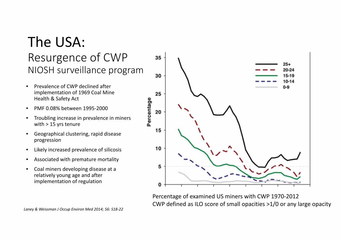
The USA:Resurgence of CWP NIOSH surveillance program
• Prevalence of CWP declined after implementation of 1969 Coal Mine Health & Safety Act
• PMF 0.08% between 1995‐2000
• Troubling increase in prevalence in miners with > 15 yrs tenure
• Geographical clustering, rapid disease progression
• Likely increased prevalence of silicosis
• Associated with premature mortality
• Coal miners developing disease at a relatively young age and after implementation of regulation
Percentage of examined US miners with CWP 1970‐2012CWP defined as ILO score of small opacities >1/0 or any large opacity
Laney & Weissman J Occup Environ Med 2014; 56: S18‐22

Coal workers’ pneumoconiosis in Great Britain, 2005‐2015

Australia

Back to Basics: mechanisms of lung clearance

Dust exposure, dust retention and radiographic change: mechanisms• Coal dust inhalation releases cytokines including TNF alpha, IL‐1, IL‐6, superoxides incl catalase
• Oxidative stress plays a role • Level of bioavailable iron in different mining regions may be important
• Genetic susceptibility likely important
• Inherent capability of lungs to clear dust is good
• Significant redundancy in lung capacity

Coal mine dust lung diseases (CMDLD): NOT one disease. A spectrum of lung disorders
• Classical coal workers pneumoconiosis
• Silicosis• Dust‐related diffuse fibrosis• Mixed dust pneumoconiosis• Rheumatoid pneumoconiosis (Caplan’s syndrome)
• Chronic obstructive pulmonary disease (COPD)
• Diesel exhaust exposure, nitrogen oxides
• Toxic gases from coalmine fires etc
"The first priority and concern of all in the coal or other mining industry must be the health and safety of its most precious resource – the miner." (Federal Coal Mine Health and Safety Act of 1969, USA, amended 1977.)
W Raymond Parkes: “The term “black lung” is uninformative……and should have no place in medical terminology”.
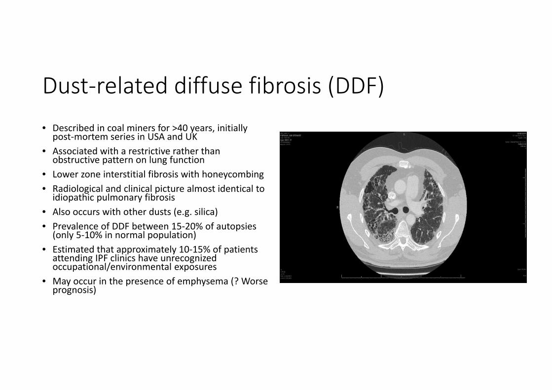
Dust‐related diffuse fibrosis (DDF)• Described in coal miners for >40 years, initially post‐mortem series in USA and UK
• Associated with a restrictive rather than obstructive pattern on lung function
• Lower zone interstitial fibrosis with honeycombing• Radiological and clinical picture almost identical to idiopathic pulmonary fibrosis
• Also occurs with other dusts (e.g. silica)• Prevalence of DDF between 15‐20% of autopsies (only 5‐10% in normal population)
• Estimated that approximately 10‐15% of patients attending IPF clinics have unrecognized occupational/environmental exposures
• May occur in the presence of emphysema (? Worse prognosis)

Surveillance• Need to have a practical approach• Current system insensitive for full spectrum of coal dust lung diseases
• ? Add in measurement of DLCO when assessing lung function
• Major confounding factor is obstructive airway disease
• Also ? add HRCT scanning in those with abnormal CXRs or greater than projected trajectory of lung function decline
• Better information is required about local conditions in Australia and correlation with actual personalized dust levels
• Evidence base for changes required
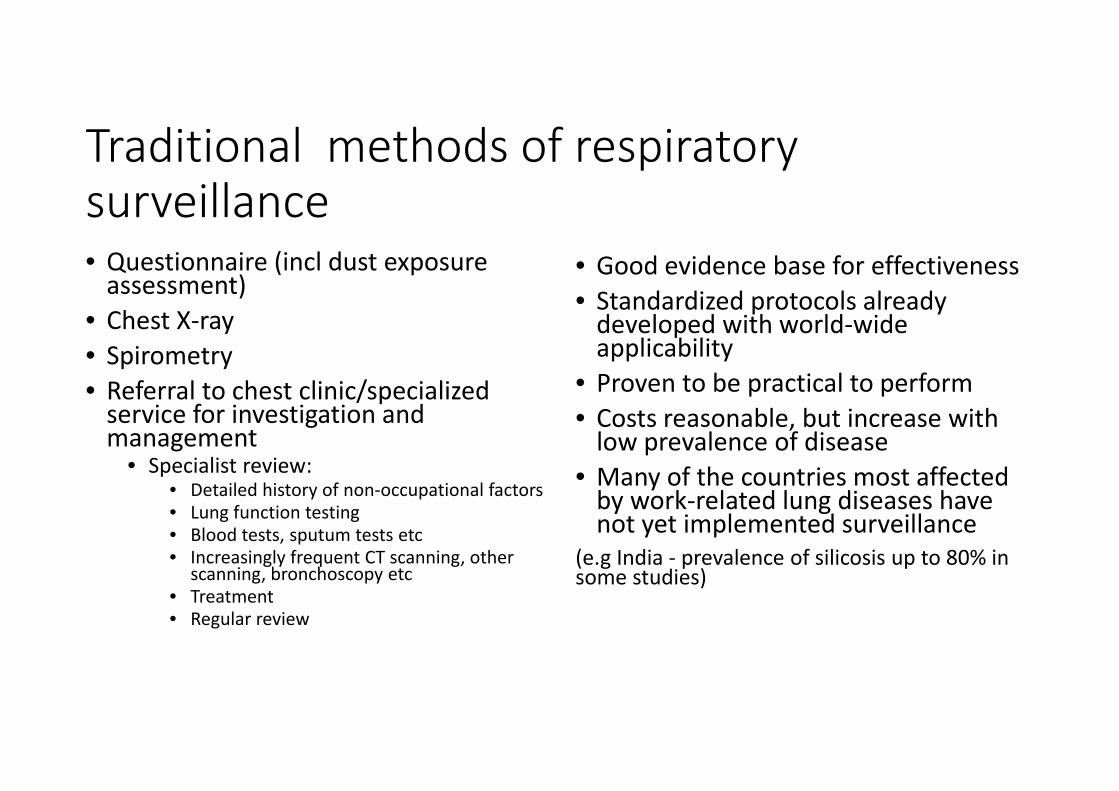
Traditional methods of respiratory surveillance• Questionnaire (incl dust exposure assessment)
• Chest X‐ray• Spirometry• Referral to chest clinic/specialized service for investigation and management
• Specialist review:• Detailed history of non‐occupational factors • Lung function testing• Blood tests, sputum tests etc• Increasingly frequent CT scanning, other
scanning, bronchoscopy etc• Treatment • Regular review
• Good evidence base for effectiveness• Standardized protocols already developed with world‐wide applicability
• Proven to be practical to perform• Costs reasonable, but increase with low prevalence of disease
• Many of the countries most affected by work‐related lung diseases have not yet implemented surveillance
(e.g India ‐ prevalence of silicosis up to 80% in some studies)
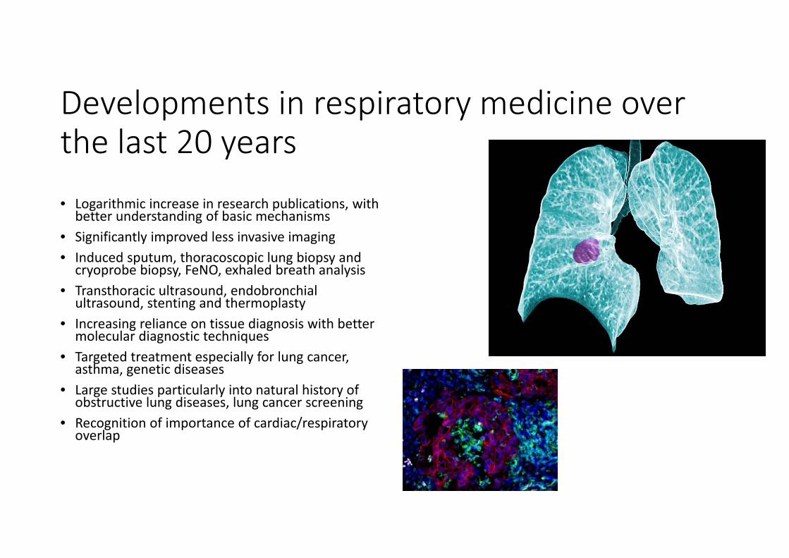
Developments in respiratory medicine over the last 20 years• Logarithmic increase in research publications, with better understanding of basic mechanisms
• Significantly improved less invasive imaging• Induced sputum, thoracoscopic lung biopsy and cryoprobe biopsy, FeNO, exhaled breath analysis
• Transthoracic ultrasound, endobronchialultrasound, stenting and thermoplasty
• Increasing reliance on tissue diagnosis with better molecular diagnostic techniques
• Targeted treatment especially for lung cancer, asthma, genetic diseases
• Large studies particularly into natural history of obstructive lung diseases, lung cancer screening
• Recognition of importance of cardiac/respiratory overlap

Chest radiography compared with CT
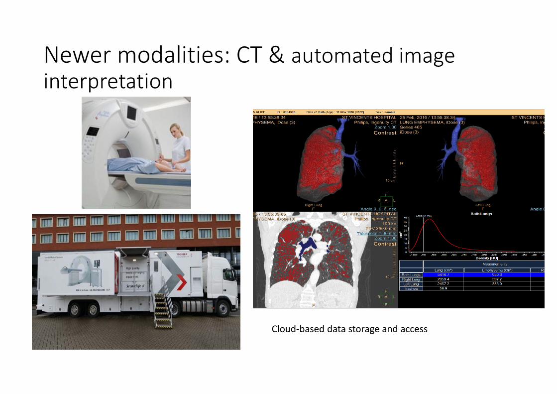
Newer modalities: CT & automated image interpretation
Cloud‐based data storage and access

Recommendations regarding HRCT/CXR in Occupational Lung Disease: American College of Radiologists Appropriateness Criteria: Occupational Lung Diseases (J Thoracic Imaging 2016;31: W1‐3)
• Evidence‐based guidelines reviewed every 3 years by multidisciplinary expert panel
• Summary:• Analogue radiography replaced by digital radiography
• Chest CT without contrast suffices for routine analysis in most scenarios
• HRCT useful in silicosis, coal dust exposure, suspected pneumoconiosis, asbestos exposure, suspected interstitial lung disease or mesothelioma
• CT is more sensitive than chest radiography in detecting lung abnormalities (e.g. Spyratos et al 2012, chest radiography detected abnormalities in 21% of employees, HRCT in 67%)
• MRI may be an alternative in pneumoconiosis but is not yet recommended (one study only available)
• PET/CT useful in excluding extra‐pleural disease in mesothelioma
PET/CT in silicosis

Abbreviations: RO=rounded opacities; IR=irregular opacities; GG=ground glass opacities;HC=honeycombing; EM=emphysema; LO=large opacities; and PL=pleural abnormalities. * Positive findings: more than four readers agreed on the presence of the respective finding in both rounds of the trial readings.
Reliability of the proposed international classification of high resolution computed tomography for occupational and environmental respiratory diseases. Suganuma et al. J Occ Health 2009; 51(3): 210‐222
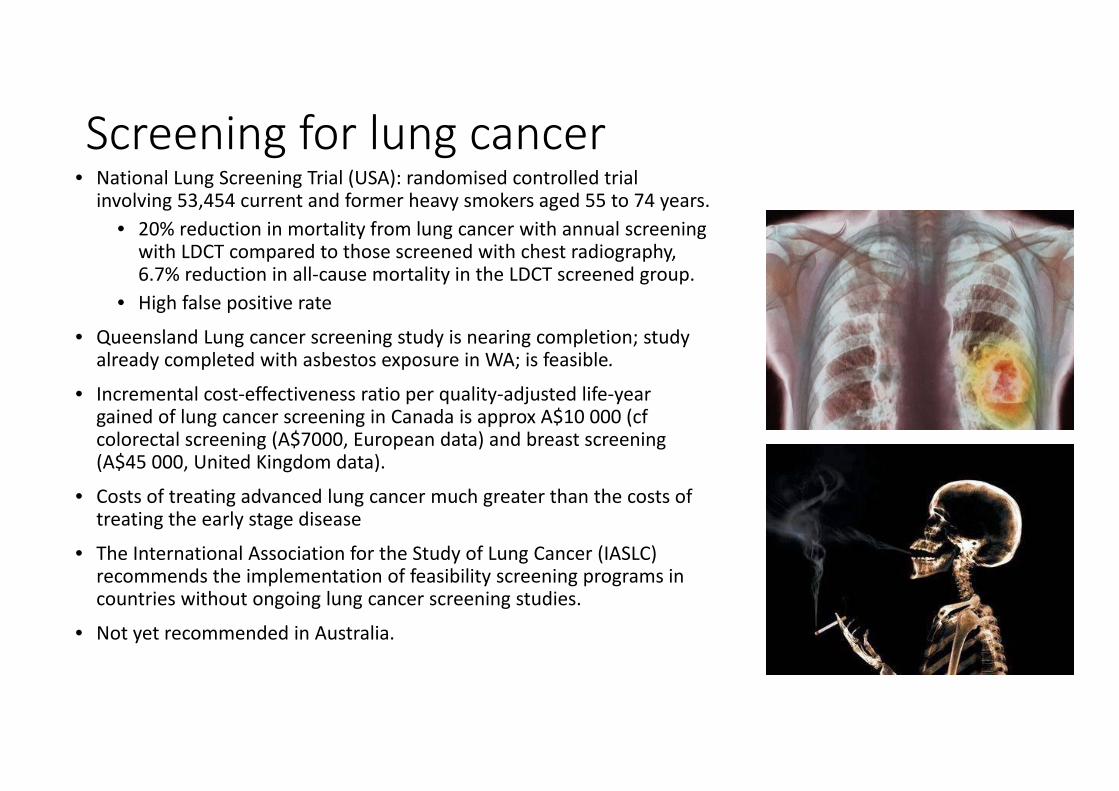
Screening for lung cancer• National Lung Screening Trial (USA): randomised controlled trial involving 53,454 current and former heavy smokers aged 55 to 74 years.
• 20% reduction in mortality from lung cancer with annual screening with LDCT compared to those screened with chest radiography, 6.7% reduction in all‐cause mortality in the LDCT screened group.
• High false positive rate
• Queensland Lung cancer screening study is nearing completion; study already completed with asbestos exposure in WA; is feasible.
• Incremental cost‐effectiveness ratio per quality‐adjusted life‐year gained of lung cancer screening in Canada is approx A$10 000 (cfcolorectal screening (A$7000, European data) and breast screening (A$45 000, United Kingdom data).
• Costs of treating advanced lung cancer much greater than the costs of treating the early stage disease
• The International Association for the Study of Lung Cancer (IASLC) recommends the implementation of feasibility screening programs in countries without ongoing lung cancer screening studies.
• Not yet recommended in Australia.

HRCT scanning in respiratory surveillance for occupational lung disease: less well evaluated• Pneumoconiosis – not as much evidence to show that detection has a beneficial effect on outcome as with lung cancer, but likely. Increasing evidence base.
• Particularly useful with borderline findings, discrepancy between lung function and CXR, or with pleural changes
• Important to have a standardized approach (now possible with the International Classification of HRCT for Occupational and Environmental Diseases (ICOERD))
• Certain to pick up non‐occupational lung and other disorders especially lung cancer, obstructive lung disease
• Currently more expensive than CXR• Could allow opportunities to:
• Reduce exposures• Aid smoking cessation• Allow vaccination for reduction of respiratory infections• Increase health knowledge about other diseases (e.g. cardiac)
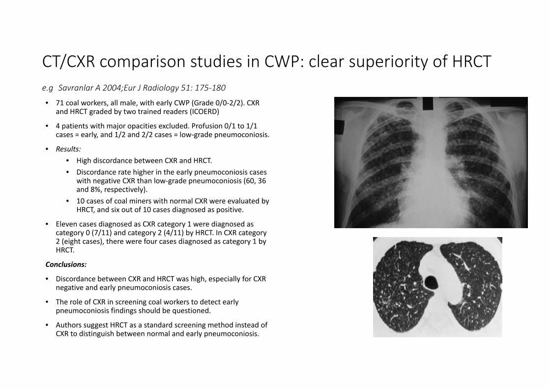
CT/CXR comparison studies in CWP: clear superiority of HRCT e.g Savranlar A 2004;Eur J Radiology 51: 175‐180
• 71 coal workers, all male, with early CWP (Grade 0/0‐2/2). CXR and HRCT graded by two trained readers (ICOERD)
• 4 patients with major opacities excluded. Profusion 0/1 to 1/1 cases = early, and 1/2 and 2/2 cases = low‐grade pneumoconiosis.
• Results:• High discordance between CXR and HRCT. • Discordance rate higher in the early pneumoconiosis cases
with negative CXR than low‐grade pneumoconiosis (60, 36 and 8%, respectively).
• 10 cases of coal miners with normal CXR were evaluated by HRCT, and six out of 10 cases diagnosed as positive.
• Eleven cases diagnosed as CXR category 1 were diagnosed as category 0 (7/11) and category 2 (4/11) by HRCT. In CXR category 2 (eight cases), there were four cases diagnosed as category 1 by HRCT.
Conclusions:
• Discordance between CXR and HRCT was high, especially for CXR negative and early pneumoconiosis cases.
• The role of CXR in screening coal workers to detect early pneumoconiosis findings should be questioned.
• Authors suggest HRCT as a standard screening method instead of CXR to distinguish between normal and early pneumoconiosis.

Savranlar A 2004;Eur J Radiology 51: 175‐180
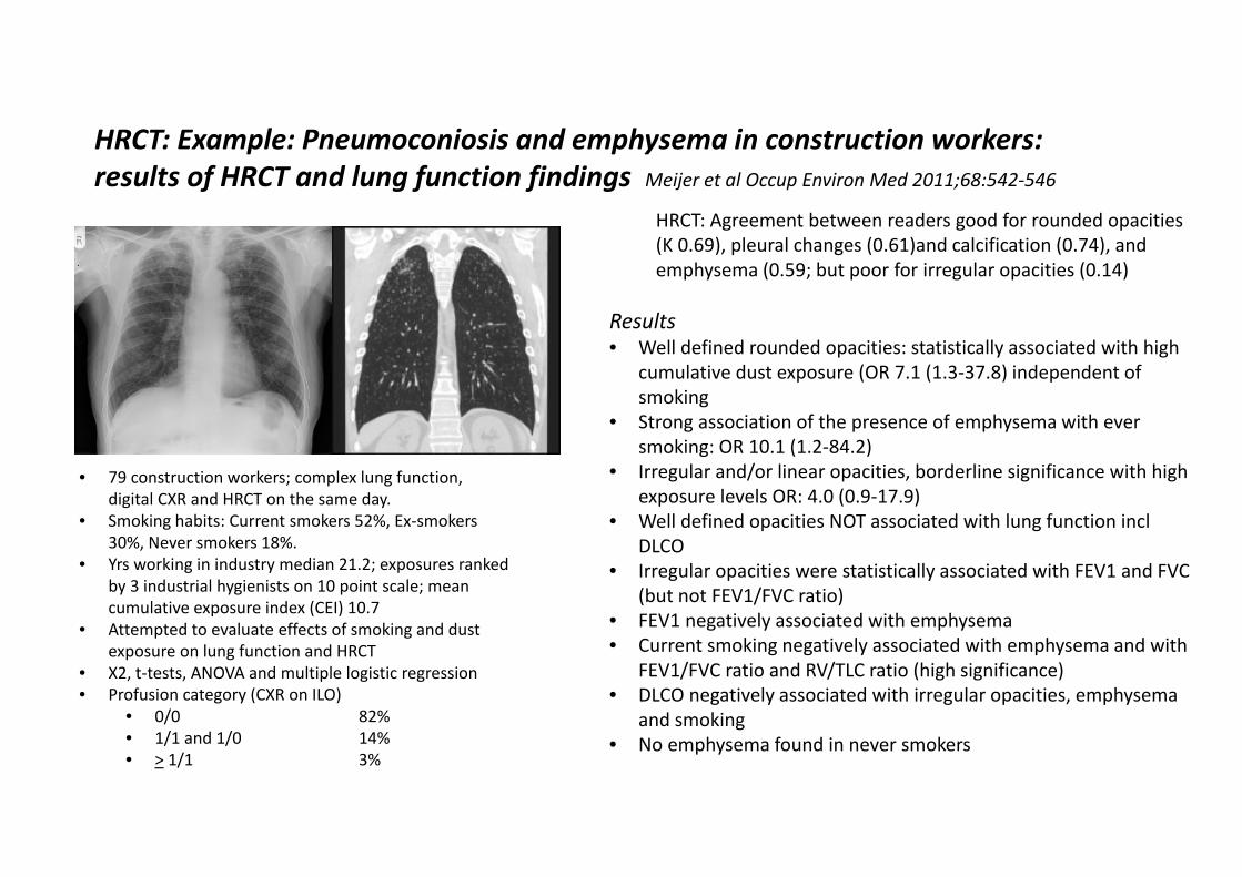
• 79 construction workers; complex lung function, digital CXR and HRCT on the same day.
• Smoking habits: Current smokers 52%, Ex‐smokers 30%, Never smokers 18%.
• Yrs working in industry median 21.2; exposures ranked by 3 industrial hygienists on 10 point scale; mean cumulative exposure index (CEI) 10.7
• Attempted to evaluate effects of smoking and dust exposure on lung function and HRCT
• X2, t‐tests, ANOVA and multiple logistic regression• Profusion category (CXR on ILO)
• 0/0 82%• 1/1 and 1/0 14%• > 1/1 3%
HRCT: Example: Pneumoconiosis and emphysema in construction workers: results of HRCT and lung function findings Meijer et al Occup Environ Med 2011;68:542‐546
HRCT: Agreement between readers good for rounded opacities (K 0.69), pleural changes (0.61)and calcification (0.74), and emphysema (0.59; but poor for irregular opacities (0.14)
Results• Well defined rounded opacities: statistically associated with high
cumulative dust exposure (OR 7.1 (1.3‐37.8) independent of smoking
• Strong association of the presence of emphysema with ever smoking: OR 10.1 (1.2‐84.2)
• Irregular and/or linear opacities, borderline significance with high exposure levels OR: 4.0 (0.9‐17.9)
• Well defined opacities NOT associated with lung function inclDLCO
• Irregular opacities were statistically associated with FEV1 and FVC (but not FEV1/FVC ratio)
• FEV1 negatively associated with emphysema• Current smoking negatively associated with emphysema and with
FEV1/FVC ratio and RV/TLC ratio (high significance)• DLCO negatively associated with irregular opacities, emphysema
and smoking• No emphysema found in never smokers

Screening for occupational lung disorders using HRCT: summary
Pneumoconiosis/ARD• Clear evidence from many studies that HRCT is more sensitive than chest radiography for asbestos‐related pleural disease
• Used also in several other studies for silicosis, other pneumoconioses
• Finnish Institute of Occupational Health (FIOH 2014) recommends LDCT screening of workers with any asbestos exposure and a smoking history equal to the NLST study for lung cancer detection
• No large studies yet reported using LDCT and/or complex lung function for pneumoconiosis
Other respiratory diseases• Good evidence that CT much more sensitive for diagnosis of COPD than CXR
• Miners have higher incidence of obstructive than restrictive disease
• Current definition of COPD relies on lung function, but this is non‐specific, as COPD is now known to be heterogeneous disease
• Can quantify extent of emphysema, detect and measure airway abnormalities and diagnose co‐existing disease e.g. bronchiectasis
• Sensitive for detection of wide variety of conditions but needs expert interpretation
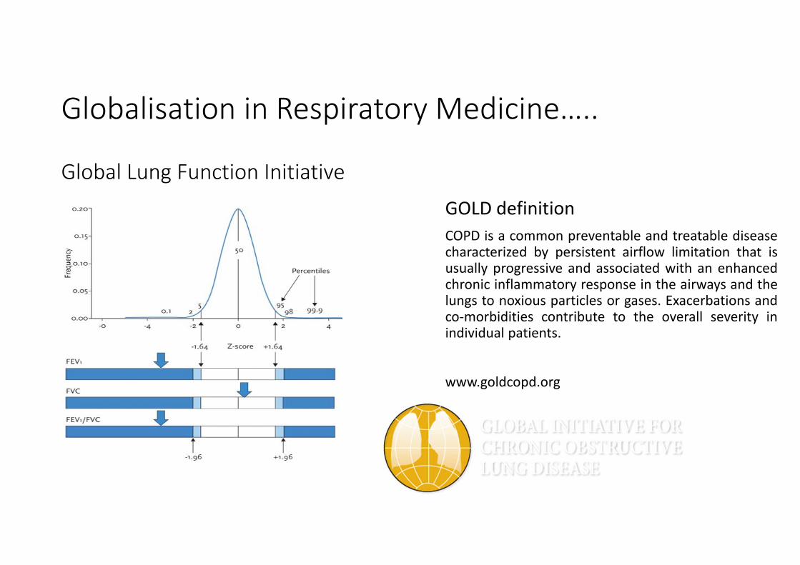
GOLD definitionCOPD is a common preventable and treatable diseasecharacterized by persistent airflow limitation that isusually progressive and associated with an enhancedchronic inflammatory response in the airways and thelungs to noxious particles or gases. Exacerbations andco‐morbidities contribute to the overall severity inindividual patients.
www.goldcopd.org
Globalisation in Respiratory Medicine…..
Global Lung Function Initiative

COPD in coal miners Reminder of definitions: • Chronic bronchitis
• Chronic productive cough for more than 3 months of the year for two successive years
• Emphysema• A chronic, irreversible disease of the lungs
characterized by abnormal enlargement of airspaces within the lungs and accompanied by destruction of the tissue lining the walls of the airspaces
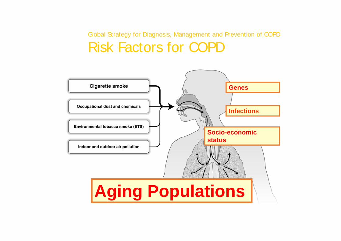
Global Strategy for Diagnosis, Management and Prevention of COPD
Risk Factors for COPD
Genes
Infections
Socio-economic status
Aging Populations© 2015 Global Initiative for Chronic Obstructive Lung Disease

Rapid changes in understanding of diseases
• Heterogeneity of diseases increasingly recognized e.g. asthma, COPD, IPF
• Better understanding of the multiple factors affecting progress of disease
• New techniques allowing better earlier diagnosis e.g respiratory PCR, FeNO
• Better understanding of basic pathophysiology potentially allowing individualized treatment
• More revolutionary technology on its way eg. whole genome testing
• Globalization of the research environment with large studies in Respiratory Medicine

Lots of LARGE lung studies: new information about asthma, COPD and longitudinal lung function
Obstructive Lung Disease• Longitudinal population based study of childhood asthma
• Evaluation of COPD Longitudinally to Identify Predictive Surrogate Endpoints (ECLIPSE)
• National Health & Nutrition Study (NHANES)
• Copenhagen City Heart Study
• Lovelace Smokers Cohort
• Framingham Offspring Cohort
• UK Household Longitudinal Survey
• Health Survey for England
• European Community Respiratory Health Survey (ECRHS)
• Tasmanian Longitudinal Health Study
COPD treatment trials • TORCH study (Towards a Revolution in COPD Health)
• UPLIFT study (Understanding Potential Long‐term Impacts on Function with Tiotropium)
• POET‐COPD study (Prevention of Exacerbations with Tiotropium)
• ISOLDE study (Inhaled Steroids in Obstructive Lung Disease)
• TIOSPIR study• NETT trial (National Emphysema Treatment Trial)• COPD Gene study

Diagram showing the loss of the lower limit of normal of FEV1:FVC ratio with ageing. The blue shaded portion represents elderly patients who are potentially over‐diagnosed and the orange shaded portion represents younger adults who are potentially underdiagnosed with obstructive lung disease. FEV1=forced expiratory volume in 1 s. FVC=forced vital capacity.
Lung function: method of defining abnormality is important……

Natural history of lung function and development of chronic obstructive pulmonary disease
Rennard SI, Drummond MB. Lancet. 2015 May 2; 385(9979): 1778–1788.

New information from non‐occupational studies• Spirometry: not perfect
• FEV1 <80%, FEV/FVC ratio 0.7 not an ideal way of assessing airflow obstruction• Normal FEV1/FVC ratio declines with age in healthy adults• Use of ratio results in up to 50% over‐diagnosis in those over 50 yrs, and underestimates the degree of airflow
obstruction in younger age groups• With good quality spirometry, ethnicity specific lower limit of normal (LLN) is probably more accurate e.g. UK
Longitudinal Health Survey: 22.2% COPD with ratio, 13.1% using LLN, 2.8% with physician‐diagnosed COPD.• NHANES III supports using the age, sex and race‐specific lower limits of normal (LLNs) (a ratio less than the fifth
percentile of the distribution of Z scores) correctly identified persons with an increased risk of death and prevalence of respiratory symptoms
• Still debated…..• Prediction algorithm available for coal dust exposed workers for spirometry
• Smoking: NOT the only factor involved in COPD• ECHRS (15,000 persons aged 20‐56 yrs) followed for 9 years: current smokers RR 4.5 for airflow obstruction• Early onset asthma associated with the development of COPD independently of smoking OR 21.0 for those with asthma
developing at age less than 10 yrs (current smokers RR 23.7)• Smoking interacts with asthma to increase the risk of COPD and the effect may be synergistic • Atopy is also a risk factor for accelerated decline in lung function (Weiss AJRCCM 2000)• Rate of decline greater in women• Significant genetic determinants
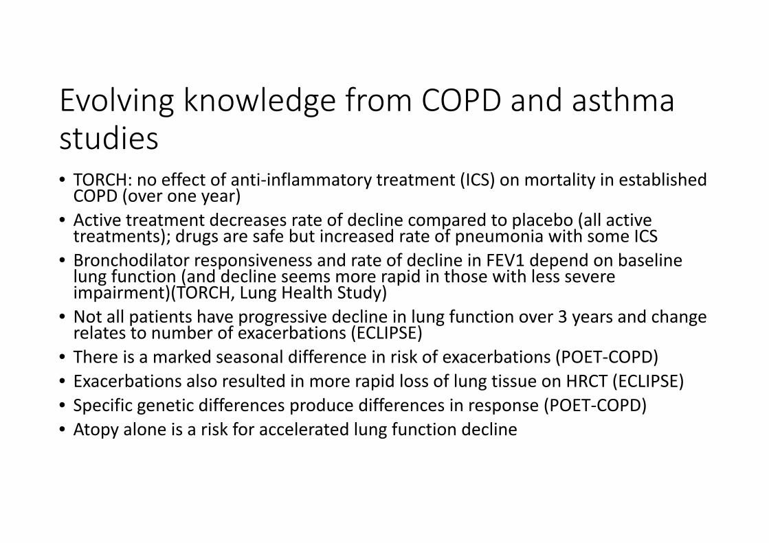
Evolving knowledge from COPD and asthma studies• TORCH: no effect of anti‐inflammatory treatment (ICS) on mortality in established COPD (over one year)
• Active treatment decreases rate of decline compared to placebo (all active treatments); drugs are safe but increased rate of pneumonia with some ICS
• Bronchodilator responsiveness and rate of decline in FEV1 depend on baseline lung function (and decline seems more rapid in those with less severe impairment)(TORCH, Lung Health Study)
• Not all patients have progressive decline in lung function over 3 years and change relates to number of exacerbations (ECLIPSE)
• There is a marked seasonal difference in risk of exacerbations (POET‐COPD)• Exacerbations also resulted in more rapid loss of lung tissue on HRCT (ECLIPSE)• Specific genetic differences produce differences in response (POET‐COPD)• Atopy alone is a risk for accelerated lung function decline

MESA Lung Study: JEM predictive of airflow limitation
Adjusted odds ratios (ORs) for the association of airflow limitation with occupational exposures ascertained using questionnaire and NIOSH job‐exposure matrix (JEM)
Exposure All Males Females Never‐smokers
n=3508; airflow limitation=196 n=1782; airflow limitation=115 n=1726; airflow limitation=81 n=1581; airflow limitation=32
OR (95% CI) OR (95% CI) OR (95% CI) OR (95% CI)
Dust 1.23 (0.88–1.72) 1.21 (0.77–1.90) 1.23 (0.74–2.06) 1.98 (0.88–4.49)
Vapor‐gas 1.55 (1.06–2.26) 1.55 (0.97–2.47) 1.36 (0.70–2.65) 2.71 (1.09–6.71)
Fumes 1.39 (0.97–1.99) 1.47 (0.93–2.32) 1.28 (0.71–2.33) 4.15 (1.74–9.87)
Severity of VGDF
None 1.0 1.0 1.0 1.0
Mild 1.18 (0.79–1.75) 1.24 (0.74–2.08) 1.09 (0.58–2.06) 1.64 (0.57–4.72)
Moderate 1.48 (0.94–2.34) 1.22 (0.65–2.31) 1.82 (0.92–3.61) 2.64 (0.85–8.20)
Severe 2.13 (1.21–3.74) 1.57 (0.72–3.46) 2.99 (1.29–6.93) 14.75 (4.06–53.63)
X2‐trend, p‐value 7.5, p < 0.01 1.3, p = 0.25 7.0, p < 0.01 13.9, p < 0.001
Years exposure VGDF, %
None
≤15 1.31 (0.88–1.94) 1.60 (0.95–2.71) 0.98 (0.51–1.86) 2.42 (0.90–6.52)
>15 1.31 (0.88–1.94) 1.10 (0.64–1.91) 1.63 (0.91–2.95) 3.31 (1.26–8.69)
X2‐trend, p‐value 2.0, p = 0.16 0.3, p = 0.60 2.3, p = 0.13 6.2, p < 0.05
No. of VGDF agents‡
0 1.0 1.0 1.0 1.0
1 1.40 (0.93–2.11) 1.28 (0.71–2.32) 1.54 (0.87–2.73) 1.82 (0.63–5.22)
2 1.40 (0.81–2.40) 1.45 (0.71–2.98) 1.35 (0.57–3.19) 3.64 (1.12–11.92)
3 1.69 (1.06–2.70) 1.62 (0.91–2.88) 1.53 (0.63–3.68) 5.07 (1.54–16.69)
X2‐trend, p‐value 5.1, p < 0.05 2.9, p = 0.09 1.4, p = 0.23 8.8, p < 0.01
Dust JEM
Low 1.0 1.0 1.0 1.0
Med 1.08 (0.60–1.94) 0.85 (0.40–1.81) 1.80 (0.69–4.74) 1.99 (0.49–8.11)
High 2.35 (1.10–5.04) 2.28 (1.03–5.07) ‐‐ 1.87 (0.16–21.66)
X2‐trend, p‐value 3.3, p = 0.07 2.3, p = 0.13 1.0, p = 0.32 0.89, p = 0.34
Organic dust JEM
Low 1.0 1.0 1.0 1.0
Medium 1.39 (0.70–2.77) 0.83 (0.31–2.20) 2.87 (1.05–7.87) 3.04 (0.67–13.69)
High 2.31 (0.93–5.74) 2.23 (0.87–5.76) ‐‐ ‐‐
X2‐trend, p‐value 3.8, p = 0.05 1.5, p = 0.21 3.3, p = 0.07 0.4, p = 0.53

Lung function and more…
Body plethysmography, fractional exhaled nitric oxide, electronic nose
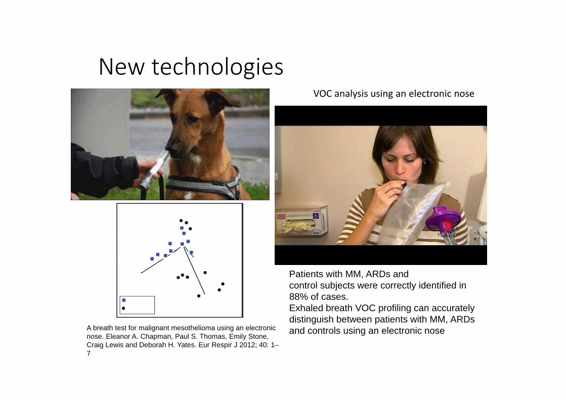
New technologies
Patients with MM, ARDs andcontrol subjects were correctly identified in 88% of cases.Exhaled breath VOC profiling can accurately distinguish between patients with MM, ARDs and controls using an electronic noseA breath test for malignant mesothelioma using an electronic
nose. Eleanor A. Chapman, Paul S. Thomas, Emily Stone, Craig Lewis and Deborah H. Yates. Eur Respir J 2012; 40: 1–7
VOC analysis using an electronic nose

Practical issues: important, especially in Australia
Population distribution in AustraliaMines in Australia
Role of surveillance: can this be improved?
Symptoms
• Standardized questionnaires• Many well validated respiratory disease questionnaires now available
Imaging• CT scanning clearly shown to be superior to chest X rays but standardization of technique and interpretation not yet accepted & widely agreed
• Mobile CT scanners now available; low doses and computerized algorithms
Lung function• Spirometry well validated but needs to be performed properly and longitudinal results are far superior to single measurements
• Measurement of DLCO the most sensitive measure of emphysema and again serial measurements preferable
• However, lung function is not specific and therefore the whole clinical picture needs to be considered
• Disruption/anxiety/cost of surveillance measures need also to be taken into account

Recommendations for control of coal workers’ pneumoconiosis (CWP) Thoracic Society of Australia and New ZealandZosky G, Hoy R, Silverstone E, Brims F,Miles S, Johnston A, Yates DH. Coal workers’ pneumoconiosis: an Australian perspective. Med J Aust 2016; 204 (11): 414‐418.
• Goal: Eliminate CWP in Australia• 1. Exposure limits and monitoring protocols
• Standardise across Australia and harmonise to international recommendations
• 2. Screening• Develop and implement a national screening program for at‐risk workers• Questionnaire, imaging, lung function testing
• 3. Medical workforce training• 4. A centralised occupational lung disease register

Respiratory surveillance:conclusions
• Recent rapid and novel developments in understanding respiratory disorders • Application of new techniques and technology have not yet achieved mainstream adoption in occupational medicine but will come
• Current methods used will underestimate dust related disorders (and systemic effects)• Surveillance is secondary rather than primary prevention; dust control is key; current methods will not pick up all coal dust related disorders
• Modified approach probably most feasible; evidence base needed• Advantages of increased surveillance need to be balanced against potential disadvantages• COPD likely to become increasingly recognized and as part of the spectrum of dust‐related disorders
• Greater understanding of mechanisms and pathology will allow more targeted treatment, as for other lung diseases
• Alignment and collaboration within the global medical world is essential

In 20 years……