screening marine-derived endophytic fungi for xylan-degrading enzymes
TRANSCRIPT
Special section:
CURRENT SCIENCE, VOL. 109, NO. 1, 10 JULY 2015 112
*For correspondence. (e-mail: [email protected]; [email protected]) †These authors contributed equally to the article.
Screening marine-derived endophytic fungi for xylan-degrading enzymes N. Thirunavukkarasu1, Ben Jahnes2,†, Arthur Broadstock3,†, M. B. Govinda Rajulu4, T. S. Murali5, Venkat Gopalan2,3,6,* and T. S. Suryanarayanan4,* 1Postgraduate and Research Department of Botany, Ramakrishna Mission Vivekananda College, Chennai 600 004, India 2Department of Microbiology, 3Department of Chemistry and Biochemistry, The Ohio State University, Columbus, OH 43210, USA 4Vivekananda Institute of Tropical Mycology, Ramakrishna Mission Vidyapith, Chennai 600 004, India 5Department of Biotechnology, School of Life Sciences, Manipal University, Manipal 576 104, India 6Center for RNA Biology, The Ohio State University, Columbus, OH 43210, USA
Marine-derived fungi surviving as symptomless endo-phytes in seaweeds and seagrasses were screened for production of xylan-degrading enzymes. Of the eight endophyte isolates obtained from five different sea-grasses and another eight from six different marine algae, half of them exhibited xylanase activity in an agar plate assay. Further examination of these lead candidates using spectrophotometric assays revealed that Trichoderma harzianum, endophytic in the brown alga Sargassum wightii, had the maximum secreted xy-lanase and xylosidase activity. Moreover, this fungus could grow in NaCl-containing media (up to 1.2 M NaCl), and inclusion of 0.26 M NaCl in the media elic-ited a two- and three-fold increase in extracellular xylanase and xylosidase activity respectively. These findings highlight the potential of prospecting marine-derived fungal endophytes to identify novel cell-wall degrading enzymes of value to the biofuel industry. Keywords: Biomass deconstruction, marine-derived fungi, Trichoderma harzianum, xylan-degrading enzymes.
Introduction
LIGNIN, cellulose and hemicellulose are three polymers that make up the lignocellulosic biomass (LCB). While cellulose is the major component, the relative amount of the three polymers varies depending on the type of bio-mass. Food, fibre and fuel industries depend on the eco-nomical and efficient conversion of cellulose and hemicellulose polymers to their constituent monomers1–3. However, deconstruction of LCB remains a challenging goal. For example, the cellulase-catalysed conversion of cellulose (a glucose homopolymer) to glucose is impeded by hemicellulose that acts as a physical barrier4–6. The focus of this study is the enzymatic hydrolysis of hemi-cellulose, an objective critical both for complete depoly-
merization of cellulose in LCB and for the generation of useful end-products from hemicellulose. Hemicellulose refers broadly to branched non-cellulosic polymers and differs in make-up within and across higher plants/algae1,7–9. Xylans, linear polymers of 1,4-linked -D-xylopyranosyl residues, are the major constituents of hemicellulose in angiosperms contributing up to 30% of the cell-wall content. Xylans are typically hetero-polymers with side-chain substitutions containing D-glucoronopyranosyl (with or without methylation), D-galactopyranosyl, L-arabinofuranosyl, acetyl, feruloyl, and p-coumaroyl groups, whose presence varies depending on the source. Given this heterogeneous composition, xylan degradation requires the concerted and synergistic action of main chain-deconstructing glycohydrolases and side chain-deconstructing esterases. However, xylanases and xylosidases are the principal enzymes in this repertoire. Xylanases cleave xylan polymers to release short xylooligosaccharides (e.g. xylopentaose), which in turn are cleaved by xylosidases to release xylose. Although it has been appreciated for some time that xylanases are produced by a wide range of organisms spanning marine algae, protozoans, crustaceans, insects, plants and fungi8, bioprospecting for improved variants that fulfil a niche requirement has motivated studies of bacteria and fungi from deep-sea hydrothermal vents10, the Antarctic11, marine sediments12, insect gut13, alimentary canal of ruminant animals14, compost/soil15,16 and decaying wood/ litter17. Because xylan-degrading enzymes from marine sources are less studied compared to those from terrestrial organisms and filamentous fungi produce xylanases at much higher levels than bacteria18,19, we investigated xylanases and xylosidases produced by marine-derived fungi (MDF) surviving as endophytes in marine angio-sperms and seaweeds. There were two reasons that inspired our focus on MDF. First, they have not been studied for their cell wall-restructuring catalytic potential, although marine fungi have been investigated for salt-tolerant, extracellular enzymes20. Fungi of the marine ecosystem are broadly
Fungal endophytes – biology and bioprospecting
CURRENT SCIENCE, VOL. 109, NO. 1, 10 JULY 2015 113
grouped into obligate marine fungi and MDF. The former grow and sporulate in the marine environment, whereas the latter are either terrestrial or freshwater forms that may be present in the marine habitat21. The MDF exist either as saprotrophs in wood and marine sediments, or as parasites and symbionts of various marine organisms such as algae, fish, coral, molluscs, sponges and sea-grasses. There are many studies that highlight the syn-thetic ability of endophytic MDF to generate bioactive secondary metabolites22–25, but few address their potential for producing industrially important enzymes26. This situation mirrors the fact that endophytes associated with terrestrial plants have been studied as a rich source of metabolites with bioactivities27–29, and only recently been explored as an excellent source of enzymes3,30,31. Second, since some seagrasses contain up to 40% xylan32, we pos-tulated that endophytic fungi residing in seagrasses might have evolved efficient mechanisms for xylan degradation. We report here that indeed MDF isolated as endophytes from marine algae and seagrasses produce xylanases and xylosidases, thus underscoring the need to study these fungi for their biocatalyst inventory.
Materials and methods
Sources of endophytes
We screened the secretomes of 16 marine-derived endo-phytic fungi for xylanase activity. These fungi were isolated from algae and sea grasses33,34 growing in Kodiyakkarai (10170N, 79490E), Keezhakkarai (9.2314N, 78.7844E) and Mandapam (9.2800N, 79.1200E), along the southeast coast of India. While the genera of these fungi were identified largely on the basis of their spore characteristics, two of them (644 and 516) were further identified at the species level by analysis of their ribosomal internal transcribed spacer (ITS) and one (562) was identified by analysing its translation elonga-tion factor-1 (EF1) DNA sequence; two of them (580 and 568) had been identified before using their ITS sequence (Table 1). Representative cultures of these fungi have been deposited with either the Microbial Type Culture Collection (MTCC), Chandigarh or the National Fungal Culture Collection of India (NFCCI) Pune, with the following accession numbers: Curvularia tuberculata 1 (NFCCI 2434), Phomopsis sp. 1 (MTCC10345), Trichoderma sp. 1 (MTCC10344). (Although brown al-gae do not belong to the plant kingdom, we use the term ‘endophytes’ in this study to denote the symptomless endosymbiotic fungi associated with them.)
DNA extraction, amplification and sequencing of ITS and EF1 DNA
Using a standard phenol–chloroform-based extraction method, genomic DNA was isolated from mycelia grown
on potato dextrose agar (PDA) medium for a period of 7 days. While ITS1-5.8S-ITS2 regions of fungal rDNA were amplified by PCR using the fungal-specific primers ITS4 and ITS5 (ref. 35), the EF1-coding DNA was ampli-fied using primers EF-728 and EF2 (refs 36 and 37). These amplicons (~500–600 bp in both cases) were electropho-resed on a 1% (w/v) agarose gel, excised and extracted, and subsequently sequenced using either ITS4 or EF728 in an automated sequencer (Applied Biosystems 3130 Genetic Analyzer). The sequences obtained were used as queries in BLASTn (default settings) to find the closest match in GenBank and in MycoBank databases; the same hits were obtained regardless of the database used. These ITS and EF1 sequences were then submitted to Gen-Bank and their accession numbers are listed in Table 1.
Qualitative plate assay to assess secreted xylanase activity37
Endophyte isolates were grown in oat spelt xylan medium (OX; 9 g oat spelt xylan (Sigma), 5 g yeast extract, 1 g NaNO3, 1 g KH2PO4, 1 g peptone, 0.3 g MgSO47H2O, 1000 ml water, pH 5.5) for 5 days at 31C with shaking (120 rpm in a REMI orbital rotary shaker). The culture fluid was filtered through a Whatman #1 filter paper, cen-trifuged at 9600 g for 20 min at 4C, and the supernatant was used as the enzyme input for the plate assay described below. Oat spelt xylan (9 g) and agar (18 g) were dissolved in 1000 ml of 100 mM sodium phosphate (pH 4.5, 5.5 or 6.5), and autoclaved before being dispensed into petri dishes. After setting of the agar, a cork borer (5 mm diameter) was used to create the diffusion wells (Figure 1). To each well, 10 l of culture supernatant (crude enzyme) was loaded, and the agar plate incubated over-night at 31C. Formation of a clear zone around the well was used to score a given endophyte secretome as posi-tive for xylanase activity38. Ten microlitres of uninocu-lated OX medium in lieu of a crude extract served as a negative control (Figure 1).
Preparation of crude secretome extracts for xylanase and xylosidase assays
Endophyte isolates were grown in birch wood xylan (BX) medium (10 g birch wood xylan, 5 g yeast extract, 1 g NaNO3, 1 g KH2PO4, 1 g peptone, 0.3 g MgSO47H2O) for 5 days at 31C in an orbital shaker (120 rpm). The culture fluid was then filtered through a Whatman #1 fil-ter paper, and the filtrate centrifuged at 9600 g for 20 min at 4C. The supernatant was dialysed against water (with three changes over 16–18 h) at 4C and lyophilized. The protein amount in these dried secretomes was measured using either the Bradford or the bicinchoninic acid assay, with bovine serum albumin serving as the standard.
Special section:
CURRENT SCIENCE, VOL. 109, NO. 1, 10 JULY 2015 114
Table 1. Summary of endophytes displaying xylanase activity
Isolate from seagrass (1–8) Xylanase activity Accession numbers for ITS Fungus (isolate number) or algal (9–16) species (pH 4.5, 5.5, 6.5) or EF1 sequences
Aphanocladium sp. (508) Thalassia sp. – Aspergillus terreus (644) Cymodocea serrulata + KP122964 Cladosporium sp.1 (505) Thalassia sp.1 – Curvularia tuberculata (503) Thalassia sp.1 – Curvularia tuberculata (516) Syringodium sp. + KP122965 Gonatophragmium mori (622) Thalassia sp. + Nodulisporium sp. (679) Thalassia sp. 3 + Torulomyces sp. (683) Syringodium sp. + Cladosporium sp.1 (547) Ulva lactuca – Drechslera sp. 1 (583) Sargassum wightii – Drechslera sp. 2 (610) Turbenaria sp. – Fusarium oxysporum (580) Sargassum wightii + KC254033* Nigrospora sp. (538) Jania adherens – Phoma sp. 1 (605) Sargassum sp. – Phomopsis sp. (562) Portieria hornemonii + KP204449 Trichoderma harzianum (568) Sargassum wightii + KC330218*
*These species were identified in an earlier study33. +, Positive; –, Negative.
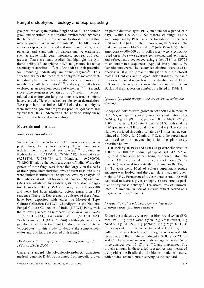
Figure 1. Agar plate assay for secreted xylanase activity. Culture fil-trates of endophytes were tested for their ability to hydrolyze oat spelt xylan impregnated in an agar plate. Endophytes were scored as positive if their filtrates resulted in a zone of clearance, as observed here with Aspergillus terreus (644), Nodulisporium sp. (679) and Torulomyces sp. (683). Uninoculated culture filtrate served as the negative control. While three representative species are shown here, similar studies were conducted to obtain the data summarized in Table 1.
Enzyme assays
Xylanase assays: Xylanase activity was assayed as descri-bed elsewhere39. The underlying principle is that xylan degradation releases xylose, which in turn can reduce 3,5-dinitrosalycilic acid (DNS) to 3-amino, 5-nitrosalicylic acid that absorbs at 540 nm. Using a standard curve gener-ated with known quantities of xylose, the amount of xylose released in an enzymatic assay is easily deter-mined. We employed two variants of this xylanase assay. For the end-point measurements (Figure 2), we used a 2 ml assay mixture containing 1.8 ml of 1% (w/v) birch wood
xylan in 50 mM sodium citrate (pH 5.3) and 0.2 ml of secretome (crude enzyme). The reaction was initiated by incubation at 50C for 5 min, and immediately added to 3 ml of 1% DNS prior to boiling for 10 min and subse-quent addition of 1 ml 40% (w/v) potassium tartrate. The Abs540 of these solutions was measured using a Unico SpectroQuest 4802 spectrophotometer. For the kinetic assays at 37C (Figures 3 and 5), we used a final concentration of 50 mM sodium acetate (pH 5.5) and 0.5% (w/v) birch wood xylan. A 360 l reaction mixture contained 0.5% (w/v) birch wood xylan in 50 mM sodium acetate (pH 5.5). The substrate in buffer (310 l) and the secretome resuspended in phosphate-buffered saline (PBS; ~30 to 300 g in 50 l) were indi-vidually pre-incubated for 10 min at 37C, before they were mixed to initiate the reaction. Five aliquots were withdrawn over 15 or 75 min (depending on strong or weak activity), and each 72 l aliquot was mixed with 72 l 1% (w/v) DNS and boiled for 10 min. The boiled samples were cooled and added to 72 l 40% (w/v) potassium tartrate. The final volume (216 l for each time point) was divided into two equal parts to enable two independent measurements at 540 nm by a Flexstation 3 Multi-Mode Microplate Reader (Molecular Devices, Sunnyvale, USA). For each secretome, a specific blank was generated to ensure that xylose carryover (if any) was accounted for. Rather than performing a time course after addition of a pre-incubated crude secretome to the substrate, the blanks entailed immediate boiling with 1% (w/v) DNS. The sub-sequent steps were the same as those described above for the corresponding test assays. Xylosidase assays: -Xylosidase activity was deter-mined by measuring the amount of p-nitrophenol (pNP)
Fungal endophytes – biology and bioprospecting
CURRENT SCIENCE, VOL. 109, NO. 1, 10 JULY 2015 115
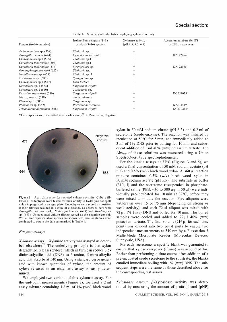
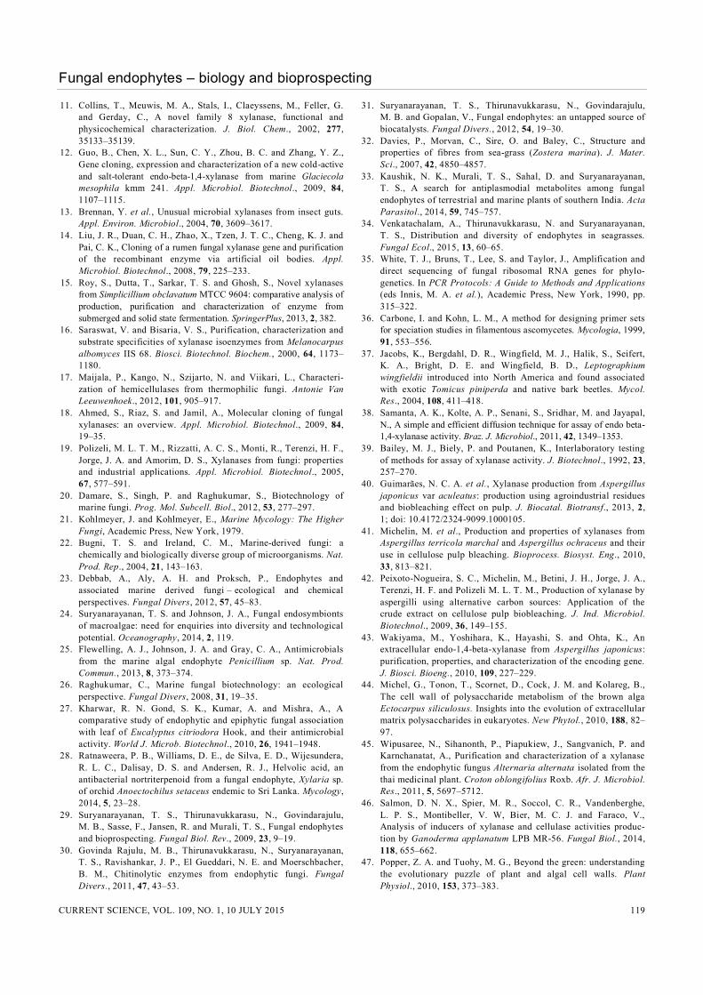
Figure 2. Effect of culture age and media pH on xylanase activity secreted by T. harzianum. Single time-point meas-urements were used to determine the xylanase activities in filtrates of cultures grown in the presence of 1% (w/v) xylan, either at pH 5.5 for 1–8 days (a) or at three different pH values for 5 days (b). The activities are normalized against the highest value observed in each comparison panel. From end-point measurements, we determined xylanase specific activi-ties at 50C to exhibit less than twofold variation and a maximum of 46 U/mg (e.g. pH 4.5). The standard deviation of the mean from three independent xylanase activity measurements did not exceed 24% (with the exception of the measurement for day 1, 44%).
released from p-nitrophenyl--D-xyloside (pNPX; Sigma, St Louis, USA). All kinetic assays were per-formed at 37C in a final concentration of 33 mM sodium acetate (pH 5.2) and 3.3 mM pNPX. The substrate was pre-incubated at 37C for 7 min in a volume of 696 l, and the reaction initiated by adding a defined amount of secretome (~25–65 g protein resuspended in 24 l PBS). Four aliquots were withdrawn every 15 min during a 1 h incubation (Figure 3); for the experiments examining effect of NaCl in the media (Figure ), only three aliquots were withdrawn during a 45 min incubation. At each time point, the reactions were terminated by adding an equal volume of 1 M sodium carbonate. The amount of pNP formed was determined based on the absorbance at 405 nm. For each time point, the final quenched reaction (360 l) was divided into three equal parts to allow three independent measurements at 405 nm by a Flexstation 3 Multi-Mode Microplate Reader. As a negative control, we incubated the substrate without enzyme and sub-tracted the values for uncatalysed breakdown of pNPX in our calculations of xylosidase activities.
Additional comments related to enzyme assays: The dried secretomes were re-suspended in PBS and centri-fuged at 16,000 g for 10 min to remove particulate material. The resulting supernatant was used in the enzyme assays. Although there was variability in the protein content of se-cretomes obtained from the different species, we used the same amount of total protein in our individual assays. In all assays, one unit is defined as the amount of en-zyme that produces 1 mol of product per minute. Linear regression (Excel) analysis of the product formed as a function of time was used to calculate the initial velocity
(r2 0.96 for xylanase and r2 0.98 for xylosidase). Mean and standard deviation values were calculated from three independent assays, and the means used to calculate the relative specific activities (Figures 3 and 4). Since we used two versions of the xylanase assay at the two research sites (in India and the US), there are some differences in the specific activities determined (Figures 2 and 3). These differences likely stem from end-point (Figure 2) versus kinetic assays (Figures 3 and 4), as well as the concentration of substrate and temperature of the assay. However, these variations do not influence the inferences drawn as each set of data was used to make comparisons only within that group.
Effect of culture age and pH on extracellular xylanase activity of Trichoderma harzianum
Trichoderma harzianum was inoculated in 10 ml BX medium and incubated at 31C in an orbital shaker (120 rpm). For evaluating the effect of culture age, the filtrate was obtained from T. harzianum cultures grown for 1, 2, 3, 4, 5, 6, 7 and 8 days. For assessing the effect of media pH, T. harzianum was cultured in BX medium (pH 4.5, 5.5 or 7.5) for five days before the filtrate was collected. In these two instances, end-point assays at 50C were used to determine the xylanase activity.
Effect of NaCl on the growth of T. harzianum
To study the effect of NaCl on the growth of T. harzianum, a plug of mycelium (5 mm diameter) was cut from the margin of a colony growing actively on Czapek–Dox agar (CDA) medium (2 g NaNO3, 1 g KH2PO4, 0.5 g
4
Special section:
CURRENT SCIENCE, VOL. 109, NO. 1, 10 JULY 2015 116
MgSO4, 0.5 g KCl, 0.01 g FeSO4, 30 g sucrose, 1000 ml water). This mycelial plug was placed on CDA amended with 0%, 1.5%, 2.5%, 3.5% or 7% (w/v) NaCl and incu-bated at 26C for 25 days with periodic measurements of the colony diameter. The mean values reported were cal-culated from data obtained in three replicates (Figure 5).
Effect of including NaCl on extracellular xylanase and xylosidase activity of T. harzianum
To examine the effect of including NaCl in the media, T. harzianum was cultured for five days in BX medium
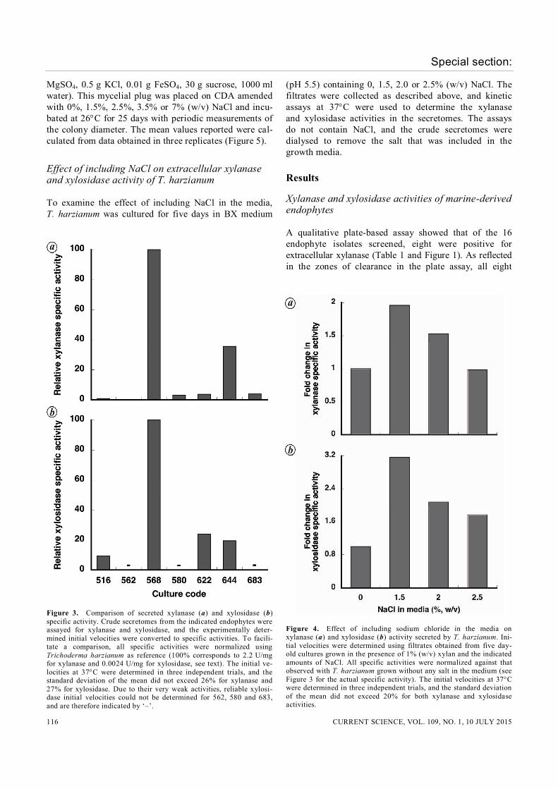
Figure 3. Comparison of secreted xylanase (a) and xylosidase (b) specific activity. Crude secretomes from the indicated endophytes were assayed for xylanase and xylosidase, and the experimentally deter-mined initial velocities were converted to specific activities. To facili-tate a comparison, all specific activities were normalized using Trichoderma harzianum as reference (100% corresponds to 2.2 U/mg for xylanase and 0.0024 U/mg for xylosidase, see text). The initial ve-locities at 37C were determined in three independent trials, and the standard deviation of the mean did not exceed 26% for xylanase and 27% for xylosidase. Due to their very weak activities, reliable xylosi-dase initial velocities could not be determined for 562, 580 and 683, and are therefore indicated by ‘–’.
(pH 5.5) containing 0, 1.5, 2.0 or 2.5% (w/v) NaCl. The filtrates were collected as described above, and kinetic assays at 37C were used to determine the xylanase and xylosidase activities in the secretomes. The assays do not contain NaCl, and the crude secretomes were dialysed to remove the salt that was included in the growth media.
Results
Xylanase and xylosidase activities of marine-derived endophytes
A qualitative plate-based assay showed that of the 16 endophyte isolates screened, eight were positive for extracellular xylanase (Table 1 and Figure 1). As reflected in the zones of clearance in the plate assay, all eight
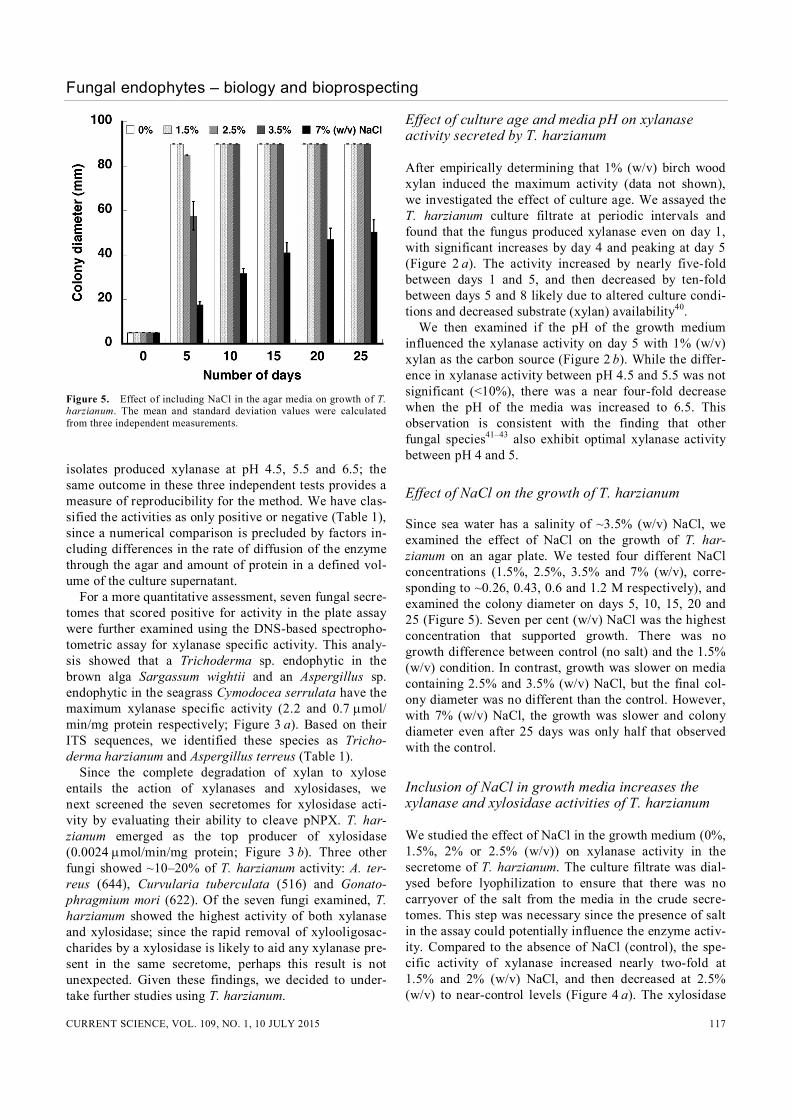
Figure 4. Effect of including sodium chloride in the media on xylanase (a) and xylosidase (b) activity secreted by T. harzianum. Ini-tial velocities were determined using filtrates obtained from five day-old cultures grown in the presence of 1% (w/v) xylan and the indicated amounts of NaCl. All specific activities were normalized against that observed with T. harzianum grown without any salt in the medium (see Figure 3 for the actual specific activity). The initial velocities at 37C were determined in three independent trials, and the standard deviation of the mean did not exceed 20% for both xylanase and xylosidase activities.
Fungal endophytes – biology and bioprospecting
CURRENT SCIENCE, VOL. 109, NO. 1, 10 JULY 2015 117
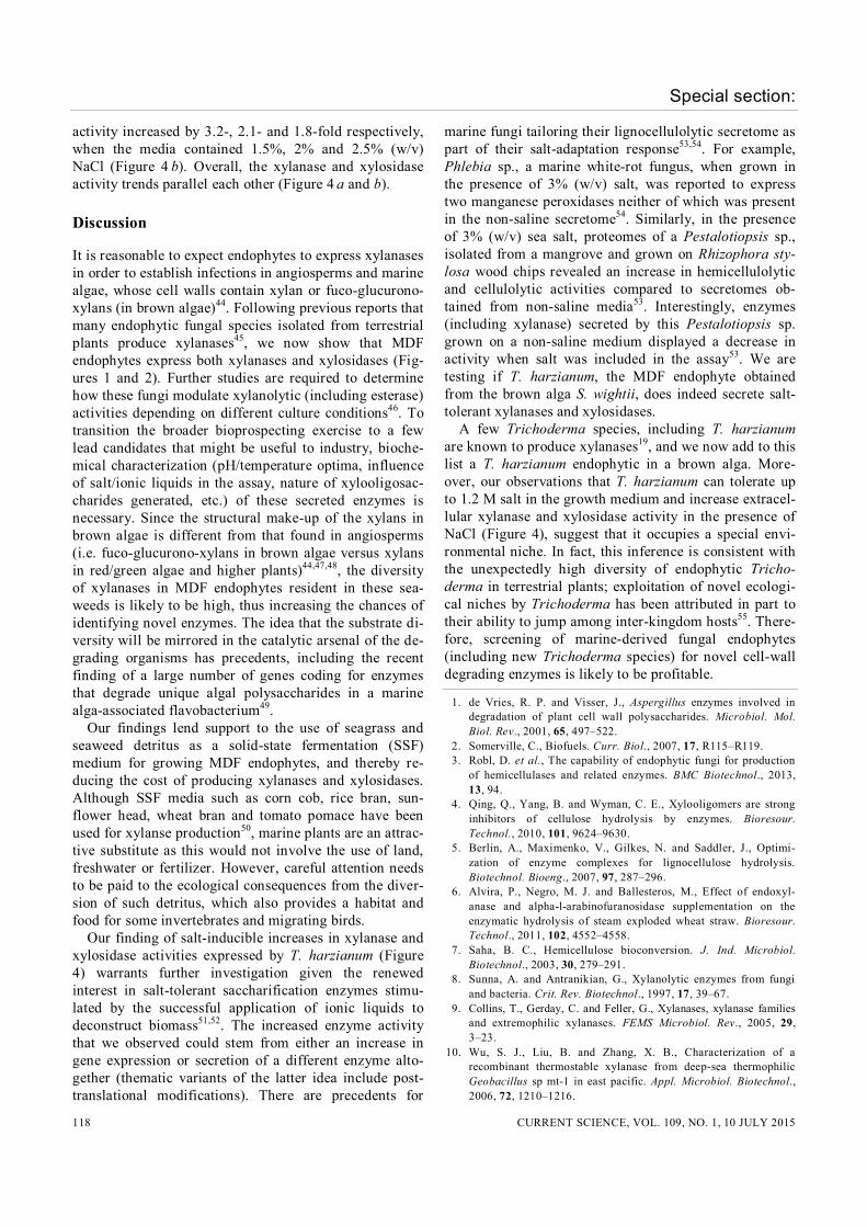
Figure 5. Effect of including NaCl in the agar media on growth of T. harzianum. The mean and standard deviation values were calculated from three independent measurements. isolates produced xylanase at pH 4.5, 5.5 and 6.5; the same outcome in these three independent tests provides a measure of reproducibility for the method. We have clas-sified the activities as only positive or negative (Table 1), since a numerical comparison is precluded by factors in-cluding differences in the rate of diffusion of the enzyme through the agar and amount of protein in a defined vol-ume of the culture supernatant. For a more quantitative assessment, seven fungal secre-tomes that scored positive for activity in the plate assay were further examined using the DNS-based spectropho-tometric assay for xylanase specific activity. This analy-sis showed that a Trichoderma sp. endophytic in the brown alga Sargassum wightii and an Aspergillus sp. endophytic in the seagrass Cymodocea serrulata have the maximum xylanase specific activity (2.2 and 0.7 mol/ min/mg protein respectively; Figure 3 a). Based on their ITS sequences, we identified these species as Tricho-derma harzianum and Aspergillus terreus (Table 1). Since the complete degradation of xylan to xylose entails the action of xylanases and xylosidases, we next screened the seven secretomes for xylosidase acti-vity by evaluating their ability to cleave pNPX. T. har-zianum emerged as the top producer of xylosidase (0.0024 mol/min/mg protein; Figure 3 b). Three other fungi showed ~10–20% of T. harzianum activity: A. ter-reus (644), Curvularia tuberculata (516) and Gonato-phragmium mori (622). Of the seven fungi examined, T. harzianum showed the highest activity of both xylanase and xylosidase; since the rapid removal of xylooligosac-charides by a xylosidase is likely to aid any xylanase pre-sent in the same secretome, perhaps this result is not unexpected. Given these findings, we decided to under-take further studies using T. harzianum.
Effect of culture age and media pH on xylanase activity secreted by T. harzianum
After empirically determining that 1% (w/v) birch wood xylan induced the maximum activity (data not shown), we investigated the effect of culture age. We assayed the T. harzianum culture filtrate at periodic intervals and found that the fungus produced xylanase even on day 1, with significant increases by day 4 and peaking at day 5 (Figure 2 a). The activity increased by nearly five-fold between days 1 and 5, and then decreased by ten-fold between days 5 and 8 likely due to altered culture condi-tions and decreased substrate (xylan) availability40. We then examined if the pH of the growth medium influenced the xylanase activity on day 5 with 1% (w/v) xylan as the carbon source (Figure 2 b). While the differ-ence in xylanase activity between pH 4.5 and 5.5 was not significant (<10%), there was a near four-fold decrease when the pH of the media was increased to 6.5. This observation is consistent with the finding that other fungal species41–43 also exhibit optimal xylanase activity between pH 4 and 5.
Effect of NaCl on the growth of T. harzianum
Since sea water has a salinity of ~3.5% (w/v) NaCl, we examined the effect of NaCl on the growth of T. har-zianum on an agar plate. We tested four different NaCl concentrations (1.5%, 2.5%, 3.5% and 7% (w/v), corre-sponding to ~0.26, 0.43, 0.6 and 1.2 M respectively), and examined the colony diameter on days 5, 10, 15, 20 and 25 (Figure 5). Seven per cent (w/v) NaCl was the highest concentration that supported growth. There was no growth difference between control (no salt) and the 1.5% (w/v) condition. In contrast, growth was slower on media containing 2.5% and 3.5% (w/v) NaCl, but the final col-ony diameter was no different than the control. However, with 7% (w/v) NaCl, the growth was slower and colony diameter even after 25 days was only half that observed with the control.
Inclusion of NaCl in growth media increases the xylanase and xylosidase activities of T. harzianum
We studied the effect of NaCl in the growth medium (0%, 1.5%, 2% or 2.5% (w/v)) on xylanase activity in the secretome of T. harzianum. The culture filtrate was dial-ysed before lyophilization to ensure that there was no carryover of the salt from the media in the crude secre-tomes. This step was necessary since the presence of salt in the assay could potentially influence the enzyme activ-ity. Compared to the absence of NaCl (control), the spe-cific activity of xylanase increased nearly two-fold at 1.5% and 2% (w/v) NaCl, and then decreased at 2.5% (w/v) to near-control levels (Figure 4 a). The xylosidase
Special section:
CURRENT SCIENCE, VOL. 109, NO. 1, 10 JULY 2015 118
activity increased by 3.2-, 2.1- and 1.8-fold respectively, when the media contained 1.5%, 2% and 2.5% (w/v) NaCl (Figure 4 b). Overall, the xylanase and xylosidase activity trends parallel each other (Figure 4 a and b).
Discussion
It is reasonable to expect endophytes to express xylanases in order to establish infections in angiosperms and marine algae, whose cell walls contain xylan or fuco-glucurono-xylans (in brown algae)44. Following previous reports that many endophytic fungal species isolated from terrestrial plants produce xylanases45, we now show that MDF endophytes express both xylanases and xylosidases (Fig-ures 1 and 2). Further studies are required to determine how these fungi modulate xylanolytic (including esterase) activities depending on different culture conditions46. To transition the broader bioprospecting exercise to a few lead candidates that might be useful to industry, bioche-mical characterization (pH/temperature optima, influence of salt/ionic liquids in the assay, nature of xylooligosac-charides generated, etc.) of these secreted enzymes is necessary. Since the structural make-up of the xylans in brown algae is different from that found in angiosperms (i.e. fuco-glucurono-xylans in brown algae versus xylans in red/green algae and higher plants)44,47,48, the diversity of xylanases in MDF endophytes resident in these sea-weeds is likely to be high, thus increasing the chances of identifying novel enzymes. The idea that the substrate di-versity will be mirrored in the catalytic arsenal of the de-grading organisms has precedents, including the recent finding of a large number of genes coding for enzymes that degrade unique algal polysaccharides in a marine alga-associated flavobacterium49. Our findings lend support to the use of seagrass and seaweed detritus as a solid-state fermentation (SSF) medium for growing MDF endophytes, and thereby re-ducing the cost of producing xylanases and xylosidases. Although SSF media such as corn cob, rice bran, sun-flower head, wheat bran and tomato pomace have been used for xylanse production50, marine plants are an attrac-tive substitute as this would not involve the use of land, freshwater or fertilizer. However, careful attention needs to be paid to the ecological consequences from the diver-sion of such detritus, which also provides a habitat and food for some invertebrates and migrating birds. Our finding of salt-inducible increases in xylanase and xylosidase activities expressed by T. harzianum (Figure 4) warrants further investigation given the renewed interest in salt-tolerant saccharification enzymes stimu-lated by the successful application of ionic liquids to deconstruct biomass51,52. The increased enzyme activity that we observed could stem from either an increase in gene expression or secretion of a different enzyme alto-gether (thematic variants of the latter idea include post-translational modifications). There are precedents for
marine fungi tailoring their lignocellulolytic secretome as part of their salt-adaptation response53,54. For example, Phlebia sp., a marine white-rot fungus, when grown in the presence of 3% (w/v) salt, was reported to express two manganese peroxidases neither of which was present in the non-saline secretome54. Similarly, in the presence of 3% (w/v) sea salt, proteomes of a Pestalotiopsis sp., isolated from a mangrove and grown on Rhizophora sty-losa wood chips revealed an increase in hemicellulolytic and cellulolytic activities compared to secretomes ob-tained from non-saline media53. Interestingly, enzymes (including xylanase) secreted by this Pestalotiopsis sp. grown on a non-saline medium displayed a decrease in activity when salt was included in the assay53. We are testing if T. harzianum, the MDF endophyte obtained from the brown alga S. wightii, does indeed secrete salt-tolerant xylanases and xylosidases. A few Trichoderma species, including T. harzianum are known to produce xylanases19, and we now add to this list a T. harzianum endophytic in a brown alga. More-over, our observations that T. harzianum can tolerate up to 1.2 M salt in the growth medium and increase extracel-lular xylanase and xylosidase activity in the presence of NaCl (Figure 4), suggest that it occupies a special envi-ronmental niche. In fact, this inference is consistent with the unexpectedly high diversity of endophytic Tricho-derma in terrestrial plants; exploitation of novel ecologi-cal niches by Trichoderma has been attributed in part to their ability to jump among inter-kingdom hosts55. There-fore, screening of marine-derived fungal endophytes (including new Trichoderma species) for novel cell-wall degrading enzymes is likely to be profitable.
1. de Vries, R. P. and Visser, J., Aspergillus enzymes involved in degradation of plant cell wall polysaccharides. Microbiol. Mol. Biol. Rev., 2001, 65, 497–522.
2. Somerville, C., Biofuels. Curr. Biol., 2007, 17, R115–R119. 3. Robl, D. et al., The capability of endophytic fungi for production
of hemicellulases and related enzymes. BMC Biotechnol., 2013, 13, 94.
4. Qing, Q., Yang, B. and Wyman, C. E., Xylooligomers are strong inhibitors of cellulose hydrolysis by enzymes. Bioresour. Technol., 2010, 101, 9624–9630.
5. Berlin, A., Maximenko, V., Gilkes, N. and Saddler, J., Optimi-zation of enzyme complexes for lignocellulose hydrolysis. Biotechnol. Bioeng., 2007, 97, 287–296.
6. Alvira, P., Negro, M. J. and Ballesteros, M., Effect of endoxyl-anase and alpha-l-arabinofuranosidase supplementation on the enzymatic hydrolysis of steam exploded wheat straw. Bioresour. Technol., 2011, 102, 4552–4558.
7. Saha, B. C., Hemicellulose bioconversion. J. Ind. Microbiol. Biotechnol., 2003, 30, 279–291.
8. Sunna, A. and Antranikian, G., Xylanolytic enzymes from fungi and bacteria. Crit. Rev. Biotechnol., 1997, 17, 39–67.
9. Collins, T., Gerday, C. and Feller, G., Xylanases, xylanase families and extremophilic xylanases. FEMS Microbiol. Rev., 2005, 29, 3–23.
10. Wu, S. J., Liu, B. and Zhang, X. B., Characterization of a recombinant thermostable xylanase from deep-sea thermophilic Geobacillus sp mt-1 in east pacific. Appl. Microbiol. Biotechnol., 2006, 72, 1210–1216.
Fungal endophytes – biology and bioprospecting
CURRENT SCIENCE, VOL. 109, NO. 1, 10 JULY 2015 119
11. Collins, T., Meuwis, M. A., Stals, I., Claeyssens, M., Feller, G. and Gerday, C., A novel family 8 xylanase, functional and physicochemical characterization. J. Biol. Chem., 2002, 277, 35133–35139.
12. Guo, B., Chen, X. L., Sun, C. Y., Zhou, B. C. and Zhang, Y. Z., Gene cloning, expression and characterization of a new cold-active and salt-tolerant endo-beta-1,4-xylanase from marine Glaciecola mesophila kmm 241. Appl. Microbiol. Biotechnol., 2009, 84, 1107–1115.
13. Brennan, Y. et al., Unusual microbial xylanases from insect guts. Appl. Environ. Microbiol., 2004, 70, 3609–3617.
14. Liu, J. R., Duan, C. H., Zhao, X., Tzen, J. T. C., Cheng, K. J. and Pai, C. K., Cloning of a rumen fungal xylanase gene and purification of the recombinant enzyme via artificial oil bodies. Appl. Microbiol. Biotechnol., 2008, 79, 225–233.
15. Roy, S., Dutta, T., Sarkar, T. S. and Ghosh, S., Novel xylanases from Simplicillium obclavatum MTCC 9604: comparative analysis of production, purification and characterization of enzyme from submerged and solid state fermentation. SpringerPlus, 2013, 2, 382.
16. Saraswat, V. and Bisaria, V. S., Purification, characterization and substrate specificities of xylanase isoenzymes from Melanocarpus albomyces IIS 68. Biosci. Biotechnol. Biochem., 2000, 64, 1173–1180.
17. Maijala, P., Kango, N., Szijarto, N. and Viikari, L., Characteri-zation of hemicellulases from thermophilic fungi. Antonie Van Leeuwenhoek., 2012, 101, 905–917.
18. Ahmed, S., Riaz, S. and Jamil, A., Molecular cloning of fungal xylanases: an overview. Appl. Microbiol. Biotechnol., 2009, 84, 19–35.
19. Polizeli, M. L. T. M., Rizzatti, A. C. S., Monti, R., Terenzi, H. F., Jorge, J. A. and Amorim, D. S., Xylanases from fungi: properties and industrial applications. Appl. Microbiol. Biotechnol., 2005, 67, 577–591.
20. Damare, S., Singh, P. and Raghukumar, S., Biotechnology of marine fungi. Prog. Mol. Subcell. Biol., 2012, 53, 277–297.
21. Kohlmeyer, J. and Kohlmeyer, E., Marine Mycology: The Higher Fungi, Academic Press, New York, 1979.
22. Bugni, T. S. and Ireland, C. M., Marine-derived fungi: a chemically and biologically diverse group of microorganisms. Nat. Prod. Rep., 2004, 21, 143–163.
23. Debbab, A., Aly, A. H. and Proksch, P., Endophytes and associated marine derived fungi – ecological and chemical perspectives. Fungal Divers, 2012, 57, 45–83.
24. Suryanarayanan, T. S. and Johnson, J. A., Fungal endosymbionts of macroalgae: need for enquiries into diversity and technological potential. Oceanography, 2014, 2, 119.
25. Flewelling, A. J., Johnson, J. A. and Gray, C. A., Antimicrobials from the marine algal endophyte Penicillium sp. Nat. Prod. Commun., 2013, 8, 373–374.
26. Raghukumar, C., Marine fungal biotechnology: an ecological perspective. Fungal Divers, 2008, 31, 19–35.
27. Kharwar, R. N. Gond, S. K., Kumar, A. and Mishra, A., A comparative study of endophytic and epiphytic fungal association with leaf of Eucalyptus citriodora Hook, and their antimicrobial activity. World J. Microb. Biotechnol., 2010, 26, 1941–1948.
28. Ratnaweera, P. B., Williams, D. E., de Silva, E. D., Wijesundera, R. L. C., Dalisay, D. S. and Andersen, R. J., Helvolic acid, an antibacterial nortriterpenoid from a fungal endophyte, Xylaria sp. of orchid Anoectochilus setaceus endemic to Sri Lanka. Mycology, 2014, 5, 23–28.
29. Suryanarayanan, T. S., Thirunavukkarasu, N., Govindarajulu, M. B., Sasse, F., Jansen, R. and Murali, T. S., Fungal endophytes and bioprospecting. Fungal Biol. Rev., 2009, 23, 9–19.
30. Govinda Rajulu, M. B., Thirunavukkarasu, N., Suryanarayanan, T. S., Ravishankar, J. P., El Gueddari, N. E. and Moerschbacher, B. M., Chitinolytic enzymes from endophytic fungi. Fungal Divers., 2011, 47, 43–53.
31. Suryanarayanan, T. S., Thirunavukkarasu, N., Govindarajulu, M. B. and Gopalan, V., Fungal endophytes: an untapped source of biocatalysts. Fungal Divers., 2012, 54, 19–30.
32. Davies, P., Morvan, C., Sire, O. and Baley, C., Structure and properties of fibres from sea-grass (Zostera marina). J. Mater. Sci., 2007, 42, 4850–4857.
33. Kaushik, N. K., Murali, T. S., Sahal, D. and Suryanarayanan, T. S., A search for antiplasmodial metabolites among fungal endophytes of terrestrial and marine plants of southern India. Acta Parasitol., 2014, 59, 745–757.
34. Venkatachalam, A., Thirunavukkarasu, N. and Suryanarayanan, T. S., Distribution and diversity of endophytes in seagrasses. Fungal Ecol., 2015, 13, 60–65.
35. White, T. J., Bruns, T., Lee, S. and Taylor, J., Amplification and direct sequencing of fungal ribosomal RNA genes for phylo-genetics. In PCR Protocols: A Guide to Methods and Applications (eds Innis, M. A. et al.), Academic Press, New York, 1990, pp. 315–322.
36. Carbone, I. and Kohn, L. M., A method for designing primer sets for speciation studies in filamentous ascomycetes. Mycologia, 1999, 91, 553–556.
37. Jacobs, K., Bergdahl, D. R., Wingfield, M. J., Halik, S., Seifert, K. A., Bright, D. E. and Wingfield, B. D., Leptographium wingfieldii introduced into North America and found associated with exotic Tomicus piniperda and native bark beetles. Mycol. Res., 2004, 108, 411–418.
38. Samanta, A. K., Kolte, A. P., Senani, S., Sridhar, M. and Jayapal, N., A simple and efficient diffusion technique for assay of endo beta-1,4-xylanase activity. Braz. J. Microbiol., 2011, 42, 1349–1353.
39. Bailey, M. J., Biely, P. and Poutanen, K., Interlaboratory testing of methods for assay of xylanase activity. J. Biotechnol., 1992, 23, 257–270.
40. Guimarães, N. C. A. et al., Xylanase production from Aspergillus japonicus var aculeatus: production using agroindustrial residues and biobleaching effect on pulp. J. Biocatal. Biotransf., 2013, 2, 1; doi: 10.4172/2324-9099.1000105.
41. Michelin, M. et al., Production and properties of xylanases from Aspergillus terricola marchal and Aspergillus ochraceus and their use in cellulose pulp bleaching. Bioprocess. Biosyst. Eng., 2010, 33, 813–821.
42. Peixoto-Nogueira, S. C., Michelin, M., Betini, J. H., Jorge, J. A., Terenzi, H. F. and Polizeli M. L. T. M., Production of xylanase by aspergilli using alternative carbon sources: Application of the crude extract on cellulose pulp biobleaching. J. Ind. Microbiol. Biotechnol., 2009, 36, 149–155.
43. Wakiyama, M., Yoshihara, K., Hayashi, S. and Ohta, K., An extracellular endo-1,4-beta-xylanase from Aspergillus japonicus: purification, properties, and characterization of the encoding gene. J. Biosci. Bioeng., 2010, 109, 227–229.
44. Michel, G., Tonon, T., Scornet, D., Cock, J. M. and Kolareg, B., The cell wall of polysaccharide metabolism of the brown alga Ectocarpus siliculosus. Insights into the evolution of extracellular matrix polysaccharides in eukaryotes. New Phytol., 2010, 188, 82–97.
45. Wipusaree, N., Sihanonth, P., Piapukiew, J., Sangvanich, P. and Karnchanatat, A., Purification and characterization of a xylanase from the endophytic fungus Alternaria alternata isolated from the thai medicinal plant. Croton oblongifolius Roxb. Afr. J. Microbiol. Res., 2011, 5, 5697–5712.
46. Salmon, D. N. X., Spier, M. R., Soccol, C. R., Vandenberghe, L. P. S., Montibeller, V. W, Bier, M. C. J. and Faraco, V., Analysis of inducers of xylanase and cellulase activities produc-tion by Ganoderma applanatum LPB MR-56. Fungal Biol., 2014, 118, 655–662.
47. Popper, Z. A. and Tuohy, M. G., Beyond the green: understanding the evolutionary puzzle of plant and algal cell walls. Plant Physiol., 2010, 153, 373–383.
Special section:
CURRENT SCIENCE, VOL. 109, NO. 1, 10 JULY 2015 120
48. Kloareg, B. and Quatrano, R. S., Structure of the cell-walls of marine-algae and ecophysiological functions of the matrix polysaccharides. Oceanogr. Mar. Biol., 1988, 26, 259–315.
49. Mann, A. J. et al., The genome of the alga-associated marine flavobacterium Formosa agariphila kmm 3901(t) reveals a broad potential for degradation of algal polysaccharides. Appl. Environ. Microbiol., 2013, 79, 6813–6822.
50. Umsza-Guez, M. A., Diaz, A. B., de Ory, I., Blandino, A., Gomes, E. and Caro, I., Xylanase production by Aspergillus awamori under solid state fermentation conditions on tomato pomace. Braz. J. Microbiol., 2011, 42, 1585–1597.
51. Datta, S. et al., Ionic liquid tolerant hyperthermophilic cellulases for biomass pretreatment and hydrolysis. Green Chem., 2010, 12, 338–345.
52. Shi, J. et al., One-pot ionic liquid pretreatment and saccharifica-tion of switchgrass. Green Chem., 2013, 15, 2579–2589.
53. Arfi, Y., Chevret, D., Henrissat, B., Berrin, J. G., Levasseur, A. and Record, E., Characterization of salt-adapted secreted ligno-cellulolytic enzymes from the mangrove fungus Pestalotiopsis sp. Nature Commun., 2013, 4, 1810.
54. Kamei, I., Daikoku, C., Tsutsumi, Y. and Kondo, R., Saline-dependent regulation of manganese peroxidase genes in the hypersaline-tolerant white rot fungus Phlebia sp. strain mg-60. Appl. Environ. Microbiol., 2008, 74, 2709–2716.
55. Chaverri, P. and Samuels, G. J., Evolution of habitat preference and nutrition mode in a cosmopolitan fungal genus with evidence of interkingdom host jumps and major shifts in ecology. Evolution, 2013, 7, 2823–2837.
ACKNOWLEDGEMENTS. We are grateful to Dr T. Ezeji, Ohio Ag-ricultural Research and Development Center (OARDC), USA, for a gift of birch wood xylan. T.S.S. thanks the Secretary, Ramakrishna Mission Vidyapith, Chennai, for providing research facilities. N.T. thanks the Department of Science and Technology, Government of India for funds through the SERB project (SB/EMEQ-005/2013). T.S.M. thanks Manipal University (MU), and the TIFAC-CORE in Pharmacogeno-mics at MU for facilities and support. Biofuel-related research in VG’s laboratory is supported by funds from OARDC (award to T. Ezeji and VG).