scleroderma and central nervous system...
TRANSCRIPT
410
Scleroderma and Central NervousSystem Vasculitis
Rajiv Pathak, MD, and Andrew J. Gabor, MD
We describe a patient with scleroderma (CREST syndrome) and central nervous systemvasculitis. While angjography demonstrated segmental symmetrical arterial narrowing charac-teristic of vasculitis, results of leptomeningeal biopsy were normal. There was no evidence ofsystemic vasculitis, renal failure, or malignant hypertension previously thought to be required toexplain central nervous system dysfunction in patients with scleroderma. Signs and symptomsattributable to vasculitis were reversible with aggressive immunosuppressive therapy. (Stroke1991^2:410-413)
Scleroderma (progressive systemic sclerosis) is amultisystem disease affecting the skin, lungs,kidneys, vascular system, myocardium, ner-
vous system, and gastrointestinal tract. Most casereports and texts describing the effects of the disor-der on the nervous system have focused on theperipheral nervous system and the cranial nerves.Central nervous system (CNS) dysfunction secondaryto the vascular effects of scleroderma is recognizedand considered a rare manifestation of the disor-der.1-3 When cerebral ischemia is a manifestation ofscleroderma, it is generally associated with evidenceof renal failure and severe hypertension.4-5 We de-scribe a patient with CREST syndrome (a variantof scleroderma characterized by calcinosis cutis,Raynaud's phenomenon, esophageal reflux, sclero-dactyly, and telangiectasia) and CNS vasculitis whowas treated with high-dose immunosuppressive ther-apy, resulting in dramatic clinical improvement.
Case ReportA 45-year-old right-handed woman with previously
diagnosed CREST syndrome and primary biliarycirrhosis was admitted to the University of California,Davis Medical Center in late February 1988 becauseof severe frontotemporal headaches of 3 days' dura-tion. The intense headaches were characterized bysudden onset and were throbbing in character. Twodays prior to admission a computed tomogram (CTscan) of the head showed a small area of subarach-noid hemorrhage in the left parietal region.
From the Department of Neurology, University of California,Davis, Sacramento, Calif.
Address for correspondence: Andrew J. Gabor, MD, Depart-ment of Neurology, University of California, Davis Medical Cen-ter, Sacramento, CA 95817.
Received October 15, 1990; accepted November 27, 1990.
At age 21 years the patient developed Raynaud'sphenomenon. Medications for this afforded no relief,and in 1966 a bilateral sympathectomy was per-formed. Bilateral subclavian bypass surgery was per-formed in 1978 in continuing attempts to improvecirculation in the upper extremities. The symptoms ofRaynaud's phenomenon persisted, however, and thepatient lost the distal portions of two fingers of eachhand. No further symptoms of peripheral ischemiaoccurred. In 1980 she developed esophageal reflux,and abnormal liver function tests were recorded.Four liver biopsies at different institutions followed,resulting in a diagnosis of primary biliary cirrhosis.Calcific subcutaneous nodules were first noted in1987, resulting in the diagnosis of CREST syndrome.The patient was discovered to be hypertensive in1974 and had been receiving 80 mg/day propranolol.Her blood pressure 3 months prior to admission wasrecorded as 148/94 torr.
At the time of admission, the patient's bloodpressure was 122/60 ton, her temperature was 36°C,her pulse was 64/min, and respirations were 16/min.The patient had telangiectasias on her face, chest,and extremities. Subcutaneous nodules could be pal-pated over her extremities and hips. Bilateral carotidbruits and a systolic murmur were present. Her liverwas palpable 2 cm below the costal margin, and herspleen was enlarged.
She was fully alert and oriented. There was noevidence of dysarthria or dysphasia. The results offunduscopic examination were normal. A left lateralrecrus palsy due to an old eye injury was present. Theremainder of the neurological examination was nor-mal. A repeat CT scan was normal, and spinal fluidexamination revealed a protein content of 77 mg%with no cells and no evidence of xanthochromia orincreased pressure.
To determine if the patient had an intracranialaneurysm, cerebral angiography was performed the
by guest on June 8, 2018http://stroke.ahajournals.org/
Dow
nloaded from

Pathak and Gabor Sclerodenna and CNS Vascnlitis 411
morning after admission. The study had to be discon-tinued after bilateral carotid injections, however,because the patient developed a severe headache anddisorientation associated with hypertension (bloodpressure of 230/130 torr).
The angiogram demonstrated 50% stenosis of theleft common carotid artery at its origin. Slight nar-rowing was present in the left internal carotid arteryjust above the carotid bulb. No irregularities werenoted in either carotid siphon. These findings wereinterpreted as characteristic of atherosclerotic dis-ease. Narrowing of several distal medium-sized andsmall arteries was seen in the anterior and middlecerebral artery distributions on the left and in a smalldistal branch of the middle cerebral artery on theright. An occluded medium-sized branch of the mid-dle cerebral artery was also seen on the left. Bothposterior cerebral arteries filled from the carotidinjections and showed no abnormalities. These find-ings were reported as evidence of arterial spasm orvasculitis.
During angiography, the patient became confused,disoriented, and unable to follow two-step com-mands. After angiography her blood pressure contin-ued to fluctuate (230-109/120-55 torr) for 12 hours,and elevations in pressure were accompanied bysevere throbbing headaches despite antihypertensivetherapy (propranolol, sublingual and oral nifedipine,and intravenous hydralazine). A generalized convul-sive seizure occurred, and she was treated withphenobarbital. Another spinal fluid examination onlyconfirmed an elevated protein content of 83 mg%with no evidence of xanthochromia.
The patient's blood pressure was controlled(135/86 torr), but her confusion and disorientationdid not improve. Because of the absence of clinicalimprovement and despite the uncertainty of theradiological diagnosis, 100 mg i.v. methylpred-nisolone every 4 hours was started 48 hours afterangiography because of the suspicion of CNS vascu-litis. Within 6 hours her mental status began toimprove, and within the next 14 hours the patient wasalert, oriented, and free of headache with a normalmental status. The steroid dosage was reduced toprednisone 60 mg/day. She continued to do well, and2 days later angiography was performed again tovisualize the vessels of the basilar circulation.
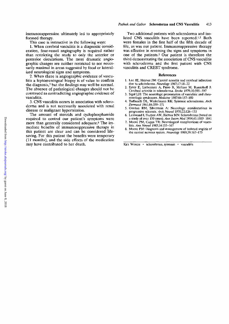
The angiogram showed multiple long segments ofsmooth, symmetrical narrowing in large and medium-sized vessels of the posterior circulation; the mostprominent of these were in the superior cerebellararteries near their origins (Figure 1). The 0.5-1.5-cm-long narrowed arterial segments were thought tobe characteristic of vasculitis as opposed to nonspe-cific arterial spasm such as might occur in associationwith subarachnoid hemorrhage.
Skin biopsy was performed to document systemicvasculitis and showed epidermal and dermal changesassociated with scleroderma, but no evidence ofvasculitis. The patient remained free of symptoms
and was discharged 3 days later receiving 60 mgprednisone and 12.5 mg captopril per day.
Two weeks later the prednisone dosage was re-duced to 50 mg/day following a tapering program.Two days later the patient began to have episodes ofnumbness of her right arm and shoulder, each epi-sode lasting 1-5 minutes. She was brought to theemergency room on March 29, 1988, following anepisode of numbness of her right face, arm, and legassociated with expressive aphasia. Examination con-firmed the aphasia and hemianalgesia, and a bloodpressure of 186/130 torr was recorded. Methylpred-nisolone 100 mg i.v. was administered, resulting inresolution of her neurological symptoms within 30minutes; her blood pressure returned to baselinelevels (140/90 torr). During her first week of hospi-talization methylprednisolone was replaced withprednisone 60 mg/day.
The results of a leptomeningeal and cortical biopsyobtained to confirm the presence of CNS vasculitiswere normal. Because of cushingoid facies, nausea,vomiting, depression, and intermittent confusion (allconsidered to be side effects of steroid therapy) aswell as the negative biopsy results, the prednisonedosage was tapered to 50 mg/day. No further neuro-logical symptoms occurred for 2 days, and the patientwas discharged.
During the first week at home the patient againbegan to experience episodes of right facial andupper extremity paresthesias associated with expres-sive aphasia. She continued to have clinical symptomsof ischemia, and now hyperglycemia was added to theother side effects of steroid therapy. Because of poortherapeutic response to moderate-dosage steroid ther-apy and the unacceptable side effects of higher dos-ages of steroids, the patient was started on cyclophos-phamide 100 mg/day and the steroid dosage wasreduced to 40 mg/day. Over the next 1.5 months thecyclophosphamide dosage was gradually increased to175 mg/day and the prednisone dosage was graduallydecreased to 20 mg/day. The patient became symp-tom-free after starting cyclophosphamide therapy andremained so for 2 months. In early August, however,she began to experience 1-2-minute episodes ofnumbness of the right angle of her mouth and herright hand. The cyclophosphamide dosage was in-creased to 200 mg/day, and the prednisone dosage wasgradually tapered to 10 mg/day. Episodes of neuro-logical deficit did not recur, but 1.5 months later awhite blood cell (WBC) count of 2,300/mm3 and atotal platelet count of 61,000/mm3 required discontin-uation of the cyclophosphamide and the prednisonedosage was increased to 60 mg/day.
Within 2 weeks after discontinuing cyclophospha-mide therapy the patient was experiencing frequentfalls and numbness of her left face and hand. Beingunable to restart cyclophosphamide therapy, we in-creased the prednisone dosage to 120 mg/day. InNovember her WBC count was 7,000/mm3 andmonthly cyclophosphamide pulse therapy was initi-ated (700 mg/mo). The prednisone dosage was de-
by guest on June 8, 2018http://stroke.ahajournals.org/
Dow
nloaded from

412 Stroke Vol 22, No 3 March 1991
FIGURE 1. Vertebral angiogram demonstrating multiple areas of symmetrical segmental narrowing of superior cerebellar arteries(1, arrows). Narrowed portions of arteries are 0.5-1.5 cm long. Distal branch of superior cerebellar artery terminates abruptly (2,arrow). Other examples of segmental arterial narrowing and abruptly terminating vessels could also be identified on originalroentgenograms.
creased to 60 mg/day and then tapered at a rate of2.5 mg/wk. On this regimen the patient had only twobrief episodes of right facial numbness during thenext 3 months.
In January 1989 the patient was seen in the emer-gency room because of right abdominal pain, fever,chills, and a cough. Her WBC count was 27,000/mm3,and she had severe metabolic acidosis. She did notimprove, and ultimately an exploratory laparotomywas performed in search of a source of sepsis. Duringsurgery the patient became hypotensive and sufferedirreversible cardiac arrest. Permission for autopsy wasdenied.
Pertinent laboratory data included rheumatoidfactor positive at 1:160, with normal complementlevels and absent Sjogren's antibodies SS-A andSS-B; results of an antinuclear antibody (ANA)screen were negative. Her liver function tests re-vealed an alkaline phosphatase level of 600-700units/1, an aspartic transaminase level of 100 units/1,a y-glutamic transaminase level of 1,700 units/1, atotal bilirubin content of <1.0 mg%, a cholesterolcontent of 471 mg%, and a triglyceride content of 101mg%. Her blood urea nitrogen and creatinine levels
were always normal, as was her plasma renin level.Her erythrocyte sedimentation rate was normal orslightly elevated (20-39 mm/hr). Creatinine clear-ance was elevated at 168 ml/min. Thyroid functiontests and a-fetoprotein levels were normal. Assay forhepatitis B surface antigen was negative; the rapidplasma renin test and microhemagglutination test forTreponema pallidum were normal.
DiscussionPrimary CNS involvement in scleroderma is rare
and unusual,3-6 leading some to argue that CNSdysfunction is either secondary to end-organ failureor, if vasculitis is present, it is associated with evi-dence of systemic vasculitis.4-5 The rarity of CNSvasculitis associated with scleroderma in addition tothe underestimated significance of the angiographicdata resulted in diagnostic uncertainty and delayedappropriate medical intervention for our patient.Further uncertainty resulted from the subsequentnormal findings on leptomeningeal biopsy, which hadbeen recommended to confirm the angiographic find-ings. Only the repeated, prompt, dramatic benefits of
by guest on June 8, 2018http://stroke.ahajournals.org/
Dow
nloaded from
Pathak and Gabor Scleroderma and CNS Vasculitis 413
immunosuppression ultimately led to appropriatelyfocused therapy.
This case is instructive in the following ways:1. When cerebral vasculitis is a diagnostic consid-
eration, four-vessel angiography is required ratherthan restricting the study to only the anterior orposterior circulations. The most dramatic angio-graphic changes are neither restricted to nor neces-sarily maximal in areas suggested by focal or lateral-ized neurological signs and symptoms.
2. When there is angiographic evidence of vascu-litis a leptomeningeal biopsy is of value to confirmthe diagnosis,7 but the findings may well be normal.The absence of pathological changes should not beconstrued as contradicting angiographic evidence ofvasculitis.
3. CNS vasculitis occurs in association with sclero-derma and is not necessarily associated with renaldisease or malignant hypertension.
The amount of steroids and cyclophosphamiderequired to control our patient's symptoms weremore than generally considered adequate.8 The im-mediate benefits of immunosuppressive therapy inthis patient are clear and can be considered life-saving. For this patient the benefits were temporary(11 months), and the side effects of the medicationmay have contributed to her death.
Two additional patients with scleroderma and iso-lated CNS vasculitis have been reported.12 Bothwere females in the first half of the fifth decade oflife, as was our patient. Immunosuppressive therapywas effective in reversing the signs and symptoms inone of the patients.2 Our patient is therefore thethird demonstrating the association of CNS vasculitiswith scleroderma and the first patient with CNSvasculitis and CREST syndrome.
References1. Lee JE, Haynes JM: Carotid arteritis and cerebral infarction
due to scleroderma. Neurology 1967;17:18-222. Estey E, Lieberman A, Pinto R, Meltzer M, Ransohoff J:
Cerebral arteritis in scleroderma. Stroke 1979;10:595-5973. Sigal LH: The neurologic presentation of vasculitic and rheu-
matologic syndromes. Medicine 1987;66:157-1804. Tuffanelli DL, Winkelmann RK: Systemic scleroderma. Arch
Dermatol 1961;84:359-3715. Gordon RM, Silverstein A: Neurologic manifestations in
progressive sclerosis. Arch Neurol 1970;22:126-1336. Leinwand I, Duryee AW, Richter MN: Scleroderma (based on
a study of over 150 cases). Ann Intern Med 1954;41:1003-10417. Moore PM, Cupps TR: Neurological complications of vascu-
litis. Ann Neurol 1983;14:155-1678. Moore PM: Diagnosis and management of isolated angiitis of
the central nervous system. Neurology 1989;39:167-173
KEY WORDS • scleroderma, systemic • vasculitis
by guest on June 8, 2018http://stroke.ahajournals.org/
Dow
nloaded from
R Pathak and A J GaborScleroderma and central nervous system vasculitis.
Print ISSN: 0039-2499. Online ISSN: 1524-4628 Copyright © 1991 American Heart Association, Inc. All rights reserved.
is published by the American Heart Association, 7272 Greenville Avenue, Dallas, TX 75231Stroke doi: 10.1161/01.STR.22.3.410
1991;22:410-413Stroke.
http://stroke.ahajournals.org/content/22/3/410World Wide Web at:
The online version of this article, along with updated information and services, is located on the
http://stroke.ahajournals.org//subscriptions/
is online at: Stroke Information about subscribing to Subscriptions:
http://www.lww.com/reprints Information about reprints can be found online at: Reprints:
document. Permissions and Rights Question and Answer available in the
Permissions in the middle column of the Web page under Services. Further information about this process isOnce the online version of the published article for which permission is being requested is located, click Request
can be obtained via RightsLink, a service of the Copyright Clearance Center, not the Editorial Office.Stroke Requests for permissions to reproduce figures, tables, or portions of articles originally published inPermissions:
by guest on June 8, 2018http://stroke.ahajournals.org/
Dow
nloaded from